T
T
H
H
È
È
S
S
E
E
En vue de l'obtention du
D
D
O
O
C
C
T
T
O
O
R
R
A
A
T
T
D
D
E
E
L
L
’
’
U
U
N
N
I
I
V
V
E
E
R
R
S
S
I
I
T
T
É
É
D
D
E
E
T
T
O
O
U
U
L
L
O
O
U
U
S
S
E
E
Délivré par l'Université Toulouse III - Paul Sabatier Discipline ou spécialité : Biologie Moléculaire
JURY
Pr Pierre-Emmanuel Gleizes, Professeur d'université,Toulouse-Président Dr Olivier BENSAUDE, Directeur de recherche INSERM, Paris-Rapporteur
Pr Manuel ECHEVERRIA, Professeur d'université, Perpignan-Rapporteur Dr Céline VERHEGGEN, Chargé de recherche CNRS, Montpellier-Examinateur Dr Tamás KISS, Directeur de recherche INSERM, Toulouse-Directeur de thèse
Ecole doctorale : Biologie-Santé-Biotechnologies
Unité de recherche : Laboratoire de Biologie Moléculaire Eucaryote Directeur(s) de Thèse : Tamás KISS
Rapporteurs : Dr Olivier BENSAUDE
Pr Manuel ECHEVERRIA
Présentée et soutenue par Natalia PINZON RESTREPO Le 16 Décembre 2011
Titre : Characterization of regulatory noncoding RNAs:
the U1 small nuclear RNA
PREFACE: SMALL NUCLEAR NON-CODING RNA FUNCTION IN EVERY ASPECT OF GENOME EXPRESSION, MAINTENANCE AND ARCHITECTURE
4 CHAPTER I: SMALL NUCLEAR SPLICEOSOMAL RNAS: ARCHITECTURE, BIOGENESIS AND FUNCTION
8
A. PRECURSOR mRNA SPLICING 8
B. SnRNA BIOGENESIS 14
Integrated transcription and 3’-end processing 14
Nuclear export 16
Sm core assembly 18
Nuclear import 18
Completion of snRNP maturation in the nuclear Cajal bodies 19
C. REGULATION OF SPLICING 20
Multiple links between transcription and splicing 22 Multifunctional proteins: critical to link splicing to other steps of gene expression? 24 FET family of proteins and their role on coupling transcription and splicing 24 CHAPTER II: THE EXPANDING UNIVERSE OF NONCODING RNA: NONCODING RNAs ARE IMPORTANT REGULATORS CONTROLLING mRNA PRODUCTION
28
A. NOVEL FUNCTIONS IN mRNA PRODUCTION 28
The many faces of the U1 snRNA 28
U1 snRNA and transcription 30
U1 snRNA and polyadenylation 32
U2 snRNA and 3’ end processing 36
Small noncoding RNAs as regulators of splicing 38
Other examples of multifunctional RNAs 40
B. TRANSCRIPTIONAL REGULATION 42
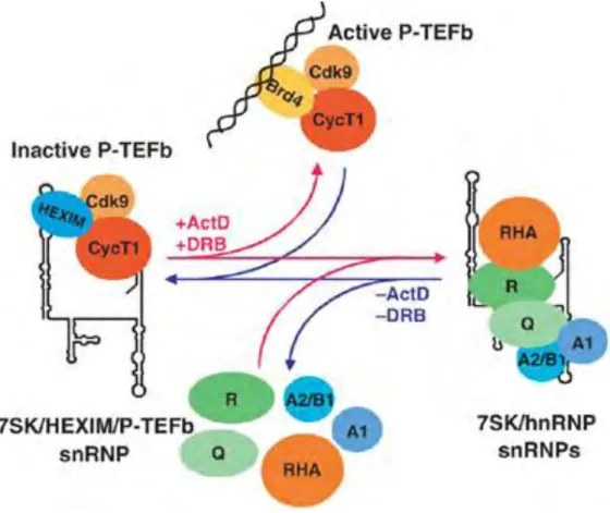
The 7SK small nuclear snRNA 42
CHAPTER III: BOX C/D SMALL RIBONUCLEOPROTEINS 45
A. STRUCTURE AND FUNCTION OF BOX C/D RNAs 45
Function of Box C/D RNAs 45
The architecture of box C/D RNPs 47
B. BIOGENESIS 49
Organization of box C/D snoRNA genes 49
Assembly of box C/D RNPs 51
Trafficking 52
C. BIOLOGICAL SIGNIFICANCE OF THE MODIFICATIONS 54
tRNA modifications 56
The Wooble position 56
Archeal box C/D sRNPs guide the methylation of tRNA 59
FIRST PART OF THE RESULTS: DISCOVERY OF A NOVEL U1-TAF15 snRNP 60
I. PUBLICATION 60
II. U1-TAF15 snRNP ADDITIONAL ANALYSIS 63
Definition of U1 snRNA elements directing TAF15 binding 63 Structural analysis of U1 snRNA associated with TAF15 67
Purification of U1-TAF15 snRNP 69
Kinetics of U1-TAF15 localization to the perinucleolar caps 71
SECOND PART OF THE RESULTS: STUDY OF TWO NOVEL BOX C/D RNPs 73
Identification of WDR79-associated RNAs by deep-sequencing 73
Characterization of CD8 RNA 75
Sequence analysis and predicted function of human CD8 RNA 75 Detection and structural characterization of CD8 RNA 77 In vivo expression and mutation analysis of CD8 RNA 79
CD8 RNA is a small Cajal body-specific RNA 81
Characterization of CD5 RNA 83
Sequence analysis and predicted function of human CD5 RNA 83
CD5 is a small Cajal body-specific RNA 85
Association of CD8 and CD5 RNAs with WDR79 89
MATERIALS AND METHODS 91
DISCUSSION I: A NOVEL U1-TAF15 snRNP 94
Biogenesis of the U1-TAF15 particle. 94
Protein composition of the U1-TAF15 particle. 95
LIST OF FIGURES
number name page
FIG1 RNPs in Gene Expression and Its Regulation. 5
FIG2 Examples of small regulatory ncRNAs and historic view of noncoding RNA. 6
FIG3 Different families of noncoding RNAs. 7
FIG4 Catalytic reactions and conserved sequence elements of splicing. 9 FIG5 Protein composition and snRNA secondary structures of the major human spliceosomal snRNPs. 11
FIG6 Pre-mRNA splicing by the U2-type spliceosome. 12
FIG7 Biogenesis of small RNPs in mammals. 15
FIG8 Schematic structure of precursor spliceosomal snRNAs and minimal sequences necessary for SMN binding in the cytoplasm. 17
FIG9 Domain structure of FET and SARFH/CABEZA proteins. 25
FIG10 Metazoan pre-mRNA 3′ end processing/polyadenylation machinery. 33 FIG11 Mechanisms of alternative splicing regulation by small ncRNAs. 39 FIG12 A model for regulation of the nuclear level of active P-TEFb by dynamic and reversible remodelling of 7SK snRNPs. 44 FIG13 Schematic structure and main function of box C/D RNAs. 46 FIG14 Box C/D snoRNPs and sRNPs core protein composition and trans-acting factors associated during the biogenesis pathway. 48
FIG15 Processing of intronic snoRNAs. 50
FIG16 Mechanism of codon recognition. 56
FIG17 Effect of modifications at position 34 and 37 on tRNA dynamics and accommodation on the ribosome. 58 FIG18 The intron-containing pre-tRNAmethylation guide sequences and tRNATrp in some Archea carries both box C/D 2’-O-Trp
sequences in the same RNA molecule. 59 FIG19 U1 snRNA loop sequences are not essential for TAF15 binding. 64 FIG20 U1 snRNA stem-loops 1, 2, 3 and 4, sequences surrounding the Sm site and the splice binding site are not essential for TAF15 binding. 65 FIG21 Structural comparison of U1 snRNAs associated with TAF15 and Sm proteins. 68
FIG22 Purification of U1-TAF15 snRNP. 70
FIG23 Kinetics of U1-TAF15 snRNP localization to perinucleolar caps. 72 FIG24 Identification and analysis of WDR79-associated RNAs by deep sequencing. 74 FIG25 The human CD8 RNA is predicted to direct U2 modification. 76 FIG26 Human CD8 RNA is a relatively short box C/D scaRNA with a well-defined secondary structure. 78 FIG27 Schematic structure of the pGL/CD8 expression vector. 80 FIG28 CD8 and CD8 Del2 localizes to Cajal bodies, but CD8 Del1+2 and CD8 Del1 localize to the nucleolus. 82 FIG29 The human CD5 RNA is predicted to direct 2’-O-methylation of cytidine 34 at the Wobble position of tRNA-Met elongator 84
FIG30 Characterization of CD5 RNA. 85
FIG31 Human CD5 RNA accumulates in Cajal bodies. 88
PREFACE: SMALL NUCLEAR NON-CODING RNA FUNCTION IN EVERY ASPECT OF GENOME EXPRESSION, MAINTENANCE AND ARCHITECTURE
The central dogma of biology, until not long ago, held that genetic information stored on DNA molecules was translated into the final protein products through RNAs as intermediate molecules. Proteins were considered to be the principal effectors of metabolism as well as responsible for the construction and diversity of organisms. During the past decade, an additional level of complexity in the regulation of genome expression was added, implicating new classes of RNA molecules called noncoding RNA (ncRNA). These ncRNAs are also often referred to as functional RNAs in that, although they do not contain the capacity to encode proteins, they a function as RNA molecules.
The most abundant ncRNAs have been extensively studied, and were found to be involved in relatively generic functions in cells. As we can see in Figure 1, ncRNAs form tight complexes with proteins that have catalytic activities, act as chaperones, protect them from degradation or meditate interactions with other complexes. These RNA-protein complexes are named ribonucleoproteins particles (RNPs). Our research group has a long-standing interest in understanding the structural and functional diversity of the complex and fascinating world of human small nuclear RNPs (snRNPs). In fact, ribosomal (rRNA) and transfer RNAs (tRNA) participate in messenger RNA (mRNA) translation, and the signal recognition particle RNA (SRP RNA) functions in the translocation of the membrane and extracellular proteins into the endoplasmic reticulum. ncRNA also function in RNA processing: like spliceosomal RNAs (snRNAs), RNase P RNA, U7 snRNA, RNase MRP small nucleolar RNA (snoRNA), box C/D and box H/ACA RNA snoRNAs and small Cajal body-specific scaRNAs, and guide RNAs for mRNA editing. Eukaryotic chromosome end synthesis is templated by the telomerase RNA and 7SK RNA is implicated in transcription elongation regulation. microRNAs (miRNAs) are important regulators of gene expression, since they have been implicated in regulation of translation and heterochromatin formation and maintenance (Figure 1).
Today, with the ability to analyze the RNA products of the genome in even greater depth, it has become clear that most of the eukaryotic genome is transcribed (Clark et al., 2011). This pervasive transcription produces a bulk of noncoding RNAs unknown until now and the remaining question is whether these transcripts are functional or they represent transcriptional noise. An increasing number of reports suggest regulatory roles for ncRNAs in systems ranging from bacteria to mammals, with reports of their involvement in nearly every cellular process (Figure 2). Novel classes of noncoding RNAs, such as piRNAs or long noncoding RNAs have recently emerged. These studies are changing our vision of the genomes, and suggest that the universe of noncoding RNAs extends far beyond the boundaries of what we had previously imagined (for reviews see (Amaral et al., 2008; Orom and Shiekhattar, 2011; Rederstorff and Huttenhofer, 2010; Ulveling et al., 2011)).
Figure 1. RNPs in Gene Expression and Its Regulation. RNPs play extensive
roles in gene expression and its regulation. Here, the major activities of RNPs are depicted during eukaryotic gene expression. Following transcription by RNA Polymerases II (RNA Pol II), pre-mRNAs are bound by diverse proteins, such as hnRNP and SR (serine-arginine-rich) proteins. Pre-mRNAs, containing exons (red) and introns (pink), are subjected to processing by a range of RNPs that include uridine-rich (U-rich) small nuclear RNPs (U snRNPs) that make up the spliceosome. Certain RNAs such as pre-transfer RNAs and mRNA transcripts encoding histones also undergo processing by specific RNPs (RNase P and U7 snRNP, respectively). Small nucleolar RNPs (snoRNPs) and small Cajal body RNPs (scaRNPs) mediate maturation of RNA components of RNPs such as ribosomal RNAs (transcribed by RNA Polymerase I, RNA Pol I) and snRNAs, respectively. Small RNAs can form microRNPs that regulate translation. In certain organisms, RNA-induced transcriptional silencing (RITS) complexes, which contain small-interfering RNAs, mediate heterochromatin formation and maintenance. Telomerase, a box H/ACA scaRNP, replenishes the terminal telomeric repeats of chromosomes to maintain genomic stability. In the cytoplasm, the ribosome is the key RNP that directs the translation of mRNA into protein. It also functions with the signal recognition particle (SRP) RNP to direct protein translocation into the endoplasmic reticulum (ER). tRNAs also form complexes in the cytoplasm with aminoacyl-tRNA (aa-tRNA) synthetases, which charge tRNAs with the corresponding amino acid, and with translation elongation factor eEF1A. Adapted from (Wahl et al., 2009).
Figure 2. A. Examples of small regulatory ncRNAs. B. A historic view of noncoding RNA. Key dates in the discovery of noncoding RNA are indicated,
from the first ribosomal RNA (rRNA) to the first description of the regulatory function of micro RNAs. Correlated with the discovery of pervasive transcription, an increasing number of functions involving non coding RNA have been described, including novel functions for well characterized RNAs, such as tRNAs, snoRNAs, snRNAs or even mRNAs. Adapted from (Ulveling et al., 2011).
Examples of the various levels of regulation of eukaryotic gene expression by small ncRNAs.
RNA maturation and processing
Spliceosomal snRNAs, U7 snRNA, RNA of RNaseP, box C/D and box H/ACA RNAs (sRNAs, snoRNAs, scaRNAs), guide RNAs Transcription
7SK, B2, Alu Translation rRNA, tRNA
Gene expression regulation miRNAs, siRNAs, piRNAs Genome maintenance Telomerase RNA
Several classes of ncRNAs have been proposed, according to their function, structure, subcellular localization and biogenesis (Figure 3). Given the vast diversity of the RNA world, the present manuscript will focus on our current knowledge of small nuclear ncRNAs ranging from 60 to 300 nucleotides (nt) in length. In fact, our purpose was to contribute to the ongoing expansion of the functions involving small nuclear RNAs. Our work has focused on two particular classes of ncRNAs: the nucleoplasmic spliceosomal snRNAs and the nucleolar and Cajal body-specific box C/D 2’-O-methylation guide RNAs.
I will first give an overview on the function of non-coding spliceosomal snRNAs. Their biogenesis as well as the multiple links between transcription and splicing will be discussed. The second chapter will focus on the expansion of the functions involving small non-coding RNAs in mRNA production. Finally, the third part of this manuscript will concentrate on the expanding class of box C/D RNA modification guide RNAs.
Figure 3. Different families of noncoding RNAs. Class I represents functional
RNAs wich possess little protein coding potential, because they carry no long open reading frames. Class II represents RNAs translated into proteins. Note that the maturation steps of ncRNAs are not indicated. Recently, RNAs with both protein coding capacity and activity as functional ncRNAs have been reported. Inversely, RNA primarily classified as non-protein-coding RNA has been showed to hold coding capacities (Ulveling et al., 2011). Therefore, categorization of some RNA molecules into class I or II may be challenging. sRNA: small RNA ; lRNA: long RNA ; siRNA: small interfering RNA ; miRNA: microRNA ; snRNA: small nuclear RNA ; snoRNA: small nucleolar RNA ; scaRNA: small Cajal body-specific RNA ; pre-mRNA: precursor messenger RNA
CHAPTER I:
SMALL NUCLEAR SPLICEOSOMAL RNAs: ARCHITECTURE,
BIOGENESIS AND FUNCTION
A. PRECURSOR mRNA SPLICING
Precursor mRNA introns have been found in all examined eukaryotes, and it is thought that they were present in the last common eukaryote ancestor. Introns of precursor mRNAs are removed in a process called splicing.
Strikingly, the signal sequences in pre-mRNAs which define the exon regions are short: they comprise the 5’ splice site (ss), the 3’ ss and the branch site (BS). The BS is typically located 18-40 nucleotides upstream from the 3'ss and in higher eukaryotes is followed by a polypyrimidine tract (PPT). The consensus sequences found in the budding yeast Saccharomyces cerevisiae exhibit a higher level of conservation than those in metazoans, probably because this organism exhibits less alternative splicing events. Additional, cis-acting pre-mRNA elements include exonic and intronic splicing enhancers (ESEs and ISEs) or silencers (ESSs and ISSs). They are typically short and diverse in sequence and modulate both constitutive and alternative splicing by binding regulatory proteins that either stimulate or repress the assembly of spliceosomal complexes at an adjacent splice site (see regulation of splicing p 19) (for reviews see (Wang and Burge, 2008) (Chen and Manley, 2009)). Therefore, the splicing machinery is able to recognize functional 5’ and 3’ splice sites and, after removal of the intronic sequences, ligate them together.
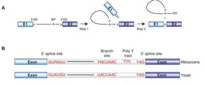
In the first step of splicing, the 2’ hydroxyl of an adenosine in the BS attacks the 5’ splice site (exon-intron junction). As a consequence, the 5’ exon is cleaved off, and the 5’ end of the intron is ligated to the BS via the 2’ hydroxyl group. In the second trans-esterification reaction, the 3’ hydroxyl of the 5’ exon attacks the 3’ splice site, which results in the ligation of the two exons and release of the intron lariat. (Figure 4) Under the appropriate conditions, the chemical steps of splicing are reversible (Cheng and Tseng, 2008)
Figure 4. Catalytic reactions and conserved sequence elements of splicing. A.
Schematic representation of the two-step mechanism of pre-mRNA splicing. Boxes and solid lines represent the exons (E1, E2) and the intron, respectively. The branch site adenosine is indicated by the letter A and the phosphate groups (p) at the 5′ and 3′ splice sites, which are conserved in the splicing products, are also shown. B. Conserved sequences found at the 5′ and 3′ splice sites and branch site of U2-type pre-mRNA introns in metazoans and budding yeast (S. cerevisiae). Y = pyrimidine and R = purine. The polypyrimidine tract is indicated by (Yn). From (Will and Luhrmann, 2011)
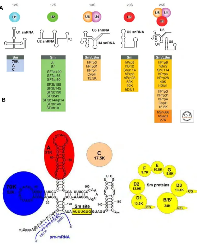
The machinery catalyzing the splicing reaction is called the spliceosome, an immense RNP estimated to be several megadaltons. Here again, non-coding RNAs play a fundamental role: this large complex contains five uridine-rich snRNAs: the U1, U2, U4, U5 and U6 spliceosomal snRNAs. The U snRNAs range in length from 107 to 210 nucleotides, and are associated with several proteins to form small nuclear ribonucleoprotein particles (snRNPs) (Figure 5). The spliceosome also contains an impressive amount of non-snRNP splicing factors. Proteomic analyse indicate that over 170 proteins associate with the human spliceosome at some point during the splicing process (for reviews see (Wahl et al., 2009) (Will and Luhrmann, 2011)). In vitro 100 proteins constitute the core spliceosome minimally required for activity in yeast (Fabrizio et al., 2009).
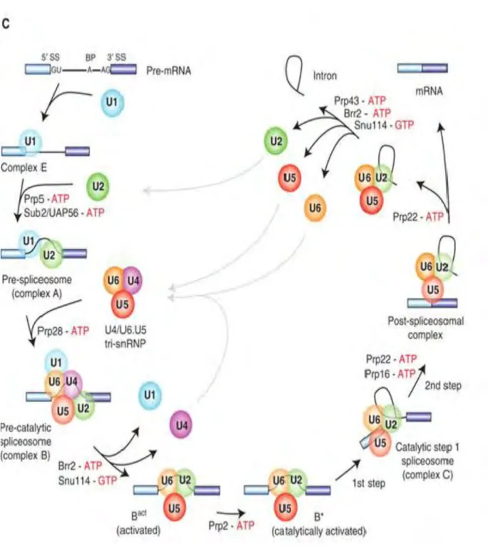
The basic mechanisms of spliceosome assembly are well known (Figure 6). The process begins with base-pairing of the 5’ arm of U1 snRNA with the 5’ splice site (Figure 5 and 6) and the binding of SF1/mBBP to the branch point. This step is ATP independent and this stage of assembly is called the E’ complex. The E’ complex can be converted into the E complex by the recruitment of U2AF to the polypyrimide tract. All further steps of spliceosome assembly are ATP or GTP dependent. In a subsequent step, SF1 is replaced by the U2 snRNP at the branch site forming a short U2-BS duplex in which the branch adenosine is bulged out, specifying its 2′ OH as the nucleophile for the first catalytic step of splicing. At this step the complex is named A complex or pre-spliceosome. The preassembled U4/U6-U5 tri-snRNP is then recruited, generating the pre-catalytic B complex. At this step, major RNA-RNA and RNA-protein rearrangements occur. Within the U4/U6.U5 tri-snRNP, the U6 and U4 snRNAs are extensively base-paired with each other. After association of the tri-snRNP with the A complex, the U4/U6 interaction is disrupted, releasing the U4 snRNA. The 5′ end of U6 snRNA base pairs with the 5′ss, displacing the U1 snRNA, and extensive U6/U2 snRNA base pairing occurs. This process gives rise to the activated spliceosome: the Bact complex. Subsequent
catalytic activation by the DEAH-box RNA helicase Prp2, generates the B*
complex, which catalyzes the first of the two steps of splicing. This yields the C complex, which in turn catalyzes the second trans-esterification step. The
spliceosome then dissociates and, after additional remodeling, the released snRNPs take part in additional rounds of splicing.
Figure 5. Protein composition and snRNA secondary structures of the major human spliceosomal snRNPs. A. All seven Sm proteins (B/B’, D3, D2, D1, E, F,
and G) or LSm proteins (Lsm2-8) are indicated by “Sm” or “LSm” at the top of the boxes showing the proteins associated with each snRNP. The U4/U6.U5 tri-snRNP contains two sets of Sm proteins and one set of LSm proteins. Note that several
snRNAs undergo structural rearrangements during splicing B. Schematic structure
of the human spliceosomal snRNP. The Sm proteins form a heptameric ring and
bind to the Sm site of U1 snRNA. The U1-specific U1-70K and U1-A proteins specifically recognize the terminal stem-loop (SL) structures of first and second hairpins of the U1 sn RNA. Binding of U1-C protein is supported mainly by protein-protein interactions. The U1snRNA exhibits 9 nt of full complementarity to the 5’splice site on the pre-mRNA. Adapted from (Will and Luhrmann, 2011).
A
Figure 6. Pre-mRNA splicing by the U2-type spliceosome. Canonical
cross-intron assembly and disassembly pathway of the U2-dependent spliceosome. For simplicity, the ordered interactions of the snRNPs (indicated by circles), but not those of non-snRNP proteins, are shown. The various spliceosomal complexes are named according to the metazoan nomenclature. Exon and intron sequences are indicated by boxes and lines, respectively. The stages at which the evolutionarily conserved DExH/D-box RNA ATPases/helicases Prp5, Sub2/UAP56, Prp28, Brr2, Prp2, Prp16, Prp22 and Prp43, or the GTPase Snu114, act to facilitate conformational changes are indicated. (From (Will and Luhrmann, 2011) )
Because of the enormous difficulty of studying entire spliceosomes, studies have been made from spliceosomes stalled at one particular stage of their assembly, giving a snap-shot of each of its sub-complexes. They have revealed that besides the extensive RNA-RNA and RNA-protein rearrangements occurring during splicing, the protein composition of spliceosome is highly dynamic as well. In fact there is a remarkable exchange of proteins from one stage of splicing to the next.
Combination of biochemical, structural and kinetic studies is currently enhancing our knowledge into the compositional and morphological changes during spliceosome activation and catalysis. Current models suggest that: 1) the dynamics and complexity of the spliceosome is important for: accurate splice site recognition,
sustaining a catalytically active structure and for the regulation and flexibility necessary for alternative splicing. (Will and Luhrmann, 2011) 2) every step of
spliceosome assembly is kinetically efficient and reversible: and no single assembly step irreversibly commits a particular pair of splice sites to splicing. Rather, commitment to splicing increases as spliceosome assembles (Hoskins et al., 2011).
B. SnRNA BIOGENESIS
Because ncRNAs typically function as ribonucleoprotein complexes and not as naked RNAs, understanding of the biogenesis of ncRNPs is crucial for comprehending their regulation and function.
Biogenesis of spliceosomal snRNPs is critical for gene expression in eukaryotes. SMN, the complex that assembles Sm cores on snRNAs, plays a key role in snRNP biogenesis. SMN deficiency, the main cause of spinal muscular atrophy (SMA), alters the repertoire of snRNAs and causes widespread and cell-type specific defects in pre-mRNA splicing (for reviews see (Battle et al., 2006; Chari et al., 2009; Patel and Bellini, 2008)).
Integrated transcription and 3’-end processing
Among the snRNAs, U1, U2, U4, and U5 of the major spliceosome and U11, U12, and U4atac of the minor spliceosome are transcribed by RNA Pol II. In contrast, U6 and U6atac snRNAs are generated by RNA Pol III. The snRNA genes are usually repeated and several pseudogenes can be found in the genome. Two functionally important sequence elements are contained within the core promoter region of Pol II and Pol III snRNA genes. The first located approximately at position −55 relative to the transcription start site is called the proximal sequence element (PSE). The second promoter element termed distal sequence element (DSE) is located around position −220. In addition to these two common sequence elements, Pol III snRNA genes contain a TATA box about 25 base pairs upstream from the transcription initiation site, which is in fact responsible for the recruitment of Pol III-specific transcription factors (for reviews see (Egloff et al., 2008; White, 2011)).
snRNA transcription by Pol II is initiated upon binding of Oct1 and STAF1 to different sites in the DSE, followed by the recognition of the PSE by PTF, an snRNA gene-specific transcription factor. The general transcription factors TFIIA, TFIIB, TFIIE, TFIIF, TBP, TAF100 and possibly also TFIIH then join the promoter and allow eventually recruitment of RNA Pol II. Efficient transcription initiation also requires the large multi-protein Integrator complex. This complex binds to the C-terminal domain (CTD) of Pol II phosphorylated at serine 2 and serine 7 positions. The requirement of this double mark for efficient transcription on a class of genes supports the existence of a CTD code (Baillat et al., 2005; Egloff et al., 2007; Egloff et al., 2010).
Integrator subunits Int11 and Int9 display sequence homology with CPSF-7 and CPSF-100 cleavage and polyadenylation factors of pre-mRNAs, respectively. Transcription termination occurs via an endonucleolytic cleavage at the 3’-box mediated by the Integrator. Therefore, the Integrator complex appears to travel along with the Polymerase to the 3’-box during transcription, integrating snRNA genes transcription initiation and termination.
The synthesis of the Pol III-transcribed U6 snRNA has been described as a model for Pol III transcription. As other Pol III genes, it requires Oct-1, STAF1 and SNAPc, but also the recruitment of the Pol III-specific general transcription factor TFIIIB to the TATA box. The U6 snRNA acquires a posttranscriptional methyl group on the γ-phosphate of the first nucleotide in a reaction involving a methyltransferase termed methylphosphate-capping enzyme (MePCE). The 5’-mono-methyl cap protects the U6 snRNA against exonucleolytic degradation. Transcription termination occurs at the poly-U termination signal, recognized by the La protein. La binding protects the 3’ end from degradation and promotes Lsm snRNP assembly on the 3’ end of the U6 snRNA. Lsm assembly promotes the targeting of the PolII-snRNAs to the nucleolus, where they undergo site-specific modifications. For review see (Kiss, 2004).
Active transcription of snRNA genes often occurs in association with Cajal bodies. This association is mediated by the nascent snRNA transcripts (Frey and Matera, 2001).
Nuclear export
In contrast to the U6 snRNA that has an exclusively nuclear biogenesis, U1, U2, U4 and U5 snRNAs biogenesis requires a complicated nucleocytoplasmic trafficking process that is directed by transport factors.
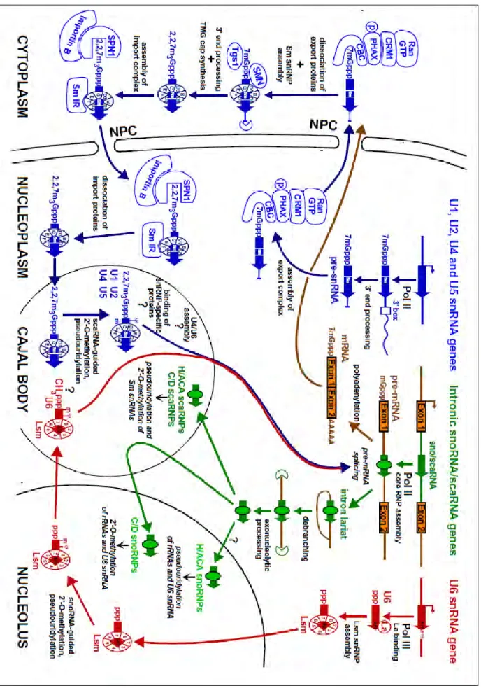
Like other Pol II-transcribed RNAs, nascent Pol II primary transcripts of snRNAs (pre-snRNAs) acquire cotranscriptionally a 7-monomethyl guanosine (7mGpppG) cap. The RNA cap structure recruits the cap binding complex (CBC) proteins, CBC20 and CBC80. The CBC bound to the RNA recruits an adaptor protein, PHAX, which then recruits the nuclear export receptor exportin 1 (CRM1). PHAX, also required for snoRNA targeting to the Cajal bodies (Boulon et al., 2004; Watkins et al., 2004), recognizes specific features of small RNAs, notably length, m7G-cap and three-dimensional structure without sequence specificity. Therefore, distinguishing between snRNAs and snoRNAs to target them to specific biogenesis pathways may involve contacts with other proteins (Masuyama et al., 2004; Mourao et al., 2010; Ohno et al., 2002). PHAX is phosphorylated in the nucleus by the CK2 kinase and dephosphorylated in the cytoplasm by the phosphatase 2A, ensuring the directionality of the export (Kitao et al., 2008). Additional 3’ end processing is performed by an unknown endonuclease, and the proper pre-snRNAs, containing the 7mG-cap and 3’ trailer sequences are exported to the cytoplasm, where the export proteins dissociate (Figure 7 and 8).
The U snRNA precursors concentrate in Cajal bodies prior to export, and the interaction with PHAX, but not CRM1, is required for the efficient exit of U snRNAs from Cajal bodies (Suzuki et al., 2010).
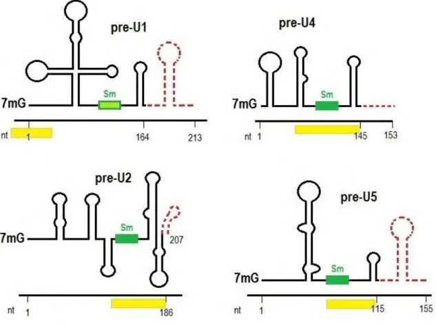
Figure 8. Schematic structure of precursor spliceosomal snRNAs and minimal sequences necessary for SMN binding in the cytoplasm. The mature form of
each snRNA is depicted by black line and the precursor sequences are shown by dashed red lines. Nucleotide size numbers for the mature and pre-snRNAs are indicated on the x-axis. Green boxes represent Sm sites. The non-canonical Sm site of U1 is shaded in light green. The snRNP code is shown as yellow bars beneath each snRNA. The precursors were detected by High-Throughput sequencing of Gemin5-bound RNA fragments (Yong et al., 2010).
Sm core assembly
In the cytoplasm, the SMN complex, comprised of the SMN, Gemin 2-8 and unrip proteins, assembles Sm cores in an ATP-dependent manner on each pre-snRNA. This process requires several assembly intermediate steps and post-transcriptional modifications of the factors involved, ensuring that the formation of the highly stable Sm core is effectuated only on proper snRNAs.
To initiate the process, pICln forms a complex with D1/D2 and F/E/G Sm proteins (6S complex), inducing the formation of a closed pentamer ring that cannot bind to the snRNAs. Independently, pICln binds B/D3 Sm proteins. At this stage, the methylosome/PRMT5 or PRMT7 make symmetrical dimethylation on SmB, SmD1 and SmD3 arginine residues, enhancing their association with SMN. The Sm proteins are transferred from the pICln complexes to the SMN complex. The SMN subunit Gemin2 binds to the D1/D2/F/E/G pentamer and holds it together, preventing their assembly on RNAs. Gemin 2 is also capable of binding the pentamer independently of pICln (Zhang et al., 2011). Immediately after their export to the cytoplasm, the SMN peripheral subunit Gemin5, via its WD repeat domain, recognizes specific sequence and structural features of the pre-snRNAs: the snRNP code (Lau et al., 2009; Yong et al., 2004; Yong et al., 2010; Yong et al., 2002). This snRNP code comprises the Sm site [A(U)5-6G] and an adjacent 3’ terminal stem-loop
structure in the snRNAs. U1 snRNA (wich has a divergent Sm site) is an exception, since the snRNP code comprises stem-loop 1 in the precursor U1 snRNAs (Yong et al., 2002) (Figure 8). After the recognition of snRNAs by Gemin 5, the SMN complex catalyzes Sm-ring loading to the Sm site of snRNAs.
Nuclear import
The assembly of the Sm core is necessary for additional maturation steps required for further nuclear import. The m7G cap is hypermethylated to the mature 2,2,7-trimethylguanosine (TMG) cap by the long cytoplasmic isoform of the methyltransferase Tgs1 (Girard et al., 2008; Mouaikel et al., 2002). The snRNAs undergo further 3’ end processing by the exosome in yeast, or an unknown exonuclease in higher eukaryotes (Houseley et al., 2006).
Interestingly, the TMG is a characteristic hallmark of all Pol II-specific snRNAs, some snoRNAs and the telomerase RNA. Tgs1 binds to the m7G cap and further nucleotides of these small RNAs, and also interacts with Sm proteins or snoRNP proteins. It has been shown that the distinction between these two classes of RNAs by Tgs1 is made two isoforms: a long cytoplasmic isoform functioning in snRNAs cap hypermethylation and a short nuclear isoform functioning in snoRNA hypermethylation (Girard et al., 2008).
The associated Sm proteins together with the TMG cap structure constitute bipartite nuclear localization signals that bind adaptor proteins capable of recruiting importin β. The TMG cap is recognized by the adaptor protein snurportin-1 (SNP1) and the SMN complex bound to the Sm proteins is thought to be the second adaptor. Recently, coimmunoprecipitation studies showed that the WDR79 protein, also denoted TCAB1 or WRAP53, mediates SMN-importin β interaction in the cytoplasm, and the authors proposed that the targeting of SMN to the nuclear Cajal bodies is dependent on this factor. Nevertheless, no cytoplasmic snRNP retention is observed after WDR79 or SMN components depletion (Mahmoudi et al., 2010), suggesting that SMN-importin β interaction may not be essential for nuclear import of snRNPs.
Completion of snRNP maturation in the nuclear Cajal bodies
After nuclear import the nascent snRNPs accumulate in the nucleoplasmic Cajal bodies (CB) where their maturation is completed. In this nuclear compartment the snRNAs undergo site-specific nucleotide modifications (Jady et al., 2003), final assembly with specific proteins (Nesic et al., 2004), and RNA structural rearrangements (Stanek et al., 2003). The role of Cajal bodies in the final steps of snRNPs assembly is clearly established, since ongoing U snRNP biogenesis is essential for their maintenance (Lemm et al., 2006). Recently coilin, a core component of Cajal bodies, has been shown to be necessary for snRNP assembly in Zebrafish (Strzelecka et al., 2010).
The SMN complex, with the exception of unrip and Gemin 5 subunits (Battle et al., 2007; Carissimi et al., 2005), is also present in the nuclear Cajal bodies. Several U snRNP proteins are imported to the nucleus independently of their cognate snRNAs, indicating that the final assembly into mature snRNPs occurs in the nucleus. The SMN complex interacts with several snRNP-specific proteins, suggesting that it could function in the final assembly of mature spliceosomal particles. Recently, the crystal structures of the U1 and the U4 snRNPs at high resolution have been resolved (Leung et al., 2011; Pomeranz Krummel et al., 2009; Weber et al., 2010). Their comparison revealed structural differences between the U1 and the U4 snRNAs emerging from the central hole of Sm proteins. These structural differences, together with the snRNAs sequences, are thought to contribute to the selectivity of the binding of particle-specific proteins.
Our work demonstrated that the U1 snRNA can form a particle that does not contain Sm proteins, but this particle still depended on the Sm site for its biogenesis. These data suggests that a portion of the U1 snRNA dissociates from snRNP proteins and associates in an independent RNP (Jobert et al., 2009). This process is most probably chaperoned by the SMN complex in the nucleus, since the Sm cores on snRNAs are highly stable structures (see discussion).
An additional role has been proposed for Cajal bodies in the assembly of spliceosomal subunits. In fact, CB are the meeting place for U4, U5 and U6 snRNAs where the formation of the U4/U6 di-snRNP and U4/U6.U5 tri-snRNP occurs. The assembly of the U4/U6 di-snRNP, stimulated by LSm proteins bound to U6 snRNA, requires the action of the factor SART3/p110 (Stanek et al., 2003).
C. REGULATION OF SPLICING
In summary, the spliceosome is a macromolecular machine that faces major challenges in the cell. First, it has to specifically recognize the authentic splice sites within a multitude of potential splice sites found in a pre-mRNA. Then it has to bring them together to promote the two trans-esterification reactions, even if the 5’ and 3’ splice sites are located thousands of nucleotides appart. Besides this remarkable accuracy, it still has to keep enough flexibility to the choice of splice sites during alternative splicing. This is why the spliceosome exhibits a particularly dynamic catalytic cycle: its constituents are recruited stepwise to the pre-mRNA substrate, are remodeled multiple times in order to properly locate reactive sites on the pre-mRNA and to generate functional active sites, and are finally disassembled in an ordered manner.
As we can see, the spliceosome being a highly flexible RNP machine, it can be regulated at almost any step of its assembly and in many ways (See Table 1). In
vivo, all steps of mRNA processing (therefore spliceosome assembly) are intimately
coupled (for recent reviews see (Moore and Proudfoot, 2009; Pandit et al., 2008; Perales and Bentley, 2009)).
Because our work revealed connections between the U1 snRNA, known to act on splicing, and the transcription initiation factor TAF15, the multiple connections between transcription and splicing will be discussed in detail. As an example of molecular machinery capable of connecting splicing and transcription, the FET family of proteins will be presented. In the second chapter, an overview of the emerging role of small non-coding RNAs on the regulation of splicing will be considered.
Table 1.
HOW CAN SPLICING BE REGULATED?
An « RNA splicing code » regulating splice site
recognition and selection
(pre-mRNA sequence elements recruiting enhancers or
inhibitors, secondary structures folding on the
pre-mRNA affecting splice sites selection, modifications of
the pre-mRNA)
(Chen and Manley, 2009; Long and Caceres, 2009; Tu et al., 2000; Wang and Burge, 2008; Zhong et al., 2009)
Protein factors regulating U1-U2 pairing to the splice
sites, or U4/U6.U5 tri-snRNP recruitment
Regulation of splicing by transcription and chromatin
structure:
1.
Recruitment model
2.
Kinetic model
3. Epigenetic mechanisms
(nucleosome positioning marking intron-exon
junctions, exon-enrichment in histone variants,
nucleosome density varies according to splice site
strength, induction of RNA Pol II pausing,
chromatin-remodeling complexes or chromatin-binding proteins
interacting with spliceosome components)
(Allemand et al., 2008; Bentley, 2005; Das et al., 2006; Kornblihtt et al., 2004; Munoz et al., 2010; Pandit et al., 2008)
(Luco et al., 2011)
Tissue specific splicing factors
(Chen and Manley,2009; Luco et al., 2011; Luco and Misteli, 2011)
Constitutive splicing factors exhibiting tissue specificity
activities
Post-translational modifications of splicing factors
Relocalization of splicing factors following specific
signals
(Luco and Misteli, 2011) (Paronetto et al., 2011)
Multiple links between transcription and splicing
Cotranscriptional splicing appears as a very intuitive concept. For example, the largest human gene, dystrophin (2400 kb), would require 16 h to be transcribed. It is difficult to imagine then that splicing would occur only after transcription is completed, and in fact, this is not the case (Tennyson et al., 1995). Many imaging studies have shown that splicing occurs at the site of transcription (Beyer and Osheim, 1988; Wuarin and Schibler, 1994; Zhang et al., 1994). Cotranscriptional splicing is predominant in human cells, even if intron removal can also be completed after the end of transcription (Schmidt et al., 2011). Nevertheless, the fact that splicing occurs at the site of transcription is not a proof of functional coupling between these two processes. Coupling implies that transcription and splicing affect each other, one process determining the efficiency or outcome of the other.
The first data suggesting functional connections between transcription and splicing came from the observation that introns greatly enhance gene expression. In mice, expression of four different pairs of chimeric genes, differing only by the presence or absence of introns, was up to 100 fold more efficient from the intron-containing constructs (Brinster et al., 1988). Furthermore, introns and/or their splicing have been found to enhance almost every step of gene expression, from transcription to translation (Abad et al., 2010; Juneau et al., 2006; Le Hir et al., 2003; Lynch and Kewalramani, 2003; Nott et al., 2004; Rose et al., 2008; Skoko et al., 2011; Wang et al., 2007; Zhao and Hamilton, 2007; Zhu et al., 2010). The enhancing effect has been attributed to proteins recruited during splicing that can promote mRNA stability, export, and even translation.
However, there seems to be several mechanisms by which transcription can increase splicing efficiency and vice versa. One possibility is that direct interactions between splicing components and the transcription machinery account for this effect: a process known as the recruitment model. In fact, many splicing factors directly or indirectly interact with the transcription machinery, in particular with the RNA Pol II carboxy terminal domain (CTD), and regulate splicing (Auboeuf et al., 2004a; Cramer et al., 1999; Das et al., 2007; Fong and Zhou, 2001). For instance, only U1 snRNP, PSF and p54nrb interact with RNA Pol II CTD independently of its phosphorylation status, and probably in the absence of ongoing transcription (Das et al., 2007; Pandit et al., 2008). This suggests that coupling is not dependent on preassembled complexes, but rather on the increasing of commitment to splicing on the nascent pre-mRNA. Consistent with this model, it has been demonstrated that the promoter used to drive transcription can influence alternative splicing by differential recruitment of splicing/transcription/coactivators factors (Auboeuf et al., 2004a; Auboeuf et al., 2004b; Auboeuf et al., 2002; Cramer et al., 1999; Nogues et al., 2002; Pagani et al., 2003).
The crosstalk between transcription and splicing seems to be a complex and regulated process that occurs in concert with all the signals that affect splicing (Table 1). In fact, another transcription-dependent mechanism affecting splicing, not mutually exclusive, has been proposed. The kinetic model proposes that the transcription elongation rate can affect subsequent splicing decisions. The strongest evidence supporting this model is that a slow mutant of RNA Pol II increases the rate of alternative exon inclusion on transfected reporter minigenes (de la Mata et al., 2003). More recently, this effect was demonstrated on endogenous genes: the RNA Pol II elongation rate can change in response to DNA damage and affect splicing events (Munoz et al., 2009). A change in kinetics of Pol II was also shown after T cells stimulation, or after membrane depolarization of neural cells, affecting splicing of endogenous genes (Batsche et al., 2006; Schor et al., 2009). The mechanisms changing the kinetics of Pol II transcription can be the CTD phosphorylation-dependent pausing, or chromatin modifications affecting the chromatin accessibility. According to this model, in vivo transcription kinetic analyses in yeast revealed the existence of an RNA Pol II pausing in yeast that is splicing dependent (Alexander et al., 2010b). Still, the study of this model was challenged by the fact that measuring changes in RNA Pol II elongation or processivity on a given gene is difficult. Most of the studies cited before assessed Pol II processivity in an indirect way by measuring the abundance of distal versus proximal pre-mRNAs with respect to the transcription start site. In this respect, novel methods of imaging transcription elongation are being used to study cotranscriptional splicing, showing that the rate of splicing can induce a delay in the release of the pre-mRNA from the transcription site (Brody et al., 2011; Darzacq et al., 2007). Recently, a novel method to monitor transcription at nucleotide resolution called NET-seq has been developed. This technique will probably allow going further on the investigation of this model by assessing directly the RNA Pol II processivity (Churchman and Weissman, 2011).
However, it remains unclear to what extent RNA Pol II kinetics can be modulated and controlled in vivo. Several studies suggest that slower and faster RNA Pol II mutants can be deleterious to growth in Drosophila and yeast (Howe et al., 2003) (unpublished results from Christine Guthrie’s laboratory). Therefore, it would be more likely that RNA Pol II kinetics is regulated locally. In fact, there is strong accumulating evidence that chromatin structure indirectly acts on splicing (see Table 1 for references). On this issue I will discuss only the potential role of non-coding RNAs on regulating chromatin structure and influencing splicing decisions (see Chapter II). I will also discuss the growing evidence for multifunctional proteins (notably the FET family of proteins) on the regulation of splicing and its coordination with other steps of gene expression.
Recently, a new hypothesis aiming to explain the enhancing effect of splicing on transcription has been proposed. Niu and Yang stated that interaction of DNA topoisomerase I with the splicing factor SF2/ASF inhibits its activity in removing
negative supercoiling of DNA generated by transcription. As a consequence, DNA is topologically opened upstream of the passage of the transcription machinery, favoring the access of RNA Pol II for later rounds of transcription (Niu and Yang, 2011). However, there is still no experimental support for this hypothesis.
Multifunctional proteins: critical to link splicing to other steps of
gene expression?
It was recently found that SR proteins, known for their critical role on spliceosome assembly regulation, can be implicated in other steps of gene expression (for review see (Zhong et al., 2009)). Even if it is still unknown if the other functions of SR proteins are dependent on their function on splicing, it is possible that multifunctional proteins are capable of coordinating all the steps of functional mRNA biosynthesis. In this respect, multifunctional proteins could be key factors for the integration of multiple levels of gene expression. One interesting family of multifunctional proteins is the FET (previously TET) family of proteins. I would like to introduce the main features of these proteins and their effects on splicing regulation.
FET family of proteins and their role on coupling transcription and splicing
A new subfamily of RRM (RNA Recognition Motif) -containing proteins, named the FET family, is an interesting model for proteins capable of accomplishing many tasks during gene expression.
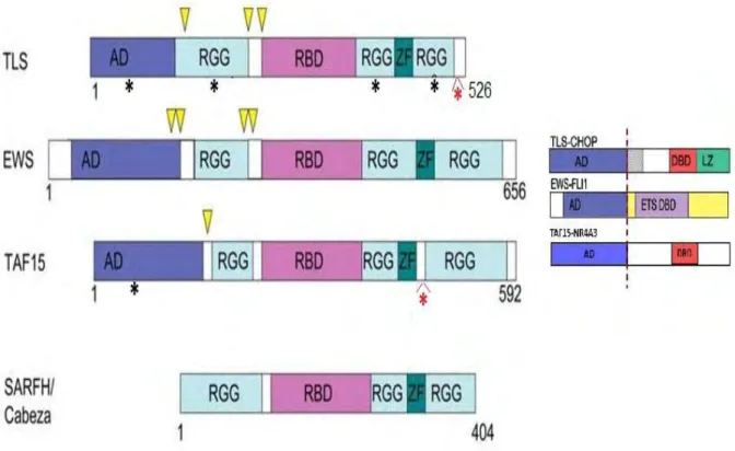
FET (FUS/TLS, EWS, TAF15) proteins constitute an ubiquitously expressed family of nuclear proteins that share extensive sequence similarity (Figure 9). The CABEZA/SARFH protein in Drosophila also shares some sequence similarity with FET proteins (Stolow and Haynes, 1995). Translocations in TLS, EWS and TAF15 genes lead to the expression of oncogenic fusion proteins, involved in uncontrolled transcriptional activation. The expression of this chimeric aberrant transcription factors seems to be the primary cause of several human cancers (for reviews on the chimeric fusion proteins and their associated phenotypes see (Kovar, 2011; Law et al., 2006; Tan and Manley, 2009)). Even if the chimeric fusion proteins are implicated in the apparition of human sarcomas, leukemias, and recently Amyotrophic Lateral Sclerosis, the functions of the endogenous proteins in the cell are still poorly understood. However, diverse associations of FET proteins with components of the transcriptional and splicing machineries suggest that they can play a role in coupling these two steps of gene expression. Nevertheless, it is important to note that FET proteins may be involved in a wide variety of processes including microRNA processing, mRNA transport, genome surveillance and DNA repair. It is also important to keep in mind that each member of the FET family may also have distinct, non-redundant functions.
Figure 9. Domain structure of FET and SARFH/CABEZA proteins. Schematic view of TLS-CHOP, EWS-FLI1, and TAF15-NR4A3 fusion proteins generated in human cancers. The N-terminal region is rich in glutamine, serine and tyrosine
aminoacids and has been shown to function as a transcription activation domain (AD) when fused to the DNA binding domain of some transcription factors (Bertolotti et al., 1999; Ohno et al., 1993; Prasad et al., 1994; Sanchez-Garcia and Rabbitts, 1994). The Arg-Gly-Gly (RGG) repeats in EWS display cis and trans repression of a broad range of activation domains (Alex and Lee, 2005). The central region contains a conserved RNA Binding Domain (RBD or RRM motif). The RBD domain of FET proteins can bind RNA and also single and double stranded DNA in vitro (Bertolotti et al., 1996). The C-terminal region contains RGG repeats, initially described as an RNA-binding motif in the hnRNP U protein (Kiledjian and Dreyfuss, 1992). Nevertheless, in the FET proteins the RGG repeats are not as closely spaced as in other RNA-binding proteins and the function of this domain is still unknown. FET proteins contain a Cys2/Cys2-type zinc finger (ZF) motif, which acts as a sequence-specific nucleic acid-binding motif as well as a sequence-specific module for protein recognition (Gamsjaeger et al., 2007). Characteristic chromosomal translocations associated with various sarcomas and leukemias lead to chimeric fusions of the activation domain of the FET genes to the DNA binding domain (DBD) of potent transcription factor genes. Arrowheads indicate the gene breakpoints. The structure of TLS-CHOP, EWS-FLI1 and TAF15-NR4A3 oncogenic fusion proteins is shown in the right. Asterisks designate domains that display mutations associated with familial Amyotrophic Lateral Sclerosis. Red asterisks indicate the regions where most mutations identified are clustered (Kwiatkowski et al., 2009; Lagier-Tourenne et al., 2010; Ticozzi et al., 2011; Vance et al., 2009).
Several studies suggest a role for FET proteins in transcription. The activation domain of FET proteins activates RNA Pol II transcription of reporter genes (see Figure 9). Because the fusion proteins lacking the RNA-binding region behave as aberrant transcription factors and this domain displays transcriptional repression activities; it is thought that the RNA binding capacity of FET proteins regulate their activity on RNA Pol II transcription. All FET proteins copurify with TFIID in different subcomplexes of this general transcription factor (Bertolotti et al., 1996; Bertolotti et al., 1998). Therefore, it is possible that they can influence transcription initiation and promoter choice within TFIID. FET proteins also bind RNA Pol II directly: for example TAF15 is associated to Rbp3, Rbp5, Rbp7 subunits of RNA Pol II (Bertolotti et al., 1996; Bertolotti et al., 1998; Zhou and Lee, 2001). Besides contacting the transcription machinery, FET proteins can also interact with transcriptional activators or repressors. EWS associates with several transcriptional activators such as Oct4, Brn3a and the CREB-binding protein (Araya et al., 2003; Lee et al., 2005; Thomas and Latchman, 2002), and regulate their activity. EWS also binds the transcriptional repressor/splicing factor SF1 (Zhang et al., 1998). TLS/FUS interacts with the DNA binding domain of various hormone receptors and is proposed to regulate their transcriptional effects (Powers et al., 1998).
As mentioned before, the RNA-binding capacity of FET proteins seems to modulate their transcriptional activity. Consistent with this, Wang et al. reported that DNA damage signals could induce a set of single stranded, low-copy-number ncRNAs, transcribed from the 5’ regulatory region of the CCND1 gene. These ncRNAs could allosterically modulate the activity of TLS. The modified TLS in turn inhibited CREB-binding protein and p300 histone acetyltransferase activities, which subsequently inhibited CCND1 transcription (Wang et al., 2008).
Taken together, these studies suggest a dual role of FET proteins on transcription by RNA Pol II: a transcriptional activity via their N-terminal domain, and a regulation of the activation domain via their RNA-binding C-terminal domain.
Recently, TLS has been shown to inhibit specifically RNA Pol III transcription
in vitro and in vivo (Tan and Manley, 2010). Therefore, FET proteins seem to have a
much broader role on gene expression than thought before.
Several studies suggest a role for FET proteins on splicing as well. All FET proteins have been found in functional spliceosome subcomplexes by different approaches (Calvio et al., 1995; Jurica and Moore, 2003; Kameoka et al., 2004; Rappsilber et al., 2002; Zhou et al., 2002). Both EWS and TLS/FUS proteins interact with several splicing factors (Knoop and Baker, 2000; Lerga et al., 2001; Shin and Manley, 2002; Yang et al., 1998; Yang et al., 2000). Furthermore, it has been shown that transient overexpression of TLS can induce alternative splicing changes in cotransfected reporters (Hallier et al., 1998; Yang et al., 1998).
More recently, transcriptome-wide studies have shown that EWS can regulate alternative splicing events of many genes relevant for response after DNA damage (Dutertre et al., 2010; Paronetto et al., 2011). By a combination of in vivo and in vitro experiments, the laboratory of Valcárcel shows that EWS associates directly to its RNA targets, preferentially in the alternatively regulated exons. ChIP and EMSA experiments also detected an association of EWS to the alternatively spliced regions of its genomic targets, in an RNA-independent manner. These associations decreased upon UV treatment, correlating with a partial relocalization of EWS into the nucleolus and alternative splicing changes. Taken together, these results suggest that EWS plays a role in splicing changes relevant for the DNA damage response. Upon UV treatment, EWS dissociates from its RNA and genomic targets and localizes transiently to the nucleolus, contributing to the cellular response to genotoxic stress.
According with this study, a transcriptome-wide identification of RNA target sites of the three FET proteins revealed that they crosslink with pre-mRNA, preferentially to the 5’ and 3’ splice sites ((Paronetto et al., 2011) Markus Landthaler laboratory unpublished results). Therefore, it is possible that FET proteins can bind pre-mRNA after activation of transcription and regulate spliceosome assembly through direct interactions with splice sites. Even if the role of EWS and FUS/TLS in splicing regulation is beginning to be well documented, the role of TAF15 in splicing is unknown. However, TAF15 crosslinks preferentially with 3’ splice sites in the pre-mRNA, and it binds specifically the U1 snRNA, so it is possible that it can function in direct or indirect splicing regulation (Markus Landthaler laboratory unpublished results and (Jobert et al., 2009)).
The many functions of FET proteins suggest that they are capable of affecting and probably connecting many cellular processes.
CHAPTER II:
THE EXPANDING UNIVERSE OF NONCODING RNA:
NONCODING RNAs ARE IMPORTANT REGULATORS
CONTROLLING mRNA PRODUCTION
A. NOVEL FUNCTIONS IN mRNA PRODUCTION
The many faces of the U1 snRNA
In vertebrates, the U1 snRNA is the most extensively characterized and according to the general believe, the structurally and functionally best understood ncRNA (see Chapter I).
However, particular features of the U1 snRNA indicate that it may have other functions in addition to splicing. To form the catalytic core of the spliceosome, the snRNPs come together in 1:1 stoichiometry (Wahl et al., 2009), but U1 is more abundant than the other snRNAs (about 5 fold more abundant than U4 snRNA), and the potential role of this excess is not known. In the mammalian nucleoplasm, in addition to be found with the other splicing snRNPs in speckles, a portion of U1 is more widely distributed (Carmo-Fonseca et al., 1992; Carmo-Fonseca et al., 1991a; Carmo-Fonseca et al., 1991b). Moreover, at an early stage of transcription inhibition, the U1 snRNA, but not the other spliceosomal RNAs, concentrate in perinucleolar caps (Carmo-Fonseca et al., 1992; Carmo-Fonseca et al., 1991a; Carmo-Fonseca et al., 1991b; Jobert et al., 2009). Strikingly, in an intronless gene, the U1 snRNP is the only U snRNP to be recruited to the transcription sites (Brody et al., 2011; Spiluttini et al., 2010), and it crosslinks to mRNA even in the absence of spliceosome formation (Wassarman and Steitz, 1993). Even biogenesis of the U1 snRNP seems to have specific features. In fact, the U1 snRNA possesses a non-canonical Sm site not functionally interchangeable with the Sm site of other snRNAs (Battle et al., 2006). Also, the recognition of snRNAs by SMN typically requires the Sm site and one stem-loop immediately downstream of it. U1 is different, since the minimal recognition sequences comprise the stem-loop near the 5’ end (Figure 8).
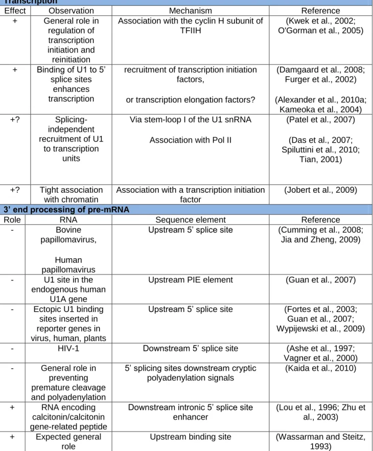
A summary of reports suggesting spliceosome-independent functions for the U1 snRNA are listed in Table 2.
Table 2. The U1 snRNA has regulatory roles independent of its canonical role in splicing. (For details see text)
Transcription
Effect Observation Mechanism Reference
+ General role in regulation of transcription initiation and reinitiation
Association with the cyclin H subunit of TFIIH (Kwek et al., 2002; O'Gorman et al., 2005) + Binding of U1 to 5’ splice sites enhances transcription
recruitment of transcription initiation factors,
or transcription elongation factors?
(Damgaard et al., 2008; Furger et al., 2002) (Alexander et al., 2010a;
Kameoka et al., 2004) +? Splicing-independent recruitment of U1 to transcription units
Via stem-loop I of the U1 snRNA Association with Pol II
(Patel et al., 2007) (Das et al., 2007; Spiluttini et al., 2010; Tian, 2001) +? Tight association with chromatin
Association with a transcription initiation factor
(Jobert et al., 2009) 3’ end processing of pre-mRNA
Role RNA Sequence element Reference
- Bovine
papillomavirus, Human papillomavirus
Upstream 5’ splice site (Cumming et al., 2008; Jia and Zheng, 2009)
- U1 site in the endogenous human
U1A gene
Upstream PIE element (Guan et al., 2007)
- Ectopic U1 binding sites inserted in reporter genes in virus, human, plants
Upstream 5’ splice site (Fortes et al., 2003; Guan et al., 2007; Wypijewski et al., 2009)
- HIV-1 Downstream 5’ splice site (Ashe et al., 1997;
Vagner et al., 2000) - General role in
preventing premature cleavage and polyadenylation
5’ splicing sites downstream cryptic polyadenylation signals
(Kaida et al., 2010)
+ RNA encoding
calcitonin/calcitonin gene-related peptide
Downstream intronic 5’ splice site enhancer
(Lou et al., 1996; Zhu et al., 2003)
+ Expected general role
Upstream binding site (Wassarman and Steitz, 1993)
U1 snRNA and transcription
Recent transcriptome-wide studies (Kaida et al., 2010), reported a general reduction of all signals from a transcript after U1 snRNA knockdown, probably reflecting a general downregulation of RNA Pol II transcription. This result is particularly resonant with several data suggesting a role of U1 in transcription regulation.
Consistent with this, the U1 snRNA was shown to associate with the Cdk7 subunit of TFIIH, a general transcription initiation factor (Kwek et al., 2002). Cdk7 was shown to phosphorylate the C-terminal repeats of the largest Pol II subunit (CTD). This phosphorylation permits the polymerase to escape from the promoter and engage in productive elongation (Akoulitchev et al., 1995; Hengartner et al., 1998; Jiang et al., 1996). Analysis of the TFIIH-dependent stages of transcription in a reconstituted system demonstrated that U1 snRNA stimulates the rate of reinitiation by RNA Pol II (Kwek et al., 2002). Deletion analysis of U1 snRNA in a reconstituted Cdk7 pull-down assay, showed that stem-loop II is essential for interaction with Cdk7. Furthermore, functional assays implicated the TFIIH-U1 snRNA interaction in the regulation of the activity of the TFIIH associated kinase, Cdk7 (O'Gorman et al., 2005).
Furger and colleagues demonstrated that 5’ splice sites proximal to the promoter of HIV constructs enhanced their expression at the transcriptional level. They demonstrated by complementary mutations, FISH and run-on analysis that the recruitment of U1 snRNA to the splice donor accounted for the transcriptional enhancement (Furger et al., 2002). They also showed that this effect was generalizable to other transcripts.
More recently, U1-mediated enhancement of transcription was analyzed in greater detail (Alexander et al., 2010a; Damgaard et al., 2008). Damgaard and colleagues demonstrated again a direct correlation between 5’ss/U1 snRNA pairing and stimulation of transcription, even in the absence of splicing. They also suggest that this effect is general as β-globin gene constructs harboring mutations in the cap-proximal 5’ss showed decreased levels of expression. By ChIP analysis, they revealed that increased transcription was not due to changes on the accessibility of chromatin but rather to an increase in the recruitment of general transcription factors.
Alexander et al. analyzed the accumulation of Env-HIV mRNA from a construct harboring WT or mutant splice sites. In a series of elegant experiments, they show again that U1 recruitment to the splice donor, even in the absence of splicing, increased transcription. The effect of U1 on transcription was independent from the promoter or SR-binding sites from exonic sequences. U1 Sm site was necessary for rescuing transcription, indicating that the U1 snRNA has to undergo
efficient biogenesis in order to increase transcription. A mutant harboring a deletion of the stem-loop II couldn’t rescue transcription either. However, mutations of the U1-70K and U1-A binding sites snRNA were not significant for enhancement of transcription. Nevertheless, the only presence of an artificial intron in the proximity of the promoter, and therefore U1 snRNP recruitment, couldn’t account for any enhancement of transcription, demonstrating that the local mRNA context is important for the transcriptional effect. Interestingly, the elongation activator Tat was able to partially rescue the expression of Env in the absence of U1 recruitment. Given the known function of HIV Tat protein in productive elongation, it is possible that U1 snRNA recruitment is required for efficient mRNA elongation. The authors proposed a model for U1 enhancement of transcription elongation: possibly via its interaction with the elongation factor Tat-SF1 (Fong and Zhou, 2001).
The major noncoding RNA associated with RNA Pol II is the U1 snRNA (Das et al., 2007; Spiluttini et al., 2010; Tian, 2001). In a mitotic cell-extract, were transcription is mainly absent, the U1 snRNA associates with RNA Pol II, suggesting that the association is independent of the nascent transcript and splicing (Spiluttini et al., 2010). Imaging studies revealed that U1 snRNA, U1-70K and ASF/SF2, but not the other spliceosomal snRNPs, are recruited to the transcription sites in the absence of splicing. The authors proposed that direct or indirect interaction of the U1 snRNA with RNA Pol II, is responsible for this observation. One could imagine that pre-association of U1 with the transcription machinery has a role in coupling of splicing and transcription, or it could influence other functions of U1.
Recently, systematic analysis of the components of the human basal transcription machinery led to the surprising discovery that a subfraction of HeLa U1 snRNA specifically interacts with the TATA-binding protein (TBP)-associated factor 15 (TAF15) (see results) (Jobert et al., 2009). This work also highlights the possibility that the U1 snRNA can have other functions within the cell, and I will further discuss about this issue in the discussion.
It is important to note that in mammals the cap-binding protein complex CBC, is implicated in U1 snRNP recognition of the 5’ splice site of the proximal intron. Therefore, U1 plays a key role in coupling the 5’ capping and splicing of precursor mRNAs (Lewis et al., 1996).
U1 snRNA and polyadenylation
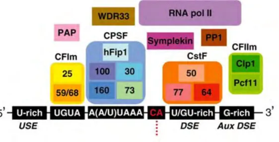
Nearly all mature eukaryotic mRNAs, with histone mRNAs being the only significant exception, contain a 3’-terminal poly-adenosine (poly(A)) tail that is important for mRNA stability, export, and translation. The poly(A) tail is posttranscriptionally added to pre-mRNAs in the nucleus by a two-step reaction called cleavage and polyadenylation. In the first step, the pre-mRNA is endonucleolytically cleaved into two molecules at the cleavage site. In the second step, a poly(A) tail is added to the 3′ end of the upstream fragment by the enzyme poly(A) polymerase, whereas the downstream pre-mRNA fragment is degraded. The reaction mechanism is relatively well understood, and the major pre-mRNA sequence elements as well as the components of the cleavage/polyadenylation machinery have been identified in both S. cerevisiae and mammalian cells (Figure
10) (for reviews, see (Danckwardt et al., 2008; Millevoi and Vagner, 2010)).
Numerous connections between the splicing and the polyadenylation machinery have been reported, and these two processes are functionally interdependent. For example, the replication-dependent histone genes lack introns, and their mRNAs are non-polyadenylated. However, insertion of an intron into a histone gene results in the formation of a polyadenylated histone mRNA, illustrating the tight coupling between splicing and polyadenylation (Pandey et al., 1990).
In a vast majority of transcripts, regulation of poly(A) tail addition involves the choice between two or more poly(A) sites, resulting in mRNAs which may differ in their stability, localization, transport and translation efficiency (for review see (Neilson and Sandberg, 2010)). A second, less common way to control poly(A) tail addition is by an “on–off” switch mechanism whereby a single poly(A) site is either active or inactive. The efficiency of poly(A) signal recognition will then determine the level of protein expression. Indeed, transcripts that are not processed at the 3’ end will be degraded or not transported to the cytoplasm.
Figure 10. Metazoan pre-mRNA 3′ end processing/polyadenylation machinery.
Known factors and cis-elements contributing to 3′ end processing of metazoan pre-mRNAs. The position of the different factors takes into account the RNA-binding specificity of each factor and, where possible, the protein contacts within the machinery. The sequence elements that comprise the poly(A) signals are indicated by black rectangles, and the site of cleavage [and subsequent poly(A) tail addition] is shown by a red dotted-line. From (Millevoi and Vagner, 2010)