Department of BiochemistryN
º……./SNV/2019
THESIS
Presented by
SaoudiSoulef
For the fulfillment of the requirements for the degree of
DOCTORATE 3
rdcycle
IN BIOLOGY
Special filed: BIOCHEMISTRY
Topic
Effect of treatment with fruits extracts on some biochemical and
antioxidant parameters in rats
Presented publically in: 05/12/2019
JURY:
President:ArrarLekhmici Pr. UFA Setif 1
Supervisor: KhennoufSeddik Pr. UFA Setif 1
Examiners: DahamnaSaliha Pr. UFA Setif 1 CharefNoureddine Pr. UFA Setif 1
Necib Youcef Pr. Univ Constantine 1
Acknowledgements
In the first, I would like to thank of all almighty God for giving the health, the patience, the power and the will for carry this thesis and complete my research.
This work has been realized in the Laboratory of Phytotherapy Applied to Chronic Diseases and was funded by the Ministry of Higher Education and Scientific Research.
I would warmly like to thank my supervisor KHENNOUF Seddik Professor in the Faculty of Nature and Life Sciences, University Ferhat Abbas Setif, Algeria for his leadership, his great patience, for the judicious advice and for his acceptance to direct and guide this work.
I would like to thank specially Hosni Karim director of laboratory of natural substances, National institute of research and physic-chemical analysis, SidiThabet 2020, Tunisia. Who have been given a chance to work in his laboratory. Thank you for the identification of phenolic compounds and for his help in provision of research facilities.
I would like also to show my sincere appreciation to member of jury for your accepted to read, to evaluate my work and to give notes and comment in order to make it a better exposition. Thank you to PrArrarLekhmici, PrDahamnaSaliha, PrCharefNourddine and PrNecibyoucef.
My sincere gratitude to DrAmranMonirahead of biochemistry laboratory of cancer Hospital of Setif for providing all the facilities to evaluate biochemical parameters.
I would like also to thank DrKrach work in the laboratory of ANAPTH in Central laboratory of University Hospital of Setif for reading and given the comment in histopathology of tissue. I thank DrBenchikhDalila, DrBentaharAssiaand DrBenchik Fatima for their help and encourangement.
My thanks are also extended to my colleagues: Nadjet, Sihem, Soulef, especially Nozha, who work with me throughout this period. I thank all the members of laboratory.
Finally, special thanks to my parents and my husband for their patience, encouragement, love and their support to achivemy objective in this research. I would like also to thank my brothers, sisters and all the members of my family.
List of publication
1. Saoudi S, Khennouf S, Amira S, Dahamna S.2017. Polyphenols contents and antioxidant
activity of Fragariaananassa, Maluscommunis, Musa paradisiacafruit extracts. Revues des
regions arides. 43(3): 601-606.
2. Saoudi S, Khennouf S, Mayouf N, Amira S, Dahamna S, Hosni Karim.2020.
Phytochemical Screening and in vivo and in vitro evaluation antioxidant capacity of
Fargariaananassa, Prunusarmeniacaand Prunuspersicafruits growing in Algeria.Progress in Nutrition. 22(1): 1-18.
List of communications
1.Saoudi S, Khennouf S, Amira S,Dahamna S. Polyphenols contents and antioxidant activity
of Fragariaananassa, Maluscommunis, Musaparadisiaca fruit extracts. Poster.5th International Meeting on Plant Biotechnology in Arid and Oasis Zones. 19-21 december, 2016, Zarzis, Tunis.
2.Saoudi S, Khennouf S, Amira S, Dahamna S, Mayouf N. Radical Scavenging, Reducing
Power and Chelating Properties of MalusCommunis. Poster.International congress of phytotherapy. 29-1 may, 2017, Monastir, Tunis.
3.Saoudi S, Khennouf S, Amira S, Dahamna S, Mayouf N, Arrar L. Bioactive compounds
and antioxidant activity of methanol extract of Musa paradisiaca. Poster.8 International sientific days on the valorisation of bioresources. 5-7 may, 2017, Monastir, Tunis.
4.Saoudi S, Khennouf S, Amira S, Dahamna S, Mayouf N, Arrar L. Polyphenols contents,
antioxidant and analgesic activity of Prunusarmeniaca fruit extract. Poster. 1st national days on the tree 25 october, 2017, M‘Sila. Algeria.
5.Saoudi S, Khennouf S, Dahamna S, Mayouf N, Amira S. La teneur en polyphenols et
l‘activité antioxydante des fruits de Malus communis, Vitisvinifera. Poster. 3 rd Internationalcongres of SAN. 28-30 november, 2017, Constantine, Algeria.
6. Saoudi S, Khennouf S, Amira S, Dahamna S. Effect of Maluscommunis against OH. , H2O2
induced cell damage. Poster.International congress of clinical genetic. 24-25 may, 2017, Setif, Algeria.
7. Saoudi S, Khennouf S, Amira S, Dahamna S, Mayouf N. Antioxidant and
anti-inflammatory activity of Fragariaananassa fruit extract. Poster. International seminar on medicinal plants .17-18 january, 2018, El-Oued, Algeria.
8.Saoudi S, Khennouf S, Amira S, Dahamna S, Mayouf N. Evaluation of bioactive cofruit
extract. Poster.At third Afirca International Allelopathy Congress.24- 26 November, 2018, Blida, Algeria.
9.Saoudi S, Khennouf S, Amira S, Dahamna S, Mayouf N. Antioxidant in vivo and in vitro
of Punicagranatum. Poster. 1st International seminar on food security and sustainable development in semi-arid Environment .8 -10 december, 2018, Setif, Algeria.
List doctorialday
1. Saoudi S, Khennouf S,Amira S,Dahamna S. polyphénol et activité antioxydante de l‘extrait
des fruits de Musa paradisiaca. Poster.Doctorial day. 31 may, 2016, Setif, Algeria.
2.Saoudi S, Khennouf S,Amira S,Dahamna S, Mayouf N. AntioxIdant activity, Sugars, tannin
and flavonols in Methanol extract of Musa paradisiaca. Poster.Doctoriale day. 09 may, 2017, Setif, Algeria.
3.Saoudi S, Khennouf S, Mayouf N. AntioxIdant activity, polyphenols, flavonoids and
flavonols in Methanol extract of Prunuspersica. Poster.Doctorial day. 09 may, 2018, Setif, Algeria.
صخلملا
شػأقاحىاىيعحٞىاعجسذقحطشْىاحْٞٞجسملأاعاّ٘لاانيَذ ,حٝ٘يخىاذاّ٘نَىاحفاماثٝشقرسَذسا عاشٍلااٍْشٞثنىاثاذحاٞفإسٗدشسفٝاٍَ فذيىاًذجاٍَٖٗذثٝجذسملأىجداؼٍحٞجساخداَ٘ثَسجىاذٝٗضرّئفازٖىٗ، .جساؼىاحْٞٞجسملأاعاّ٘لاإزٕذؼعا شثرعذ ٕزٖفذٖذ .جشحىا سٗزجىا ٓزٖى ٛذسمأرىاسشؼىاَْييقرّأْنَٞٞرىاٗ حٞجساخىاجذسملأاذاداؼَىاٍَٖاسذظَٖما٘فىا حساسذىا حٞؽاشْىاٗحٞىْ٘ٞفىاذاثمشَىاؼعثفٝشعذ ازم٘ذاْٞذٗشثىا ٘ذاٝشنسىاٗ غاتذىا ٗ خاذّٝ٘٘فلافىاٗ هْ٘فىا خاذٝذع ٙ٘رحٍشٝذقرىىإ فلارخا كإْ ُاٞئاَٞنىا وٞيحرىاشٖظأ .شئاضجىا ٜف حنيٖرسَىاٖما٘فىاٍْذٝذعييٞىّ٘اثَٞىاظيخرسَىذثنىاحَٞسٗجذسملأىجداؼَىا رعٝٗ .ٔما٘فىا ِٞت شٞثم حىٗاشفىا ضيخرسٍ ُا جئارْىا خشٖظأ .خاظيخرسَىا وم ٜف د٘جَ٘ىا ٜسٞئشىاثمشَىانٞيغىاؼَح شث جشثرعٍحَٞنٝ٘رحٝ ذجٗاَم .ُاٍشىا ضيخرسٍ ٜف جذجا٘رٍ دّامشثملأا غاتذىا حَٞم اَْٞت .خاذّٝ٘٘فلافىا ٗ هْ٘فىا خاذٝذع ٍِ ىا حٞؽاشْىا شٝذقذ ٌذ .ِٞرىا ساَث ٜف خاٝشنسيى ٜىاعىا ٙ٘رحَىا ساثرخا هاَعرسات جذسملأى جداؼَ DPPH ٗ ABTS ٗ ًاظّٗ حٞعاجسلاا جسذقىا ٗ ذٝذحىا بلاخرسا β -.لٞرٞتسات٘ٞرىا غَح ٗ ذٝذحىا دّٞاٞس٘ٞذٗ ُاذٗسام حىٗاشفىا حٖماف ُأ ذجٗ اَم .ذٝذحىا بلاخرسا ٜف جسذقىا ٜف وؼفلأا ِٞرىا شثرعٝ اَْٞت .جشحىا سٗزجىا ؽاقرىا ٚيع جسذقىا ٜف وؼفلاا ٜٕ حٖماف ليَذ خاعشجت اٝ٘ٞح جذسملأى جداؼَىا حٞؽاشْىا شٝذقذ ٌذ .خاذٞثٞيىا جذسمأ ؾٞثثذ ٚيع حٞىاع جسذق حىٗاشفىا 200 ٗ ػم/عٍ 000 جذَى خاظيخرسَىات حيٍاعَىا ذعت ُارشجىا ذْع ػم/عٍ 11 داٖجلاا خاششؤٍ ٗ اٍصلاثىا ٜف جذسملأى جداؼَىا جسذقىا ٚيع اٍ٘ٝ ىا ٗ ذثنىا ٜف ٛذسمأرىا ذْع خاظيخرسَىا عَٞج ُا جئارْىا خشٖظأ .اٍصلاثىاتحٞئاَٞم٘ٞثىا خاششؤَىا غعت ٚيع ٗ ٚين حعشج 000 هاَعرسات حٝذثنىا حَٞسىا شٝذقذ ٌذ حساسذىا ٓزٕ ٜف .ٛذسمأرىا داٖجلاا خاششؤٍ ٚيع اٝ٘ق اشٞثأذ اٖى ػم/عٍ شٞثم ونشت شثؤٝ ةمشَىا ازٕ ُا جئارْىا خشٖظا .ُ٘تشنىا ذٝس٘يم ٜعاتس وم خاظيخرسَت حيٍاعَىا خدأ اَم .ذثنىا ٚيع ِٞرعشجىا ءاطعا ٙدأ .ةمشَىا ازٖى ًاسىا شٞثأرىا ٍِ حٞينىاٗ ذثنىا جٞسّ حٝاق٘ىىإ ٔما٘فىا 200 ٗ 000 ضيخرسٍ ٍِ ػم/عٍ ٞثىا خاششؤَىا غعت ٚيعٗ ٛذسمأرىا داٖجلإا خاششؤٍ ٚيع شٞثأرىا ٚىإٗ ذثنىا ٚيع ٜئاقٗ شٞثأذ ساٖظا ٚىإ ِٞرىا حٞئاَٞم٘ ٜقاٗٗ جذسملأى داؼٍ ؽاشّ ليرَذ حيَعرسَىا ٔما٘فىا خاظيخرسٍ ُأ حساسذىا ٓزٕ جئارّ ٍِ ضيخرسّٗ .اٍصلاثىا ٜف .خاذّٝ٘٘فلافىا ٗ ٜىْ٘ٞفىا ٙ٘رحَىات اطثذشٍ حطشّلأى ةيحاتفملا تاملكلا : .حٝذثنىا حَٞسىا ،ٛذسمأرىاداٖجلاا،خاذّٝ٘٘فلافىا ،هْ٘فىا خاذٝذع،ٔما٘فىا خاظيخرسٍRésumé
Les espèces réactives oxygénées (ERO) ont une grande capacité d‘endommager presque tous les types de constituants cellulaires. Ce qui explique leur implication dans l‘induction de plusieurs pathologies. La supplémentation de l‘organisme par des antioxydants exogènes s‘avère très utile pour lutter contre ces espèces nocives. Les fruits sont une source importante d'antioxydant exogène qui peut minimiser les dommages oxydatifs. Le but de cette étude est d'évaluer les teneurs en polyphénols, flavonoïdes, sucres, tannin et protéines, l'identification de certains composés phénoliques, l'activité antioxydante et l'hépatotoxicité d'extraits méthanoliques de plusieurs fruits consommes en Algérie. L'identification par UPLC et HPLC / MS a montré une forte différence entre les extraits de fruits. L'acide gallique est le principal composé dans tous les extraits de fruits études. Les résultats ont révélé que l‘extrait de
Fargariaananassacontient une grande quantité de polyphénol, de flavonoïde et de protéine.
Le contenu élevé de tannin se trouve dans l‘extrait de Punicagranatum. Tandis que, le contenu élevé de sucres est trouvé dans le Ficus. L'activité antioxydante a été évaluée in
vitropar les tests de DPPH, d l'ABTS, radical hydroxyle, pouvoir réducteur, chélation des ions
ferreux, système β-carotène / acide linoléique, la méthode de thiocyanate ferrique et la méthode à l'acide thiobarburique. Les fraises présentent l‘extrait le plus active dans l‘activité scavenger des radicaux libres. Cependant, les figues sont le meilleur chélateur comparé aux autres fruits. Fargaria peut inhiber la peroxydation lipidique de différentes manières. L'activité antioxydantein vivoa été évaluée par la voie orale chez le rat pendant 14 jours(200 et 600 mg / kg) sur la capacité antioxydante plasmatique (PAC), les marqueurs du stress oxydatif dans le foie et les reins et certains paramètres biochimiques. Les résultats ont montré que tous les extraits à 600 mg / kg ont un bon effet contre le stress oxydatif. Dans la présente étude, l‘effet hépatoprotecteur des extraits de fruits a été évalué chez des rats intoxiqués par CCl4. Les résultats ont montré que cet agentinduisait une hépatotoxicité remarquable. Tous les
extraits de fruits ont un effet protecteur important et réduisent les dommages aux tissus hépatiques et rénaux. L'administration de 200 et 600 mg / kg de Ficus carica a montré un effet hépatoprotecteur important, il améliore les marqueurs du stress oxydant (GSH et MDA) et les paramètres biochimiques hépatiques, les résultats sont confirmés par un examen histologique. En conclusion, les extraits de fruits examinés dans cette étude ont montré une très bonne activité antioxydante, liée à leur teneur en polyphénols et flavonoïdes.
Abstract
Reactive oxygen species (ROS) have high potential to damage almost all types of cellular constituents, which explains their involvement in the induction a number of pathologies. The supplementation of the body by exogenous antioxidants seems to be very helpful to fight these harmful species.Fruits are an important source of exogenous antioxidant which can minimize oxidative damage. The aim of this studyis the evaluationpolyphenols, flavonoids, tannin, sugars and protein contents, the identification of some phenolic compounds, the antioxidant and hepatotoxicity activity of methanolic extracts of the several fruits consumed in Algeria.The identification of phenolic compounds by UPLC and HPLC/MS showed deference between fruits extracts. Gallic acid is the major compounds present in all fruits extracts. The results revealed that Fargariaananassacontains high amount of polyphenols, flavonoids and protein. The high level of tannin was found in Punicagranatum.While, the high level of sugars was found in Ficus.The antioxidant activity was evaluated in vitro using the DPPH, ABTS, hydroxyl radicals,reducing power, ferrous ion chelating tests, β-carotene/ linoleic acid model system, ferric thiocyanate method and thiobarburic acid method. Strawberries extracts were the best one can scavengers of free radicals.However Ficusextracts was the better chelator compared to other fruits. Fargariacan inhibit lipid peroxidation by deferent ways. The antioxidant activity of extracts at doses of 200 and 600 mg/kgin vivowas assessed in rats for 14 days, on plasma antioxidant capacity (PAC), oxidative stress markers in liver and kidney and on some biochemical parameters. Results showed that all extracts at 600 mg/kg were effectives against stress oxidative. The hepatoprotective effect of fruits extracted was evaluated in rats intoxicated by CCl4. Results showed that CCl4induced
remarkable hepatotoxicity. All fruits extracts have an important protective effect and reduce hepatic and renal tissues damage induced by this toxic agent. The administration of 200 and 600 mg/kg of Ficuscaricashowed a significant hepatoprotective effect, it improve the oxidative stress markers (GSH and MDA) and hepatic biochemical parameters and this results confirmed by histological examination. In conclusion, Fruit extracts examined in this study showed a very high antioxidant activity and hepatoprotective properties which wererelated to their contents in polyphenols and flavonoids.
Lists of tables
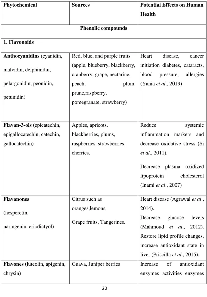
Table 1: Sources and Potential Effects on Human Health of Some Phytochemicals………20
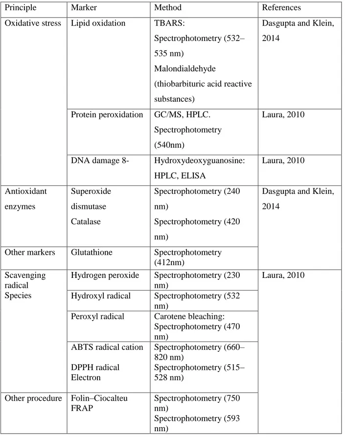
Table 2: Methods for assessing antioxidant activity in vivo and in vitro………26
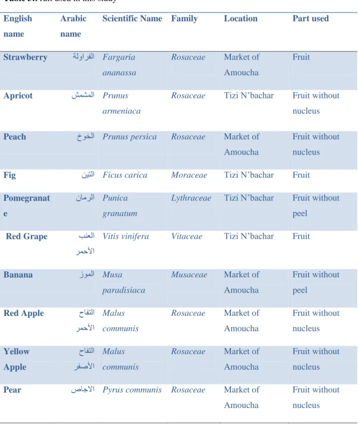
Table 3: Fruit used in this study………...34
Table 4: Total phenolics, flavonoids and tannins contents in fruits extract……….55
Table 5: Total sugars, protein contents of fruits extract………...57
Table 6: Retention time, maximum UV absorption (λmax), mass spectral data and tentative identification of the phenolic compounds in the methanol extract of Fargariaananassa……67
Table 7: Retention time, maximum UV absorption (λmax), mass spectral data and tentative identification of the phenolic compounds in the methanol extract of Prunusarmeniaca…….68
Table 8: Retention time, maximum UV absorption (λmax), mass spectral data and tentative identification of the phenolic compounds in the methanol extract of Prunuspersica………...70
Table 9: Retention time, maximum UV absorption (λmax), mass spectral data and tentative identification of the phenolic compounds in the methanol extract of Ficuscarica……….71
Table 10: Retention time, maximum UV absorption (λmax), mass spectral data and tentative identification of the phenolic compounds in the methanol extract of Punicagranatum……....72
Table 11: Retention time, maximum UV absorption (λmax), mass spectral data and tentative identification of the phenolic compounds in the methanol extract of Vitisvinifera…………....73
Table 12: Retention time, maximum UV absorption (λmax), mass spectral data and tentative identification of the phenolic compounds in the methanol extract of Musa paradisiaca…….73
Table 13: Retention time, maximum UV absorption (λmax), mass spectral data and tentative identification of the phenolic compounds in the methanol extract of Maluscommunis……….74
Table 14: Retention time, maximum UV absorption (λmax), mass spectral data and tentative identification of the phenolic compounds in the methanol extract of Maluscommunis………75
Table 15: Retention time, maximum UV absorption (λmax), mass spectral data and tentative identification of the phenolic compounds in the methanol extract of Pyruscommunis……....76
Table 16:Comparison between different fruits extracts in plasma antioxidant capacity using DPPH radical and reducing power activity………...97
Table 17:The effect of fruits extracts on hepatic MDA and GSH level and renal MDA and GSH level in mal Wister rats treated for 15 days………...100
Table 18: Effect of fruits extracts on hepatic biochemical parameters in male Wistar rats treated for 15 days………...102
Table 19: Effect of fruits extracts on renal and some biochemical parameters in male Wistar
rats treated for 15 days………...……….103
Table 20:Effect of fruits extracts on plasma antioxidant capacity. ………..105 Table 21: The effect of fruits extracts on hepatic MDA and GSH level and renal MDA and
GSH level in mal Wister rats treated for 7 days……….107
Table 22: Effect of fruits extracts on biochemical parameters in male Wistar rats treated for 7
days after injected by CCl4………...109
Table 23: Effect of fruits extracts on renal function and some biochemical parameters in male
List of figures
Figure 1: Overview of the reactions leading to the formation of ROS……….8
Figure 2: Overview of free radical damage ………11
Figure 3: Anatomy of the liver and hepatic microstructures………...28
Figure 4: Anatomy of kidney………...30
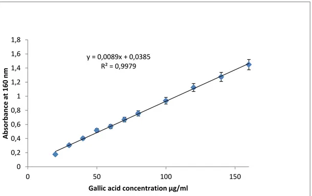
Figure 5:Standard curve of gallic acid for the determination of total polyphenols in fruits extracts. ………...……….36
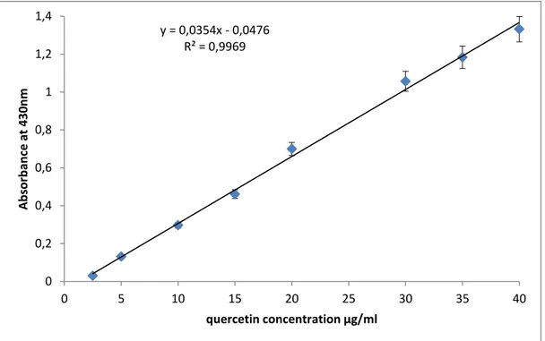
Figure 6:Standardcurve of quercetin for the determination of flavonoids in fruits extracts………...………...37
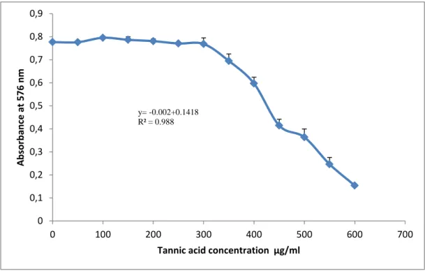
Figure 7:Standard curve of tannic acid for the determination of tannins in fruits extracts………..38
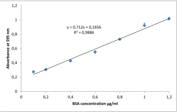
Figure 8:Standardcurve of BSA for the determination of protein in fruits extracts. ………...………39
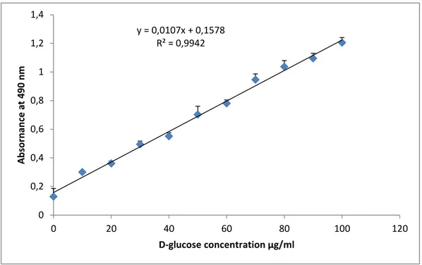
Figure 9:Standardcurve of D-glucose for the determination of sugars in fruits extracts. Each ………...40
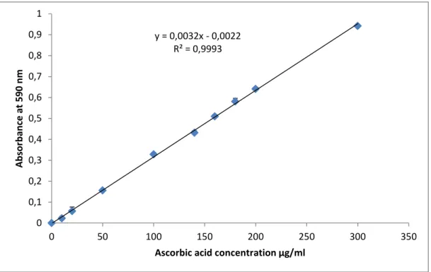
Figure 10: Standard curve of ascorbic acid for the determination of Total Antioxidant Capacity in fruits extracts. ………...………42
Figure 11: Standard curve of reduced glutathione (GSH)………..…………..52
Figure 12: Malondialdehyde assay………..53
Figure 13: UPLC chromatogram of Fargariaananassa fruit methanol extract………..59
Figure 14: UPLC chromatogram of Prunusarmeniaca fruit methanol extract………...60
Figure 15: UPLC chromatogram of Prunuspersica fruit methanol extract……….60
Figure 16: UPLC chromatogram of Maluscommunis fruit methanol extract………..61
Figure 17: UPLC chromatogram of Pyruscommunis fruit methanol extract………...61
Figure 18: UPLC chromatogram of Ficuscarca fruit methanol extract………..62
Figure 19: UPLC chromatogram of Punicagranatum fruit methanol extract………...63
Figure 20: UPLC chromatogram of Vitisvinifera fruit methanol extract………64
Figure 21: UPLC chromatogram of Musa paradisiaca fruit methanol extract………...65
Figure 22: Total antioxidant capacity of fruits extracts………...77
Figure 23: A comparison between different fruits extract in DPPH free radical scavenging activity………...79
Figure 24: A comparison between different fruits extract in ABTS free radical scavenging activity………...80
Figure 25: A comparison between different fruits extract in metal chelating activity………83 Figure 26: A comparison between different fruits extract in hydroxyl radical scavenging
activity………...85
Figure 27: Reducing power of different fruits extracts………87 Figure 28: A comparison between different fruits extracts in reducing power assay………..88 Figure 29: Antioxidant activities of different plant extracts measured by β-carotene bleaching
method………...90
Figure 30: Antioxidant activities of different extracts measured by FTC method……….…..92
Figure 31: Antioxidant activities of different plant extracts measured by TBA
method………...94
Figures 32:Histopatological of liver………..112 Figures 33:Histopatological of kidney………...116
List of abbreviations
ABTS: 2, 2′-azino-bis (3-ethylbenzthiazoline-6-sulfonic acid) AD: Alzheimer‘s disease
ALP: Alkaline phosphatase
ALT:Alanine aminotransferase
AST: Aspartate aminotransferase
ATP: Adenosine triphosphate
BHT: Butylatedhydroxytoluene DAD: Diode array detector
DNA: Deoxyribonucleic acid
DPPH: 2, 2-diphenyl-1-picryl-hydrazyl
EDTA:Ethylene diamine tetra acetic acid
ELISA:Enzyme-linked immunosorbent assay FAD: Flavin adenine dinucleotide
FRAP:Ferric reducing ability of plasma
GAE: Gallic acid equivalents
GC/MS:Gas Chromatography coupled to Mass Spectrometer
GGT:γ-Glutamyltransferase GPX: Glutathione peroxidase
GR: Glutathione reductase
GSH: Glutathione
GSSG: Glutathione disulfure oxidized
GST: Glutathione-S- transferase H2O2: Hydrogen peroxide. I%: Inhibition percentage.
IC50%: Inhibitory concentration for 50% of activity
MDA:Malondialdehyde
NAD: Nicotinamide adenine dinucleotide.
NOS: Nitric oxide synthase
NOX: NADPH oxidase.
PDA: Photodiode array detector
QE: Quercetin equivalents RNS: Nitrogen reactive species
ROS: Oxygen reactive species
SEM: Standard error of the mean
SOD: Superoxide dismutase
TAE: Tannic acid equivalents TAC: Total antioxidant capacity
AU: Uric acid
XDH: Xanthine dehydrogenase
List of contents
Introduction
………..1Review of literature
1.Oxidative stress
.………...3 1.1 Definition………..3 1.2 Free radicals………..31.3 Formation of free radicals……….3
1.3.1 Internal sources of free radicals………..3
1.3.2 External sources of free radicals……….5
1.4 Oxidative stress and pathologies associated to free radicals ………8
1.5 Antioxidant ………...12
1.5.1 Enzymatic antioxidants……….12
1.5.2 Non enzymatic antioxidants………..13
1.6 Some fruits rich in flavonoids and poyphenols………...19
1.6.1 Antioxidant activity of polyphenols and flavonoids in fruits ………..22
1.6.2 Effects of polyphenols and flavonoids on some biochemical parameters ………...23
1.7 Assay of antioxidant activity in vitro………..……24
1.8 Assay of antioxidant activity in vivo………...25
2. Study the toxicity of extracts………27
2.1 Hepatoprotective andHepatotoxicity of plants extracts and natural product ………...….27
2.1.1 Structure and function of the liver………...…27
2.1.3 Hepatoprotectives agents………..29
2.2 Nephroprotective and nephrotoxicity of plants extracts and natural product …………....29
2.2.1 Structure and function of the kidney……….29
2.2.2 Nephrotoxicity………..31
2.2.3 Nephroprotectiveproperties of natural products………...31
Materials and methods
1. Materials……….331.1 Fruits samples……….33
1. 2 Fruits used in this study……….33
1.3 Animals………...35
1.4Chemicals………35
2. Methods………35
2.1 Preparation of fruits extracts………...35
2.2Determination of total polyphenols……….35
2.3 Determination of total flavonoids ………..36
2.4 Determination of total tannin……….37
2.5 Determination of protein in fruit extracts ………..38
2.6 Total soluble content in fruit extracts ………39
2.7 Determination of polyphenols and flavonoids by UPLC-DAD………..40
2.8 Determination of polyphenols and flavonoids by HPLC/MS……….40
2.9 determination of the in vitro antioxidant activity of fruits extracts………41
2.9.1 Phosphomolybdate assay………...41
2.9.3 Free radical scavenging ability by ABTS .+………..42
2.9.4 Ferrous ion chelating activity of the extracts ………43
2.9.5 Hydroxyl radical scavenging assay……….43
2.9.6 Reducing power………..44
2.9.7 β-Carotene bleaching assay……….44
2.9.8 Ferric thiocyanate (FTC) method………44
2.9.9Thiobarbituric acid (TBA) method………..45
2.10 Determination of the in vivo antioxidant activity of fruits extracts………..45
2.10.1 Experimental design……….45
2.11 Effect of fruit extract on hepatotoxicity induced by CCl4………46
2.11.1 Effect of fruits extracts on biochemical parameters………..47
2.11.2 Histopathological examination of liver and kidney ……….50
2.11.3 Effect of extracts on plasma antioxidant capacity using DPPH radical…………50
2.11.4 Effects of extracts on plasma reducing power……….…...51
2.11.5 Preparation of liver and kidney homogenate………51
2.11.6 Effect of extracts on GSH concentration ……….51
2.11.7 Effect of extracts on lipid peroxidationin tissues homogenate ………52
2.12 Statistical analysis……….53
Result and discussion 1. Total polyphenols, flavonoids and tannin content in the extracts……….54
2. Sugars and protein content in the extracts………....56
3.1 Identification of different polyphenolic and flavonoids in fruits extracts by UPLC- DAD.
………...58
3.2 Identification of different polyphenolic and flavonoids in fruits extracts by HPLC-PDA/MS (LC/MS)………65
4. In vitroAntioxidant activity of fruits extracts ………...76
4.1 Total antioxidant capacity ………. 76
4.2 DPPH and ABTS Radical scavenging activity………...78
4.3 Metal chelating activity of extracts ………...82
4.4 Hydroxyl radical scavenging activity of fruits extracts………..84
4.5 Reducing power capacity of fruits extracts……….86
4.6 Antioxidant activity of fruit extracts using β-carotene bleaching assay……….89
4.7 Antioxidant activity of fruit extracts using FTC method………90
4.8 Thiobarbituic acid (TBA) assay ……….92
5. In vivoAntioxidant activity of fruit extracts ………...95
5.1 Plasma antioxidant capacity………95
5.1.1 Plasma antioxidant capacity using DPPH radical scavenging activity in rats …………95
5.1.2 Effects of extracts on plasma reducing power in rats ……….96
5.2 Effect of extracts on lipid peroxidation in liver and kidney homogenate ………..98
5.3Effect of fruits extracts on GSH level in liver and kidney homogenate………...……..98
5.4 Effect of fruits extracts on biochemical parameter and in vivo antioxidant activity ……101
5.4.1 Effect fruit extract on hepatic biochemical parameters………..101
5.4.2 Effect of fruits extracts on renal and some biochemical parameters……….101
6. Examination of hepatotoxicity and Nepherotoxicity induced by CCl4and protective effect of fruit extract……….104
6.1 Plasma antioxidant capacity………..104
6.1.1 Effect of extracts on plasma antioxidant capacity using DPPH radical and reducing power after CCl4 induced toxicity ……….……….104
6.2 Effect of fruits extracts on GSH and MDA levelin liver and kidney ………..106
6.3 Effect of fruits extracts on hepatic biochemical parameter after hepatotoxicity induced by CCl4……….108
6.4 Effect of fruits extracts on renal function and some biochemical parameter in rats after CCl4 injection ……….109
6.6 Histopatological examination of liver and kidney and the protective effect of fruit extracts ………....112
6.6.1 Histopatological of liver……….112
6.6.2 Histopatological of kidney……….115
Conclusionand perspectives
………..………...119Introduction
Oxygen is a very importantmolecule in the body. It is used in many biological systems such as mitochondria, which reduce the O2in the synthesis of the adenosine triphosphate
(ATP), during this reaction free radicals were product.
Free radicals are instable molecules contain one or more unpaired electron in their externe orbital. Free radicals are produced by endogenous or exogenous sources.Reactive species are compounds related to two types ofmolecules, the reactive oxygen species (ROS) and the reactive nitrogen species (RNS). Free radical produced by the body in the low level but overproduction of these molecules induced oxidativestress.
Oxidative stress is a status of an excessive production of reactive oxygen species in the organism and the low concentration of antioxidant. It has been demonstrated that reactive oxygen species (ROS) are key players in the hepatic and kidney pathophysiologic changes caused by chemicals.
The oxidative stress is associated with various diseases such as hypertension, cardiovascular disease, atherosclerosis, diabetes, cancer and arthritis. To deal with these species, the body is equipped with an effective defense system, which includes: enzymes such as superoxide dismutase (SOD), catalase (CAT), glutathione peroxidase (GP), and glutathione reductase (GR); high-molecular-weight antioxidants such as albumin, ceruloplasmin, and ferritin; and low-molecular-weight antioxidants such as ascorbic acid, α-tocopherol, β-carotene, glutathione (GSH) and uric acid.
Carbon tetrachloride (CCl4) has been widely used for the induction of liver and kidney
damage in experimental animals. CCl4 is converted by the cytochrome P450 in the CCl3OO*
radical. This induced an increase of the level of peroxyde radical. This radical is responsible for lipid peroxidation.
Polyphenols effects on health are based on the results obtained from bioactivity studies, which in turn, has increased the interest in the consumption of foods rich in polyphenols. Such bioactive effects include antioxidant properties, prevention of oxidative stress associated diseases like cardiovascular, neurodegenerative diseases and cancer and their role in long term health protection by reducing the risk of chronic and degenerative diseases. Fruits and vegetables are rich sources of some micronutrients (vitamins, minerals), fibers and a wide array of phytochemicals that individually or in combination benefit human health.Several studies have linked vegetable consumption, especially fruits with a reduced risk for cancer
and cardiovascular disease, thus characterization of polyphenols is essential to increase the knowledge on fruits contents and their related beneficial effects.
The present study was conducted in order to identify polyphenols compounds and evaluate the in vivo and in vitro antioxidant activity and the protective effect of some fruits growing in Algeria and consumed largely in Setif on some oxidative stress indicators and some biochemical parmeters.
The aim of this study is:
Phytochemical analysis of fruit extracts by UPLC/DAD and HPLC/MS (LC/MS) to identify bioactive compounds in these fruits.
Determination of polyphenols, flavonoids, tannin, sugars, and protein contents in deferent methanolic fruits extracts.
Evaluation of the in vitro antioxidant activity of extracts using different assay (DPPH, ABTS, reducing power, chelation of iron, β-carotene and peroxidation lipidic).
Evaluation of the in vivo antioxidant activity of fruits extracts.
Evaluation of MDA and GSH levels in the liver and kidney of rats, treated with fruits extracts. Evaluation of some biochemical parameters levels in plasma of rats treated with fruits extracts Evaluation of protective effect of fruits extracts against hepatotoxicity and nephrotoxicity induced by CCl4.
3
1 Oxidative stress
1.1 Definition of oxidative stress
Oxidative stress defined as a disturbance in the balance between the production of reactive oxygen and nitrogen species and antioxidant defenses (Cabello-Verrugio et al., 2017). This disturbance is resulted from the overproduction of free radicals and the low level of antioxidant (Gammaz et al., 2018).
1.2 Free radicals
Free radicals are defined as molecules formed in the human body under physiologic and pathologic conditions, and having an unpaired electron in the outer orbit, generally is unstable and very reactive (Fang et al., 2002; Labo et al., 2010) such as oxygen reactive species (superoxide, hydroxyl, peroxyl (RO2•), alkoxyl (RO•), and hydroperoxyl (H2O2•) radicals.
Examples for species nitrogen reactive (Nitric oxide and nitrogen dioxide (•NO2)). ROS and RNS include radical and non-radical species (Evans and Halliwll, 2001). Oxygen and nitrogen radicals can be transformed to non-radical, such as hydrogen peroxide, hypochlorous acid (HOCl), hypobromous acid (HOBr) and peroxynitrite (ONOO) (Kumar, 2011).
1.3 Formation of free radical
Free radicals are molecules occur by several mechanisms including endogenous and exogenous sources.A potential endogenous source of reactive oxygen species is the mitochondrial electron transport chain, inflammation, excessive exercise, ischemia, infection, cancer and aging (Hrycay and Bandiera, 2015). Exogenous free radical are the result of air and water pollution, cigarette smoke, alcohol, transition metals, certain drugs, industrial solvents, and radiation (Hrycay and Bandiera, 2015).
1.3.1 Internal sources of free radicals
ROS and RNS mainly produced by NADPH oxidases (Nox), Xanthine oxidoreductase, mitochondrial respiratory chain enzymes and NO synthases (NOS) (Liguori et al., 2018).
A) Mitochondrial respiratory chain enzymes
Mitochondria have important role in the number of fundamental function such as: respiration and oxidative energy production, regulation of the intracellular calcium concentration and
4
control of the fatty acid 𝛽-oxidation. The major role of mitochondria is the production of energy. They used about 95% of oxygen to produce ATP by the oxidizing substances contained in food by transfer of electrons to electron carriers such as NAD+ and FAD (Navarro and Boveris, 2007).
The electron is transformed by the Complexes I and II carriers in the inner mitochondrial membrane associated with the formation of ROS. The first ROS produced by the
mitochondria is the superoxide radical anion by 2% of oxygen. This converted to H2O2 in the
presence of superoxide dismutase (SOD). The hydrogen peroxide is turned in the hydroxyl radical via the Fenton reaction (Mohamed et al., 2015; Di Meo et al., 2016). As described in the following reactions:
O2 + e- O2●-
2O2●- + 2H+ SOD H2O2
Fe2+ + H2O2 OH ●
+ OH- + Fe+3
B) NADPH oxidases (Nox)
NADPH oxidase (NOX) plays a pivotal role in the production of ROS, it catalyze the conversion of oxygen to superoxide (Muzza and Fugazzola, 2017)
2O2 + NADPH 2O2.- + NADP+ + H+
NADPH oxidase (NOX) is a transmembrane enzyme and there are a number of isoforms: Fibroblasts, endothelial cells, vascular smooth muscle cells, cardiac monocytes and thyroid tissue nonphagocytic (NOX1-5 and Duox1-2).Muscle cells and fibroblasts account for the majority of O2•‐ produced in the normal vessel wall . The different NOX isoforms
in humans are involved in a wide range of cellular processes, including apoptosis, host defence, cellular signal transduction, oxygen sensing and angiogenesis. Although NOX is ubiquitously expressed the distribution of different isoforms is cell or tissue specific, allowing each NOX a distinct physiological and pathological function (Giardino et al., 2017).
C) Xanthine oxidoreductase
Xanthine oxidoreductase is a flavoprotein that contains molybdenum, nonheme iron and labile sulfur. The enzyme is present in two forms, one with dehydrogenase activity (xanthine dehydrogenase XDH) and the other with oxidase activity (Xanthine oxidase XO). XDH can
5
be converted to XO either by reversible thiol oxidation or by irreversible proteolytic cleavage (Hare et al., 2008). During the reactions of the oxidation of hypoxanthine to xanthine, and the oxidation of xanthine to uric acid, XOR normally produces two molecules of hydrogen peroxide and two molecules of superoxide. This enzyme plays an important role in the catabolism of purines in some species including human (Laurindo, 2018).
Hypoxanthine + H2O + O2 → xanthine + H2O2 Xanthine + H2O + O2 → uric acid + H2O2.
D) Nitric oxide synthases (NOS)
Nitric oxide (NO•) and L-citrulline are produced from a guanidine nitrogen of L‐arginine via electron transfer from NADPH in two successive steps. The enzyme responsible for this reaction exists in three isoforms: neuronal (nNOS, type I, NOS‐I or NOS‐1), endothelial (eNOS, type III, NOS‐III or NOS‐3) and inducible (iNOS, type II, NOS‐II or NOS‐2). Although all three NOS isoforms (two constitutively-expressed isoforms; neuronal (nNOS) and endothelial (eNOS) and one inducible isoform; (iNOS) produce identical products, the function of the NO varies widely in terms of physiological functions due to the varied localization of the isoforms within different cell populations of the body (Miller, 2004). nNOS and eNOS are constitutively expressed, but their activity is regulated by the
intracellular Ca2+ concentration. nNOS exhibits NADPH‐diaphorase (NADPH‐d) activity
(Mohammed et al., 2015).
1.3.2 External sources
Reactive oxygen species can be produced by exogenous processes. The environmental sources include ionizing radiation and pollutants such as chemicals that promote the formation of superoxides such as quinones, nitroaromatic and herbicides (Elkhateeb and Alshammary, 2018)
A) Cigarette smoke
Cigarette smoke contained 4800 compounds, approximately 400-500 compounds present in the gaseous phase of Cigarette smoke. Its composition is complex, it contains carbon monoxide, nitrogen oxide, hydrogen cyanide as well as many volatile organic compounds and particulate matter. Various compounds may be associated with particulate matter, such as nicotine, tobacco-specific nitrosamines and polyaromatic hydrocarbonsmany (Baker, 2006)
6
.The vapor phase and particules matter of cigarette smoke contains free radicals including reactive oxygen species, reactive nitrogen species, superoxide, hydroxyl radicals, nitric oxide and hydrogen peroxide. These compounds can enter the bloodstream and generating ROS especially through interact with enzyme such as NADPH oxidase, then the result is stress oxidative is increased (Tostes et al., 2008). Semiquinone radicals associated with tar can reduce oxygen to produce superoxide as well as hydrogen peroxide and hydroxyl radicals. Moreover cigarette contains metal ion which can generate hydroxyl radicals from hydrogen peroxide (Pryor, 1997). It damages the liver, causes cirrhosis, damages the heart, causes myocardiopathy and damages the brain. All these free radicals that damage the lung so that almost every smoker gets bronchitis and emphysema by time (Pham-Huy et al., 2008).
B) Metal ions
Heavy metal ions, such as iron, copper, cadmium, Mercury, can induce the generation of reactive radicals and cause cellular damage via depletion of enzyme activities through lipid peroxidation and reaction with nuclear proteins and DNA (Birben et al., 2012).
C) Air pollution
Air pollution contains gaseous phase, particulate matter, Ozone, nitrogen oxide, carbon monoxide and sulfurdioxide. Particulate matter can penetrate deep into the respiratory tract and can cause cytotoxicity by inducing oxidative stress, which may lead to oxidative damage of DNA, mutagenesis, and stimulation of proinflammatory factors. In addition to oxidative stress (due to oxygen free radicals), air pollution may also produce nitrosative stress (stress caused by reactive nitrogen species such as nitrous oxide) (Oh et al., 2011).
D) Radiation
Ionizing radiation such as UV, X or gamma ray produced hydroxyl radical, superoxide and organic radicals (Gaston, 2016). Free radicals, especially reactive oxygen species, can be generated by UV radiation that can damage DNA. Therefore, air pollution may act in synergy with UV radiation of sunlight in causing skin damage through the production of oxidative stress (Burke and Wei, 2009; Pfeifer and Besarantinia, 2012).
7 E) Dietary factors
Additives, alcohol, foods that have been barbecued, fried, grilled, foods that have been browned or burned, hydrogenated vegetable oils, processed foods containing high levels of lipid peroxides can also produce free radicals (Birben et al., 2012).
Alcohol consumption and alcoholism is one important cause of an acute illness and chronic disease worldwide. It is also observed that excess levels of ROS resulting in oxidative stress and have been implicated in a variety of human diseases.Many studies have demonstrated that alcohol increases lipid peroxidation as well as the modification of proteins. There are many processes and factors involved in causing alcohol induced free radical generation and oxidative stress (Elkhateeb and Alshammary, 2018).
Alcohol induced increases in the activity of the enzyme cytochrome P450 2E1 (CYP2E1),
which metabolizes alcohol and other molecules and generates ROS in the process. Alcohol induced increases in the levels of free iron in the cell (iron that is not bound to various proteins), which can promote ROS generation (Elkhateeb and Alshammary, 2018).
Biochemical reactions generating an alcohol derived radical (the hydroxyethyl radical and also in the conversion of the enzyme xanthine dehydrogenase into a form called xanthine oxidase, which can generate ROS. It has also been observed that alcohol deplete GSH levels, particularly in the mitochondria, which normally are characterized by high levels of GSH needed to eliminate the reactive oxygen species generated during the various activities of the respiratory chain (Mukherjee, 2014).
Fast foods are characterized as quick, easily accessible and cheap alternatives to home-cooked meals such as foods that have been barbecued, fried, grilled, foods that have been browned or burned. Fast foods are a part of the new life. Fast foods contain high level of sugars, fat and salt (sodium). If the human consume high level of carbohydrates the blood sugar is increased. Frequent spikes in blood sugar may be a contributing factor in insulin resistance and type 2 diabetes. Excess sodium in fast foods also increases risk of developing osteoporosis (thin, fragile bones) (Elkhateeb and Alshammary, 2018).
High calories and fat induced High cholesterol and high blood pressure. It is considered a factor of heart disease (Young and Woodside, 2001).
8
Figure 1: Overview of the reactions leading to the formation of ROS. Green arrows represent
lipid peroxidation. Blue arrows represent the Haber–Weiss reactions and the red arrows represent the Fenton reactions. The bold letters represent radicals or molecules with the same behavior (H2O2). SOD refers to the enzyme superoxide dismutase and CAT refers to the
enzyme catalase. Adapted from Ferreira et al., 2009 and Flora ,2009.
1.4 Oxidative stress and pathologies associated to free radicals
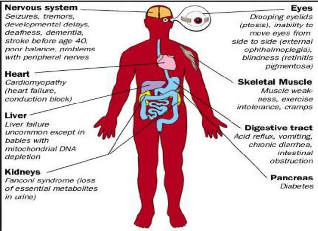
Free radical have role important in human body, it is product normally with low amount by various physiological mechanisms. Excess formation of free radical can initiate series of chemical reaction and cause damage to cellular components such as: damage of DNA, oxidations of polydesaturated fatty acids in lipids, oxidations of amino acids in proteins, oxidatively inactivate specific enzymes by oxidation of co-factors (Das Sarma et al., 2010; Shastri et al., 2018) and production of proinflammatory and anti-inflammatory cytokines (Birben et al., 2012). However, when the cell can‘t produce the antioxidant for repair the damage the oxidative stress occur. The important relationship between oxidative stress and a wide variety of human diseases has placed this stress factor at the forefront of diseases research. Indeed, diseases such as rheumatoid arthritis, Alzheimer‘s disease, Parkinson‘s disease, cardiovascular disease, diabetes, and cancer are all related to oxidative stress (Liguori
et al., 2018).
SOD
CAT CAT
9 1) Cancer:
It is one of the leading causes of death in humans. Free radicals cause different types of chemical changes in DNA, thus they could be mutagenic and involved in the etiology of cancer. Cancer cells in particular, in comparison to normal cells, have higher levels of ROS and are more susceptible to mitochondrial dysfunction due to their higher metabolic rate. Cancer cells display elevated levels of oxidative stress indicators due to the activation of oncogenes and loss of tumor suppressors. ROS by altering the growth signals and gene expression, cause continuous proliferation of cancer cells. ROS can damage DNA by inducing base modifications, deletions, strand breakage and chromosomal rearrangements (Cairns et
al., 2011; Acuna et al., 2012).
2) Alzheimer’s disease
Free radical damage induced strong damage in Nervous tissue due to high content of lipids especially polyunsaturated fatty acids. In Alzheimer‘s disease (AD) biochemical and histological studies have provided evidence for increased levels of oxidative stress and membrane lipid peroxidation. Alterations in levels of antioxidant enzymes such as catalase and CuZn- and Mn-SOD in neurons in AD patients increased stress. Peroxidation of 4-hydroxynonenal (4-HNE) in the cerebrospinal fluid of AD patients increase lipid peroxidation. Iron (Fe2+) likely contributes to increase lipid peroxidation in AD. Lipid peroxidation may promote neuronal death in AD by multiple mechanisms that include impairment of the function of membrane ion-motive ATPases (Na+/K+-ATPase and Ca2+ -ATPase), glucose transporters and glutamate transporters. Lipid peroxidation leads to production of hydroxynonenal that appears to play a central role in the neurotoxic actions of amyloid β-peptide (Sivanandham, 2011).
3) Parkinson’s disease
Parkinson‘s disease (PD) is the second most common neurodegenerative disorder in the world. Extensive postmortem studies have provided evidence to support the involvement of oxidative stress in the pathogenesis of PD; in particular, these include alterations in brain iron content, impaired mitochondrial function, alterations in the antioxidant protective systems (most notably superoxide dismutase (SOD) and reduced glutathione (GSH)) and evidence of oxidative damage to lipids, proteins, and DNA. Iron can induce oxidative stress and intranigral injections have been shown to induce a model of progressive parkinsonism. The nature of the free radical species responsible for cell death in PD remains unknown, but there
10
is evidence of involvement of hydroxyl radical, peroxynitrite and nitric oxide (Jenner and Olanow, 1996).
4) Rheumatoid Arthritis (RA)
Several lines of evidence suggest a role for oxidative stress in the pathogenesis of RA. Both ROS and RNS damage cartilage. Tissue injury in inflammation results in NO.production by
articular chondrocytes and synovial fibroblasts and elevated levels of NO.are observed in the
serum and synovial fluid of RA patients. The free radicals particularly NO. and O2•-, inhibit the synthesis of matrix components like proteoglycans by chondrocytes and also damage the extracellular matrix through activation and up regulation of matrix metalloproteinases (Vasanthi et al., 2009; Phaniendra et al., 2015).
5) Diabetes
Diabetes mellitus (type 1 and 2) is a metabolic disease associated with increased formation of free radicals and decreased antioxidant potential. Increased intracellular glucose leads to an increased RONS production, which exceeds the antioxidant capability of the cell to neutralize them. RONS induce, in this way, the activation of three important molecular pathways involved in hyperglycemia induced oxidative tissue: activation of protein kinase C (PKC), increased hexosamine pathway flux and increased polyol pathway flux (Liguori et al., 2018).
6) Cardiovascular disease
NADPH oxidases (Nox) are the important source of ROS. It is catalyze the transfer of electrons from NADPH to molecular oxygen to generate O2∙−. Important roles have been
shown for NADPH oxidases in redox signaling events involved in hypertension, atherosclerosis, endothelial activation, and angiogenesis, well as in endothelial dysfunction. The close functional association between NADPH oxidase and the renin-angiotensin system may be of particular relevance in linking oxidative stress to hypertension. Nox2 and Nox4 are the most abundant NADPH oxidases in the heart and are expressed in cardiomyocytes, endothelial cells and fibroblasts. The over production of ROS induced the development of CVDs, particularly atherosclerosis (Puddu et al., 2008; Violi et al., 2009).
Oxygen free radicals are highly reactive compounds causing peroxidation of lipids and proteins and are thought to play an important role in the pathogenesis of reperfusion abnormalities including myocardial stunning, irreversible injury and reperfusion arrhythmias (Das Sarma et al., 2010).
11 7) Aging
Aging is the progressive loss of tissue and organ function over time. The free radical theory of aging, later termed as oxidative stress theory of aging, is based on the structural damage-based hypothesis that age-associated functionallosses are due to the accumulation of oxidative damage to macromolecules (lipids, DNA and proteins) by RONS. The exact mechanism of oxidative stress-induced aging is still not clear, but probably increased RONS levels lead to cellular senescence, a physiological mechanism that stops cellular proliferation in response to damages that occur during replication (Carocho et al., 2018).
8) Kidney disorders
Mitochondrial free radical production induces lipid peroxidation during myohemoglobinuria. Iron catalyzed free radical formation and lipid peroxidation are accepted mechanisms of heme protein-induced acute renal failure. However, the source of those free radicals which trigger lipid peroxidation in proximal tubular cells remains unknown (Das sarma, 2010).
12
1.5 Antioxidants
The term antioxidant was defined as the molecule which can give an electron to free radicals to neutralize them and reduces their damage capacity. This molecule is a stable substance and protects cell against any damage effect of free radicals (Carocho andFerreira, 2013).
The damaging effect of free radicals is limited by numerous cellular antioxidant defense mechanisms in the body. Common antioxidants include: enzymes such as SOD, CAT and glutathione system, non-enzymes such as vitamins (C, E), carotenoids, flavonoids, glutathione and other minerals antioxidants (copper, ferritin, zinc, manganese, selenium etc.) (Lozovoy et
al., 2013).
1.5.1 Enzymatic antioxidants
A) Superoxide dismutase (SOD)
Superoxide dismutase is a group of metalloenzymes which are essential to protect cells under aerobic conditions. This enzyme catalyzes the transfer of an electron from one molecule of the superoxide anion to dioxygen, which rapidly combines with two hydrogen ions to produce hydrogen peroxide. There are three forms of SOD in the human body:
• SOD1 in the cytoplasm: copper and zinc (Cu-Zn-SOD).
• SOD2 in the mitochondria: Manganese superoxide dismutase (Mn-SOD) plays a crucial role in antioxidant responses to environmental stress.
• SOD3 extracellular: copper and zinc (Cu-Zn-SOD) (Docampo and Moreno, 2017).
B) Catalase (CAT)
Catalase (CAT) is a 240 kilodalton (kDa) tetrameric protein. CAT is a common antioxidant enzyme present almost in all living tissues that utilize oxygen. This enzyme is found mainly in mitochondria and peroxisomes. The enzyme was originally regarded as a monofunctional peroxisomal enzyme that efficiently catalyzes the dismutation of hydrogen peroxide to water and oxygen and uses either iron or manganese as a cofactor. In the last year catalase is considered a multifunctional enzyme, which exhibits not only classic catalase activity, but also peroxidase and oxidase functions such as: the formation of compound I, degradation of NO. Catalase has protective effects in the cardiovascular system and delayed the development of atherosclerosis (Castaldo et al., 2016).
13 C) Glutathione system
This system includes three enzymes: GSH reductase, GSH peroxidase (GPx) and glutathione-S-transferase (GST)(Lumb, 2017).
1. GSH reductase (GR)
GSH reductaseis a group of flavoproteins and it is homodimer containing FAD coenzyme in its active site that catalyzes the transfer of electrons from NADPH to FAD, ultimately to the reactive disulfide bridge center that reduces GSSG to GSH. This enzyme maintains GSH levels in the cell (Gregg and Prchal, 2018).
2. GSH peroxidase(GPx)
The enzyme activity depends on a micronutrient cofactor known as selenium. The biochemical function of GPx is to reduce free hydrogen peroxide to water and to reduce lipid hydroperoxides to their corresponding alcohols and in turn oxidizes glutathione to glutathione disulfide (Ighodaro and Akinloye, 2017). There are four isoforms of GPx
1. Cellular GPx (GPx1): reduces H2O2 and fatty acid peroxides
2. Gastrointestinal GPx (GPx2): is localized in the gastrointestinal epithelial cells.
3. Extracellular GPx (GPx3): is the only member of the GPx family that exists in the extracellular compartment, and is believed to be the most important extracellular antioxidant enzyme in mammals
4. Phospholipids hydroperoxide GPx (GPx4): esterified lipids are reduced by membrane-bound GPx-4 (Tabet and Tyouz, 2007).
3. GSH-S-transferase (GSTs)
This is another antioxidant enzyme family, which inactivate secondary metabolites, such as unsaturated aldehydes, epoxides, and hydroperoxides. Three major families of GSTs have been described: cytosolic GST, mitochondrial GST, and membrane-associated microsomal GST (Birben et al., 2012). GST used the GSH reduced by GSH reductase to neutralize diverse electrophilic and reactive compounds by conjugating them with GSH. Glutathione-S-transferases also metabolize H2O2 and many other endogenous hydroperoxides (Hrycay and
Bandiera, 2015).
1.5.2 Non enzymatic antioxidants A) Glutathion (GSH)
GSH is a tripeptide including Glutamate, cysteine and glycine. This tripeptide is formed by two steps. The first, γ-glytamylcysteine by γglytamylcysteine ligase (GSH1) then the GSH sentethase add glycine to dipeptide. GSH can regenerated from the oxidized GSSH in the
14
presence of GR. GSH is present in plants in the cytosol, chloroplast, mitochondria and nucleus. It is the major antioxidant in these cell compartments. Protective roles of glutathione against oxidative/nitrosative stress are that it can act as a co-factor for several detoxifying enzymes, participate in amino acid transport across plasma membrane and scavenge hydroxyl radical and singlet oxygen directly (Csiszár et al., 2016).
B) Vitamin C (Vit C)
Vit C is a major water soluble antioxidant in the body. It quenchs the activity of some free radicals by donating electron such as hydroxyl and superoxide, and scavenge other ROS such as singlet oxygen, superoxide, hydroxyl, water soluble peroxyl radical, and hypochlorous acid. Vit C has an important role in the regeneration of vitamin E and protects biomembranes from peroxide damage. It increases the level of GSH in the cell. Fresh fruits and vegetables contain high amounts of vitamin C (Marin et al., 2018).
C) Vitamin E
Vitamin E is a lipid soluble vitamin. It is an important antioxidant having various physiological functions, including the maintenance of plasma membrane integrity, cell signaling and cell cycle regulation, cell adhesion, platelet aggregation, smooth muscle cell proliferation and immune function. The major role of vit E is re-evaluated by the European Food Safety Authority expert panel(Raederstorff et al., 2015). It can scavenge free radicals by the hydroxyl group specifically with the acyl chains of polyunsaturated fatty acids in the membrane. There are four isoform of vit E: α-tocopherol, β-tocopherol, γ-tocopherol and δ-tocopherol (Temiz et al., 2018).
D) Carotenoids
Carotenoids are groups of pigments found in most fruits and vegetables, plants, algae and photosynthetic bacteria and more than 600 different carotenoids have been identified and characterized. Carotenoids include β-carotene, lutein, zeaxanthin, lycopene, astaxanthin and canthaxanthin. Lycopene showed potent antioxidant activity in vitro and reduces serum cholesterol in animal studies (Müller et al., 2016). Carotenoids have the ability to neutralize singlet oxygen and reactive oxygen species, protect against UV-induced peroxidation and reduce the formation of lipofuscin and associated oxidative-stress induced damage (Eggersdorfer and Wyss, 2018).
15 E) Coenzyme Q10
CoQ10 is an ubiquinone, it is a lipid-soluble benzoquinone. CoQ10 is considered as intracellular antioxidant that protects membrane phospholipids, mitochondrial membrane proteins, and low-density lipoproteins from free radical-induced oxidative damage. Coenzyme Q10 is added to support mitochondrial functions such as shuttling electrons, serving as a potent antioxidant and working as an electron transport chain to generate ATP (Mousavinejed
et al., 2018).
F) Uric acid (UA)
Uric acid is a heterocyclic organic compound. It is the end product of purine metabolism or purine nucleotides (guanosine and adenosine) catabolism in humans. In rats and mice, UA is converted by the enzyme uricase to produce allantoin. This is 100 times more water-soluble than UA and consequently has more efficient urinary excretion route than UA. Various studies have shown that uric acid is a potent antioxidant against oxidative stress. It is capable particularly of neutralizing superoxide, peroxyl, hydroxyl radicals (OH), hypochlorous acid (HOCl) and protects vascular endothelium from external oxidative stress (Banihani, 2018). Uricases also prevents peroxynitrite induced protein nitrasation, lipid and protein peroxidation and used a cofactor for endothelial nitric oxide synthase (Ndrepepa, 2018).
G) Selenium
It is a trace mineral that supports the body in many ways.Selenium has unique multi-functional function: catalytic, structural, and regulatory. It activates the action of many enzymes, vitamins, hormones and thus ensures the normal functioning of various biological systems. Selenium is one of the most important antioxidants in the human body. The biochemical functions of selenium are determined by the selenium proteins that contain selenocysteine, which have antioxidant functions and include enzymes such as glutathione peroxidase, a potent free radical scavenger. Selenium acts to destroy peroxides and thus protects lipid membranes like vitamin E (Sobolev et al., 2018).
H) Zinc
It is an essential mineral for human health, it is considered as co-factor for over 300 enzymes and 2000 transcription factors. This mineral is a mediator of cellular signaling. In addition, the zinc has an important role in the antioxidant defense system. This molecule protects the cell against the oxidative damage. It also inhibits the NADPH-oxidase enzyme, regulation of
16
GSH peroxidase and the expression of metallothioneim, used as co-factor for SOD and reduces chronic inflammation and hyperglycemia (Marreiro et al., 2017).
I) Polyphenols
Polyphenols are important group of natural compounds they contained heterogeneous group of molecules with feature structure. Polyphenols are constituted of aromatic ring and several hydroxyl groups. They are synthetized by two pathways, the first is the Shikimic acid pathway and the second is the acetic acid pathway. Based on the chemical structure, polyphenols are divided into five groups as flavonoids, phenolic acid, stilbenes, lignans and tannins (Belščak-Cvitanović et al., 2018)
1. Flavonoids
Flavonoids came from the Latin word « flavus» wich means yellow. They are the biggest group of polyphenol present in the plant with low molecular weight. Generally they have a common C6-C3-C6flavone skeleton, which contained two aromatic ring (A and C) and heterocyclic (B). Depending on the oxidation and the saturation in the C ring flavonoids mainly are divided into flavanols, flavones, flavonones, isoflavone, flavonol and flavanonol. In fact, the majority of flavonoids exist in fruits and vegetables as glycosides (Vicente and Boscaiu, 2018).
2. Tannin
Tannins are water-soluble phenolic compounds having a molecular weight between 500 and 3000 Dalton. Tannins are classified into two major groups on the basis of their structure: hydrolysable and condensed tannins.
a) Hydrolysable tannins: Hydrolysable tannins are compounds containing a central core of glucose or another polyol esterified with gallic acid, also called gallotannins, or with hexahydroxydiphenic acid, also called ellagitannins (Sarni-Manchado and cheynier, 2006).
b) Condensed tannins: Condensed tannins are oligomers or polymers composed of flavan-3-ol nuclei. They are also called proanthocyanidins, because they are decomposed to anthocyanidins in heated ethanol solutions (Dangletter, 2007).
They characteristic properties of tannin as include forming of insoluble complexes with proteins, polysaccharides, nucleic acids and alkaloids. Within this general character, tannins exhibit a number of various bioactivities, which are often related to their antioxidant activity (Koleckar et al., 2008)
17
Antioxidant activity study of tannins in vitro and in vivo showed that they are effective scavengers of free radicals and they inhibit the tissues oxidation better than vitamin C, vitamin E and β-carotene. The basic concept of free radical scavenging activity of polyphenols, including tannins, is the ability of the antioxidant to donate electron to a free radical and produce a more stable and therefore less harmful radical structure. DPPH
(1,1-diphenyl-2-(2,4,6-trinitrophenyl) hydrazyl) or ABTS ((2,
2-azinobis)3-ethyl-2,3-dihydrobenzothiazol-6-sulphonic acid) radicals are often used for in the vitro evaluation of free radical activity of plants extracts (Hatano et al., 2002). Procyanidins are the most active on the basis of scavenging ABTS radical, hypochlorous acid, or in FRAP test for the evaluation of reducing power (Koleckar et al., 2008).
In vitro conditions have been shown that condensed tannins have a preference for neutralizing
the hydroxyl free radical (OH.). It has been demonstrated that they have the capacity to act as noncompetitive inhibitors of the enzyme xanthine oxidase, one of the biggest generators of free radicals in cellular metabolism. Additionally, they act as scavengers of free radicals and inhibitors of superoxides formation and nitric oxide synthases (NOS) (Fogliani et al., 2005; Koleckar et al., 2008). Chandranayagam et al (2013) demonstrated that the gavage of tannin with dose of 30-300 mg/kg protect the liver.
3. Phenolic acids
Phenolic acids belong to a highly diversified group of phytochemicals, phenolics which are found in all foods of plant origin in the human diet. Phenolic acids are non-flavonoid polyphenolic compounds which can be further divided into two main types: benzoic acid and cinnamic acid derivatives based on C1–C6 and C3–C6 backbones. Hydroxybenzoic acids include gallic, p-hydroxybenzoic, vanillic, syringic, and protocatechuic acids. The hydroxycinnamic acids commonly found in foods and beverages are p-coumaric, caffeic, ferulic, sinapic and cinnamic acids. While fruits and vegetables contain many free phenolic acids, in grains and seed particularly in the bran or hull—phenolic acids are often in the bound form . These phenolic acids can only be freed or hydrolyzed upon acid or alkaline hydrolysis, or by enzymes (Nazck and Shahidi, 2006; Chandrasekara and Shahidi, 2010).
In the process of free radical scavenging, phenolic acids produce phenolic radicals and they can be stabilized by establishing intra-molecular hydrogen bonds and extending delocalization and conjugation of the electrons enhanced by resonance stabilization (Chen et al., 2015).