Any correspondence concerning this service should be sent to the repository administrator:
DOI:10.1007/s11104-013-1648-6
Official URL:
http://dx.doi.org/10.1007/s11104-013-1648-6
This is an author-deposited version published in: http://oatao.univ-toulouse.fr/ Eprints ID: 9737To cite this version:
Compant, Stéphane and Muzammil, Saima and Lebrihi, Ahmed and Mathieu,
Florence Visualization of grapevine root colonization by the Saharan soil isolate
Saccharothrix algeriensis NRRL B-24137 using DOPE-FISH microscopy.
(2013) Plant and Soil, vol. 370 (n° 1-2). pp. 583-591. ISSN 0032-079X
O
pen
A
rchive
T
oulouse
A
rchive
O
uverte (
OATAO
)
OATAO is an open access repository that collects the work of Toulouse researchers and makes it freely available over the web where possible.
Visualization of grapevine root colonization by the Saharan
soil isolate Saccharothrix algeriensis NRRL B-24137
using DOPE-FISH microscopy
Stéphane Compant&Saima Muzammil&
Ahmed Lebrihi&Florence Mathieu
Abstract
Background and aim There is currently a gap of
knowledge regarding whether some beneficial bacteria isolated from desert soils can colonize epi- and endophytically plants of temperate regions. In this study, the early steps of the colonization process of one of these bacteria, Saccharothrix algeriensis NRRL B-24137, was studied on grapevine roots to determine if this beneficial strain can colonize a non-natural host plant. An improved method of fluorescence in situ hybridization (FISH), the double labeling of oligonu-cleotide probes (DOPE)-FISH technique was used to visualize the colonization behavior of such bacteria as
well as to determine if the method could be used to track microbes on and inside plants.
Methods A probe specific to Saccharothrix spp. was
firstly designed. Visualization of the colonization be-havior of S. algeriensis NRRL B-24137 on and inside roots of grapevine plants was then carried out with DOPE-FISH microscopy.
Results The results showed that 10 days after
inocu-lation, the strain could colonize the root hair zone, root elongation zone, as well as root emergence sites by establishing different forms of bacterial structures as revealed by the DOPE-FISH technique. Further obser-vations showed that the strain could be also endophyt-ic inside the endorhiza of grapevine plants.
Conclusions Taking into account the natural niches of
this beneficial strain, this study exemplifies that, in spite of its isolation from desert soil, the strain can establish populations as well as subpopulations on and inside grapevine plants and that the DOPE-FISH tool can allow to detect it.
Keywords Saccharothrix algeriensis NRRL B-24137 . Grapevine . Colonization . DOPE-FISH
Introduction
Plant growth-promoting bacteria as well as biocontrol bacteria could be isolated from the phyllosphere, anthosphere, carposphere, as well as the caulosphere. The majority of these bacterial microsymbionts are,
DOI 10.1007/s11104-013-1648-6
S. Compant
:
S. Muzammil:
A. Lebrihi:
F. Mathieu LGC UMR 5503 (CNRS/INPT/UPS), Département Bioprocédés et Systèmes Microbiens, ENSAT-INP de Toulouse, Université de Toulouse,1 Avenue de l’Agrobiopôle, B.P. 32607, 31326 Castanet-Tolosan Cedex 1, France A. Lebrihi
Université Moulay Ismail,
Marjane II, BP 298, Meknès, Morocco
Present Address:
S. Compant (*)
Bioresources Unit, Health & Environment Department, AIT Austrian Institute of Technology GmbH,
3430 Tulln, Austria
however, epiphytics and colonize the rhizosphere (Lugtenberg and Kamilova 2009). A subset of the rhizosphere microflora can also enter inside plants, establishing subpopulations, and proliferate as endo-phytes. This has been well described with cultivable approaches as well as with metagenomic and micros-copy analysis (Rosenblueth and Martínez-Romero
2006; Hallmann and Berg 2007; Compant et al.
2010a; Reinhold-Hurek and Hurek 2011). Currently, there is a gap in knowledge regarding plant growth-promoting agents or biocontrol strains colonizing the rhizosphere as well as the endosphere of plants. Particularly, some microbes could be isolated from harsh environments such as desert soils (Compant et al. 2010b), and could act as plant growth-promoting bacteria or biocontrol agents once applied on a crop. Harsh environments might provide a rich source of beneficial bacteria, and it is becoming increasingly evident that microbes from soil and plants growing in harsh environments, such as desert soil, may repre-sent an enormous untapped genetic reservoir for plant improvement. It has been recently postulated that transferring these microbes from native plants to non-host plants promises a revolutionary biotechnolo-gy to rapidly improve plant germplasms (Barrow et al.
2008). However, the microbial colonization of such strains on non-host plants should be studied, and also visualization of the process of colonization should be done if any application is carried out on crops growing in temperate conditions (Compant et al. 2010b). Additionally, visualization of plant colonization by these bacteria could allow to better understand niches of colonization of plant-associated bacteria.
In correlation to the research of beneficial microbes from harsh environments, such as desert soils, an actinomycete member of the Actinosynnemataceae family was isolated from a desert soil in a palm grove of Adrar in Algeria. The strain was identified as
Saccharothrix algeriensis NRRL B-24137 (Zitouni 1995; Zitouni et al. 2004). The strain NRRL B-24137 is known as secreting various secondary me-tabolites, such as dithiolopyrrolones, with broad bio-active activities (Lamari et al. 2002a,b; Zitouni et al.
2005). The strain can also act as a biocontrol agent. It reduces Botrytis cinerea infection on grapevine plants under greenhouse at 25 °C (Muzammil2012) but also even under high temperature conditions (Muzammil et al. 2011). However, colonization processes of this strain should be studied to understand where the
beneficial strain could be localized following soil ap-plication to know its niches of colonization. Moreover, this needs to be determined before to study mecha-nisms of plant resistance responsible of reduction of phytopathogen infections (postulate described by van Loon et al. (1998)).
To visualize colonization and to track microbes on and inside plants various tools, such as gfp, dsred, and
gus markers as well as derivatives, could be used
(Larrainzar et al. 2005). However, this implies that the microbe will be transformed before application, which is difficult to achieve for some microorganisms. Other methods were used to monitor the colonization behavior of bacteria, such as the DNA-intercalating stains acridine orange, SYTO® dyes, 4′,6-diamidino-2-phenylindole, protein, or lipid stains. However, to be sure to visualize specific taxa of bacteria on and or inside plants, immunostaining with fluorescent-labeled antibodies or fluorescence in situ hybridization (FISH) method (Amann et al. 1990; Wagner et al.
2003) should be used. They can, indeed, provide high specificity to detect given bacterial species or strains (Brandl and Monier 2005; Brandl 2009). For one of these techniques, FISH, visualization can however suffer from some limited advantages for some micro-organisms (Wagner et al. 2003). Different improve-ments have been published then to increase the signal in FISH (Wagner and Haider2012). In 2010, Stoecker et al. described, for instance, the use of double labeling of oligonucleotide probes (DOPE)-FISH correspond-ing to 5′- and 3′-double-labeled probes instead of single-labeled probes for FISH. It has been demon-strated that double-labeled probes strongly increase in situ accessibility of rRNA target sites, and increase fluorescent signal. Moreover, this technique provides more flexibility for probe design (Stoecker et al.2010) and can allow visualizing microorganisms that could not be well detected by single FISH (due to low fluorescent signal) or that are difficult to transform, such as some filamentous microbes.
In this study, we created a specific probe for
Saccharothrix spp., and we used DOPE-FISH
tech-nique to monitor the early colonization process of the beneficial strain NRRL B-24137 on grapevine plants, both in the rhizosphere and in the root endosphere of plants. This was a prerequisite to better understand the interaction between a bacterium isolated from a harsh environment, such as from a Saharan soil, and a non-natural host.
Materials and methods Bacterial culture
S. algeriensis NRRL B-24137 was used throughout
this work. This strain was grown at 30 °C on ISP 2 (International Streptomyces Project 2) solid medium (pH7.0) containing per liter of distilled water: 4 gD(+) glucose (Acros organics), 10 g malt extract (Fluka), 4 g yeast extract (Fluka), and 18 g agar (Sigma). Eight days after growing on plate, aerial mycelium +spores of strain NRRL B-24137 were harvested in phosphate buffered saline (PBS) and concentration was adjusted to 5.107CFUmL−1.
Plant material
Grapevine plants, harboring as a graft part Vitis
vinif-era L. cv. Cabernet Sauvignon clone 15 and 44–53 M
(Vitis riparia×Malègue) as a rootstock were provided by “Pépinières Colombie Vendries” (Camparnaud, France). These grapevine plants were used because it has been previously described that the strain NRRL B-24137 could be beneficial once applied on them and reduce B. cinerea infection (Muzammil et al. 2011). These grapevine plants were stored at 4 °C in a dark cold chamber for at least 2 weeks before being treated with 0.05 % cryptonol for 15 h at ambient temperature (20–25 °C). Plants were then surface sterilized with 1.6 % bleach (10 min) and 70 % ethanol (30 min) before rinsing with sterile tap water. Then, they were planted in two times autoclaved soil containing 1/3 perlite, 1/3 potting soil, and 1/3 sand. Plants were allowed to grow in a phytotronic growth chamber (16-h photoperiod, 20–25 °C night–day, and 70 % relative humidity) and watered with sterile tap water.
Plant inoculation
One month after planting, grapevine plants were del-icately separated from their soils. The root systems were dipped in a bacterial solution of S. algeriensis NRRL B-24137 (or PBS) for 3 min before plants are placed in pots filled with soils (as described before). Plants were then allowed to grow for 10 days (time allowing resistance to B. cinerea described in Muzammil (2012)) before sampling of the plant parts for microscopic analysis.
Probe design for Saccharothrix spp. and labeling To create probes specifics to Saccharothrix spp., the partial 16S rRNA gene sequence of S. algeriensis NRRL B-24137 (accession: AY054972.2 GI: 134034183) was used. The design of 16S rRNA probes was made by using the Stellaris™ FISH Probes software. The specificity of probes created was then checked on National Center for Biotechnology Information (NCBI), Silva, Green genes blast (Altschul et al.1997) or on Probe Check at microbial-ecology.net(Loy et al.2007). ΔG, FA, FAm, as well as the hybridization efficiency were calculated according to Yilmaz and Noguera (2004), (2007), and Yilmaz et al. (2006) and the Tm was calculated by using Tm¼ 64:9 þ 41! G þ C " 16:4ðð Þ length= Þ according to Loy et al. (2007). These parameters were evaluated for different temperatures of hybridization.
Among all probes designed, one found as specific to Saccharothrix spp. was then purchased at Genecust (Luxemburg) with an aminomodifier C6 at 5′ and 3′ positions (for DOPE-FISH). The probe was then la-beled with dylight488 fluorochrome (Piercenet) en-abling green fluorescence under UV light.
DOPE-FISH microscopy
To visualize the rhizosphere/rhizoplane colonization by the strain NRRL B-24137, roots of grapevine were cut in small parts 10 days post-inoculation with the strain NRRL B-24137. The samples were then fixed overnight at 4 °C in a paraformaldehyde solution (4 % in PBS) in Eppendorf tubes. The samples were rinsed twice with PBS before to be treated with 1 mg/mL lysozyme at 37 °C during 15 min. They were then rinsed with PBS and dehydrated in an ethanol series (50–99.9 %; 30 min for each step). The DOPE-fluorescence in situ hybridization was then carried out according to Compant et al. (2011) by using 15 ng/μL of a probe specific to Saccharothrix spp., labeled at both 5′ and 3′ with the DyLight 488 fluoro-chrome. Following a DOPE-FISH hybridization at 51 °C, a posthybridization step was carried out at 52 °C and samples were rinsed with sterile distillated water (prewarmed at 51 °C). The samples were then kept in dark during at least 1 day. The samples were then observed using epifluorescence microscope (BH2, Olympus, Japan) under a UV light and pictures were taken with a camera (TCC-3.3ICE-N, Tucsen, China).
In parallel to the rhizosphere colonization study, a possibility of endophytism was evaluated for S.
algeriensis NRRL B-24137. For this, root samples
were treated as described before, except that after the ethanol series, the samples were included in LR white resin according to the manufacturer instruc-tions. Embedded tissues were then sliced with a mi-crotome and glass knives. Slices of 1–1.5 μm were deposited on microscopic slides previously treated with 70 % ethanol. The DOPE-FISH was then carried out as described before. Following the steps of DOPE-FISH hybridization, posthybridization, and rinsing, the slides containing slices were kept in dark during at least 1 day. Slices on slides were then observed using epifluorescence microscope (BH2, Olympus, Japan) under UV light and pictures were taken, as described before. All experiments have been repeated on three independent times with similar re-sults on ten plants each time. More than 20 slices were used per plant to visualize the colonization process.
Results
Design of a probe specific to four species of Saccharothrix
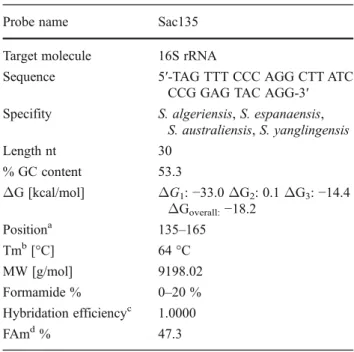
Different probes with lengths of 20 and 25 nt were designed. However, no probe with these lengths was specific to Saccharothrix spp. Therefore, additional probes of 30 nt were created. Among them, a probe named Sac135 (Table1) was designed and checked on probe check, Silva, green genes, and NCBI databases. Data revealed that the probe is specific to four species of Saccharothrix including S. algeriensis. The probe has a Percent G-C content of 53.3, is at position 135 according to the Escherichia coli gene numbering, has an exp Td (Tm) of 64 °C; a ΔG1, −33.0 kcal/mol; a
ΔG2, 0.1 kcal/mol; a ΔG3, −14.4 kcal/mol; a
ΔGoverall, −18.2 kcal/mol; and a FAm of 47.3 % at
51 °C of hybridization with 0.9M Na+ (Table 1). At 46 °C which is used for the majority of FISH exper-iments, this probe could not be used due to an ineffi-cient hybridization and a high Fam according to Yilmaz and Noguera (2004, 2007) and Yilmaz et al. (2006). This probe was then used with a formamide concentration of 20 % as hybridization efficiency at 0– 20 % of formamide was of 1.0000 (Table1).
Grapevine root surface colonization by S. algeriensis NRRL B-24137
Colonization of grapevine plants by the strain NRRL B-24137 was evaluated by DOPE-FISH analysis using the Sac135 probe. Grapevine colonization by strain NRRL B-24137 was firstly evaluated at the root hair zone level. Without hybridization with the probe, no green fluores-cence signal was recorded under the UV wavelength (data not shown). After hybridization, the bacterium was visual-ized as spores or as germinated spores, in close contact to the surface of root hairs (Fig. 1a). Hyphae were also visualized as colonizing externally the basis of root hairs (Fig.1b). Packs (or pellets) of hyphae as well were addi-tionally visualized at the root hair level (Fig. 1c–d). Although some bacteria were isolated on the surface of the root hair zone, in some other few parts some rhizodermal cells were completely filled with bacteria. This was demonstrated with strong green fluorescence signal (Fig.1f), whereas root cell fluorescence was nor-mally low yellow autofluorescent on roots of plants treated with NRRL B-24137 (Fig.1e) or control treatment (data not shown).
Table 1 The probe and specificity of the probe related to
Saccharothrix spp
Probe name Sac135 Target molecule 16S rRNA
Sequence 5′-TAG TTT CCC AGG CTT ATC CCG GAG TAC AGG-3′ Specifity S. algeriensis, S. espanaensis,
S. australiensis, S. yanglingensis Length nt 30 % GC content 53.3 ΔG [kcal/mol] ΔG1: −33.0 ΔG2: 0.1 ΔG3: −14.4 ΔGoverall:−18.2 Positiona 135–165 Tmb[°C] 64 °C MW [g/mol] 9198.02 Formamide % 0–20 % Hybridation efficiencyc 1.0000 FAmd% 47.3
aProbe position according to the E. coli gene numbering bMelting temperature dissociation
cHybridation efficiency at 0 % of formamide dMelting formamide concentration (%)
ΔG, formamide percent, hybridation efficiency, and FAm per-cent calculated for 51 °C and 0.9M Na+
Ten days post-inoculation, the bacterium was also found at root emergence sites as hyphae (Fig. 1g–h). No spores, or germinated spores, were however visual-ized. Only this type of actinobacterial form was noticed in all analyzed samples.
Colonization by the strain NRRL B-24137 was eval-uated not only at the root hair zone and emergence site levels but also at the root elongation zone. The bacteri-um was visualized as spores or germinated spores (Fig.2a–b) on the rhizoplane. Visualization of the pro-cess of colonization revealed that the bacterium could be also detected as hyphae near some cells of the rhizodermis (Fig. 2c). Colonization of parts of some rhizodermal cells (Fig. 2d) but also the whole outline of some cells on the rhizoplane was noticed as filled by bacteria (Fig.2e). In contrast to the root hair zone, root emergence site and root elongation zone, no detection of the strain NRRL B-24137 was reported on any of the analyzed samples at the root tip level (data not shown). Endorhizal colonization by S. algeriensis NRRL B-24137 The strain NRRL B-24137 was easily visualized inside roots in LR white sections and in highest numbers in
comparison to some parts of the root surfaces. It was visualized as hyphae inside the endorhiza of grapevine plants, 10 days post-inoculation, crossing from the rhi-zoplane (Fig.3a) to inside the rhizodermis (Fig.3a–b). The filamentous form of the strain was also visualized between the rhizodermis and the exodermis (Fig.3b). Additionally, hyphae of the strain were visualized intercellularly between some cortical cells in the cortical zone (Fig. 3c). A pack of hyphae was also noticed intercellularly (Fig. 3d) and some cells corresponding to the strain were further visualized, intracellularly, in parts of the cortical cell layers (Fig.3e).
Discussion
In the present study, a probe was designed for
Saccharothrix spp. The DOPE-FISH technique has
been used because of a low signal intensity, using single-labeled probe, for some microorganisms (Stoecker et al. 2010). This probe and the DOPE-FISH tool have allowed to visualize the early coloni-zation process of S. algeriensis NRRL B-24137 on and inside roots of grapevine plants. Since the strain
a
b
RH RH RHc
RHd
RHZ RH RHZg
h
Se R Se R RHZe
Uncolo-nizedf
10 µm 10 µm 10 µm 15 µm 50 µm 50 µm 50 µm 15 µm Fig. 1 Microphotographies of the grapevine root colo-nization by S. algeriensis NRRL B-24137 by DOPE-FISH microscopy showing (arrows) spores or germi-nated spores (a), hyphae (b), a pack (or pellet) of hyphae (c–d) colonizing root hairs and some rhizodermal cells not filled (e) or completely filled (f) at the root hair level as well as hyphae at root emergence sites (g–h). RHZ root hair zone, RH root hair,was isolated from a harsh environment, it could not be expected that it could colonize grapevine plants under climatic chamber conditions. However, it was shown in this study that the bacterium could colonize the rhizoplane of the root system of grapevine plants. The strain could colonize the root hair zone, the root elongation zone as well as root emergence site but not root tips.
Pathways of colonization have been described for different kinds of bacteria colonizing the surfaces of root systems of their hosts (Hallmann2001; Hardoim et al. 2008; Compant et al. 2010a) as well on the grapevine rhizoplane (Compant et al. 2005, 2008). The strain NRRL B-24137 was not visualized at the root tip level, suggesting that there is no colonization of such root parts during the process of colonization. However, we cannot exclude that some bacteria, not
well attached and colonizing root tips (therefore not well colonizing), were not fixed and therefore displaced. At the same time, various researches have demonstrated colonization of root tips by beneficial microbes after same kind of fixation (Chin-A-Woeng et al. 1997; Herschkovitz et al. 2005; Buddrus-Schiemann et al. 2010) and different ways of root surface colonization can be done by beneficial micro-organisms (Hallmann2001). It has been described that some bacteria will colonize preferentially the root tips, whereas for others their population pass from 0 to some populations from the tips to away and colonize rather the root hair zone. Niches providing high nutri-ent availability including zones of root exudation such as root hairs can be colonized by r strategists, whereas nutrient-poor or depleted niches will tend to be colo-nized by K strategists (Semenov et al. 1999). This
a
b
c
d
Rh Rh Rh Rh Rhe
5 µm 10 µm 20 µm 35 µm 35 µm Fig. 2 Microphotographies of the grapevine root colo-nization by S. algeriensis NRRL B-24137 by DOPE-FISH microscopy at the root elongation zone showing (arrows) spores or germi-nated spores (a–b), pack of hyphae (c) on the rhizo-plane. Parts (d) or the com-plete outline (e) of some rhizodermal cells were filled with bacteria. Rh rhizoplaneRh Co
a
b
c
d
e
Co Co Ex Co Ex Rh 35 µm 15 µm 15 µm 10 µm 10 µm Fig. 3 Microphotographies of LR white sections of grapevine root colonization by S. algeriensis NRRL B-24137 and using by DOPE-FISH microscopy showing (arrows), inside the endorhiza, colonization with hyphae from the rhizodermis (a) to the exodermis (a–b), as well as some cortical cells colonized intercellularly (c– d) and intracellularly (e). Rh rhizoplane, Ex exodermis,could explain why the strain NRRL B-24137 has not been detected at the root tip level, depending of its strategy to colonize grapevine plants.
The strain NRRL B-24137 was not visualized inside root hairs, but only at the surfaces of root hairs. It has been recently established that some bacteria could col-onize root hair internally (Prieto et al.2011; Mercado-Blanco and Prieto2012), but this depends of the strain and plant–microbe interactions.
Several studies have examined the colonization process by beneficial bacteria (reviewed in Reinhold-Hurek and Hurek 1998; Compant et al.
2010a; Hardoim et al. 2008). Some can be systemic colonizers whereas others could be restricted to some root parts (Benizri et al. 2001; Hallmann
2001). Although we studied early colonization pro-cess by NRRL B-24137, we detected only coloni-zation of this strain up to several cortical cell layers, and not in the vascular system. This sug-gests that the strain NRRL B-24137 will be restrict-ed to root internal parts. However, experiments were done only 10 days post-inoculation and it may be possible then that the bacterium could reach the vascular system in longer colonizations, al-though this needs to be demonstrated.
The strain used in this study is an actinobacterium that could form spores and hyphae as well as pellets of hyphae (Zitouni 1995, 2004). It was, therefore, not surprising to see that during the colonization different forms of the strain could be visualized. Different studies have described colonization by ac-tinomycetes (see for instance Coombs and Franco
2003; Merzaeva and Shirokikh 2006). In the study of Tokala et al. (2002), a Streptomyces sp. was visualized as spores or hyphae during the coloniza-tion of pea plants. Coombs and Franco (2003) de-scribed the colonization of a Streptomyces sp. in wheat caryopses. Merzaeva and Shirokikh (2006) described colonization dynamics of Streptomyces,
Micromonospora and Streptospangium members.
Other related publications can be found regarding plant colonization by actinomycetes, especially non-Streptomycetes members (described in El-Tarabily and Sivasithamparan 2006). However, this has been never determined with the DOPE-FISH technique and a Saccharothrix member.
Although more work is needed to better understand the interaction between S. algeriensis NRRL B-24137 and grapevine plants, this study shows therefore that the
strain, isolated from a Saharan soil, could colonize epi- and endophytically roots of grapevine plants as well as establish different kinds of forms during the colonization process. Although single FISH could be used to monitor the colonization process of actinobacteria, the DOPE-FISH microscopy was also used to increase the signal and to monitor the plant–microbe interaction, for which sometimes the use of other techniques restrict the visualization. This study also suggests that DOPE-FISH tech-nique could be used to monitor plant colonization by some bacteria, such as for some specific actinomycetes.
Acknowledgments The authors are grateful to the Higher Education Commission of Pakistan for according PhD scholar-ships to Ms. Saima Muzammil. Additional thanks for the plat-form of microscopy of INRA Castanet-Tolosan (France) for allowing us the use of microtome and glass knives. We would like also to acknowledge Dr. Günter Brader and Ms. Helen Smith (AIT, Austria) for proof reading of the manuscript.
References
Altschul SF, Madden TL, Schaffer AA, Zhang J, Zhang Z, Miller W, Lipman DJ (1997) Gapped BLAST and PSI-BLAST: a new generation of protein database search pro-grams. Nucl Acids Res 25:3389–3402
Amann RI, Binder BJ, Olson RJ, Chisholm SW, Devereux R, Stahl DA (1990) Combination of 16S rRNA-targeted oli-gonucleotide probes with flow cytometry for analyzing mixed microbial populations. Appl Environ Microbiol 56:1919–1925
Barrow JR, Lucero ME, Reyes-Vera I, Havstad KM (2008) Do symbiotic microbes have a role in plant evolution, perfor-mance and response to stress? Com Integr Biol 1:69–73 Benizri E, Baudoin E, Guckert A (2001) Root colonization by
inoculated plant growth-promoting rhizobacteria. Biocontrol Sci Technol 11:557–574
Brandl MT (2009) Applying fluorescence microscopy to the investigation of the behavior of foodborne pathogens on produce. In: Postek MT, Newbury DE, Platek SF, Joy DC (eds) Proc. SPIE 7378, pp 73782A1-73782A5
Brandl MT, Monier J-M (2005) Methods in microscopy for the visualization of bacteria and their behavior on plants. In: Gorny JR, Yousef AE, Sapers GM (eds) Microbiology of fruits and vegetables. CRC Press, New York, pp 595–619 Buddrus-Schiemann K, Schmid M, Schreiner K, Welzl G,
Hartmann A (2010) Root colonization by Pseudomonas sp. DSMZ 13134 and impact on the indigenous rhizo-sphere bacterial community of barley. Microb Ecol 60:381–393
Chin-A-Woeng TFC, de Priester W, van der Bij AJ, Lugtenberg BJJ (1997) Description of the colonization of a gnotobiotic
tomato rhizosphere by Pseudomonas fluorescens biocon-trol strain WCS365, using scanning electron microscopy. Mol Plant Microbe Interact 10:79–86
Compant S, Duffy B, Nowak J, Clément C, Ait Barka E (2005) Use of plant growth-promoting bacteria for biocontrol of plant diseases: principles, mechanisms of action, and future prospects. Appl Environ Microbiol 71:4951–4959 Compant S, Kaplan H, Sessitsch A, Nowak J, Ait Barka E,
C l é m e n t C ( 2 0 0 8 ) E n d o p h y t i c c o l o n i z a t i o n o f
Burkholderia phytofirmans strain PsJN in Vitis vinifera
L.: from the rhizosphere to inflorescence tissues. FEMS Microbiol Ecol 63:84–93
Compant S, Clément C, Sessitsch A (2010a) Plant growth-promoting bacteria in the rhizo- and endosphere of plants. Their role, colonization, mechanisms involved and pros-pects for utilization. Soil Biol Biochem 42:669–678 Compant S, van der Heijden M, Sessitsch A (2010b) Climate
change effects on beneficial plant–microbes interactions. FEMS Microbiol Ecol 73:197–214
Compant S, Reiter B, Colli-Mull JG, Gangl H, Sessitsch A (2011) Endophytes of grapevine flowers, berries and seeds: identification of cultivable bacteria, comparison with other plant parts, and visualization of niches of colonization. Microb Ecol 62:188–197
Coombs JT, Franco CMM (2003) Visualization of an endophyt-ic Streptomyces species in wheat seed. Appl Environ Microbiol 69:4260–4262
El-Tarabily KA, Sivasithamparam K (2006) Non-streptomycete actinomycetes as biocontrol agents of soil-borne fungal plant pathogens and as plant growth promoters. Soil Biol Biochem 38:1505–1520
Hallmann J (2001) Plant interactions with endophytic bacteria. In: Jeger MJ, Spence NJ (eds) Biotic interactions in plant– pathogen associations. CABI, Wallingford, pp 87–119 Hallmann J, Berg B (2007) Spectrum and population dynamics
of bacterial root endophytes. In: Schulz BJE, Boyle CJC, Sieber TN (eds) Microbial root endophytes. Springer, Berlin, pp 15–31
Hardoim PR, van Overbeek LS, van Elsas JD (2008) Properties of bacterial endophytes and their proposed role in plant growth. Trends Microbiol 16:463–471
Herschkovitz Y, Lerner A, Davidov Y, Rothballer M, Hartmann A, Okon Y, Jurkevitch E (2005) Inoculation with the plant-growth-promoting rhizobacterium Azospirillum brasilense causes little disturbance in the rhizosphere and rhizoplane of maize (Zea mays). Microb Ecol 50:277–288
Lamari L, Zitouni A, Boudjella H, Badjin B, Sabaou N, Lebrihi A, Lefebvre G, Seguin E, Tillequin F (2002a) New dithiolopyrrolones antibiotics from Saccharothrix sp. SA 233: I. Taxonomy, fermentation, isolation and biological activities. J Antibiot 55:696–701
Lamari L, Zitouni A, Dob T, Sabaou N, Lebrihi A, Germain P, Seguin E, Tillequin F (2002b) New dithiolopyrrolone an-t i b i o an-t i c s f r o m S a c c h a ro an-t h r i x s p . S A 2 3 3 . I I . Physiochemical properties and structure elucidation. J Antibiot 55:702–707
Larrainzar E, O’Gara F, Morrissey JP (2005) Applications of autofluorescent proteins for in situ studies in microbial ecology. Annu Rev Microbiol 59:257–277
Loy A, Maixner F, Wagner M, Horn M (2007) probeBase —an online resource for rRNA-targeted oligonucleotide
probes: new features 2007. Nucleic Acids Res 35: D800–D804
Lugtenberg B, Kamilova F (2009) Plant-growth-promoting rhizobacteria. Annu Rev Microbiol 63:541–556
Mercado-Blanco J, Prieto P (2012) Bacterial endophytes and root hairs. Plant Soil 361:301–306
Merzaeva OV, Shirokikh IG (2006) Colonization of plant rhizo-sphere by actinomycetes of different genera. Microbiology 75:226–230
Muzammil S (2012) Saccharothrix algeriensis NRRL B-24137: biocontrol properties, colonization and induced systemic resistance towards Botrytis cinerea on grapevine and
Arabidopsis thaliana. PhD thesis, INP-ENSAT,
Castanet-Tolosan, France, pp. 117–126.
Muzammil S, Compant S, Yu Z, Mathieu F, Lebrihi A (2011)
Saccharothrix algeriensis NRRL B-24137: a new
endo-phyte with high potential to protect grapevine towards
Botrytis cinerea in case of high temperature conditions.
In: Oeno 2011—Actes de colloques du 9e symposium international d’oenologie de Bordeaux. Dunod, in press. Prieto P, Schilirò E, Maldonado-González M, Valderrama R,
Barroso-Albarracín JB, Mercado-Blanco J (2011) Root hairs play a key role in the endophytic colonization of olive roots by Pseudomonas spp. with biocontrol activity. Microbial Ecol 62:435–445
Reinhold-Hurek B, Hurek T (1998) Life in grasses: diazotrophic endophytes. Trends Microbiol 6:139–144
Reinhold-Hurek B, Hurek T (2011) Living inside plants: bacte-rial endophytes. Curr Opinion Plant Biol 14:435–443 Rosenblueth M, Martínez-Romero E (2006) Bacterial
endo-phytes and their interaction with hosts. Mol Plant-Microbe Interact 19:827–837
Semenov AM, van Bruggen AHC, Zelenov VV (1999) Moving waves of bacterial populations and total organic carbon along roots of wheat. Microbiol Ecol 37:116–128 Stoecker K, Dorninger C, Daims H, Wagner M (2010)
Double-labeling of oligonucleotide probes for fluorescence in situ hybridization (DOPE-FISH) improves signal intensity and increases rRNA accessibility. Appl Environ Microbiol 76:922–926
Tokala RK, Strap JL, Jung CM, Crawford DL, Salove H, Deobald LA, Bailey FJ, Morra MJ (2002) Novel plant-microbe rhizosphere interaction involving Streptomyces
lydicus WYEC108 and the pea plant (Pisum sativum).
Appl Environ Microbiol 68:2161–2171
Van Loon LC, Bakker PAHM, Pieterse CMJ (1998) Systemic resistance induced by rhizosphere bacteria. Annu Rev Phytopathol 36:453–483
Wagner M, Haider S (2012) New trends in fluorescence in situ hybridization for identification and functional analyses of microbes. Curr Opinion Biotech 23:96–102
Wagner M, Horn M, Daims H (2003) Fluorescence in situ hybridisation for the identification and characterisation of prokaryotes. Curr Opin Microbiol 6:302–309
Yilmaz LS, Noguera DR (2004) Mechanistic approach to the problem of hybridization efficiency in fluorescent in situ hybridization. Appl Environ Microbiol 70:7126–7139 Yilmaz LS, Noguera DR (2007) Development of
thermodynam-ic models for simulating probe dissociation profiles in fluorescence in situ hybridization. Biotechnol Bioengin 96:349–363
Yilmaz LS, Okten HE, Noguera DR (2006) All regions of the 16S rRNA of Escherichia coli are accessible in situ to DNA oligonucleotides with sufficient thermodynamic af-finity. Appl Environ Microbiol 72:733–744
Zitouni A (1995) Les genres Nocardiopsis et Saccharothrix (Actinomycetales) dans les sols sahariens: taxonomie numérique, extraction, purification et caractérisation de quelques antibiotiques synthétisés. Magister de microbiologie, E.N.S. de Kouba, Algeria
Zitouni A, Lamari L, Boudjella H, Badji B, Sabaou N, Gaouar A, Mathieu F, Lebrihi A, Labeda DP (2004) Saccharothrix
algeriensis sp. nov., isolated from Saharan soil. Int J Syst
Evol Microbiol 54:1377–1381
Zitouni A, Boudjella H, Lamari L, Badji B, Mathieu F, Lebrihi A, Sabaou N (2005) Nocardiopsis and Saccharothrix gen-era in Saharan soils in Algeria: isolation, biological activ-ities and partial characterisation of antibiotics. Res Microbiol 156:984–993