HAL Id: tel-03224177
https://pastel.archives-ouvertes.fr/tel-03224177
Submitted on 11 May 2021HAL is a multi-disciplinary open access
archive for the deposit and dissemination of sci-entific research documents, whether they are pub-lished or not. The documents may come from teaching and research institutions in France or abroad, or from public or private research centers.
L’archive ouverte pluridisciplinaire HAL, est destinée au dépôt et à la diffusion de documents scientifiques de niveau recherche, publiés ou non, émanant des établissements d’enseignement et de recherche français ou étrangers, des laboratoires publics ou privés.
Mécanismes d’action de complexes polypyridyle du
Ruthénium (II) utilisés comme agents
chimiothérapeutiques ou photosensibilisateurs pour la
photothérapie dynamique
Marta Jakubaszek
To cite this version:
Marta Jakubaszek. Mécanismes d’action de complexes polypyridyle du Ruthénium (II) utilisés comme agents chimiothérapeutiques ou photosensibilisateurs pour la photothérapie dynamique. Chimie thérapeutique. Université Paris sciences et lettres, 2020. Français. �NNT : 2020UPSLC001�. �tel-03224177�
Préparée à l’École Nationale Supérieure de Chimie de Paris
Mechanisms of action of Ru(II) Polypyridyl Complexes
as Photodynamic Therapy Photosensitizers and
Chemotherapy Agents
Soutenue parMarta JAKUBASZEK
Le 19 Mai 2020 Ecole doctorale n° 406Chimie Moléculaire
de Paris Centre
SpécialitéChimie moléculaire
Composition du jury : Christian, GAIDDON Directeur de recherche,Université de Strasbourg Président & rapporteur
Mélanie, HAMON
Directrice de recherche,
Institut Pasteur Examinateur
Pascal, BIGEY
Maître de conférences,
Chimie ParisTech, PSL Examinateur
Bruno, GOUD
Directeur de recherche,
Institut Curie, PSL Examinateur
Caroline, MAAKE
Professeur,
Université de Zurich, Suisse Rapporteur
Gilles, GASSER
Professeur,
Acknowledgements
I greatly appreciated the involvement, help and scientific guidance of my supervisor Dr. Gilles Gasser through the three years of my PhD. Thank you Gilles for your motivation and patience. I will always be thankful for giving me an opportunity to work in your group in beautiful Paris. A big thank you also goes to my second supervisor Dr Bruno Goud and his group at the Institut Curie. I am very grateful for all the kind words, guidance, advice and encouragement.
During my PhD, I had the chance to work with many people. Thank you all for this stimulating and friendly environment. It is not always easy to be a biologist in a chemistry lab, but you made this a cheerful journey!
I wish to say thank you to all the collaborators with whom I have worked. Without them, this work would not have been possible.
Mazzarine and Chloé: you were the best students that I had the chance to teach. Working with you was a pleasure. I want to thank you for all of your hard work and commitment.
I want to thank all the people that helped me with data acquiring and analysis:
Dr. Bruno Saubamea and Dr. Patricia Le-Baccon from the microscopy cores.
Dr. Coralie Guerin, Dr. Lea Guyonnet and Dr. Annick Viguier from the flow cytometry core. Mickaël Tharaud for the ICP-MS measurements.
Dr. Johanne Seguin for introducing me to the world of in vivo.
Warm thanks go to Francine, Ali, Didier and Patrick Guezo. Merci!
Finally, importantly, I would like to thank the ERC for financially supporting this work (Consolidator Grant PhotoMedMet (GA 681679)).
Podziękowania
Chciałabym z całego serca podziękować wszystkim tym, którzy zainspirowali mnie bym podjęła studia doktoranckie, jak również tym, którzy byli dla mnie wsparciem w tej wędrówce. Mamo, Tato dziękuję Wam przede wszystkim za miłość, wsparcie w moich decyzjach i zachęcanie mnie do pokonywania własnych barier. Nauczyliście mnie by próbować wszystkiego, co oferuje życie. Dzięki temu mogę się rozwijać a namacalnym dowodem na to jest ten manuskrypt. Jesteście zawsze przy mnie, gdy tego potrzebuję i wiem, że mogę na was liczyć w każdej sytuacji. Kocham Was!
Piotrze, dziękuję Ci za Twoją miłość, motywację i cierpliwość. Jesteś moją podporą. Dzięki Tobie nie oszalałam w tym trudnym okresie.
Dominiko, dziękuję Ci serdecznie za dobre słowa i wsparcie. Nikt nie zrozumie wyzwania, jakim jest doktorat tak dobrze jak osoba, która też bierze udział w tej przygodzie.
Dziękuję również mojej grupie przyjaciół i znajomych. Olgo, Asiu, Liliano, Kingo, Wojtku, Dawidzie i Kubo jesteście wspaniali. Dziękuję!
Table of contents
RÉSUMÉ ...1 SUMMARY ...9 CHAPTER 1- MECHANISMS OF ACTION OF RU(II) POLYPYRIDYL COMPLEXES IN
LIVING CELLS UPON LIGHT IRRADIATION ... 17
CHAPTER 2- EVALUATION OF THE POTENTIAL OF COBALAMIN DERIVATIVES
BEARING RU(II) POLYPYRIDYL COMPLEXES AS PHOTOSENSITISERS FOR
PHOTODYNAMIC THERAPY ... 65 SUPPLEMENTARY INFORMATION ... 85
CHAPTER 3- SYNTHESIS AND CHARACTERIZATION OF AN EPIDERMAL GROWTH
FACTOR RECEPTOR SELECTIVE RU(II) POLYPYRIDYL-NANOBODY CONJUGATE AS A PHOTOSENSITIZER FOR PHOTODYNAMIC THERAPY ... 101
SUPPLEMENTARY INFORMATION ... 141
CHAPTER 4- SYSTEMATIC INVESTIGATION OF THE ANTIPROLIFERATIVE ACTIVITY OF
A SERIES OF RUTHENIUM TERPYRIDINE COMPLEXES ... 157 SUPPLEMENTARY INFORMATION ... 189
CHAPTER 5- RATIONALLY DESIGNED LONG-WAVELENGTH ABSORBING RU(II)
POLYPYRIDYL COMPLEXES AS PHOTOSENSITIZERS FOR PHOTODYNAMIC THERAPY
... 233
SUPPLEMENTARY INFORMATION ... 259
CHAPTER 6- RUTHENIUM-INITIATED POLYMERIZATION OF LACTIDE: A ROUTE TO
REMARKABLE CELLULAR UPTAKE FOR PHOTODYNAMIC THERAPY OF CANCER ... 371 SUPPLEMENTARY INFORMATION ... 393
CHAPTER 7- A RUTHENIUM(II) COMPLEX CONTAINING A REDOX-ACTIVE
SEMIQUINONATE LIGAND AS POTENTIAL CHEMOTHERAPEUTIC AGENT: FROM
SYNTHESIS TO IN VIVO STUDIES ... 425 SUPPLEMENTARY INFORMATION ... 481
CHAPTER 8- A MALTOL-CONTAINING RUTHENIUM POLYPYRIDYL COMPLEX AS A
POTENTIAL ANTICANCER AGENT... 509 SUPPLEMENTARY INFORMATION ... 551
CHAPTER 9- INCREASING THE CYTOTOXICITY OF RU(II) POLYPYRIDYL COMPLEXES
BY TUNING THE ELECTRONIC STRUCTURE OF DIOXO LIGANDS ... 579 SUPPLEMENTARY INFORMATION ... 639
CHAPTER 10- NOVEL RUTHENIUM (II) POLYPYRIDYL COMPLEXES WITH FLAVONOID
LIGANDS AS ANTICANCER DRUG CANDIDATES ... 705 SUPPLEMENTARY INFORMATION ... 745
1
Résumé
Cette thèse de doctorat a pour dessain d’évaluer d’un point de vue chimique, mais surtout biologique les complexes polypyridyle Ru (II). Ces complexes métalliques peuvent être utilisés comme photosensibilisateurs (PS) pour la thérapie photodynamique (PDT), ou encore comme agents chimiothérapeutiques dans le traitement du cancer. La PDT est un traitement alternatif ou complémentaire à la chirurgie, la chimiothérapie ou la radiothérapie.Ses nombreux avantages lui confèrent un intêret dans le traitement actuel du cancer. Son contrôle spatial et temporel est particulièrement intéressant, ce qui conduit à cibler les tumeurs tout en préservant les tissus sains. De plus, les résistances à répétition et les effets secondaires graves provoqués par la chimiothérapie incitent le monde scientifique à rechercher de nouveaux médicaments candidats anticancéreux. Les complexes de ruthénium sont l'un des groupes les plus prometteurs de médicaments candidats à base de métaux (comme chimiothérapeutiques ou PS) en raison de leurs multiples états d'oxydation stables. Cette thèse décrit un aperçu des modes d'action connus des complexes polypyridyle Ru (II) comme PS pour la PDT et introduit de nouveaux complexes, qui peuvent être utilisés pour des traitements PDT réguliers et ciblés. En outre, cette thèse se concentre également sur la caractérisation d'une nouvelle classe de complexes Ru générés comme agents anticancéreux potentiels pour la chimiothérapie, par coordination de différents dioxoligands au noyau métallique. Cette thèse est composée de 11 chapitres et leur contenu est brièvement décrit ci-dessous
Chapitre 1
Ce premier chapitre se concentre sur l'introduction de complexes polypyridyle Ru (II) en tant que PS pour la PDT et, en outre, décrit le ou les mécanismes connus d'action de ces composés dans des cellules / souris vivantes lors d'une irradiation lumineuse. Malheureusement, à ce jour,
2
il y’a peu d’études décrivant le ou les mode (s) d'action de ces composés. Dans ce chapitre, seules les études biologiques, traitant de la phototoxicité et de la localisation cellulaire de certains complexes Ru (II), sont passées en revue, à partir des résultats obtenus avec le TLD-1433, le PS du groupe McFarland actuellement en essai clinique. À la fin de ce chapitre, une classification des complexes Ru (II) en fonction de leur localisation cellulaire est fournie. Il convient de noter que seuls les complexes de polypyridyle Ru (II) saturés de manière coordonnée et inertes par substitution sont discutés dans ce chapitre.
Chapitre 2
Cette section décrit la synthèse, les propriétés photophysiques et l'évaluation biologique des complexes de polypyridyle Ru (II) portant une fraction cobalamine. Les PS pour PDT actuels manquent de sélectivité pour les cellules cancéreuses. Pour remédier à cet inconvénient, , ce chapitre décrit la conjugaison de deux complexes de polypyridyle de ruthénium à la vitamine B12 (cobalamine), afin de bénéficier de la solubilité et absorption active de celle-. Ainsi, nos
résultats montrent que la voie de la transcobalamine n’est probablement pas impliquée pour la libération de ces PS à base de ruthénium, soulignant la difficulté de livrer avec succès des complexes métalliques aux cellules cancéreuses.
Chapitre 3
Ce chapitre présente la synthèse, la caractérisation et l'évaluation photophysique approfondie de nanocorps comportant un complexe conjugué de polypyridyle Ru (II) sélectif pour le récepteur du facteur de croissance épidermique (EGFR) en vue d'une PDT ciblée. Actuellement, un essor pour le développement de nouveaux PS de PDT est observé, ceux actuellement approuvés nn’étant pas entièrement satisfaisant. Parmi les composés testés, les complexes polypyridyle Ru (II) de type [Ru (bipy) 2 (dppz)] 2+ et [Ru (phen) 2 (dppz)] 2+ (bipy = 2,2'-bipyridine; dppz = dipyrido [3,2-a: 2 ′, 3′-c] -phénazine, phén = 1,10-phénanthroline) ont déjà
3
été étudiés. Ces complexes ciblent sélectivement l'ADN. Cependant, comme l'ADN est omniprésent, l’objectif fût d’accroître la sélectivité de ces PS en les reliant à un vecteur de ciblage en vue de la PDT ciblée. Dans ce chapitre, les techniques ICP-MS et de microscopie confocale ont permis de démontrer que le nanocorps à base de complexe conjugué polypyridyle de Ru (II) a une sélectivité élevée pour le récepteur EGFR. Celui-ci a une importance particulière du fait qu’il soit une cible oncologique cruciale, en effet, celui-ci est surexprimé et / ou dérégulé dans une variété de tumeurs solides. Cependant, les expériences de coloration DCFH-DA ont indiqué qu'aucun ROS significatif n'était produit à l'intérieur des cellules. C'est très probablement la raison pour laquelle le complexe s'est révélé non phototoxique.
Chapitre 4
Ce chapitre présente une série de complexes Ru (II) portant des ligands de coordination 2,2´: 6´, 2´´-terpyridine (terpy), moins étudiés que les complexes basés sur la coordination des ligands bidentés donneurs de N au noyau de ruthénium. Ici, 7 complexes du type [Ru (terpy) (terpy-X)] 2+ (X = H (1), Cl (2), Br (3), OMe (4), COOH (5), COOMe ( 6), NMe2 (7)) ont été étudiés comme agents chimiothérapeutiques potentiels et PS de PDT. Les composés ont été entièrement caractérisés, y compris par cristallographie aux rayons X. Il est important de noter que six des sept complexes se sont avérés stables dans le plasma humain ainsi que photostables dans l'acétonitrile lors d'une irradiation LED continue. La détermination des valeurs de logP pour les 7 complexes a révélé leur bonne solubilité dans l'eau. Le complexe le plus prometteur 7 s'est révélé être cytotoxique dans la gamme micromolaire dans l'obscurité et avoir une certaine phototoxicité lors d'une exposition à la lumière à 480 nm dans l'épithélium pigmentaire rétinien non cancéreux (RPE-1) et le carcinome cervical humain cancéreux (HeLa) cellules.
4
Chapitre 5
Cette section présente une tentative réussie de recherche guidée grâce à une étude DFT pour un PS de PDT efficace qui aura un fort décalage vers le rouge. Malgré les récents développements de la recherche, les traitements de thérapie photodynamique utilisent la lumière bleue ou UV-A pour obtenir un effet PDT. En conséquence, la profondeur de pénétration à l'intérieur du tissu est limitée et, la possibilité de traiter des tumeurs profondes ou de grande taille est affaiblie. Grâce à cette conception rationnelle, des complexes de ruthénium avec une forte absorption dans le rouge ont pu être préparés avec succès. L'un des complexes stable dans le plasma humain ainsi que lors d'une irradiation lumineuse, s'est révélé se localiser dans le cytoplasme des cellules HeLa. Lors de l'irradiation à 595 nm cliniquement pertinente, elle a entraîné une perturbation de la respiration mitochondriale et des processus de glycolyse dans les cellules monocouches 2D. De plus, il a été démontré que le composé était également photo-cytotoxique dans les MCTS 3D, qui sont un modèle tumoral beaucoup plus approprié que les cultures monocouches. D'autres recherches sur l'efficacité in vivo de ce composé prometteur sont prévues à l'avenir.
Chapitre 6
Ce chapitre décrit la synthèse de nanoconjugués par polymérisation d’ouverture de cycle du lactide initiée par un complexe polypyridyle de Ru non phototoxique et ne pénétrant pas dans les cellules (RuOH). Ces conjugués ont ensuite été formulés en nanoparticules par nanoprécipitation, puis caractérisés par spectrométrie de résonance magnétique nucléaire (RMN), désorption-ionisation laser de matrice couplée à la spectrométrie de masse à temps de vol (MALDI-TOF MS) et par diffusion dynamique de la lumière (DLS). Enfin, leur indice photothérapeutique (λexc = 480 nm ; 3.21 J.cm-2) ainsi que celui du précurseur RuOH a été
non-5
cancéreuses d’épithélium pigmentaire rétinien (RPE-1) et leur internalisation cellulaire a été évaluée par microscopie confocale et par spectrométrie de masse à plasma à couplage inductif (ICP-MS). Ces nanoparticules ont montré des propriétés photophysiques, telles que la luminescence et le rendement de production d’oxygène singulet, supérieures à celles du complexe seul ainsi qu’une internalisation cellulaire plus importante pouvant potentiellement résulter en une meilleure phototoxicité. Globalement, cette étude montre la possibilité de transformer un PS non phototoxique en un PS actif en employant une réaction de polymérisation simple et modulable.
Chapitre 7
Ce chapitre décrit la caractérisation d’un nouveau candidat médicament anticancéreux noté [Ru(DIP)2(sq)]PF6 (Ru-sq) (DIP = 4,7-diphényl-1,10-phénantroline ; sq = ligand
semiquinonate). Le but de cette étude est de combiner le grand potentiel anticancéreux d’un complexe polypyridyle de Ru(II) avec les propriétés biologiques et redox particulières du groupement catécholate. Des résultats expérimentaux (cristallographie aux rayons X, résonance paramagnétique électronique, électrochimie) montrent que la forme semiquinonate est l’état d’oxydation prédominant du ligand dioxo de ce complexe. L’activité biologique de Ru-sq a ensuite été évaluée in vitro et in vivo, révélant le fort potentiel thérapeutique de ce complexe en tant qu’anticancéreux. En particulier, Ru-sq présente une cytotoxicité bien supérieure à celle du cisplatine (de l’ordre du nanomolaire) qui, contrairement au cisplatine, peut être expliquée en partie par l’induction d’une dysfonction mitochondriale. Les multiples cibles cellulaires de
Ru-sq peuvent être la solution pour contourner un des désavantages du cisplatine (i.e.,
l’apparition de résistances). De plus, Ru-sq a présenté une activité spectaculaire dans un modèle de sphéroïdes multicellulaires tumoraux (MCTS), menant à l’inhibition de la croissance tumorale 13 jours après traitement (20 µM). Notablement, ce composé a été bien toléré et a montré une activité prometteuse dans deux modèles in vivo différents.
6
Chapitre 8
En raison du fort potentiel exprimé par le candidat médicament anticancéreux noté Ru-sq ([Ru(DIP)2(sq)]PF6 (DIP : 4,7-diphényl-1,10-phénantroline, sq : ligand semiquinonate) décrit
dans le chapitre 7, le chapitre 8 présente une étude de relation structure-activité (SAR) incluant une gamme plus large d’analogues résultant de la coordination de différents ligands dioxo analogues du catéchol sur le même centre Ru(DIP)2. Des catéchols portant des groupements
électrodonneurs (EDG) ou électroattracteurs (EWG) ont été sélectionnés et les propriétés physicochimiques et biologiques de leur complexe ont été déterminées. Différents résultats expérimentaux démontrent que la coordination de catéchols portant des groupements électrodonneurs mène à la formation de complexes rouges profonds et positivement chargés (complexes 1-4), dans lesquels l’état d’oxydation prédominant des ligands dioxo est la forme semiquinonate portant une unique charge négative. D’autre part, les complexes comportant un ligand catéchol portant un groupement électroattracteur (complexes 5 et 6), sont des complexes neutres bleus/violets où le catéchol est doublement chargé négativement. L’évaluation biologique des complexes 1-6 a mené à la conclusion que les différences dans leurs propriétés physicochimiques ont un fort impact sur leur activité biologique. Ainsi, les complexes 1-4 présentent une cytotoxicité bien supérieure à celle des complexes 5 et 6. Le complexe 1 est le composé le plus prometteur de la série et a donc été sélectionné pour une évaluation biologique plus poussée. Outre une remarquable cytotoxicité (IC50 = 0.07-00.7 µM dans différentes lignées
cellulaires), le complexe 1 est internalisé très efficacement par les cellules HeLa en suivant un mécanisme de transport passif. De plus, son accumulation modérée dans différents compartiments intracellulaires (i.e., noyau, lysosomes, mitochondries et cytoplasme) est un avantage significatif dans la recherche d’un agent anticancéreux à modes d’action multiples. En complément, des études de la métallation de l’ADN et du métabolisme énergétique suggèrent une interaction directe du complexe 1 avec l’ADN ainsi que l’induction d’une
7
dysfonction mitochondriale. Les cibles multiples du complexe 1 ainsi que sa remarquable cytotoxicité en font un candidat médicament précieux dans le domaine de la recherche contre le cancer.
Chapitre 9
Le chapitre 9 décrit un analogue du complexe présenté dans le chapitre 7 noté
[Ru(DIP)2(mal)](PF6), portant un ligand maltol (mal), un exhausteur de goût approuvé par la FDA. Posséder un ligand approuvé par la FDA est essentiel pour un complexe dont le mécanisme d’action peut impliquer un échange de ligand. Dans ce chapitre sont décrites la synthèse et la caractérisation de [Ru(DIP)2(mal)](PF6), l’étude de sa stabilité en milieu biologique ainsi que son évaluation biologique poussée. Des tests de cytotoxicités sur différentes lignées cellulaires dans un modèle 2D ainsi que dans un modèle de sphéroïdes multicellulaires tumoraux (MCTS) de cellules HeLa ont montré que ce composé présente une activité accrue comparée au cisplatine, actuellement commercialisé, justifiant une étude plus poussée. [Ru(DIP)2(mal)](PF6) est efficacement internalisé par les cellules HeLa en suivant
une voie de transport passive, et affecte sévèrement le métabolisme mitochondrial.
Chapitre 10
Ce chapitre présente quatre nouveaux complexes monocationiques polypyridyles de Ru(II), synthétisés à partir de la formule générale [Ru(DIP)2flv]X, où DIP correspond à la
4,7-diphényl-1,10-phénantroline, flv correspond à un ligand flavonoïde (5-hydroxyflavone dans le complexe [Ru(DIP)2(5-OHF)](PF6), génistéine dans le complexe [Ru(DIP)2(gen)](PF6), chrysine dans le complexe [Ru(DIP)2(chr)](OTf), et morine dans le complexe
[Ru(DIP)2(mor)](OTf)) et X représente les contre-ions PF6- et OTf- (triflate, CF3SO3-). Ces
nouveaux composés ont été caractérisés et leur cytotoxicité contre différentes lignées cellulaires a été testée. L’activité biologique du complexe le plus prometteur [Ru(DIP)2(gen)](PF6) a
8
ensuite été étudiée. Des études du métabolisme énergétique ont montré que ce complexe affecte sévèrement la respiration mitochondriale. De plus, son accumulation préférentielle dans les cellules MDA-MB-435S (cellules de mélanome identifiées initialement comme des cellules cancéreuses de glandes mammaires ou du sein extraites d’un site métastatique situé dans un épanchement pleural), fréquemment utilisées pour l’étude des métastases, explique l’efficacité accrue du complexe dans cette lignée comparée à la lignée MCF-7 (carcinome canalaire humain).
Chapitre 11
Ce dernier chapitre contient un résumé ainsi que les conclusions finales de ces travaux de thèse portant sur l’étude du mode d’action de complexes polypyridyles de Ru(II) utilisés en tant que PS pour la PDT ou en tant qu’agent chimiothérapeutique. Il résume les désavantages actuels de ces complexes et propose des pistes d’améliorations pour cette classe intéressante de complexes organométalliques.
9
Summary
This PhD thesis aims to evaluate chemically and, more importantly, biologically Ru(II) polypyridyl complexes. These metal complexes can be used as photodynamic therapy (PDT) photosensitizers (PS) or as chemotherapeutic agents in cancer treatment. PDT is an alternative or complimentary treatment to surgery, chemotherapy or radiotherapy. Currently it draws a lot of attention due to its advantages. Especially interesting is its spatial and temporal control, which leads to targeting tumours while preserving healthy tissue. Additionally, repeatedly occurring resistances and severe side effects brought by chemotherapy urges the scientific world to search for new anticancer drug candidates. Ruthenium complexes are one of the most promising groups of metal-based drug candidates (as chemotherapeutics or PSs) owing to their multiple stable oxidation states, etc. This thesis describes an overview of the known modes of action of Ru(II) polypyridyl complexes as PDT PS and introduces new complexes that can be used in regular as well as targeted PDT. Additionally, this thesis also focuses on the characterisation of novel class of Ru complexes that were generated as potential anticancer agents for chemotherapy by coordination of different dioxoligands to the metal core. This thesis is composed of 11 chapters and their content is shortly described below.
Chapter 1
This chapter of the thesis focuses on the introduction of Ru(II) polypyridyl complexes as a class of PDT PSs and, in addition, describes known mechanism(s) of action of these compounds in living cells/mice upon light irradiation. Unfortunately, to date, there is a scarcity of studies exploring thoroughly the mode(s) of action of these compounds. In this chapter, only biological studies that show more than just the phototoxicity and the cellular localisation of some Ru(II) complexes are reviewed, starting from the results obtained with TLD-1433, the PS of the
10
McFarland group currently in clinical trial. To the end of this chapter, a classification of the Ru(II) complexes depending on their cellular localisation is provided. Of note, only coordinatively saturated and substitutionally inert Ru(II) polypyridyl complexes are discussed in this chapter.
Chapter 2
This section describes the synthesis, photophysical properties and biological evaluation of Ru(II) polypyridyl complexes bearing a cobalamin moiety. The current PDT PSs lack selectivity for cancer cells. To tackle this drawback, in view of selective cancer delivery, this chapter describes the conjugation of two ruthenium polypyridyl complexes to vitamin B12
(cobalamin) to take advantage of the solubility and active uptake of the latter. Ultimately, our results show that the transcobalamin pathway is unlikely involved for the delivery of these ruthenium-based PDT PSs, emphasizing the difficulty in successfully delivering metal complexes to cancer cells.
Chapter 3
This chapter presents the synthesis, characterization and in-depth photophysical evaluation of a nanobody-containing Ru(II) polypyridyl conjugate selective for the epidermal growth factor receptor (EGFR) in view of targeted PDT. There is currently a surge for the development of novel PDT PSs since those currently approved are not completely ideal. Among the tested compounds, Ru(II) polypyridyl complexes with a [Ru(bipy)2(dppz)]2+ and [Ru(phen)2(dppz)]2+
scaffold (bipy = 2,2'-bipyridine; dppz = dipyrido[3,2-a:2′,3′-c]-phenazine, phen = 1,10-phenanthroline) were previously investigated. These complexes selectively target DNA. However, since DNA is ubiquitous, it was of great interest to increase the selectivity of these PDT PSs by linking them to a targeting vector in view of targeted PDT. In this chapter, ICP-MS and confocal microscopy techniques allowed to demonstrate that the a
nanobody-11
containing Ru(II) polypyridyl conjugate had a high selectivity for the EGFR receptor, which is a crucial oncological target as it is overexpressed and/or deregulated in a variety of solid tumors. However, DCFH-DA staining experiments indicated that no significant ROS was produced inside the cells. This is most probably the reason why the complex was found to be non-phototoxic.
Chapter 4
This chapter presents a series of Ru (II) complexes bearing 2,2´:6´, 2´´-terpyridine (terpy) coordinating ligands, which are less investigated than the complexes based on the coordination of N-donating bidentate ligands to the ruthenium core. Herein, 7 complexes of the type [Ru(terpy)(terpy-X)]2+ (X = H (1), Cl (2), Br (3), OMe (4), COOH (5), COOMe (6), NMe2 (7)) were investigated as potential chemotherapeutic agents and PDT PSs. The compounds were characterized in-depth including by X-ray crystallography. Importantly, six of the seven complexes were found to be stable in human plasma as well as photostable in acetonitrile upon continuous LED irradiation. The determination of the logP values for the 7 complexes revealed their good water solubility. The most promising complex 7 was found to be cytotoxic in the micromolar range in the dark as well as to have some phototoxicity upon light exposure at 480 nm in non-cancerous retinal pigment epithelium (RPE-1) and cancerous human cervical carcinoma (HeLa) cells.
Chapter 5
This section presents a successful attempt of DFT guided search for an efficient PDT PS that will have a strong red shift. Currently photodynamic therapy treatments, despite the recent research developments, utilizes blue or UV-A light to obtain a PDT effect. As a result, penetration depth inside the tissue is limited and therefore, the possibility to treat deep-seated or large tumours is weakened. Thanks to this rational design, ruthenium complexes with a
12
strong red shift in their absorption profile could be successfully prepared. One of the complexes, while being stable in human plasma as well as upon light irradiation, was found to localize in the cytoplasm of HeLa cells. Upon irradiation at clinically relevant 595 nm, it led to the disturbance of mitochondrial respiration and glycolysis processes in 2D monolayer cells. Moreover, it was demonstrated that the compound was also photo-cytotoxic in 3D MCTS, which are a much more suitable tumour model than monolayer cultures. Further investigations of the in vivo efficiency of this promising compound are planned in the future.
Chapter 6
This chapter describes the synthesis of ruthenium-containing nanoconjugates from a non-cell-penetrating, non-phototoxic ruthenium(II) polypyridyl complex (RuOH), by a drug-initiated ring-opening polymerization of lactide. These conjugates were then formulated into nanoparticles by nanoprecipitation and characterized by means of nuclear magnetic resonance spectroscopy (NMR), matrix-assisted laser desorption/ionization - time of flight mass spectrometry (MALDI-TOF MS) and dynamic light scattering (DLS). Finally, their photo-therapeutic activity (λexc = 480 nm, 3.21 J.cm-2) in cancerous human cervical carcinoma (HeLa)
and non-cancerous retinal pigment epithelium (RPE-1) cells was tested alongside that of RuOH and their cellular uptake in HeLa cells was assessed by confocal microscopy and inductively coupled plasma - mass spectrometry (ICP-MS). All nanoparticles showed improved photophysical properties including luminescence and singlet oxygen generation, enhanced cellular uptake and, capitalizing on this, an improved photo-toxicity. Overall, this study demonstrates how it is possible to transform a non-phototoxic PDT PS into an active PS using an easy, versatile polymerisation.
13
Chapter 7
This chapter characterises a new chemotherapeutic drug candidate against cancer, namely [Ru(DIP)2(sq)]PF6 (Ru-sq) (DIP = 4,7-diphenyl-1,10-phenanthroline; sq = semiquinonate
ligand). The aim of this study was to combine the great potential expressed by Ru(II) polypyridyl complexes and the singular redox and biological properties associated to the catecholate moiety. Experimental evidences (e.g., X-ray crystallography, electron paramagnetic resonance, electrochemistry) demonstrated that the semiquinonate is the preferred oxidation state of the dioxo ligand in this complex. The biological activity of Ru-sq was then scrutinised
in vitro and in vivo, and the results highlight the auspicious potential of this complex as a
chemotherapeutic agent against cancer. Ru-sq was notably found to have a much higher cytotoxic activity than cisplatin (i.e. in the nanomolar range), and, contrary to cisplatin, to have mitochondrial disfunction as one of its modes of action. The multicellular targets of Ru-sq could potentially be the key to overcome one of the main drawbacks of cisplatin (i.e., the occurrence of resistance). Moreover, Ru-sq exhibited impressive activity on Multi Cellular Tumour Spheroids (MCTS) model, even leading to growth inhibition of the tumour 13 days after treatment (20 µM). Importantly, using two different in vivo models, this compound was found to be well-tolerated by mice and has very promising activity.
Chapter 8
Due to the great potential expressed by an anticancer drug candidate discussed in Chapter 7, namely Ru-sq ([Ru(DIP)2(sq)](PF6) (DIP: 4,7-diphenyl-1,10-phenanthroline, sq:
semiquinonate ligand), Chapter 8 presents a structure-activity relationship (SAR) that involves a broader range of derivatives resulting from the coordination of different catecholate-like dioxoligands to the same Ru(DIP)2 core. More in detail, catechols carrying either
14
and biological properties of their complexes investigated. Several pieces of experimental evidences demonstrated that the coordination of catechols bearing EDGs led to deep red positively charged complexes 1–4, in which the preferred oxidation state of the dioxoligand is the uninegatively charged semiquinonate. Complexes 5 and 6, on the other hand, are blue/violet neutral complexes, which carry an EWG substituted dinegatively charged catecholate ligand. The biological investigation of complexes 1–6 led to the conclusion that the difference in their physico-chemical properties has a strong impact on their biological activity. Thus, complexes
1–4 expressed much higher cytotoxicities than complexes 5 and 6. Complex 1 constitutes the
most promising compound of the series and was selected for a more in-depth biological investigation. Apart from its remarkably high cytotoxicity (IC50 = 0.07–0.7 µM in different
cancerous cell lines), complex 1 was taken up by HeLa cells very efficiently by a passive transportation mechanism. Moreover, its moderate accumulation in several cellular compartments (i.e., nucleus, lysosomes, mitochondria and cytoplasm) is extremely advantageous in the search of a potential drug with multiple modes of action. Further DNA metalation and metabolic studies pointed to the direct interaction of complex 1 with DNA and to the severe impairment of the mitochondrial function. Multiple targets, together with its outstanding cytotoxicity, make complex 1 a valuable candidate in the field of chemotherapy research.
Chapter 9
Chapter 9 focuses on structurally similar compound to the one from Chapter 7, namely
[Ru(DIP)2(mal)](PF6), carrying the flavour-enhancing agent approved by the FDA, maltol (mal). To possess an FDA approved ligand is crucial for a complex, whose mechanism of action might include ligand exchange. Herein, we describe the synthesis and characterisation of
[Ru(DIP)2(mal)](PF6), its stability in solutions and in conditions which resemble the physiological ones, and its in-depth biological investigation. Cytotoxicity tests on different cell
15
lines in 2D model and on HeLa MultiCellular Tumour Spheroids (MCTS) demonstrated that our compound has higher activity compared to the approved drug cisplatin, inspiring further tests. [Ru(DIP)2(mal)](PF6) was efficiently internalised by HeLa cells through a passive transport mechanism and severely affected the mitochondrial metabolism.
Chapter 10
This Chapter presents four novel monocationic Ru(II) polypyridyl complexes that have been synthesized with the general formula [Ru(DIP)2flv]X, where DIP is
4,7-diphenyl-1,10-phenanthroline, flv stands for the flavonoid ligand (5-hydroxyflavone in [Ru(DIP)2
(5-OHF)](PF6), genistein in [Ru(DIP)2(gen)](PF6), chrysin in [Ru(DIP)2(chr)](OTf), and morin in [Ru(DIP)2(mor)](OTf)) and X is the counterion, PF6̄, and OTf ̄ (triflate, CF3SO3̄ ),
respectively. These novel compounds were thoroughly characterised, and their cytotoxicity tested against several cancer cell lines. The most promising complex, [Ru(DIP)2(gen)](PF6), was further investigated for its biological activity. Metabolic studies revealed that this complex severely impaired mitochondrial respiration and glycolysis processes, contrary to its precursor,
Ru(DIP)2Cl2, which showed a prominent effect only on the mitochondrial respiration. In addition, its preferential accumulation in MDA-MB-435S cells (a human melanoma cell line previously described as mammary gland/breast; derived from metastatic site: pleural effusion), that are used for the study of metastasis, explained the better activity in this cell line compared to MCF-7 (human, ductal carcinoma).
Chapter 11
The last section of this thesis contains the summary and final conclusions of introduced work regarding modes of action of Ru (II) polypyridyl complexes as PDT PSs and as chemotherapy drug candidates. It recapitulates on the current drawbacks and future directions for this interesting class of metal-based complexes.
17
Chapter 1-
Mechanisms of Action of Ru(II) Polypyridyl
Complexes in Living Cells upon Light Irradiation
Marta Jakubaszeka,b , Bruno Goudb, Stefano Ferraric and Gilles Gassera,*
a Chimie ParisTech, PSL University, CNRS, Institute of Chemistry for Life and Health
Sciences, Laboratory for Inorganic Chemical Biology, Paris, France.
b Institut Curie, PSL University, CNRS UMR 144, Paris, France
c Institute of Molecular Cancer Research, University of Zurich, Zurich, Switzerland.
* Corresponding author: Email: gilles.gasser@chimie-paristech.fr; www.gassergroup.com; Tel. +33 1 44 27 56 02.
This chapter has been publish in Chemical Communications, 2018, 54, 13040-13059
Reproduced by permission of The Royal Society of Chemistry
18
Contribution to the publication:
Marta Jakubaszek made the literature research and wrote the first draft of the review.
Marta Jakubaszek 19.05.2020
19
Abstract
The unique photophysical properties of Ru(II) polypyridyl complexes make them very attractive candidates as photosensitisers in Photodynamic Therapy (PDT). However, to date, there are not many studies exploring in detail the mechanism(s) of action of such compounds in living systems upon light irradiation. This feature article provides an overview of the most in-depth biological studies on such compounds.
20
Introduction
The earliest reports on the use of light in combination with chemical entities in the field of medicine are more than 100 years old.1 Since then, this medical technique, known as photodynamic therapy (PDT), has evolved to a successful alternative or complimentary treatment to chemotherapy, radiotherapy and surgery. Nowadays PDT is an approved and common treatment in dermatology. It is used to treat acne, psoriasis, keloid scars and port wine stains, helping patients to improve their appearance and quality of life.2, 3 PDT also gives another, new perspective for cancer therapy due to its spatial and temporal control.4 This treatment modality is currently approved for a wide range of cancer types using commercially available photosensitisers such as Photofrin®, Visudyne®, Foscan® or Levulan® (see Figure 1 for structures).5, 6
21
Figure 1. Chemical structures of Photofrin®, Visudyne®, Foscan® and Levulan®.
PDT usually requires three main components, namely a photosensitiser (PS), molecular oxygen (3O
2) and light. After injection/application of the PS into/on the patient, the latter is irradiated
at a specific, defined wavelength, allowing the PS to reach its singlet excited state 1PS*. After intersystem crossing (ISC), the PS reaches an excited state, which has a triplet character (3PS*). It might then react in two different electron exchange mechanisms, resulting in the formation of very reactive singlet oxygen 1O2 (Type II) or radical anions or cations, which can further
react with oxygen producing other reactive oxygen species (ROS) like hydrogen peroxide H2O2,
superoxide O2- or hydroxyl radicals •OH (Type I). Both mechanisms, namely Types I and II,
22
eukaryotic cell or bacteria death. The ratio between these two processes depends on the PS used as well as the concentrations of molecular oxygen and other biological substrates.7 The most attractive feature of PDT is its subsistent selectivity. Indeed, areas that are affected by PDT treatments are only those where the PS has accumulated and where light is applied. Additionally, due to the short life of generated 1O2 (40 ns) and radicals, the area of action is
estimated to be only 20 nm.8
Currently used PSs are based on cyclic tetrapyrrolic structures like porphyrins, phtalocyanines or chlorins.9 Although they fill the requirements of a PS, they also have a number of drawbacks. Photofrin®, for example, exhibits poor light penetration into the tumour as well as low clearance from the patients bodies that leads to photosensitivity.10 There is therefore a need for new PSs that overcome these unwanted effects and that have a higher uptake and selectivity towards cancer cells.11 Recently designed molecules can be classified in two main classes, namely modified porphyrin-based PSs or porphyrin-free PDT systems.6 In the second class, inert Ru(II) polypyridyl complexes have raised great interest not only as alternatives to cisplatin but also as a novel PDT PSs because of their favourable photophysical properties (e.g. long excited state lifetimes, visible light absorption and two-photon excitation).6, 12-15 One of these compounds, namely TLD-1433, is currently undergoing a human clinical trial against invasive bladder cancer (Figure 2).58
Understanding the mechanism(s) of action of these compounds in living cells/mice upon light irradiation is extremely important to establish their therapeutic potential and to design new generation PSs. Unfortunately, to date, there is a scarcity of studies exploring in depth the mode(s) of action of these compounds.16 In this feature article, we review only biological studies that describe more than just the phototoxicity and the cellular localisation of some Ru(II) complexes, starting from the results obtained with TLD-1433, the PS of the McFarland group currently in clinical trial. To the end of our feature article, we have decided to classify the Ru(II)
23
complexes depending on their cellular localisation. Of note, only coordinatively saturated and substitutionally inert Ru(II) polypyridyl complexes are discussed herein.
TLD-1433 and its derivatives
In 2013, the group of prof. McFarland reported two compounds, namely 1411 and
TLD-1433 (see Figure 2).17 Both molecules were first investigated for photodynamic inactivation (PDI) of pathogenic bacteria.
Figure 2. TLD-1411 and TLD-1433
The researchers pointed out that the 2-(2’,2’’:5’’,2’’’-terthiophene)-imidazo[4,5-f][1,10]phenanthroline (IP-TT) ligand in the compounds structure might be responsible for both Type I and Type II electron exchange mechanisms. The ability of the designed complexes to work in low oxygen conditions through a Type I mechanism corroborated the advantage of these compounds. Promising results obtained in bacteria led to further examinations of the compounds. In 2015, a study on TLD-1411 and TLD-1433 as PSs suitable for anticancer PDT
in vitro and in vivo was reported by Lilge and co-workers.18 For in vitro studies, four cell lines
were used, namely CT26 and CT25.26 (respectively wild type and N-nitroso-N-methylurethane-induced mouse colon carcinoma), U87MG (human glioblastoma cell line) and
24
F98 (rat glioblastoma). The Lethal Dose to kill 50 % of the cell population (LD50) was
determined for TLD-1411 and TLD-1433 on all four cell lines in the dark and after light irradiation. Concentration of 4 µM of TLD-1411 and 1 µM of TLD-1433 effectively killed 100% of CT-26 WT and U87MG cells upon light irradiation (green LED emitting at 525 ± 25 nm; 45 J cm-2). U87MG cells were chosen to check whether these PSs could be used in hypoxic and normoxic conditions. A photodynamic effect was observed in normoxic conditions with concentrations of 18 µM (70% of cells killed). Unfortunately, TLD-1411 and TLD-1433 did not work in hypoxia conditions in human cell lines. The compounds were also tested in vivo using 8-10 week-old BALB/C mice injected with CT26.WT murine colon carcinoma. The maximum tolerated dose 50 (MTD50) values for TLD-1411 and TLD-1433 were established to
be 36 mg.kg-1 and 103 mg.kg-1, respectively. Mice treated with doses of TLD-1411 higher than MTD50 showed sign of weakness, ataxia and died a couple of days post-injection. On the
contrary, TLD-1433 when given at higher doses than MTD50 did not cause death and all
behavioural symptoms disappeared 24 h post-injection. Accumulation studies showed that both compounds were detectable in the tumour, liver and brain after 24 h. Tumour concentration of
TLD-1411 was lower than the one of TLD-1433 (4.32 µM to 16.1 µM). The efficacy of PDT
treatment was also tested using the same mouse model. Mice with grown tumours were injected with compounds and irradiated after 4 h thereafter with 190 J.cm-2 for 32 min in 30 s cycles. Tumours were significantly reduced when treated with 2 mg.kg-1 of TLD-1411 and displayed
a growth delay of 8 days. However, all tumours recurred. A higher dose of 5 mg.kg-1 of
TLD-1433 gave a tumour reduction and growth delay of 9 days. The researchers also checked
whether continuous wave (cw lasers) or pulsed lasers would give better results with the tested PSs. Cw lasers are regularly used in PDT applications. Pulsed ones have the advantage of lowering down the local tissue heating, keeping the high power density. Mice treated with
TLD-25
1411 and TLD-1433 showed significant increase in survival when higher doses of the
compounds as well as cw light source was applied.
Upregulated receptors or cell surface markers in cancer cells are useful targets for therapeutic agents. Usually, targeting mosaic is conjugated with the complex. It is also common to use the association of the serum or membrane proteins with the active compound in non-covalent manner to improve compound uptake. Ru complexes are known to associate with human serum albumin (HSA) or transferrin.19, 20 Transferrin is a 78 kDa glycoprotein necessary for chelating Fe 3+ from the serum.21 Cancer cells display upregulated levels of transferrin receptors due to their higher demand for Fe3+ to grow.22 In 2016, Lilge et al. confirmed that the uptake of
TLD-1433 as well as ROS production upon light irradiation (96 laser diode array light source; 625
nm; 90 ± 6 J cm-2) were improved in cell free environment when the complex was mixed with
transferrin.23 TLD-1433 associated with transferrin showed also lower dark cytotoxicity, probably due to enhanced Fe3+delivery to the cancer cells, and resistance to photobleaching in contrary to TLD-1433 alone.
Cell localisation of TLD-1433 and its impact on cell metabolism by changing the cellular redox balance was published in a recent study.24 Colocalisation studies performed by confocal and time-resolved laser scanning microscopy were inconclusive. Additionally, fluorescence signals of the tracking dyes vanished before the TLD-1433 signal could be detected. It is possible that redox reactions and complex activation during laser scanning could be the reason for that unexpected phenomena.
The good results obtained with TLD-1433 led to the preparation of a series of cyclometalated Ru(II) complexes similar to TLD 1433 structure (1-4, Figure 3).25
26
Figure 3. Structure of complexes 1-4
Cyclometalated Ru(II) complexes are usually more photostable and their absorption spectra is red-shifted compared to diamine Ru(II) complexes. The (photo-)toxicity of the complexes was checked in two cell lines, namely SK-MEL-28 (melanoma) and CCD-1064Sk (normal skin fibroblasts). Complexes 1-3 were found to be highly cytotoxic in the dark towards melanoma cell line and were affecting much less normal skin fibroblasts. Complex 4 did not show any cytotoxicity in the dark. Upon irradiation with visible light (400-700 nm, 34.7 mW.cm-2), all complexes appeared to be extremely cytotoxic to melanoma cells. Particularly, complex 4 had a surprising PI of more than 1100, much higher than the three other complexes. To determine if complexes 1-4 would possibly bind to DNA, a mobility shift assay was performed. Upon light irradiation with visible light, the pUC19 plasmid formed aggregates in the presence of the complexes. No single-strand nor double-strand DNA breaks were observed under these conditions. Ethidium bromide staining with or without light irradiation was impaired,
27
presumably as a result of the intercalation of complexes 1-4 into DNA, or quenching of the ethidium bromide fluorescence. Confocal microscopy and DIC images were taken to assess compounds uptake and cells morphology before and after light treatment (400-700 nm, 34.7 mW.cm-2, 50 J.cm-2). Complexes 1 and 2, which had the highest uptake in melanoma cells, as
determined by confocal microscopy, were not taken up by non-cancerous cells. Complexes 3 and 4, despite their lower uptake in melanoma cells, caused impressive changes of cell shape upon light irradiation, contrary to complexes 1 and 2.
Mitochondria targeting compounds
Mitochondria are the cell energy centres and play and important role in the intrinsic apoptotic pathway. DNA damage, metabolic stress or the presence of unfolded proteins might lead to the permeabilisation of mitochondrial outer membrane. The release of mitochondrial proteins into the cytosol (e.g. cytochrome c) activates an apoptotic signalling cascade and finally leads to cell death.26 Generation of singlet oxygen or other ROS in this organelle might trigger a rapid
apoptotic response in the targeted cell, making this cellular compartment an interesting target for PDT photosensitizers.
Two Ru(II) polypyridyl compounds that target mitochondria functionalized with tyrosine and tryptophan were designed in 2013 (Figure 4).27 Both amino acids were chosen to improve the cellular uptake of the Ru complexes.
28
Figure 4 Structures of complexes 5 and 6.
Cytotoxicity in the dark and upon light irradiation (4 h with visible light source λ~450-480 nm, 10 J cm-2) of both compounds was examined in A549 (pulmonary carcinoma) and HCT116 (colon cancer) cell line. Promising phototoxic index (PI) values in A549 cell line (>10 for complex 5 and >10 for complex 6) and in HCT116 (>9 and >10, respectively) encouraged the authors to perform further biological studies.
Singlet oxygen 1O2 production upon light irradiation was confirmed and was suggested to be
responsible for cell death. Fluorescence spectroscopy, UV-Vis absorption and isothermal titration calorimetry experiments showed that the Ru(II) complexes were able to bind CT-DNA in a non-covalent way, probably by intercalation into the DNA groove. Irradiation of pUC19 plasmid with the Ru(II) complexes led to photo-cleavage of the DNA, suggesting this mechanism as the main cause of cell death. This finding was further confirmed by single cell gel electrophoresis, which revealed DNA damage in treated A549 cells upon light irradiation. Confocal laser scanning microscopy helped identify the cellular localisation of the complexes in A549 cells. Unexpectedly, none of the compounds was found to localise in the cell nucleus. Signals from Mitotracker Green suggested the presence of the complexes in mitochondria and cell membranes. Microscopy studies after light irradiation would have been an interesting addition to the work since some of the compounds are known to modify their localisation after illumination of the cells.28 Nevertheless, singlet oxygen is known to alter the mitochondrial
29
trans-membrane potential, which might trigger the apoptotic pathway. To further investigate the molecular mechanism of cell death, western blot analysis was performed. It revealed that caspase-3, a marker of apoptosis, was found to be overexpressed in irradiated cells. The researchers concluded that the mechanism of cell death included the disruption of mitochondria membrane potential that, in turn, triggered the caspase-3-dependent apoptotic pathway.
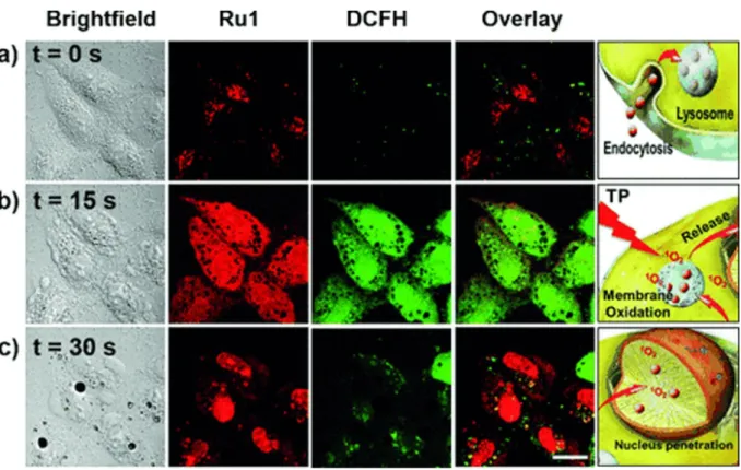
Biological evaluation on Ru(II) complexes containing pdppz ([2,3-h]dipyrido[3,2-a:2’,3’-c]phenazine) ligands was published in 2015.16 Complexes 7 and 8 were expected to bind DNA because of their extended dppz ligand, while complex 9 was used as a control (see Figure 5 for chemical structures). Experiments confirmed that complexes 7 and 8 were able to intercalate DNA in non-cell environment. Incubation of complex 8 with the plasmid pBR322 upon light irradiation (390nm, 2 J cm-2) caused single and double breaks in the DNA. Such effect was not seen with complex 7. It was shown that HeLa cells could actively uptake compounds 7 and 8- in a temperature-dependent manner. Confocal microscopy studies of complex 8 demonstrated that this compound colocalised with mitochondria and lysosomes, which clustered near the nucleus. It is possible that small amounts of 8 were also able to localise to the nucleus. Alkaline comet assay revealed DNA damage in treated and irradiated cells. ICP-MS experiments would have been an attractive addition to this study.
30
Figure 5. Chemical structures of complexes 7, 8, 9.
Cytotoxicity of the complexes in the dark and light conditions (≥400 nm, ~18 J cm-2) was examined in HeLa (cervical cancer), two mesothelioma cell lines (CRL5915 and One58), in Mutu-1 (Epstein-Barr virus-related Burkitt lymphoma) and DG-75 (Burkitt lymphoma) cell lines. Complex 7 did not show any dark or light cytotoxicity. Complex 8 was moderately cytotoxic in the dark (Inhibitory concentration 50 -IC50 values ranged from >100 to 40.2 µM).
Light irradiation of the treated cells caused phototoxic effect (IC50 values ranged from 42.8 to
8.8 µM). Pre-treatment of Hela cells with N-acetylcysteine (NAC), an established antioxidant, confirmed that ROS were involved in cell death. Hela cells were 50% more viable with the NAC treatment upon light irradiation. Real-time confocal microscopy demonstrated that HeLa cells treated with 8 displayed an apoptotic morphology upon light irradiation. Such result was confirmed by Fluorescent Activated Cell Sorting (FACS) analysis. Interestingly cell death could be prevented when cells were co-treated with VAD-fmk (inhibitor of caspases). Hence, these results demonstrated that 8 triggered apoptotic cell death in the treated cells.
31
Another set of four Ru (II) compounds that target mitochondria was synthesised by the Chao group in 2015 (see Figure 6 for structures).29
32
The triphenylphospine (TPP) present in complexes 12 and 13 adds lipophilic character to the compounds, resulting in better mitochondria targeting abilities.30 Confocal microscopy with Mitotracker Green in HeLa cell line revealed that complex 13 localises in the mitochondria. Three other compounds were found to moderately localise in that compartment. Localisation results were confirmed by ICP-MS analysis, showing that complexes 10-12 were present in higher amount in the cytoplasm (Figure 7).
Figure 7.ICP-MS quantification of the internalized Ru by the HeLa cells. Figure taken from ref 30 with permission from Elsevier.
All four compounds were designed to produce singlet oxygen not only using a one-photon but also a two-photon irradiation process. Confocal microscopy images of Hela cells taken before and after two-photon irradiation (810-830 nm for 3 minutes; 800 J.cm-1) with 2,7-dichlorodihydro-fluorescein diacetate (DCFH-DA) allowed verifying singlet oxygen production in all samples. Dark and light cytotoxicities of all compounds were tested under one-photon irradiation. Compounds were not toxic under dark conditions (IC50 >100 µM). After
irradiation (LED source; 450 nm; 12 J.cm-2), complexes 10-12 showed similar cytotoxicity, varying from 12.4 to 15.5 µM. Probably due to its high concentration in mitochondria, complex
33
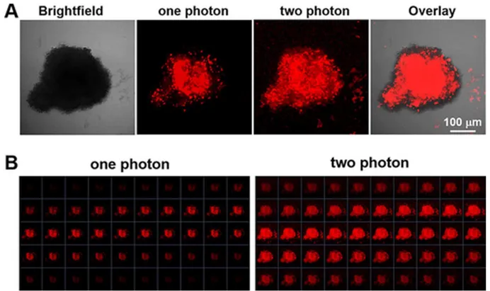
13 was found to be the most effective compound tested, with a PI >28. Since monolayer cell
cultures are not a good model for tumour treatment, HeLa multicellular tumour spheroids (MCTS) were used for further tests. Diffusion of the compounds (10 µM, 8 h treatment) was examined in 800 µm MCTSs. Treated spheroids were imaged with one-photon and two-photon z-stack microscopy. The luminescence signal of the compounds was found in all depth sections of the spheroids. Two-photon microscopy showed deeper penetration of the complexes through spheroids than one-photon microscopy, probably due to its excitation wavelength in the therapeutic window. This confirmed the high permeability of the complexes through the MCTSs. Singlet oxygen generation with DCFH-DA was also investigated in MCTS. Enrichment of the singlet oxygen signal was observed in the treated spheroids. The results showed lower signal of produced singlet oxygen in the cores of the spheroids as compared to their surface. Compounds treatment also inhibited MCTSs growth after irradiation with two-photon technique. The best results were obtained again with complex 13. All synthesised compounds exhibited good photodynamic therapy ability against the HeLa cell line. However, further investigations should include healthy cells to establish a possible therapeutic window for these compounds.
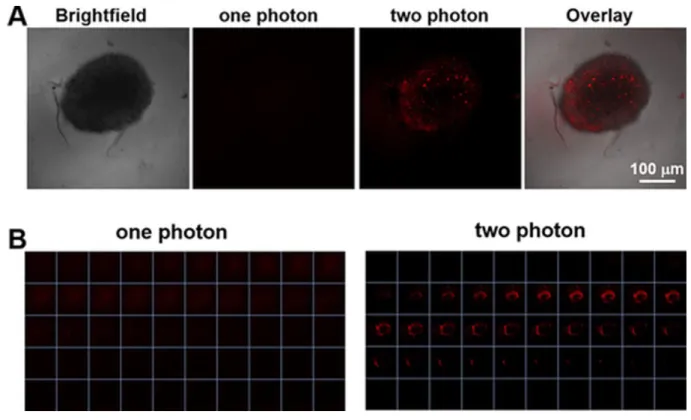
An interesting study was recently published by the same group, who designed mitochondria-localising Ru(II) complexes that can be activated by glutathione (GSH).31 The aim of the study was to improve the tumour selectivity of the Ru complexes that are used as PDT PSs. Complex
14 is a dinuclear Ru(II) complex, which is bridged by a GSH activating ligand
4,4”-azobis(2,2’-bipyridine) (Figure 8). Specific properties of the ligand cause quenching of luminescence of the Ru complex. Since intracellular concentration of GSH in cancer cells are higher than in healthy ones, the authors were hoping that the complex would be activated and transformed into complex 15 (Figure 8), and this mostly in cancer cells.
34
Figure 8. Chemical structures of complex 14 and complex 15.
For the experiments, Chao and co-workers used two cell lines, namely HeLa and LO2 (human fetal hepatocyte- healthy control). Both were cultured in monolayers as well as in MTCSs. ICP-MS and confocal microscopy experiments confirmed that the mitochondria were the main target for complex 14. As expected, LO2 cells displayed a much weaker accumulation of the complex compared to Hela. Two-photon irradiation (810 nm, 100 mW, 80 MHz, 100 fs) was used to establish ROS generation in treated 2D and 3D cell cultures. A strong green fluorescence of the ROS indicator was detected, confirming that the complex was able to permeate the MCTSs and induce single oxygen production. Cytotoxicity studies demonstrated that complex 14 was not toxic in the dark (IC50> 70 M) for both cells lines. After 15 min irradiation at 450 nm (20
mW.cm2), its cytotoxicity raised to about 5 M for HeLa and 13 M for LO2 cells. Similar
35
100 M) and became more harmful on MCTSs after light irradiation (5.71 µM). Viability of the MCTSs was checked by Calcein AM staining. Irradiation of treated cells caused loss of the fluorescent signal from the dye, suggesting cell death. It is worth noting that MCTSs treated with complex 14 at 10 uM concentration stopped growing two days after two-photon irradiation, whereas the control group treated with the same concentration of cisplatin kept growing. Of note, annexin V and propidium iodide (PI) staining showed that apoptosis was the main cause of cell death.
In 2018, Stang, Chao and coworkers prepared a tetrametallic macrocyclic structure containing Ru(II) and Pt(II) atoms, that can be used in two-photon PDT (Figure 9).32
Figure 9. Chemical structures of complex 16.
The addition of the Pt(II) moieties to the two Ru(II) complexes was made to enhance the intrinsic photophysical properties of the Ru(II) complexes. Impressive two photon absorption (TPA) cross-section values of 1371 GM were obtained, which were much higher than the one of [Ru(bpy)3]2+ itself (66 GM). Moreover, the intersystem crossing process was enhanced,
which elevated the singlet oxygen quantum yield value to 88% in mehanol, when [Ru(bpy)3]2+]
36
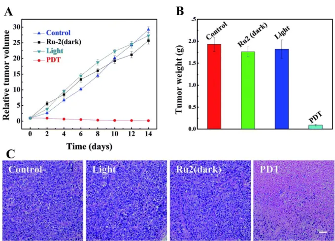
accumulating in the mitochondria and the nucleus. ICP-MS results corroborated those of microscopy, indicating that, after 2 h incubation, complex 16 (5 µM) localised in mitochondria (67 %) and in the nucleus (25 %). Cellular uptake data revealed that complex 16 entered the cells through endocytosis pathway. Cytotoxicity experiments were performed on HeLa, A549, A549R (cisplatin resistant cell line), KV (multi-resistant human oral floor carcinoma) and PC-3 (prostate cancer) cell lines. The PI values ranged between 11.6- 114 (irradiation conditions: LED source; 450 nm, 21.8 mW cm-2, 5 min). Since A549 cells displayed the highest PI, they were chosen as a model cell line for further studies. DCFH-DA staining and calcein AM/ethidium homodimer-1 (EthD-1) co-staining after two photon (TP) irradiation of the treated cells confirmed that compound 16 can generate singlet oxygen and cell death only in the irradiated area. Compound 16 caused cell apoptosis, confirmed by annexin V and PI staining as well as by elevated levels of caspase-3/7. To assess the impact of complex 16 in mitochondria and nucleus, several tests were performed. The mitochondrial membrane potential (MMP) was significantly lower in irradiated cells. TP irradiation also caused DNA fragmentation in the nucleus. Due to these promising results obtained in vitro, in vivo studies in mice were performed. To assess two photon photodynamic therapy (TP-PDT) efficacy of complex 16, A549 tumour bearing nude mice were used. The group treated with complex 16 (0.5 mg kg-1) and irradiated with TP laser (800nm, 50 mW, 20 s mm-1) did not exhibit observable weight loss. The tumour volume of the treated group was reduced by 78%, while control mice groups showed 13-fold increase in tumour mass. Additionally, examination of tumour tissue of the treated group showed pathological changes, which were not observed in other organs like liver, kidney, heart, etc.
37
Nucleus targeting compounds
Besides mitochondria, another important target for PDT PSs is the nucleus. Generation of singlet oxygen or other ROS, in close proximity to the DNA, might allow for DNA damage, and finally lead to cell death. It is known that dipyridophenazine (dppz) ligands have the ability to intercalate within DNA.33, 34 That is why in 2014 six different [Ru(bpy)2dppz]2+ derivatives
17-22 were investigated by our groups (Figure 10).35
Figure 10. Chemical structures of [Ru(bpy)2dppz]2+ derivatives.
Singlet oxygen production study showed that all compounds had a high efficacy for 1O2
production but only in hydrophobic environment. The excited state of the complexes bearing a dppz ligand are quenched very fast in the presence of water molecules.36 All synthesised complexes were found to be non-cytotoxic in the dark (IC50 >100 µM) against HeLa and
MRC-5 cells (normal lung fibroblast). Light cytotoxicity studies were performed using two different light treatments: 10 min at 350 nm (2.58 J.cm-2) and 20 min at 420 nm (9.27 J.cm-2). Among all compounds, only complexes 17 and 18 showed an interesting phototoxic effect. The PI value for complex 9 was higher than 150, while for complex 18 it was 42. The cellular uptake of the Ru compounds was investigated by High-Resolution Continuum Source Atomic Absorption Spectrometry (HR-CS AAS), showing that it correlated well with toxicity studies. The most cytotoxic complexes 17 and 18 had the highest accumulation in the HeLa cells (1.08 and 1.76 nmol Ru per mg protein). Accumulation of the compounds in the MRC-5 cell line was different
38
since only 0.76 and 0.18 nmol Ru per mg protein were determined. This indicates that the complexes penetrated the non-cancerous cell line to a lesser extent than the cancerous line. Cellular localisation of complexes 17 and 18 was investigated using confocal microscopy.
Figure 11. Cellular localisation of complex 18. Figure taken from ref 36, with permission from
John Wiley and Sons.
The first complex was difficult to detect even when cells were treated with high doses of compound. The low luminescence quantum yield is probably responsible for this result. On the other hand, complex 18 was able to accumulate preferentially in the nucleus. Because of the luminescent quenching effect of the complex in aqueous environment mentioned above, fluorescence microscopy localisation was supported by HR-CS AAS. The results showed that both complexes efficiently accumulated in the nucleus (0.43 ± 0.05 and 0.96 ± 0.06 nmol Ru per mg protein). To check if nuclear localisation and binding to DNA might have been the reason of toxicity, DNA photocleavage experiments were conducted. Treatment of pcDNA3 plasmid with complexes and irradiation at 420 nm for 20 min (9.27 J.cm-2) showed that both complex 17 and 18 were able to cleave plasmid DNA. Administration of compounds in the dark did not cause cleavage of the plasmid. In a follow up study, our groups further explored the molecular cell death mechanism of complex 18.37 Mechanistic studies on the outcome of DNA binding led to the conclusion that irradiation of the intercalated compound caused oxidative
39
damage of purines in DNA. Importantly, alkaline comet assay supported these results in living cells. Confocal microscopy images of different cell lines such as U2OS (human bone osteosarcoma), MCF-7 (mammary gland adenocarcinoma) and RPE-1 (normal retina pigmented epithelium) confirmed that the complex was mainly localised in the nucleus 35.
ICP-MS confirmed these results. The determination of the presence of specific markers of DNA damage response, analysis of DNA content and cytotoxicity studies after irradiation showed that cells underwent cell cycle arrest and loss of viability. Annexin V and PI staining experiments of interphase cells excluded classic apoptotic or necrotic cell death. Further analysis demonstrated that cell death was caused by DNA damage and endoplasmic reticulum- (ER) mediated stress response pathways. On the other hand, treatment and irradiation of mitotic cells caused death according to classic apoptotic pathways, indicating two distinct modes of cells death in interphase or mitosis and pointing to the potential of the use of these compounds in combination with established cancer therapeutics.
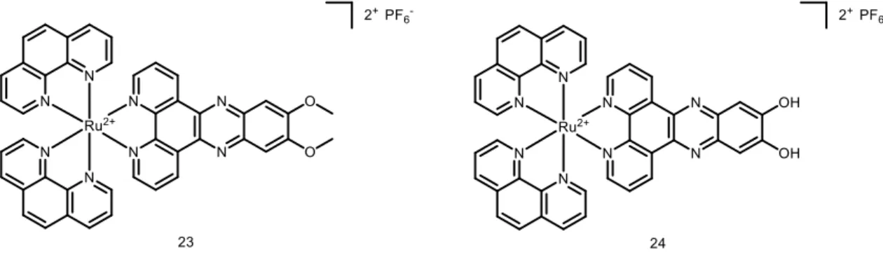
Further studies on Ru(II) polypyridyl complexes with dppz ligands were performed by our group in collaboration with the Chao group.28 Two substitutionally inert complexes, namely [Ru(phen)2(dppz-7,8-(OMe)2]2+ (complex 23) and [Ru(phen)2dppz-7,8(OH)2]2+ (complex 24)
were investigated (see Figure 12 for structures).
40
The aim of this study was to investigate if small structural differences could cause significant changes in the biological response. It is worth noting that both complexes were investigated for use in one-photon (OP) and two-photon (TP) PDT. Dark and light cytotoxicity studies on HeLa and MRC-5 cell line monolayers showed that the introduction of -OMe groups on the ligand enhanced toxicity compared to those bearing the -OH groups (decrease of the IC50 value from
16.7 ± 2.6 µM in -OH bearing compound to 3.1 ± 0.6 µM in -OMe compound in HeLa). Both complexes were also much more effective than the positive control aminolevulinic acid (5-ALA), an approved PDT PS. Interestingly, the compounds were also studied on 3D multicellular tumour spheroid to provide a comprehensive overview on how Ru(II) complexes might act in solid tumours. Surprisingly, only complex 23 was active on MCTSs upon light irradiation (LED light source; IC50 32.5 ± 6.8 µM). To further explore the mechanism of action
of the complexes, cellular localization and uptake of the compounds were studied. ICP-MS showed that the amount of complex 23 was much higher in HeLa cells than complex 24 (2.4 nmol Ru/mg protein to 0.9 nmol Ru/mg protein). This result might explain the differences between the IC50 values obtained for both complexes in the dark and upon light irradiation.
Confocal microscopy showed that the Ru complexes under study localised in different compartments of the cell. Complex 23 was found to accumulate in the nucleus and mitochondria while 24 localised in the outer cell membranes. Imaging was also performed after light irradiation. Complex 23 changed its localisation and moved completely into the nucleus, probably as result of damage generated by singlet oxygen in membranes, enabling the compound to reach the nucleus. Worthy of note, these Ru complexes might also localise in other compartments that escaped detection by confocal microscopy. Indeed, due to luminescence quenching in aqueous solution of these dppz-containing complexes, their detection is only possible in hydrophobic environment.38 OP and TP absorption was also used