UNIVERSITE
DE
GENEVE
FACULTE
DE
MEDECINE
Section de Médecine CliniqueDépartement de Pédiatrie
Thèse préparée sous la direction du Professeur Claire-Anne Siegrist
ANOMALIE DE PRODUCTION D’ANTICORPS CONTRE LES
ANTIGENES PNEUMOCOCCIQUES POLYSACCHARIDIQUES ET
PROTEIQUES CHEZ LES ENFANTS AVEC INFECTIONS
RECIDIVANTES DES VOIES RESPIRATOIRES INFERIEURES
Thèse
Présentée à la Faculté de Médecine de Genève Pour obtenir le grade de Docteur en Médecine
Par Isabelle Rochat De Le Lieu, VD Thèse N° 10420 Genève 2005
Remerciements
J’aimerais exprimer toute mon admiration et ma reconnaissance aux femmes médecins qui ont jalonné mon parcours : Professeur Claire-Anne Siegrist, qui m’a encouragée dès l’ébauche de ce projet et m’a conseillée pendant et après sa réalisation. Professeur Constance Barazzone, qui m’a prodigué un soutien constant et m’a appris la pneumologie pédiatrique. Et Docteur Antoinette Vincent, qui m’a donné le virus de la pneumologie et transmis des valeurs de vie, autant professionnelle que personnelle.
Un grand merci à l’équipe du Centre de Vaccinologie et d’Immunologie Néonatale de m’avoir si bien accueillie et fait découvrir la recherche fondamentale.
Je remercie également Professeur Suzanne Suter, qui a permis la réalisation financière de ce projet.
Table des matières
A. Résumé 5
B. Abréviations utilisées 6
C. Introduction au travail 7
1 Généralités 7
2 Déficits immunitaires et infections pulmonaires 7 3 Détection des anticorps spécifiques pour le diagnostic des
enfants avec infections récidivantes : l’expérience du Centre
de Vaccinologie de Genève 8
4 Altération de la capacité de réponse aux antigènes polysaccharidiques 8 5 Réponse immunitaire aux antigènes protéiques et polysaccharidiques 10
6 Bibliographie 11
D. Article
I. Introduction 13
II. Materials and Methods 15
2.1 Patient population 15
2.2 Determination of serum immunoglobulins, anti-tetanus and
anti-diphtheria antibodies 16
2.3 Determination of type-specific S.Pneumoniae antipolysaccharide
antibodies 17
2.4 Definition of normal responses to S.Pneumoniae PS 18 2.5 Determination of antibodies to S.Pneumoniae protein antigens 19
III. Results 21 3.1 Children with RRTI have normal or high serum Ig and responses
to protein vaccines 21
3.2 Responses to pneumococcal polysaccharides 21 3.3 Maturation of pneumococcal protein antibodies in children with
recurrent lower respiratory tract infections 23 3.4 Responses to S.Pneumoniae protein and polysaccharide antigens in children with recurrent lower respiratory tract infections 24
IV. Discussion 25
V. Acknowledgements 27
VI. References 28
VII. Tables 34
VIII. Figure legends 36
A. Résumé
Les infections respiratoires récidivantes (IRR) des nourrissons reflètent souvent un défaut de production d’anticorps contre les polysaccharides (PS) bactériens, production d’anticorps dont la maturation est lentement progressive. Dans ce travail, nous avons étudié la maturation des réponses aux antigènes pneumococciques chez 54 enfants (12 mois-12 ans) référés pour IRR persistant après 24 mois. Malgré une exposition probablement précoce et répétée aux pneumocoques, les taux d'anticorps contre les PS pneumococciques étaient faibles/absents chez 50%. 16 patients ont été vaccinés par des PS pneumococciques, mais 9 (56%) n’ont pas répondu à cette vaccination. Ce défaut d’immunité ne semble pas spécifique des PS puisque 44% des patients sans réponse aux PS avaient également des taux d’anticorps faibles/absents contre 3 protéines pneumococciques, comparé à <10% des enfants contrôles. Ainsi, les IRR n’induisent pas toujours une production appropriée d'anticorps contre les antigènes bactériens, posant la question de la contribution de ce défaut d’anticorps à la persistance des IRR.
B. Abréviations utilisées
CVI: common variable immunodeficiency Ig: immunoglobulins
PAD: polysaccharide antibody deficiency PS: polysaccharides
RRTI : recurrent respiratory tract infections URTI : upper respiratory tract infections PspA: pneumococcal surface protein A PsaA: pneumococcal surface adhesin A AOM: acute otitis media
WB: wheezy bronchitis
C. INTRODUCTION AU TRAVAIL
1. Généralités
Il n’est pas rare que le pédiatre soit confronté à des enfants « qui sont toujours malades » et qui passent d’une infection des voies aériennes à l’autre. Dans la majorité des cas, la fréquence et la nature de ces infections font partie du développement normal de l’enfant et diminuent après l’âge de 2 ans. Dans certains cas, au contraire, elles sont le témoin d’une maladie associée. En effet, de nombreux facteurs de risque et pathologies sous-jacentes peuvent favoriser les infections itératives des voies aériennes. Notamment, la présence d'un déficit immunitaire, bien que rare dans la population pédiatrique globale (1/10000), est fréquemment associée aux infections respiratoires récidivantes.1,2
2. Déficits immunitaires et infections pulmonaires
Chez l'enfant, l'expression clinique principale des déficits immunitaires se manifeste sous forme d'infections respiratoires à répétition. En effet, les mécanismes de défense contre les agents infectieux au niveau pulmonaire dépendent non seulement de phénomènes non immuns (activité mucociliaire, réflexe de toux et d’oblitération oropharyngée), de l’immunité innée (macrophages, neutrophiles présents dans la muqueuse du tractus respiratoire) mais aussi de l'immunité humorale.3,4 Parmi les déficits immunitaires primaires et secondaires, ceux touchant l'immunité humorale sont les plus fréquents.5 Leur spectre de sévérité va de l’absence quasi totale de lymphocytes B matures avec un taux d’immunoglobulines sériques anormalement bas (X-linked agammaglobulinemia) à un défaut isolé d’anticorps contre les polysaccharides avec un taux d'immunoglobulines sériques normal pour l'âge. Le syndrome de défaut sélectif en anticorps antipolysaccharides est jusqu’à 4 fois plus fréquent que les autres anomalies de l’immunité humorale.6 Il s’agit du déficit immunitaire le plus fréquemment impliqué dans les cas d’infections respiratoires récidivantes. Il est donc important, en présence d'un enfant souffrant d'infections respiratoires de fréquence et/ou de sévérité élevées, et ceci malgré un développement staturo-pondéral harmonieux, de procéder à une évaluation de son système immunitaire. Le but de cette démarche est de répondre à la question:
"L'immunité humorale de ce patient est-elle normale ou les infections à répétition sont-elles le reflet d'une atteinte de la production d'anticorps?".
La mise en évidence précoce d'un déficit immunitaire permet d'établir une prise en charge adéquate (vaccin pneumococcique conjugué, antibiothérapie alternée, immunoglobulines intraveineuses) afin de ralentir, voire d'éviter, l'apparition d'une atteinte pulmonaire chronique ainsi que l'établissement de séquelles sous forme de limitation des fonctions pulmonaires ou encore de bronchiectasies.7,8
3. Détection des anticorps spécifiques pour le diagnostic des enfants avec infections récidivantes : l’expérience du Centre de Vaccinologie de Genève L’importance de disposer de méthodes permettant d’analyser la fonction immunitaire des nourrissons et jeunes enfants a conduit le Centre de Vaccinologie (Département de Pédiatrie) à développer des microméthodes permettant la détermination quantitative par ELISA des anticorps induits en réponse à la vaccination (tétanos, diphtérie, H. influenzae du groupe B) et/ou à l’exposition (S. pneumoniae, sérotypes de base 14, 19F, 23F et sérotypes supplémentaires 1, 5 et 7). Ces analyses, validées et accréditées depuis janvier 2004, sont demandées par un nombre croissant de médecins, essentiellement de Suisse Romande mais aussi de Suisse Alémanique. En 2003, 3594 analyses ont ainsi été effectuées sur 1478 échantillons. Elles ont permis d’identifier les enfants ayant des réponses B trop faibles pour l’âge et/ou l’anamnèse vaccinale, de préciser le type de dysfonctionnement (réponses aux protéines et/ou aux polysaccharides), de proposer les vaccinations supplémentaires éventuellement nécessaires à la réactivation ou au maintien de leur immunité.
4. Altération de la capacité de réponse aux antigènes polysaccharidiques
Le premier rapport décrivant une association entre les infections respiratoires récidivantes et un déficit sélectif d’anticorps antipolysaccharides date de 1987.9 Plusieurs autres études et descriptions de cas ont été publiées par la suite, parmi lesquelles:
Dans un collectif de 45 patients âgés de 1.7 à 17.1 ans présentant des infections récidivantes des voies respiratoires (otites moyennes aigues, otorrhée, sinusites, pneumonies), 14% des patients montrent une incapacité
de réponse aux antigènes polysaccharidiques après vaccination par Pneumovax®.10
Sur 100 patients âgés de 3 à 8.4 ans présentant des infections respiratoires récidivantes (otites, sinusites, pneumonies), 6.5% ne répondent pas au vaccin pneumococcique polysaccharidique.11
Parmi 51 patients âgés de 22 à 158 mois évalués pour des infections respiratoires récurrentes (otites, sinusites), 20% présentent une incapacité de réponse aux polysaccharides capsulaires d'un vaccin Hib non conjugué.12
Ainsi, le pourcentage de patients présentant des infections respiratoires récidivantes associées à une incapacité de réponse aux antigènes polysaccharidiques varie selon les études. Il s'agit néanmoins d'un déficit immunitaire fréquent, dont les caractéristiques cliniques sont multiples et ne se limitent pas seulement aux infections invasives à S. Pneumoniae.
Rappelons qu’une des caractéristiques de ce syndrome est un taux d’immunoglobulines sériques normal pour l’âge.
L’identification d’un défaut de réponse aux antigènes polysaccharidiques repose en partie sur la mesure des taux d'anticorps d'exposition naturelle aux Pneumocoques, mais surtout sur ceux obtenus suite à la vaccination pneumococcique multivalente non conjuguée (Pneumovax®). Il est important de noter qu’un défaut de réponse aux polysaccharides ne peut être évoqué qu’après l’âge de 2 ans puisque l'ontogénèse de la réponse immunitaire aux polysaccharides est un processus tardif qui n'est terminé que vers l’âge de 24 mois.
Au contraire, la production d’anticorps contre les antigènes protéiques est effective dès les premiers mois de vie. Ceci se traduit part des taux d'anticorps contre les antigènes de vaccination (tétanos, diphtérie) avoisinant les valeurs adultes mêmes chez les jeunes enfants. Dans les différentes cohortes mentionnées précédemment, l'ensemble des patients décrits montrait une production adéquate d’anticorps contre les antigènes de vaccination de nature protéique.
5. Réponse immunitaire aux antigènes protéiques et polysaccharidiques
Chez l’enfant sain, les processus caractérisant la maturation différée de la réponse immunitaire aux antigènes polysaccharidiques par rapport aux antigènes protéiques ont été bien décrits (immaturité de la zone marginale de la rate, nombre réduit de lymphocytes B CD21, activité réduite du complément C3).10,13 Cependant, il n’est pas encore défini si ces mêmes causes expliquent également la persistance d'un déficit de réponse aux antigènes polysaccharidiques après l'âge de 24 mois.
En raison de l'apparition tardive de la capacité de réponse aux antigènes polysaccharidiques des jeunes enfants, et ainsi de leur susceptibilité accrue aux infections à Pneumocoque, de nombreux travaux se sont penchés sur la réponse aux antigènes pneumococciques de nature protéiques. Ces antigènes sont présents et communs à tous les Pneumocoques. La réponse aux protéines pneumococciques (pneumococcal surface protein (PspA), pneumococcal surface adhesin A (PsaA) et pneumolysin (Ply)) a été étudiée tant chez l'animal que chez l'adulte et l'enfant.14-16 Les travaux rapportés concernent essentiellement des enfants sains présentant des otites moyennes aigues non compliquées, ou encore des enfants souffrant d'infections invasives à Pneumocoques. Les jeunes patients souffrant d'infections respiratoires récidivantes n'ont pas encore fait l'objet de travaux de recherche publiés.
De plus, il n'existe que quelques publications décrivant à la fois la production d'anticorps antipolysaccharidiques et antiprotéiques pneumococciques. Ces travaux ne permettent pas d'établir un lien entre le défaut de production d'anticorps antipneumococciques et antiprotéiques chez un même patient.
Aucune étude n'a décrit simultanément la capacité de réponse aux polysaccharides et aux protéines pneumococciques dans un contexte d'infections respiratoires récidivantes. Notre travail a pour but d'étudier et de comparer la réponse immunitaire aux antigènes polysaccharidiques et protéiques du Pneumocoque chez les enfants d'âge préscolaire et scolaire présentant une telle symptomatologie clinique. Il s'agit d'une étude de cohorte rétrospective qui permettra de définir un état des lieux et d'établir la nécessité d'une étude prospective à large échelle servant à évaluer objectivement la réponse clinique et immunologique à une vaccination pneumococcique.
6. Bibliographie
1. Couriel J. Assessment of the child with recurrent chest infections. Br Med Bull. 2002;61:115-32.
2. Hansen IC, Shearer SW. Acute and Chronic Bronchitis. In: McMillan JA, DeAngelis DC, Feigin RD, Warshaw JB, ed. Oski's Pediatrics: Principles and
Practice. Philadelphia, PA: Lippincott, Williams and Wilkins; 1999:1211-1214.
3. Gerritsen J. Series: Basic Sciences. Host defence mechanisms of the respiratory system. Paediatr Respir Rev. 2000;1:128-34.
4. Larsen GL. Host defense systems of the lung. In: Taussig LM, Landau, LI, ed.
Pediatric Respiratory Medicine. St-Louis, Missouri: Mostby; 1999:57-75.
5. Dizon JG, Goldberg BJ, Kaplan MS. How to evaluate suspected immunodeficiency. Pediatr Ann. 1998;27:743-50.
6. Javier FC, 3rd, Moore CM, Sorensen RU. Distribution of primary immunodeficiency diseases diagnosed in a pediatric tertiary hospital. Ann
Allergy Asthma Immunol. 2000;84:25-30.
7. Dagli E. Non cystic fibrosis bronchiectasis. Paediatric Respiratory Reviews. 2000;1:64-70.
8. Callahan CW, Redding GJ. Bronchiectasis in children: orphan disease or persistent problem? Pediatr Pulmonol. 2002;33:492-6.
9. Ambrosino DM, Siber GR, Chilmonczyk BA, Jernberg JB, Finberg RW. An immunodeficiency characterized by impaired antibody responses to polysaccharides. N Engl J Med. 1987;316:790-3.
10. Rijkers GT, Sanders EA, Breukels MA, Zegers BJ. Infant B cell responses to polysaccharide determinants. Vaccine. 1998;16:1396-400.
11. Epstein MM, Gruskay F. Selective deficiency in pneumococcal antibody response in children with recurrent infections. Ann Allergy Asthma Immunol. 1995;75:125-31.
12. Herrod HG, Gross S, Insel R. Selective antibody deficiency to Haemophilus influenzae type B capsular polysaccharide vaccination in children with recurrent respiratory tract infection. J Clin Immunol. 1989;9:429-34.
13. Timens W, Boes A, Rozeboom-Uiterwijk T, Poppema S. Immaturity of the human splenic marginal zone in infancy. Possible contribution to the deficient infant immune response. J Immunol. 1989;143:3200-6.
14. Soininen A, Pursiainen H, Kilpi T, Kayhty H. Natural development of antibodies to pneumococcal capsular polysaccharides depends on the serotype: association with pneumococcal carriage and acute otitis media in young children. J Infect Dis. 2001;184:569-76.
15. Soininen A, Lahdenkari M, Kilpi T, Makela PH, Kayhty H. Antibody response to pneumococcal capsular polysaccharides in children with acute otitis media.
Pediatr Infect Dis J. 2002;21:186-92.
16. Virolainen A, Jero J, Chattopadhyay P, Karma P, Eskola J, Leinonen M. Comparison of serum antibodies to pneumolysin with those to pneumococcal capsular polysaccharides in children with acute otitis media. Pediatr Infect Dis
D. Article
I. Introduction
Respiratory tract infections are the most frequent acute illnesses of childhood.1 It is not uncommon for children up to 5 years of age to present 6-10 episodes of respiratory infections per year and therefore appear as “continually infected”. Most episodes involve the upper respiratory tract, are mild and remain self-limited. Involvement of the lower respiratory tract occurs in a subgroup of patients, may be severe and includes the risk of long term pulmonary damage such as bronchiectases. Children with recurrent lower respiratory tract infections thus represent a significant diagnostic and therapeutic challenge.
Numerous predisposing factors and underlying conditions have been associated with recurrent respiratory tract infections (RRTI).1,2 These primarily include children with antibody deficiencies, the most prevalent immunodeficiency in childhood,3 who may present with normal growth and symptom-free intervals between frequent upper and lower respiratory tract infections. Since the initial description of a patient with recurrent sino-pulmonary infections, normal immunoglobulin levels and selective absence of IgG antibody response to polysaccharide (PS) antigens,4 multiple reports have examined the association between deficient response to PS antigens and RRTI in children.5-7 Polysaccharide antibody deficiency (PAD) is a form of immunodeficiency defined on the basis of a clinical history of recurrent infections, normal serum immunoglobulin levels and antibodies to protein vaccine antigens and the inability to mount an adequate antibody response in vivo to immunization with bacterial PS (H. influenzae b, S. pneumoniae).8 The mechanisms at the basis of this
pathological condition are not well understood. The possibility that some children with PAD may be manifesting a transient delay in their immune maturation has been proposed,6 as impaired production of antibodies to PS antigens is a normal feature in children below 18-24 months of age.9 However, some patients do not outgrow these limitations and present with persisting selective PAD or subsequently progress towards hypogammaglobulinemia and common variable immunodeficiency (CVI). Identifying the children likely to outgrow a transient PAD deficiency, i.e. whose prognosis is excellent, from those likely to progress towards CVI is currently impossible. This generates significant concerns in parents and physicians, results into repeated extensive immune workup and leads to the large use of both prophylactic and therapeutic measures.
In contrast to the delayed ontogeny of anti-PS antibodies, young children are generally capable of eliciting immune responses to protein antigens already early in life. The objective of a protein-based pneumococcal vaccine that would be immunogenic in early life and whose scope would not be restricted to the few serotypes that can be included into glycoconjugate vaccines has generated interest in pneumococcal protein antigens common to most bacterial strains. These proteins include pneumococcal surface protein A (PspA), pneumococcal surface adhesin A (PsaA), pneumolysin (Ply), PspC and others (reviewed in 10). These antigens induce protection against pneumococcal nasopharyngeal carriage and invasive infections in animal models and exhibit sufficient cross-protection to be considered as potential vaccine candidates.11-13
Relatively little is known yet on the natural development of antibodies to pneumococcal proteins. It is clear, however, that antibodies to PspA and PsaA
increase as the proportion of children who have been exposed to S. pneumoniae increases.14 PsaA antibodies are induced early in response to nasopharyngeal pneumococcal exposure, being detected in 94% of previously exposed children and 80% of their mothers. Conversely, antibody responses to PspA are induced later in life and are only present in approximately 50% of healthy 2 years old children.14,15 Whether this reflects the role of pneumococcal infections versus carriage in the induction of PspA antibodies is not known. Importantly, children with documented exposure to S. pneumoniae have significantly higher antibody concentrations to PspA and PsaA than unexposed children at each time point assessed.14 Consequently, these antibodies should be prevalent in patients with RRTI, of which a significant fraction is of pneumococcal aetiology. In this study, we decided to take advantage of the availability of these pneumococcal protein antigens to compare the maturation of antibody responses to protein and PS pneumococcal antigens in a highly selected population of preschool and school age children referred to our center for evaluation of RRTI.
II. Materials and Methods 2.1 Patient population
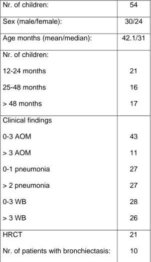
The patients of this study were referred to our pediatric pulmonology consultation between January 1st 2001 and December 31st 2002 for investigations of RRTI. We defined RRTI as at least 2 episodes of pneumonia and/or 3 episodes of bronchitis and/or chronic cough (present for more than 3 consecutive months). The medical records were probed for demographic, clinical and radiological data (Table 1). Patients were excluded from the study if they suffered from anatomic abnormalities of
the respiratory tract, recognized disease entities such as cystic fibrosis, known immune deficiency, or atopy and asthma. Fifty-four patients 12 months to 12 years of age were included. Study patients were divided into 3 age groups for the evaluation of the maturation of their antibody responses (group 1: 12 to 24 months (n = 20), group 2: 25 to 48 months (n = 17), group 3: > 48 months (n = 17)). Routine immunological investigations included the determination of total serum immunoglobulins and vaccine (tetanus, diphtheria, Hib) antibodies. All serum specimens were stored at -80°C until use for the determination of IgG antibodies to pneumococcal PS and protein antigens.
2.2 Determination of serum immunoglobulins, anti-tetanus and anti-diphtheria antibodies
Total serum IgG, IgA, IgM were measured by nephelometry (Immage, Beckman, USA) in the first specimen available, i.e. at time of enrolment. Total serum IgE and specific IgE to common respiratory allergens (Phadiatop) were measured by fluorimetric assay (UniCAP system, Pharmacia Diagnostics, Uppsala, Sweden). Normal values for age represent a mean of 2 standard deviation (SD) above and below the mean for age.16-19 Anti-tetanus and anti-diphtheria antibodies were measured by ELISA as previously described.20 Responses were considered as normal if reaching 1 IU/l one month after the most recent vaccine dose or after adjustment for the time elapsed between immunization and testing. Children with low antibody levels were given an extra dose of diphtheria-tetanus-pertussis +/- HIB vaccine to ascertain their vaccine response capacity.
2.3 Determination of type-specific S. pneumoniae antipolysaccharide antibodies
Serum antibody titers to 3 common pneumococcal serotypes (14, 19F, 23F), selected for their higher, average or lower avidity in a large cohort of non-immunodeficient patients (not shown), were measured by ELISA at time of enrolment and 4 to 6 weeks after immunization, when applicable. We used a modified ELISA protocol based on the third-generation consensus assay recently adopted by a World Health Organization expert group.21 Microtiter plates (Nunc, Roskilde, Denmark) were separately coated with each pneumococcal PS antigen (50µl/well, concentration 1µg/ml, 5µg/ml and 2µg/ml for serotype 14, 19F and 23F respectively, ATCC, LGC Promochem, France) and incubated for 5 hours at 37°C. After washing with a 0.9% NaCl solution containing 0.05% Tween 20, plates were saturated with 100µl/well of a phosphate buffered saline-0.05% Tween 20-1% bovine serum albumin (PBST-1% BSA) solution overnight at 4°C. Sera were preadsorbed for 30 minutes at room temperature (RT) with C-PS (5µg/ml, Statens Serum Institut, Copenhagen, Denmark) and with 22F capsular PS (10µg/ml, ATCC, LGC Promochem, France) to neutralize non functional antibodies. The serum standard 89SF (FDA/CBER, Bethesda, MD) was preadsorbed with C-PS only. Serum specimens were serially diluted, added to PS-coated microtiter plates (50µl/well) and incubated for 2 hours at RT. The plates were washed 4 times. Goat anti-human IgG conjugated to horseradish peroxydase (HRP, Cappel, ICN Biomedicals, Aurora, OH) in PBST-1% BSA was added to each well for 2 hours at RT. After 5 washings, the ABTS substrate was added. Optical density (OD) was read at 405 nm after 30 minutes of color development, at RT, in the dark (Vmax kinetic microplate reader, Molecular Device, Sunnyvale, CA). The OD was converted to antibody concentrations in mg/l (SOFTmax®PRO 2.6.1, Sunnyvale,
CA.) by comparison with the standard 89SF known to contain 27.8mg/l, 13mg/l and 8.1mg/l specific IgG against serotype 14, 19F and 23F respectively.22 Concentrations below the cut-off limit of the assay were assigned a value of half the cut-off value.
2.4 Definition of normal responses to S. pneumoniae PS
In the absence of well-defined normal values for anti-pneumococcal antibodies measured after CPS-22F adsorption, conservative definitions were used to ensure the identification of children with PAD. Evidence of normal exposure-driven antibody responses to pneumococcal PS antigens was defined by the presence of a minimal antibody titer of 0.5mg/l, based on protective antibody titers against invasive infections in adults (0.25-0.3mg antibody nitrogen/ml) and against nasopharyngeal colonization in children (0.2mg antibody nitrogen/ml).8 It is generally considered that at least half of the serotypes tested should have a concentration above predefined minimum values to allow for a possible lack of exposure to certain serotypes.8,23 Increasing the number of serotypes above a certain threshold does not facilitate the identification of non-responders to PS antigens,24 and our data (not shown). Thus, to allow for a possible lack of exposure to a given serotype, evidence of normal exposure-driven responses to S. pneumoniae PS was considered as demonstrated if antibodies were above threshold values for ≥ 2 of 3 selected serotypes,8,23,24
and our data (not shown).
Patients ≥ 24 months with antibody responses < 0.5mg/l to ≥ 2 serotypes were offered immunization with a PS vaccine (Pneumovax®, Merck, USA) and their responses assessed 4-6 weeks later. Evidence of a positive vaccine response was defined by a minimal post vaccination titer of 1mg/l for ≥ 2 of the 3 serotypes tested. This value is slightly lower than the one previously reported (1.3µg/ml,23), but this
was prior to the recommendation of preadsorbing sera with both C-PS and PS of serotype 22F,22 which reduces antibody titers. Evidence of normal vaccine responses to S. pneumoniae PS was again considered as demonstrated if antibodies were above threshold values for ≥ 2 of 3 selected serotypes. This definition was based on the demonstration that responses to serotypes 14 and 19F allowed differentiating between patients able to raise antibodies to pneumococal PS or not,25 in addition to our own observations of the relative immunogenicity of serotypes 1, 5, 7F, 14, 19F and 23F in high risk groups of non immunodeficient children (not shown). Patients < 24 months of age, i.e. too young to be vaccinated with the PS vaccine, remained unclassified unless they had exposure-driven titers ≥ 0.5mg/l to at least 2 serotypes.
2.5 Determination of antibodies to S. pneumoniae protein antigens
Pneumococcal surface adhesin A (PsaA) and 3 pneumococcal surface proteins A (PspA clades RX1M1, EF3296 and EF5668) were kindly donated by Aventis Pasteur (Swiftwater, USA). PspA is a highly variable protein with an N-terminal region containing a portion of amino acids responsible for eliciting cross-protective immunity. The amino acid pattern of this protective region allows classification of PspA molecules into clades and families. RX1MI belongs to family 1, clade 2, whereas EF3296 and EF5668 belong to family 2, clade 3 and 4 respectively. These 3 clades represent > 90% of pneumococcal strains that circulate in the Northern Hemisphere.11 Serum antibody levels against PspA RX1MI, EF3296 and EF5668 and against PsaA were detected by ELISA. Maxisorp 96-well plates (Nunc, Roskilde, Denmark) were separately coated with each PspA and PsaA antigens (2µg/ml in PBS, 50µl/well) and incubated overnight at RT. The plates were washed 3 times and incubated on a shaker with serial serum dilutions in PBST-2% BSA for 2 hours at RT.
The plates were then washed 4 times prior to incubation on a shaker for 2 hours at RT, with HRP conjugated goat anti-human IgG (1/1000) in PBST-2% BSA. Following 5 washes, antibodies were detected by adding the ABTS substrate. The OD was read at 405 nm after 30 minutes of color development, at RT, in the dark. It was converted to antibody titers in U/ml by comparison with a commercial preparation of purified human immunoglobulins (Endobulin®, Baxter, Switzerland) whose specific titers against each of the 4 protein antigens had been previously determined by end-point titration. To define age-appropriate antibody responses to each pneumococcal protein in our local population, responses to PsaA and to each of the 3 PspA clades were measured in a control group of 55 age-matched children recruited after informed consent among children undergoing elective surgery. These children were free of respiratory symptoms at time of bleeding.26 Exposure-driven responses of RRTI patients were considered as low if they were absent or below the lower 90% CI value obtained in age-matched control patients.
2.6 Statistical analyses
GraphPad Prism® 3.0 software was used for statistical calculations. The Mann-Whitney test for non-parametric data was used to compare immunoglobulin concentrations between immunological categories and antipolysaccharide and antiprotein antibody titers between age groups. Correlation between antibody concentrations was obtained with the Spearman rank test (rs). Pre and post
immunization antibody titers were compared using the Wilcoxon matched pairs test. All other comparisons were done with the chi-square test. A p value of less then 0.05 was considered statistically significant.
III. Results
3.1 Children with RRTI have normal or high serum Ig and responses to protein vaccines. Immunoglobulins and protein vaccine responses were assessed in 54 RRTI patients aged 12 months to 12 years (mean 42 months, median 31 months) to rule out Common Variable Immunodeficiency (CVI). Serum immunoglobulins were high (≥2 SD for age) in 22% (IgG), 30% (IgM) and 9% (IgA). One patient had low IgM and another patient low IgA values, but IgG were normal or high in all patients. This is in accordance with previous studies reporting higher rather than lower serum IgG titers in children with recurrent infections (reviewed in 27). Total IgE levels and IgE antibodies to common respiratory allergens were only increased in 4/54 patients, confirming that RRTI were essentially not caused by atopy or respiratory allergies in this cohort. All patients had normal anti-tetanus and anti-diphtheria antibody responses at baseline or normalized their antibody levels after an additional booster dose of tetanus and diphtheria toxoid vaccines (data not shown). This confirmed that all children enrolled in this study had a normal immune response capacity to protein antigens and no underlying identified immune deficiency.
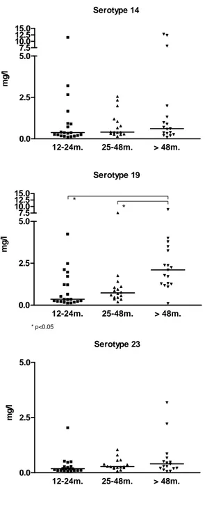
3.2 Responses to pneumococcal polysaccharides. Exposure-driven anti-PS antibodies were assessed against 3 pneumococcal serotypes (14, 19F and 23F) of high, average or lower immunogenicity, respectively (Fig 1). Serotype-specific antibodies tended to increase with age, reaching statistical significance for serotype 19F. At enrollment, 27/54 (50%) RRTI patients had IgG antibodies ≥ 0.5mg/l for ≥ 2 of the 3 serotypes tested, which was considered as evidence of normal exposure-driven responses to S. pneumoniae PS (Table II). Conversely, 27/54 (50%) RRTI patients had exposure-driven antibody concentrations < 0.5mg/l for ≥ 2 of the 3
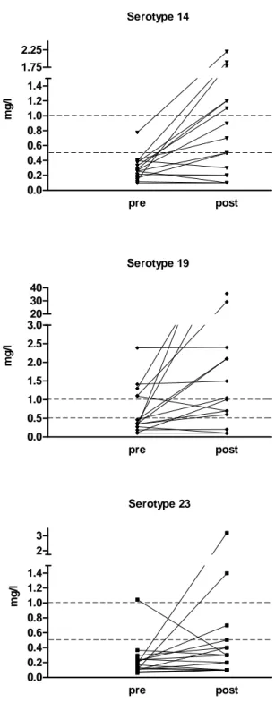
serotypes tested (Table II). Children too young to be immunized with a PS vaccine were considered as having a yet undefined response capacity to PS (Table II), except for one patient who was included in the “late responder” group given subsequent spontaneous increase in PS antibodies. Sixteen children ≥ 24 months without evidence of responses to S. pneumoniae PS were immunized with a 23-valent pneumococcal vaccine (Pneumovax®, Merck, USA) at a median age of 28 months to define their immune response capacity to bacterial PS. Post immunization antibody concentrations were significantly higher than pre immunization levels for serotypes 14 and 19F (p=0.008 and p=0.002, respectively, Fig 2). The post immunization increase in antibody titers to serotype 23F was not significant (p=0.23), confirming the lower immunogenicity of this serotype.28
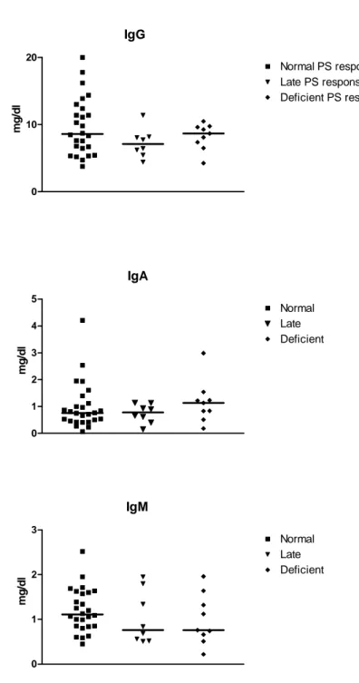
7/16 (44%) RRTI patients responded to pneumococcal PS vaccination with antibody concentrations reaching ≥ 1mg/l for ≥ 2 of the 3 serotypes tested. Without exposure-driven antibodies at time of analysis but subsequently able to respond to vaccination, they were considered as “late responders” (Table II). In contrast, PS responses remained absent or low in 9/16 (56%) patients (“non-responders”). Late responder and non-responder patients were of the same age (p=0.67) and had similar Ig values as children with normal exposure-driven PS responses (p=0.17 and 0.5, respectively, Fig 3), indicating that limited responses to S. pneumoniae PS do not result into lower serum Ig concentrations. Thus, 27/54 (50%) preschool and school-aged children referred for evaluation of RRTI had no exposure-driven responses to pneumococcal PS at time of evaluation and 9/16 failed to respond to pneumococcal PS immunization, meeting the definition of polysaccharide antibody deficiency (PAD).
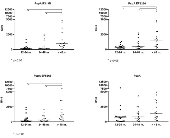
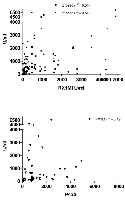
3.3 Maturation of pneumococcal protein antibodies in children with recurrent lower respiratory tract infections. IgG antibodies to 4 pneumococcal protein antigens (3 clades of PspA representing ≥ 90% of the circulating strains and PsaA) were assessed in all patients. Antibody concentrations against the 3 PspA antigens increased significantly with age (Fig 4), whereas PsaA antibody concentrations were similar in all age group, confirming their induction at an early age.29 Patient responses to individual antigens were considered as low if they were absent or below the lower 90% CI value obtained in age-matched control patients. To progress towards a definition of normal responses to pneumococcal protein antigens, the correlation between antibody concentrations to each of the 4 pneumococcal proteins was assessed. Despite their induction by a common pathogen, correlations were only modest. There was a stronger correlation among anti-PspA antibodies (RX1MI and EF3296 (rs = 0.54, p < 0.0001); RX1MI and EF5668 (rs = 0.51, p < 0.0001); EF3296
and EF5668 (rs = 0.64, p < 0.0001)), than between PspA and PsaA antibodies
(RX1MI and PsaA (rs = 0.42, p < 0.0015); EF3296 and PsaA (rs = 0.49, p = 0.0001);
EF5668 and PsaA (rs =0.5, p < 0.0001), Fig 5). This suggested considering the
presence of normal antibodies to both PspA and ≥ 2 clades of PspA as evidence of normal responses to pneumococcal proteins. Using this definition, 10/44 (23%) RRTI patients did not have evidence of age-appropriate responses to pneumococcal protein antigens despite recurrent respiratory infections of presumably frequent pneumococcal origin. This proportion was not lower (30%, p=0.37) than in age-matched control children without recurrent respiratory tract infections.
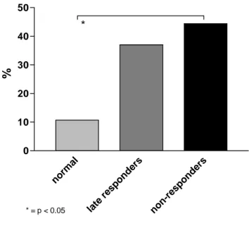
3.4 Responses to S. pneumoniae protein and polysaccharide antigens in children with recurrent lower respiratory tract infections. The unexpected absence of antibodies to pneumococcal proteins in some children with RRTI led us to assess the correlations between exposure-driven antibodies to pneumococcal protein and to PS antigens. No significant correlation was observed for any of the specific combination tested (data not shown). However, fraction of patients had low antibodies to both pneumococcal protein and PS antigens despite recurrent respiratory infections. To better characterize this subset of patients, we compared the proportion of patients without exposure-driven antibodies to pneumococcal proteins among patients with normal, delayed or deficient responses to PS (Fig 6). Exposure-driven antibodies to pneumococcal proteins were absent in 3/27 (11%) RRTI patients with normal responses to pneumococcal PS, but in 37% and 44% (p=0.03) of patients with delayed or absent responses to pneumococcal PS, respectively. This did not reflect an incapacity to respond to pneumococcal antigens as antibodies to serotype 14, 19 and 23F reached normal levels in children who received a glycoconjugate vaccine (Prevenar) after the demonstration of their unresponsiveness to Pneumovax (not shown). Thus, recurrent respiratory tract infections fail to induce circulating antibodies to both pneumococcal PS and protein antigens in a large proportion of children, despite intact responses to protein vaccines including pneumococcal glycoconjugates.
IV. Discussion
This study confirms that a large proportion (50%) of preschool and school aged patients with recurrent lower respiratory tract infections have limited exposure-driven IgG antibodies to pneumococcal PS despite normal IgG levels. It also shows that this deficiency persists in 54% of RRTI patients despite pneumococcal PS immunization. Unexpectedly, it demonstrates that a large proportion (44%) of patients with deficient responses to PS (PAD) also have absent or low exposure-driven IgG antibodies to pneumococcal proteins, failing to support the hypothesis that these patients suffer from a selective deficiency of responses to PS antigens.
The induction of antibodies to bacterial PS essentially results from nasopharyngeal colonization with S. pneumoniae, or possibly by other cross-reactive encapsulated pathogens.30 The prevalence of S. pneumoniae carriage increases with age, and even more so in conditions such as AOM or respiratory infections.31
Accordingly, antibody levels to pneumococcal PS increase with age in healthy children.32,33 This is confirmed by our observation in 50% of RRTI children of a progressive increase in exposure-driven antibody concentrations to the 3 serotypes (14, 19F and 23F) most frequently responsible for pneumococcal infections in Western Europe, in the absence of routine immunization with a glycoconjugate vaccine.34,35 Despite recurrent infections, 50% of our RRTI patients lacked antibodies to pneumococcal PS. This is in accordance with previous reports in which half of the patients presented with absent or low antibody levels at first evaluation.6,36 Absent responses to pneumococcal PS in children with RRTI despite normal Ig levels and normal responses to protein vaccines are considered as hallmarks of PAD.36-39 Using this definition, we identified 9 PAD patients among 16 RRTI children immunized with
pneumococcal PS because of absent or low exposure-driven pneumococcal antibodies, which is also in accordance with observations of others.6,36
Recurrent infections in children with PAD have essentially been attributed to a selective deficiency in their responses to PS. We show here that this may not be the case. Recurrent infections elicited antibody responses to S. pneumoniae proteins in most (77%) children. Few patients (11%) with normal responses to PS lacked antibodies to PsaA and PspA. In contrast, the absence of antibodies to pneumococcal proteins was a frequent observation in children with delayed (37%) or absent (44%) responses to PS. The absence of responses to both protein and PS pneumococcal antigens in a large proportion of patients with RRTI is unexpected. The observation (this report and 23,40) that children failing to respond to pneumococcal PS immunization appropriately respond to the same pneumococcal PS when these are conjugated to a carrier protein and given by subcutaneous injection suggests that a defective antigen presentation affects both PS and protein antigens. This could involve determinants that are intrinsic to S. pneumoniae, such as Toll-like receptor mediated recognition, or affect mucosal responses to several pathogens. Low antibody levels to PspA were reported in patients with severe invasive pneumococcal disease, both at diagnosis and during convalescence, indicating that factors may indeed limit responses to pneumococcal proteins in some patients.41 A recent study suggested that recurrent upper respiratory tract infections (URTI) result into a state of chronic inflammation42 which may affect responses to mucosal pathogens. Whether URTI were more frequent in children with defective responses to both protein and PS pneumococcal antigens is an interesting possibility.
Regardless of the exact mechanism which leaves a significant proportion of children without antibodies to both pneumococcal PS and protein antigens, this is likely to contribute to their vulnerability to RRTI. The design of this retrospective study did not allow us to objectively assess whether the pattern of responses to pneumococcal PS and protein antigens influenced the frequency and/or the type of lower respiratory tract infections. We can also not formally demonstrate whether pneumococcal PS immunization had a beneficial influence on RRTI in children who responded to immunization, whether it possibly had a deleterious influence in those who failed to respond and/or whether glycoconjugate pneumococcal immunization were beneficial in children unresponsive to PS. Anecdotal observations however suggests that this is the case and warrants prospective studies in countries where routine immunization with pneumococcal glycoconjugates is unlikely to be introduced in a near future.
V. Acknowledgements
This work was supported by research grants from the Department of Pediatrics of the Geneva University Hospital and from Aventis-Pasteur. We thank Paolo Valenti, Gianna Cadau and Christine Brighouse for excellent technical assistance. We are grateful to the Laboratory of Immunology at the University Hospital for measurements of total and allergen-specific serum IgE levels.
VI. References
1. Couriel J. Assessment of the child with recurrent chest infections. Br Med Bull. 2002;61:115-32.
2. Rubin BK. The evaluation of the child with recurrent chest infections. Pediatr
Infect Dis. 1985;4:88-98.
3. Sorensen RU, Moore C. Antibody deficiency syndromes. Pediatr Clin North
Am. 2000;47:1225-52.
4. Ambrosino DM, Siber GR, Chilmonczyk BA, Jernberg JB, Finberg RW. An immunodeficiency characterized by impaired antibody responses to polysaccharides. N Engl J Med. 1987;316:790-3.
5. Herrod HG, Gross S, Insel R. Selective antibody deficiency to Haemophilus influenzae type B capsular polysaccharide vaccination in children with recurrent respiratory tract infection. J Clin Immunol. 1989;9:429-34.
6. Sanders LA, Rijkers GT, Kuis W, Tenbergen-Meekes AJ, de Graeff-Meeder BR, Hiemstra I, Zegers BJ. Defective antipneumococcal polysaccharide antibody response in children with recurrent respiratory tract infections. J
Allergy Clin Immunol. 1993;91:110-9.
7. Epstein MM, Gruskay F. Selective deficiency in pneumococcal antibody response in children with recurrent infections. Ann Allergy Asthma Immunol. 1995;75:125-31.
8. Wasserman RL, Sorensen RU. Evaluating children with respiratory tract infections: the role of immunization with bacterial polysaccharide vaccine.
Pediatr Infect Dis J. 1999;18:157-63.
9. Stein KE. Thymus-independent and thymus-dependent responses to polysaccharide antigens. J Infect Dis. 1992;165 Suppl 1:S49-52.
10. Paton JC. Novel pneumococcal surface proteins: role in virulence and vaccine potential. Trends Microbiol. 1998;6:85-7; discussion 87-8.
11. Briles DE, Hollingshead S, Brooks-Walter A, Nabors GS, Ferguson L, Schilling M, Gravenstein S, Braun P, King J, Swift A. The potential to use PspA and other pneumococcal proteins to elicit protection against pneumococcal infection. Vaccine. 2000;18:1707-11.
12. Briles DE, Ades E, Paton JC, Sampson JS, Carlone GM, Huebner RC, Virolainen A, Swiatlo E, Hollingshead SK. Intranasal immunization of mice with a mixture of the pneumococcal proteins PsaA and PspA is highly protective against nasopharyngeal carriage of Streptococcus pneumoniae. Infect Immun. 2000;68:796-800.
13. Briles DE, Hollingshead SK, Paton JC, Ades EW, Novak L, van Ginkel FW, Benjamin WH, Jr. Immunizations with pneumococcal surface protein A and pneumolysin are protective against pneumonia in a murine model of pulmonary infection with Streptococcus pneumoniae. J Infect Dis. 2003;188:339-48.
14. Rapola S, Jantti V, Haikala R, Syrjanen R, Carlone GM, Sampson JS, Briles DE, Paton JC, Takala AK, Kilpi TM, Kayhty H. Natural development of antibodies to pneumococcal surface protein A, pneumococcal surface adhesin A, and pneumolysin in relation to pneumococcal carriage and acute otitis media. J Infect Dis. 2000;182:1146-52.
15. Samukawa T, Yamanaka N, Hollingshead S, Klingman K, Faden H. Immune responses to specific antigens of Streptococcus pneumoniae and Moraxella catarrhalis in the respiratory tract. Infect Immun. 2000;68:1569-73.
16. Ritchie RF, Palomaki GE, Neveux LM, Navolotskaia O, Ledue TB, Craig WY. Reference distributions for immunoglobulins A, G, and M: a practical, simple, and clinically relevant approach in a large cohort. J Clin Lab Anal. 1998;12:363-70.
17. Ritchie RF, Palomaki GE, Neveux LM, Navolotskaia O. Reference distributions for immunoglobulins A, G, and M: a comparison of a large cohort to the world's literature. J Clin Lab Anal. 1998;12:371-7.
18. Kjellman NM, Johansson SG, Roth A. Serum IgE levels in healthy children quantified by a sandwich technique (PRIST). Clin Allergy. 1976;6:51-9.
19. Dutau G, Bremont F, Hoff M, Nouilhan P, Moisan V, Campistron G, Regis H. [Detection of respiratory allergies using the Phadiatop test in children 1 to 6 years of age]. Ann Pediatr (Paris). 1990;37:355-9.
20. Ota MO, Vekemans J, Schlegel-Haueter SE, Fielding K, Sanneh M, Kidd M, Newport MJ, Aaby P, Whittle H, Lambert PH, McAdam KP, Siegrist CA, Marchant A. Influence of Mycobacterium bovis bacillus Calmette-Guerin on antibody and cytokine responses to human neonatal vaccination. J Immunol. 2002;168:919-25.
21. Wernette CM, Frasch CE, Madore D, Carlone G, Goldblatt D, Plikaytis B, Benjamin W, Quataert SA, Hildreth S, Sikkema DJ, Kayhty H, Jonsdottir I, Nahm MH. Enzyme-linked immunosorbent assay for quantitation of human antibodies to pneumococcal polysaccharides. Clin Diagn Lab Immunol. 2003;10:514-9.
22. Concepcion N, Frasch CE. Evaluation of previously assigned antibody concentrations in pneumococcal polysaccharide reference serum 89SF by the method of cross-standardization. Clin Diagn Lab Immunol. 1998;5:199-204.
23. Sorensen RU, Leiva LE, Giangrosso PA, Butler B, Javier FC, 3rd, Sacerdote DM, Bradford N, Moore C. Response to a heptavalent conjugate Streptococcus pneumoniae vaccine in children with recurrent infections who are unresponsive to the polysaccharide vaccine. Pediatr Infect Dis J. 1998;17:685-91.
24. Go ES, Ballas ZK. Anti-pneumococcal antibody response in normal subjects: a meta-analysis. J Allergy Clin Immunol. 1996;98:205-15.
25. Hidalgo H, Moore C, Leiva LE, Sorensen RU. Preimmunization and postimmunization pneumococcal antibody titers in children with recurrent infections. Ann Allergy Asthma Immunol. 1996;76:341-6.
26. Spicher VM, Bouvier P, Schlegel-Haueter SE, Morabia A, Siegrist CA. Epidemiology of herpes simplex virus in children by detection of specific antibodies in saliva. Pediatr Infect Dis J. 2001;20:265-72.
27. Siegrist CA. [The child with recurrent infections: which screening for immunoe deficiency?]. Arch Pediatr. 2001;8:205-10.
28. Koskela M, Leinonen M, Haiva VM, Timonen M, Makela PH. First and second dose antibody responses to pneumococcal polysaccharide vaccine in infants.
Pediatr Infect Dis. 1986;5:45-50.
29. Rapola S, Kilpi T, Lahdenkari M, Makela PH, Kayhty H. Antibody response to the pneumococcal proteins pneumococcal surface adhesin A and pneumolysin in children with acute otitis media. Pediatr Infect Dis J. 2001;20:482-7.
30. Gray BM, Dillon HC, Jr. Epidemiological studies of Streptococcus pneumoniae in infants: antibody to types 3, 6, 14, and 23 in the first two years of life. J
31. Syrjanen RK, Kilpi TM, Kaijalainen TH, Herva EE, Takala AK. Nasopharyngeal carriage of Streptococcus pneumoniae in Finnish children younger than 2 years old. J Infect Dis. 2001;184:451-9.
32. Soininen A, Pursiainen H, Kilpi T, Kayhty H. Natural development of antibodies to pneumococcal capsular polysaccharides depends on the serotype: association with pneumococcal carriage and acute otitis media in young children. J Infect Dis. 2001;184:569-76.
33. Schauer U, Stemberg F, Rieger CH, Buttner W, Borte M, Schubert S, Mollers H, Riedel F, Herz U, Renz H, Herzog W. Levels of antibodies specific to tetanus toxoid, Haemophilus influenzae type b, and pneumococcal capsular polysaccharide in healthy children and adults. Clin Diagn Lab Immunol. 2003;10:202-7.
34. Konradsen HB, Kaltoft MS. Invasive pneumococcal infections in Denmark from 1995 to 1999: epidemiology, serotypes, and resistance. Clin Diagn Lab
Immunol. 2002;9:358-65.
35. Kyaw MH, Clarke S, Edwards GF, Jones IG, Campbell H. Serotypes/groups distribution and antimicrobial resistance of invasive pneumococcal isolates: implications for vaccine strategies. Epidemiol Infect. 2000;125:561-72.
36. Akikusa JD, Kemp AS. Clinical correlates of response to pneumococcal immunization. J Paediatr Child Health. 2001;37:382-7.
37. Sanders LA, Rijkers GT, Tenbergen-Meekes AM, Voorhorst-Ogink MM, Zegers BJ. Immunoglobulin isotype-specific antibody responses to pneumococcal polysaccharide vaccine in patients with recurrent bacterial respiratory tract infections. Pediatr Res. 1995;37:812-9.
38. Finocchi A, Angelini F, Chini L, Di Cesare S, Cancrini C, Rossi P, Moschese V. Evaluation of the relevance of humoral immunodeficiencies in a pediatric population affected by recurrent infections. Pediatr Allergy Immunol. 2002;13:443-7.
39. Zora JA, Silk HJ, Tinkelman DG. Evaluation of postimmunization pneumococcal titers in children with recurrent infections and normal levels of immunoglobulin. Ann Allergy. 1993;70:283-8.
40. Barnett ED, Pelton SI, Cabral HJ, Eavey RD, Allen C, Cunningham MJ, McNamara ER, Klein JO. Immune response to pneumococcal conjugate and polysaccharide vaccines in otitis-prone and otitis-free children. Clin Infect Dis. 1999;29:191-2.
41. Virolainen A, Russell W, Crain MJ, Rapola S, Kayhty H, Briles DE. Human antibodies to pneumococcal surface protein A in health and disease. Pediatr
Infect Dis J. 2000;19:134-8.
42. Malaponte G, Bevelacqua V, Li Volti G, Petrina M, Nicotra G, Sapuppo V, Li Volti S, Travali S, Mazzarino MC. Soluble adhesion molecules and cytokines in children affected by recurrent infections of the upper respiratory tract.
VII. Tables
Table 1: Patient demographics, clinical and radiological data
Nr. of children: 54
Sex (male/female): 30/24 Age months (mean/median): 42.1/31 Nr. of children: 12-24 months 25-48 months > 48 months 21 16 17 Clinical findings 0-3 AOM > 3 AOM 0-1 pneumonia > 2 pneumonia 0-3 WB > 3 WB 43 11 27 27 28 26 HRCT
Nr. of patients with bronchiectasis:
21 10
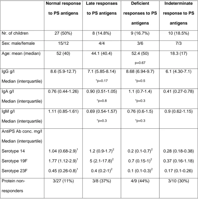
Table 2: Ig levels and responses to pneumococcal PS and proteins Normal response to PS antigens Late responses to PS antigens Deficient responses to PS antigens Indeterminate response to PS antigens Nr. of children 27 (50%) 8 (14.8%) 9 (16.7%) 10 (18.5%) Sex: male/female 15/12 4/4 3/6 7/3
Age: mean (median) 52 (40) 44.1 (40.4) 52.4 (50)
p=0.67 18.3 (17) IgG g/l Median (interquartile) 8.6 (5.9-12.7) 7.1 (5.85-8.14) *p=0.17 8.68 (6.94-9.7) °p=0.5 6.1 (4.30-7.1) IgA g/l Median (interquartile) 0.76 (0.44-1.26) 0.90 (0.51-1.05) *p=0.8 1.1 (0.7-1.4) °p=0.3 0.41 (0.27-0.78) IgM g/l Median (interquartile) 1.11 (0.85-1.61) 0.69 (0.54-1.57) *p=0.3 0.76 (0.6-1.5) °p=0.3 0.9 (0.62-1.15) AntiPS Ab conc. mg/l Median (interquartile) Serotype 14 Serotype 19F Serotype 23F 1.04 (0.68-2.9)1 1.77 (1.12-2.9)1 0.45 (0.26-0.8)1 1.2 (0.9-1.7)2 5 (2.1-17.8)2 0.4 (0.2-1)2 0.2 (0.1-0.7)2 0.7 (0.15-1)2 0.1 (0.1-0.3)2 0.28 (0.18-0.38) 0.37 (0.16-1.18) 0.17 (0.1-0.26) Protein non-responders 3/27 (11%) 3/8 (37%) 4/9 (44%) 3/10 (30%) 1
: exposure-driven antibody concentration 2
: post-immunization (pneumococcal polysaccharide vaccine) antibody concentration
*: comparison between normal and late responses
VIII. Figures legends
Figure 1: Distribution of serotype specific antipolysaccharide antibodies according to age (group 1: age 12-24 months, group 2: age 25-48 months, group 3: age > 48 months). *: p < 0.05
Figure 2: Serotype specific pre- and post-vaccination antibody levels. There is a statistically significant increase in post-vaccination antibody concentration for serotypes 14 and 19F.
Figure 3: Distribution of Ig values for age according to pneumococcal PS antibody responses
Figure 4: Distribution of pneumococcal antiprotein antibody concentration according to age (group 1: age 12-24 months, group 2: age 25-48 months, group 3: age > 48 months). *: p < 0.05
Figure 5: Correlation between PspA clades and between PsaA and PspA.
Figure 6: Distribution of protein non-responder patients according to their antibody response to polysaccharide (PS) antigens. *: p = 0.03
IX. Figures Serotype 14 12-24m. 25-48m. > 48m. 0.0 2.5 5.0 7.5 10.0 12.5 15.0 mg /l Serotype 19 12-24m. 25-48m. > 48m. 0.0 2.5 5.0 * * * p<0.05 7.5 10.0 12.5 15.0 mg /l Serotype 23 12-24m. 25-48m. > 48m. 0.0 2.5 5.0 mg /l
Figure 1: Distribution of serotype specific antipolysaccharide antibodies according to age (group 1: age 12-24 months, group 2: age 25-48 months, group 3: age > 48 months). *: p < 0.05
Serotype 14 pre post 0.0 0.2 0.4 0.6 0.8 1.0 1.2 1.4 1.75 2.25 mg /l Serotype 19 pre post 0.0 0.5 1.0 1.5 2.0 2.5 3.020 30 40 mg /l Serotype 23 pre post 0.0 0.2 0.4 0.6 0.8 1.0 1.2 1.4 2 3 mg /l
Figure 2: Serotype specific pre- and post-vaccination antibody levels. There is a statistically significant increase in post-vaccination antibody concentration for serotypes 14 and 19F.
IgG 0 10 20 Normal PS responses Late PS responses Deficient PS responsessp. mg /d l IgA 0 1 2 3 4 5 Normal Late Deficient mg /d l IgM 0 1 2 3 Normal Late Deficient mg /d l
Figure 3: Distribution of Ig values for age according to pneumococcal PS antibody responses
PspA RX1M I 12-24 m. 24-48 m. > 48.m 0 2500 5000 * * *p<0.05 7500 10000 12500 U/ m l PspA EF3296 12-24 m. 24-48 m. > 48 m. 0 2500 5000 * * *p<0.05 7500 10000 12500 U/ m l PspA EF5668 12-24 m. 24-48 m. > 48 m. 0 2500 5000 * * *p<0.05 7500 10000 12500 U/ m l PsaA 12-24 m. 24-48 m. > 48 m. 0 2500 5000 7500 10000 12500 U/ ml
Figure 4: Distribution of pneumococcal antiprotein antibody concentration according to age (group 1: age 12-24 months, group 2: age 25-48 months, group 3: age > 48 months). *: p < 0.05
0 1000 2000 3000 4000 0 1000 2000 3000 4000 EF3296 (r2= 0.54) EF5668 (r2= 0.51) 4500 6500 4500 7000 RX1MI U/ml U/ m l 0 2000 4000 6000 8000 0 1000 2000 3000 4000 RX1MI (r2= 0.42) 4500 6500 PsaA U/ m l
norm al late resp onde rs non-re spon ders 0 10 20 30 40 50 * * = p < 0.05 %
Figure 6: Distribution of protein non-responder patients according to their antibody response to polysaccharide (PS) antigens. *: p = 0.03