HAL Id: inserm-02148360
https://www.hal.inserm.fr/inserm-02148360
Submitted on 5 Jun 2019
HAL is a multi-disciplinary open access
archive for the deposit and dissemination of sci-entific research documents, whether they are pub-lished or not. The documents may come from teaching and research institutions in France or abroad, or from public or private research centers.
L’archive ouverte pluridisciplinaire HAL, est destinée au dépôt et à la diffusion de documents scientifiques de niveau recherche, publiés ou non, émanant des établissements d’enseignement et de recherche français ou étrangers, des laboratoires publics ou privés.
Using CRISPR/Cas9 Technology and Microinjection
into Zygotes
M. Crispo, A. Mulet, L. Tesson, N. Barrera, F. Cuadro, P. dos Santos-Neto,
T. Nguyen, A. Crénéguy, L. Brusselle, Ignaco Anegón, et al.
To cite this version:
M. Crispo, A. Mulet, L. Tesson, N. Barrera, F. Cuadro, et al.. Efficient Generation of Myostatin Knock-Out Sheep Using CRISPR/Cas9 Technology and Microinjection into Zygotes. PLoS ONE, Pub-lic Library of Science, 2015, 10 (8), pp.e0136690. �10.1371/journal.pone.0136690�. �inserm-02148360�
Efficient Generation of Myostatin Knock-Out
Sheep Using CRISPR/Cas9 Technology and
Microinjection into Zygotes
M. Crispo1*, A. P. Mulet1, L. Tesson3, N. Barrera2, F. Cuadro2, P. C. dos Santos-Neto2, T.
H. Nguyen3, A. Crénéguy3, L. Brusselle3, I. Anegón3*, A. Menchaca2*
1 Unidad de Animales Transgénicos y de Experimentación (UATE), Institut Pasteur de Montevideo, Montevideo, Uruguay, 2 Instituto de Reproducción Animal Uruguay, Fundación IRAUy, Montevideo, Uruguay, 3 INSERM UMR 1064, Center for Research in Transplantation and Immunology-ITUN, Nantes, France
*[email protected](MC);[email protected](IA); [email protected](AM)
Abstract
While CRISPR/Cas9 technology has proven to be a valuable system to generate gene-targeted modified animals in several species, this tool has been scarcely reported in farm animals. Myostatin is encoded byMSTN gene involved in the inhibition of muscle differenti-ation and growth. We determined the efficiency of the CRISPR/Cas9 system to editMSTN in sheep and generate knock-out (KO) animals with the aim to promote muscle develop-ment and body growth. We generated CRISPR/Cas9 mRNAs specific for ovineMSTN and microinjected them into the cytoplasm of ovine zygotes. When embryo development of CRISPR/Cas9 microinjected zygotes (n = 216) was compared with buffer injected embryos (n = 183) and non microinjected embryos (n = 173), cleavage rate was lower for both micro-injected groups (P<0.05) and neither was affected by CRISPR/Cas9 content in the injected medium. Embryo development to blastocyst was not affected by microinjection and was similar among the experimental groups. From 20 embryos analyzed by Sanger sequencing, ten were mutant (heterozygous or mosaic; 50% efficiency). To obtain liveMSTN KO lambs, 53 blastocysts produced after zygote CRISPR/Cas9 microinjection were transferred to 29 recipient females resulting in 65.5% (19/29) of pregnant ewes and 41.5% (22/53) of new-borns. From 22 born lambs analyzed by T7EI and Sanger sequencing, ten showed indel mutations atMSTN gene. Eight showed mutations in both alleles and five of them were homozygous for indels generating out-of frame mutations that resulted in premature stop codons. Western blot analysis of homozygous KO founders confirmed the absence of myostatin, showing heavier body weight than wild type counterparts. In conclusion, our results demonstrate that CRISPR/Cas9 system was a very efficient tool to generate gene KO sheep. This technology is quick and easy to perform and less expensive than previous techniques, and can be applied to obtain genetically modified animal models of interest for biomedicine and livestock.
OPEN ACCESS
Citation: Crispo M, Mulet AP, Tesson L, Barrera N, Cuadro F, dos Santos-Neto PC, et al. (2015) Efficient Generation of Myostatin Knock-Out Sheep Using CRISPR/Cas9 Technology and Microinjection into Zygotes. PLoS ONE 10(8): e0136690. doi:10.1371/ journal.pone.0136690
Editor: Reiner Albert Veitia, Institut Jacques Monod, FRANCE
Received: May 31, 2015 Accepted: August 5, 2015
Published: August 25, 2015
Copyright: © 2015 Crispo et al. This is an open access article distributed under the terms of the
Creative Commons Attribution License, which permits unrestricted use, distribution, and reproduction in any medium, provided the original author and source are credited.
Data Availability Statement: All relevant data are within the paper and its Supporting Information files. Funding: This project was financially supported by the IHU-CESTI, which received French government financial support managed by the National Research Agency, Région Pays de la Loire through Biogenouest, IBiSA program, Fondation Progreffe and TEFOR (Infrastructures d’Avenir of the French government), the integrative genomic facility of Nantes for sequencing experiments, FOCEM (MERCOSUR Structural Convergence Fund) COF 03/11 (Uruguay), and received additional support by
Introduction
Modification of the genome in large animals has always been a difficult task, with few efficient technologies available until recently [1–4]. The generation of genetically modified large animals has several advantages for different applications not only in meat, dairy or wool production but also in the generation of bioreactors to produce recombinant proteins of biomedical interest or for the production of models of human diseases [5]. Nevertheless, the massive application of these technologies has been limited due in part to the low efficiency to obtain transgenic foun-ders [6,7]. While lentiviral transgenesis does show high efficiency [8], site-specific gene inte-gration is still not possible, among other problems. Gene edition in general and in particular the generation of genetically-modified animals has experienced a major advance in the last few years due to the incorporation of novel technologies of direct microinjection into zygotes of gene-specific nucleases such as zinc finger nucleases (ZFN) [9], TALENs [10] and homing endonucleases [11,12]. Although the use of these gene-specific nucleases made possible the generation of KO animals, they are difficult to engineer. Lately, a new gene-specific nuclease is being applied with high efficiency. Known as clustered regularly interspaced short palindromic repeats (CRISPR/Cas9), this system is taken from the adaptive immune system of bacteria and archae [13], and is based in small RNAs and Cas endonucleases proteins that can induce site-specific DNA double-stranded breaks. CRISPR/Cas9 has been rapidly tested in several species to date [14–19] and promises to revolutionize the field of genome editing in general and in par-ticular transgenic vegetal and animal fields. Genetically-modified farm animals using zygote microinjection of CRISPR/Cas9 have been reported recently in pigs [20], goats [17] and sheep [21], but more information is needed to extend the generated information.
According to official data of FAO estimations, in order to feed a larger, more urban and richer human population, food production must be increased around 70 percent before the year 2050 [22]. According to meat production, the improvement of traditional animal breeds with modern technologies should be implemented. Myostatin is a member of the transforming growth factor beta (TGF-β) superfamily, involved in the inhibition of muscle differentiation and growth [23]. Previous studies have reported that the inhibition of this protein induces a significant increase in the muscle volume and mass producing more meat in animals known as double-muscle animals [24–27]. In farm animals, sheep are good models to be modified geneti-cally due to small size and short gestation period compared to cows. However, until now their use was limited due to the lack of efficient gene editing tools in livestock. In addition, the silenc-ing of this gene in breeds as Australian Merino, the most specialized breed in the production of superfine or ultrafine wool, could be an interesting model to produce more meat in a high-quality wool producing animal.
The objective of this study was to demonstrate the efficiency of the CRISPR/Cas9 gene edit-ing technology coupled to zygote microinjection in the generation of MSTN KO sheep. We show that this approach induces mutations with a high efficiency (45.5%), resulting in live off-spring carrying single-gene mono and bi-allelic mutations.
Materials and Methods
Ethics statement
All procedures that include animal handling were carried out in strict accordance with the rec-ommendations in the Guide for the Care and Use of Laboratory Animals of the National Insti-tutes of Health. Protocol # 001/2014 was approved by the Animal Care Committee of the Fundación IRAUy certified by the National Council of Animal Care of Uruguay and have therefore been performed in accordance with the ethical standards laid down in the 1964
PEDECIBA (UdelaR, Uruguay). The funders had no role in study design, data collection and analysis, decision to publish, or preparation of the manuscript. MC, IA and AM are fellows of Sistema Nacional de Investigadores (SNI, National Research Agency), Uruguay.
Competing Interests: The authors have declared that no competing interests exist.
Declaration of Helsinki and its later amendments. All surgeries were performed under strict aseptic conditions, and all efforts were made to minimize animal suffering.
Generation of the plasmid co-expressing Cas9 and sgRNA
The pX330-U6-Chimeric_BB-CBh-hSpCas9 plasmid (a gift from Feng Zhang, Addgene plasmid # 42230) was digested with BbsI, dephosphoryated using Antarctic Phosphatase (NEB, UK), and the linearized vector was gel purified. To generate the bicistronic vector (pX330-cas9-MSTN) expressing Cas9 and sgRNA against MSTN (GGCTGTGTAATGCATGCTTG), a pair of oligos for targeting myostatin exon 1 (5’-CACCGGCTGTGTAATGCATGCTTG-3’ and 5’-CACCGG CGAAGCTTACTGAGGATT-3’) was annealed, phosphorylated and ligated to a linearized vec-tor (details of the vecvec-tor plasmid is described by Cong et al [28]).
Genome editing assay in cells
The A15 astroglial sheep cell line (kindly provided by J. Chapuis, UR 892, INRA, Jouy-en-Josas, France) [29] was maintained in Dulbecco’s modified Eagle medium (DMEM) in 10% Fetal Bovine Serum, 2mM glutamine, 1% sodium pyruvate and 1% penicillin/streptomycin. Cells were transfected in 24-well plates with 2μg of pX330-cas9-MSTN co-expressing Cas9 and sgRNA against myostatin using lipofectamine LTX reagent (Life technologies, NY, USA) according to the manufacturer’s manual. Three days later, genomic DNA from transfected cells was extracted using a kit from Macherey Nagel (NucleoSpin Tissue, Duren, Germany) and quantified using the NanoDrop2000 spectrophotometer (Thermo Fisher Scientific, Illkirch, France). A260/A280 and A260/A230 ratios accounted for sample purity.
Gene mutation activity of sgRNA sequence at the target locus of MSTN exon 1 was quanti-fied using the T7EI mismatch detection assay. DNA sequence of interest was PCR-ampliquanti-fied with a high-fidelity polymerase (Herculase II fusion polymerase) using specific primers (for-ward primer 5’-tcactggtgtggcaagttgt-3’ and reverse primer 5’-aaaagctctttgccctcctc-3’). The 634bp PCR product was then denatured and slowly re-annealed (95°C, 2min; 95°C to 85°C, -2°C/sec; 85°C to 25°C, -1°C/sec) to produce homoduplex/heteroduplex mix. This was then digested by 5U of T7EI restriction enzyme (NEB) at 37°C for 30 minutes. Digestion products were separated by 2% agarose gel electrophoresis. The ratio of cleaved (255-bp and 379-bp) to uncleaved (634-bp) products was used to calculate NHEJ frequency as previously described using Image J software [30]. NHEJ frequency was calculated as follow: % gene modifica-tion = 100 X (1-(1-fracmodifica-tion cleaved)^1/2.
Production of sgRNA and Cas9 mRNA
These procedures have been described in detail elsewhere [31]. Briefly, T7 promoter was added to sgRNA template by PCR amplification of pX330-cas9-MSTN plasmid using the following primers: 5’-ttaatacgactcactataggctgtgtaatgcatgcttg-3’ and 5’-tttaaaagcaccgactcggtgcc-3’. The PCR product was purified using NucleoSpin Gel and PCR Clean-up (Macherey Nagel). It was used as the template for in vitro transcription using MEGAshortscript T7 kit (Life Technolo-gies) according to the manufacturer’s manual. Following completion of transcription, DNase I treatment was performed.
The Cas9 mRNA was transcribed using PmeI-digested Cas9 expression JDS246 plasmid (a gift from Keith Joung, Addgene plasmid # 43861) and the mMESSAGE mMACHINE T7 ULTRA Transcription Kit (Life Technologies) according to the manufacturer’s manual. Fol-lowing completion of transcription, the poly(A) tailing reaction and DNase I treatment were performed.
Both the Cas9 mRNA and the sgRNAs were purified using MEGAclear kit (Life Technolo-gies) and eluted in elution buffer.
Prediction of potential Off Target sites
Potential targets of the CRISPR sgRNA MSTN were found using the rules outlined in [32] since this is the only web tool that includes the sheep genome for predicting potential off-tar-gets. The sequences TAATGCATGCTTGTGG, TAATGCATGCTTGAGG, TAATGCATGC TTGCGG, TAATGCATGCTTGGGG (underlined is the PAM, preceded by the N13 sequence of the sgRNA MSTN) were blasted in the genome database of the sheep (http://blast.ncbi.nlm. nih.gov/Blast.cgi). Seven potential off-target sites were detected, that were amplified in the DNA samples from all the mutant lambs, and subjected to Sanger sequencing.
In vitro production of embryos
Unless otherwise indicated, chemicals were purchased from Sigma Chemical Company (St Louis, MO, USA). The embryos were produced by in vitro fertilization according to routine procedure performed in our lab as described earlier [8]. Briefly, ovaries from slaughterhouse were transported to the laboratory and cumulus oocyte complexes (COCs) were aspirated in recovery medium. The selected COCs were placed in maturation medium for 24 h in 5% CO2
in humidified air atmosphere at 39°C. Then, expanded COCs were inseminated in 100μl drops with 1 x 10⁶dose of frozen-thawed semen selected by ascendant migration on a swim up method. Fertilization was carried out in 5% CO2with humidified atmosphere at 39°C for 22 h.
Microinjection into zygotes
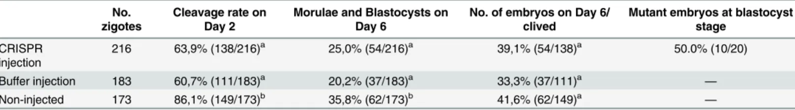
Soon after fertilization, 572 presumptive zygotes were randomly assigned to three experimental groups to be microinjected (CRISPR group, n = 216; and Buffer group, n = 183) or not (Control group, n = 173). Microinjection of CRISPR group was performed into the cytoplasm with 5 ng/μl of sgRNA and 20ng/μl of Cas9 mRNA diluted in injection buffer (10mM Tris pH 7.5, 0.1mM EDTA), while Buffer group was injected with the same procedure but with buffer alone. Lastly, injected and non-injected embryos were transferred to culture medium under mineral oil, in 5% CO2, 5% O2and 90% N2in humidified atmosphere at 39°C. Cleavage rate on
Day 2 (cleaved zygotes per total oocytes) and development rate on Day 6 (morulae and blasto-cysts per total oocytes) were recorded for all experimental groups. After Day 6, DNA from 20 CRISPR group embryos was analyzed by Sanger sequencing to detect the mutation at the MSTN gene level.
To determine the in vivo efficiency of the system, 53 blastocysts produced by CRISPR/Cas9 zygote microinjection were transferred to 29 recipient females. Only early blastocysts, blasto-cysts and expanded blastoblasto-cysts classified as excellent or good (i.e. Grade 1) [33] were trans-ferred on Day 6 after fertilization. Embryo transfer was performed by minimally invasive surgery assisted by laparoscopy to place the embryos into the cranial side of the ipsilateral uter-ine horn to the corpus luteum. Recipient ewes were previously synchronized to be on Day 6 of the estrous cycle using a standard protocol to control ovulation described previously [34].
Monitoring of fetuses and lambs
Pregnancy diagnosis and fetal development were performed on Day 30 and 105, respectively, by using B-mode ultrasonography equipped with a 5 and 3.5 MHz probe (Well-D, Shenzhen, China). Day 0 of the experiment was defined as the moment of embryo fertilization. Several parameters were measured to study the development of fetuses at Day 105 of gestation:
thoracic diameter, biparietal diameter, occipitonasal length and heart rate. At delivery, length of gestation, gender, rectal temperature, heart and respiratory rates, body weight, thoracic perimeter, biparietal diameter, crown-rump and occipitonasal length, height at withers, height at hips, width at hips and width at chest were recorded. Body weight and morphometric vari-ables were determined at birth, and 15, 30 and 60 days later.
Identification and genotyping of transgenic animals
Samples from skin and limb muscle of the lambs were taken seven days after birth and T7EI assay, western blot test and histology examinations were performed in order to identify and characterize KO founders and off-target sites. Total DNA was isolated from skin biopsies for all animals and from muscle for some animals. PCR and T7EI assay were performed using the same protocol than from cells samples. Samples were analyzed using capillary electrophoresis (Caliper, PerkinElmer, Hopkinton, MA). Genotyping of MSTN exon 1 was performed by direct sequencing of PCR amplicons using the same primers described above and in muscle biopsies by additional sequencing of isolated bacterial clones with individual amplicon sequences, as described in detail previously [30,35].
Analysis of myostatin expression
Western blotting was performed to determine the presence of myostatin in the muscle fiber. Equal amounts of total proteins were run on 12% (v/v) gel electrophoresis and electrophoreti-cally transferred to a PVDF membrane. Monoclonal mouse anti-myostatin (sc-398333, Santa-cruz) and anti-GAPDH (G8795, Sigma) antibodies were used in the western blotting. The washed membranes were incubated with 1:50000 dilution of secondary antibody linked to horseradish peroxidase (HPR). HPR activity was detected using western blot chemilumines-cence (Promega, MA, USA).
Muscle fiber histology
Samples from deltoid and biceps femoris muscles were fixed in 4% formaldehyde solution until analysis. Samples were included in paraffin, sectioned in 1 mm slides and stained with haema-toxilin-eosin to study muscle morphology. Muscle fibers (min 250 per sample) were measured with respect to their minimum Feret (MinFeret) diameter. The mean area was also calculated.
Statistical analysis
In vitro embryo development on Day 2 and Day 6 among experimental groups was compared by logistic regression. Continues variables measured during pregnancy and body growth after birth in mutant vs. wild type lambs were analyzed by one-way ANOVA or by non-parametric Kruskal Wallis test. Differences were considered significant when P<0.05.
Results
Efficiency of Cas9/sgRNA activity in sheep fetal fibroblast
Before microinjection of the sgRNA and Cas9 coding sequences into the ovine zygotes, fetal ovine fibroblasts were transfected with the pX330-cas9-MSTN plasmid co-expressing Cas9 and sgRNA, showing mutations at the targeted MSTN exon 1 in 10% of the total DNA (Fig 1). Although it is difficult to compare with other cell lines, this efficacy in vitro is comparable to what we and others have previously observed as being sufficient to obtain mutated animals by microinjection into zygotes of not only CRISPRs/Cas9 but also ZFNs or TALENs [15,35].
Thus, we proceeded to microinject ovine zygotes with both mRNAs encoding for Cas9 and sgRNA.
Analysis of
MSTN mutations in embryos microinjected with Cas9/sgRNA
mRNAs
We first determined whether zygotes microinjected with Cas9 and sgRNA mRNAs and further cultured in vitro showed normal development and MSTN mutations. From 216 injected zygotes 63.9% survived to micromanipulation and reached cleavage stage on Day 2, and 39.1% achieved morula or blastocyst stage on Day 6. Cleavage rate and development rate for control non-injected embryos were significantly higher than for CRISPR and buffer microinjected embryos (Table 1). However, no differences were found on embryo development rate among control and microinjected zygote groups after cleavage. The total number of embryos on Day 6 from cleaved embryos did not show statistical differences among groups (Table 1). Thus, microinjection resulted in some embryo lethality up to the cleavage state but Cas9 and sgRNA mRNAs microinjection had no adverse effects on subsequent in vitro embryo development.
DNA from developed CRISPR injected embryos were extracted and analyzed by Sanger sequencing. From 20 embryos analyzed, 10 showed mutations at the MSTN gene (50%). Two of those mutants were heterozygous (wt + mutated allele) and eight were mosaic presenting more than two sequences. The deletions observed ranged from 6 to 350 bp, besides just 1 bp insertions were observed.
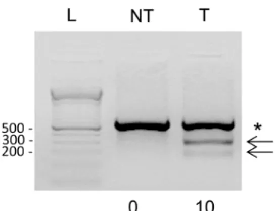
Fig 1. CRISPR-Cas9 genome editing activity in transfected ovine cells. A) Exon 1 of the ovineMSTN gene. The sequence recognized by sgRNA is in italic and bold, from nt 110–130, the PAM sequence TGG is underlined and the rest of exonic sequences in smaller font. B) Ovine cells were transfected with pX330-cas9-MSTN plasmid co-expressing Cas9 and sgRNA and three days later cellular DNA was isolated and subjected to T7EI assay for detecting mutation at the targetedMSTN exon 1. The PCR amplifies a band of 634 bp and uncleaved (asterisk) and cleaved (arrows, 255 bp and 379 bp) products are indicated. Numbers below each lane indicate the percentage of targeted indels. NT: non-transduced cells, T: transfected cells, L: 1Kb DNA. The experiment is representative of two replicates performed with similar results.
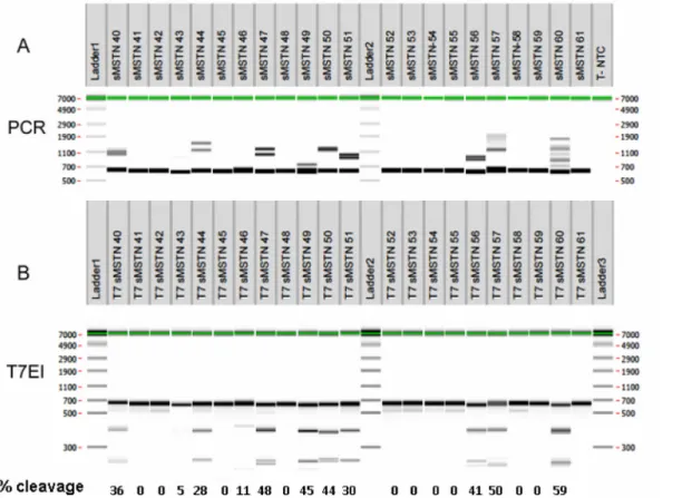
PCR and T7EI genotyping of lambs following microinjection of Cas9/
sgRNA mRNAs
From 53 Grade 1 in vitro produced sheep blastocyst transferred into 29 recipient females, 41.5% of them were detected at 30 days of gestation, 65.5% of the recipients were pregnant and 22 lambs were delivered (Table 2). From these 22 lambs, three (#43 49 and 53) died at delivery or within the first day after birth. Skin and muscle biopsies from deltoid and biceps femoris were taken from 22 lambs within 1 week after birth. DNA from the 22 skin biopsies was extracted and analyzed by PCR and T7EI assay followed by capillary electrophoresis (Fig 2). PCR analysis showed the presence of main band of the expected size in the absence of muta-tions (634 bp) and bands of apparent higher molecular weight, due to open angles formed by heteroduplexes of DNA strands with mismatches [36] in nine animals, suggesting the presence of mutations (Fig 2A). In an additional animal (#43) a single band of slightly smaller size as compared to the other animals was observed (Fig 2A).
Since T7EI digestion of amplicon allows detection of mutations when both DNA strands show mismatches, animals with WT sequences or with the same mutation in both alleles should not generate new bands and animals which have different alleles, either one WT and the other mutated or both with different mutations, should generate new bands. The T7EI assay revealed that the same nine animals with heteroduplexes in the PCR assay generated bands of lower molecular weight upon T7EI digestion (Fig 2B), confirming the presence of mutations in these animals. Animal #43 did not show new smaller bands but if both alleles con-tained the same mutation the T7EI assay was not expected to be positive. The analysis of DNA cleavage showed that several animals (#47, 49, 50, 56, 57 and 60) had around 50% cleavage cor-responding to half of the alleles being mutated and are thus not mosaic. Some animals (#40, 44, 46 and 51) had cleavage of less than 50% and are likely mosaic.
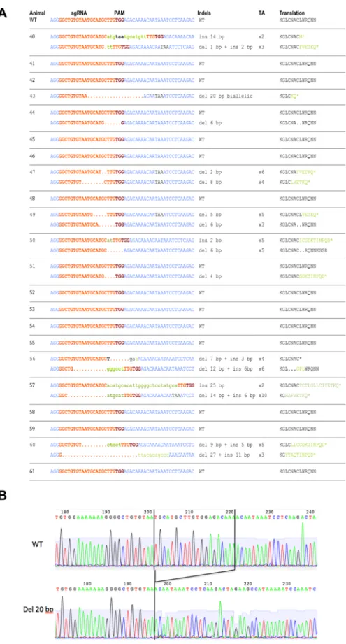
DNA sequence genotyping of lambs following microinjection of Cas9/
sgRNA mRNAs
Sequencing of PCR amplicons compassing the targeted MSTN exon 1 sequence showed that ten animals contained mutations; nine identified using capillary electrophoresis and animal
Table 1. Cleavage rate on Day 2 and development rate on Day 6 of embryos that were microinjected into cytoplasm with CRISPR/Cas9 RNA sys-tem, buffer injection solution, or non-injected embryos (Control group).
No. zigotes
Cleavage rate on Day 2
Morulae and Blastocysts on Day 6
No. of embryos on Day 6/ clived
Mutant embryos at blastocyst stage CRISPR injection 216 63,9% (138/216)a 25,0% (54/216)a 39,1% (54/138)a 50.0% (10/20) Buffer injection 183 60,7% (111/183)a 20,2% (37/183)a 33,3% (37/111)a — Non-injected 173 86,1% (149/173)b 35,8% (62/173)b 41,6% (62/149)a — For different superscripts, P<0.05.
doi:10.1371/journal.pone.0136690.t001
Table 2. Efficiency obtained with CRISPR/Cas9 system injected into cytoplasm of ovine zygotes to produceMSTN mutant lambs. Data obtained from 53 embryos transferred into 29 recipient ewes.
Embryos on Day 30
Pregnant ewes
Fetal loss Mutant/ born lambs Biallelic/ mutant lambs Homozygous/ mutant lambs CRISPR/Cas9 efficiency 41.5% (22/53) 65.5% (19/29) 0.0% (0/ 22) 45.5% (10/22) 80.0% (8/10) 50.0% (5/10) doi:10.1371/journal.pone.0136690.t002
#43 which showed an identical deletion of 20 nt in both chromosomes (Fig 3A and 3B), explaining the smaller size of the amplicons in capillary electrophoresis and the negative T7EI assay. The other 12 animals showed only WT alleles. Among the ten mutated animals, eight showed both alleles mutated (#40, 43, 47, 49, 50, 56, 57 and 60) and two only one allele (#44 and 51). These introduced mutations disrupted the coding frame of exon 1 (number of indels different of 3nt or multiples of 3nt) with the generation of premature stop codons in nine mutated animals in at least one of the two alleles. The exception was animal #44 which showed an in-frame shift mutation (Fig 3A). Newly generated stop codons (Fig 3A) are recognized by cells as premature stop codons due to the absence of other normal Cis sequences targeting these mRNAs for degradation through nonsense-mediate decay pathway resulting in the com-plete absence of the protein [36]. Some animals showed biallelic frameshift mutations intro-ducing premature stop codons (#40, 43, 47, 57 and 60), and are thus homozygous KO animals. Other animals showed one frameshift mutation and another with inframe mutations (#49, 50 and 56) and are thus heterozygous KO animals. Analogously, one animal (#51) showed one allele with a frameshift mutation and a WT allele and thus it is also a heterozygous KO animal. Finally, one animal (#44) had one allele with an inframe mutation and one wild-type allele and thus it is not a KO animal.
The DNA sequences performed in skin and muscle from the same lambs showed the same mutations (S1 Table), indicating the absence of mosaicism in ectoderm and mesoderm-derived
Fig 2. Genotype of lambs born following microinjection of sgRNA forMSTN exon 1 and Cas9. DNA was extracted from lambs’ skin biopsies one week after birth, with the exception of lambs #43 and #49 that were stillborn and were biopsied 1–2 hours after delivery. A) Results from PCR amplifying 634 bp. Additional larger bands represent formation of heteroduplex DNA double strands with mismatches between the two strands resulting in slower migration during electrophoresis. B) T7EI assay of lambs produced by zygote injection of CRISPR/Cas9 mRNA, indicating numbers of mutant animals with bands of 255, 379 and 634 bp (#40, 43, 44, 47, 49, 50, 51, 56, 57 and 60) and WT animals with only one band of 634 bp (#41, 42, 45, 46, 48, 52, 53, 54, 55, 58, 59 and 61).
Fig 3. Sequence analysis of lambs’ MSTN exon 1. The same DNAs analyzed inFig 2were PCR amplified and the amplicons directly sequenced. In some animals (#40, 47, 49, 50, 56, 57 and 60) DNA from muscle
tissues and thus indicating the action of Cas9 at the zygote stage. Thus, sgRNA and Cas9 mRNA microinjection into ovine zygotes achieved high efficient introduction of mutations resulting in inactivation of the MSTN gene.
Primers pairs were designed and the possible off target sites were amplified and sequenced in order to assess the presence of possible off target effects (S2 Table). In the animals #40 and #57 one 27 pb deletion and one 1 bp insertion were found respectively for the off target region number 3. In both cases this mutation was heterozygous. None other off target effects were identified in the seven regions assessed.
Analysis of myostatin expression
Homozygous KO lambs with two frameshift mutations should show absence of myostatin whereas heterozygous KO lambs with one coding allele should show myostatin. Western blot for myostatin analysis showed undetectable levels in homozygous KO lamb #43 and the pres-ence of myostatin in heterozygous (1 WT and 1 in-frame shift allele) lamb #44 (Fig 4) and these levels were comparable to those of lambs with 2 WT alleles (S1 Fig).
Fetal development and delivery
Fetal determinations by ultrasonography on Day 105 of gestation showed no statistical differ-ences for thoracic and biparietal diameter, occipito-nasal length or heart rate among WT, homozygous KO or heterozygous KO mutant animals (S2 Fig), indicating that the mutation did not affect in utero measurements although the MSTN gene was already mutated.
For mutant lambs, # 43 and 49 died at delivery and lamb # 57 died within first month after birth due to a wound infection. For WT lambs, #53 and 58 died at delivery and within first month after birth, respectively. The rest of the lambs developed normaly until at least 60 days of life. Length of gestation was 147.0 ± 0.9, 149.8 ± 0.6 and 149.4 ± 1.0 days for WT, homozy-gous KO and heterozyhomozy-gous KO mutant lambs respectively (P = NS). Rectal temperature at birth was 40.2 ± 0.2, 39.9 ± 0.3 and 39.9 0078 0.4°C for WT, homozygous KO and heterozygous KO mutant lambs, respectively (P = NS). Heart rate and respiratory rate did not differ among groups and was 189.8 ± 3.1 and 93.3 ± 1.8 for WT lambs, 184.0 ± 5.7 and 86.0 ± 13.1 for homo-zygous KO lambs, and 203.0 ± 11.8 and 75.0 ± 5.0 for heterohomo-zygous KO lambs, respectively.
Postnatal phenotype
Morphometric variables registered from birth to 60 days later are shown inFig 5andS3 Fig. Body weight from homozygous KO lambs was not different than WT animals at birth (P = NS) and was greater at 15 and 30 days (P<0.05) with a statistical tendency at 60 days after birth (P = 0.08). This faster growth showed by homozygous KO lambs within two months of age averaged an increase between 20 and 30%. Body weight for heterozygous KO mutant lambs
biopsies were PCR amplified and amplicons were cloned into plasmids by TA cloning and electroporated into bacteria, followed by sequencing of 8–10 bacterial clones. A) Depicts for each of the 22 delivered lambs the flanking DNA sequences (in blue) close to the targeted sgRNA sequence (in red) and the PAM sequence (violet); missing nucleotides are represented by spaces, added ones in green and small characters and stop codons are labeled in black. The column Genotype recapitulates the genotype found for each lamb. The column TA indicates the number of bacterial colonies that were sequenced for each allele of the muscle biopsies. The column Translation depicts the aminoacids translated; spaces for the missing ones, in green the ones that are new due to the shift in the coding reading frame and the* represents the stop of the aminoacid sequence due to the premature stop codons. Results are representative of two different PCR amplicons sequencing for all animals. B) A representative sequence electrophoresis, in this case the one of animal #43 which has a biallelic identical deletion of 20 nt.
Fig 4. Myostatin western blot. Muscle biopsies from two representative animals, one homozygous (homoz.) for a frameshift mutation and another heterozygous (heteroz.) for a frameshift mutation and a WT copy in the second allele (# 43 and 44, respectively, the numbers of animals correspond to those ofFig 3) analyzed by western blot using an anti-MSTN monoclonal antibody. After stripping of the anti-MSTN antibody from the membrane, an anti-GAPDH was used as loading control. One representative western blot experiment out of three performed with the same results.
was intermediate (P = NS). The rest of the variables did not show statistical differences among the three genotypes, indicating that the mutation at the MSTN gene affects body weight mainly by increasing muscle mass (S3 Fig). However, a statistical tendency (P<0.1) was found at 60 days for height and width at withers, and width at chest among homozygous KO and WT lambs, which gave a very clear phenotype in some MSTN KO lambs as shown inFig 6.
Muscle fiber histology
As expected from previous results of lambs with spontaneous MSTN mutations [37], histologi-cal analyses of muscle biopsies showed muscle fibers of lower diameter although due to the rel-atively low number of homozygous KO animals analyzed (n = 4) it did not reach statistical significance when compared to WT animals (data not shown).
Discussion
The current study demonstrates that microinjection into zygotes of sgRNA and Cas9 mRNAs was a very efficient approach to obtain myostatin KO sheep. Embryo survival and embryo development were not affected by the microinjection of sgRNA and Cas9 mRNAs into the cytoplasm. Also, no differences in fetal loss, birth rate and postnatal survival rates were detected for both groups, suggesting that mutant animals are as healthy as WT animals. These results are in agreement with our recent report where the microinjection of a GFP lentiviral
Fig 5. Body weight in lambs produced by CRISPR/Cas9 system. Significant diference (P<0.05) from 15 to 60 days after birth was found among homozygous KO and wild-type lambs.
construct into ovine zygotes did not affect embryo development, fetal growth or postnatal vari-ables when compared to WT lambs [8]. Ten lambs with mutations in the MSTN gene were obtained (45.5%), eight had biallelic mutations and five of them were biallelic out of frame shift mutations. These lambs resulted in myostatin deficiency and a double-muscle phenotype with a significant increase in body weight when compared to control wild type animals.
During recent years gene editing nucleases have revolutionized the field of transgenesis, decreasing the time, cost and effort to obtain genetically-modified animals [38]. However, the use of CRISPR/Cas9 system has not been reported in many ruminant species to date [17,38, 21]. We demonstrated that this technology does not affect embryo survival and in vitro devel-opment rate when injected into the cytoplasm of zygotes. However, the cleavage rate was affected by the embryo manipulation required for the microinjection per se, which has been reported previously in zygotes submitted to zone pellucidae break and microinjection [39]. No effect of microinjection was found in the ability of those surviving embryos 48h after fertiliza-tion to continue its development and to reach blastocyst stage on Day 6 after fertilizafertiliza-tion. In addition, the overall procedure allowed a high fertility rate since 41.5% of the microinjected embryos transferred to recipients were pregnant and developed to term. The efficiency of the
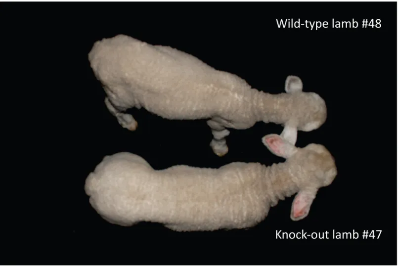
Fig 6. Phenotype of lambs 30 days after birth. The lamb at the top is wild type (#48), the lamb at the bottom is a homozygous KO (#47). Note the differences in muscle mass from the posterior limbs and loin from the homozygous KO lamb when compared to wild-type. At the time of this picture, with 30 days old the body weight was 8.750 (#48) and 11.150 kg (#47).
technique is also seen in the fact that no gestation loss occurs during pregnancy avoiding a well-known issue reported for somatic cell nuclear transfer and other techniques [40,41]. In addition, the production of mutant animals was remarkably high. We found that 50% of in vitro produced blastocysts showed some type of mutations. This result was confirmed after birth, since from 22 born lambs analyzed by T7EI assay and sequencing, ten were mutant (45.5%). Five of them (i.e. 50% of mutated ones) were biallelic KO inducing a myostatin defi-cient phenotype. The performance showed in this study with CRISPR/Cas9 system is clearly higher than the reported for other previous techniques in farm animals. In general, very low outcomes of pronuclear microinjection, several difficulties with nuclear transfer, and the tech-nical challenge to produce lentiviral vectors of good quality, have limited the spread of trans-genic technologies to new laboratories around the world [42]. Previous work has shown that mice or rats mutated in the mdx gene generated using CRISPR/Cas9 [43] or TALENs [44] respectively, showed mosaicism that resulted in some cases in absence of transmission of the mutation to the offspring. For our mutated lambs, their age and time of gestation after mating preclude this analysis. Nevertheless, the fact that both skin and muscle, developing from ecto-derm and mesoecto-derm, showed mutations and that the percentage of DNA mutated analyzed using the T7EI assay showed a high degree of mutation, it is likely that the mutations will be transmitted to the offspring by at least some of the founders. Our results show that CRISPR/ Cas9 is an easier, faster and more effective technique to produce a high proportion of mutant animals, including biallelic founders, when compared to somatic cell nuclear transfer, the only technique available up to date in sheep to perform targeted gene KO.
Off target effects using CRISPR/Cas9 system as well as ZFNs or TALENs could be a problem when trying to obtain KO animals for a specific gene. However it should be pointed out that most of these off effects have been shown in cells transfected in vitro [45] and not in geneti-cally-modified animals [15,46], possibly due to a larger amount of transfected nucleic acids as compared to microinjection. Furthermore, breeding of mutated founders with WT partners results in dilution of the potential off target mutations in the offspring. In our case, we only found one non coding region that was mutated in two animals. Thus, the potential off effects seem low in this report. If more specificity is desired, strategies have been designed to overcome this difficulty, such as the generation of Cas9 mutants able to cleave only one DNA strand [47] or shorter sgRNAs [48] that show lower off effects.
Our study describes the use of CRISPR/Cas9 gene editing system in sheep, targeting the MSTN gene to modify the phenotype related to body weight and growth rate. Myostatin mutant lambs produced in this study were significantly heavier than control lambs, and more-over mutant animals developed normally until today. Myostatin have been previously modified by genetic engineering in ruminant species using technologies such as ZFN and TALEN [49, 50]. However, the use of the CRISPR/Cas9 system is easier and is now available for livestock production. Interestingly, while no differences were found at birth in body size and weight among homozygous KO lambs and wild type counterparts, KO lambs were 20–30% heavier 60 days later with no differences in body size. The production of newborns with low body weight at birth and faster growth rate, obtaining heavy lambs showing similar body size, represent an interesting productive feature in terms of lambing ease, meat production, and dressing percent-age for livestock industry. Moreover, double muscle animals had shown a lower content of intramuscular fat, with more unsaturated fatty acid [51]. These characteristics make this meat healthier for the consumers, reaffirming the relevance of applying this technology to improve these kind of traits [52]. In addition, this mutation applied in Merino superfine animals could exemplify a new system for genetic improvement programs as never described before, with spe-cifically designed dual or multi-purpose animals to produce more meat maintaining very high quality wool production. It is interesting to point out that the use of gene editing technologies,
with the generation of mutations without introducing exogenous DNA, overcome the issue of introducing an exogenous gene into the animal genome. In this sense, a spontaneous mutation of MSTN gene that modify myostatin expression is present in some cattle breeds (e.g. Belgian Blue and Piedmont breeds) [53], as well as in sheep (e.g. Texel breed) [54] and goats [55]. Since these animals are already present in livestock production, their meat has been consumed for a long time. The easy access to CRISPR/Cas9 technology by more laboratories, together with more reasonable regulatory standards for its approval since no exogenous genes are inserted, suggests a promising future for gene editing systems in animal industry.
The CRISPR/Cas9 technology will also be likely used in the future to generate lambs harbor-ing exogenous sequences to produce recombinant proteins with much higher efficiency through the introduction of these sequences by homologous recombination into loci that are permissive for frequent and high transgene expression, such as it has been done in other species using the Rosa26 locus [35].
In conclusion, our study reports the production of healthy myostatin KO lambs using the CRISPR/Cas9 system in an efficient way to increase muscle growth and body weight. CRISPR/ Cas9 system could be easily applied to obtain mutant farm animals for several genes of produc-tive or biomedical interest.
Supporting Information
S1 Table. Comparison of skin and muscle DNA sequencing. (DOC)
S2 Table. Primers designed for the amplification of the potential off target sites.Seven loci were defined, amplified and sequenced.
(DOC)
S1 Fig. Myostatin western blot.Muscle biopsies from 2 representative animals, one heterozy-gous (heteroz.) for a frameshift mutation and a WT copy in the second allele and another WT for both alleles (#51 and 54, respectively, the numbers of animals correspond to those ofFig 3) analyzed by western blot using an anti- myostatin monoclonal antibody. After stripping of the anti-myostatin antibody from the membrane, an anti-GAPDH was used as loading control. One representative western blot experiment out of three performed with the same results. (TIF)
S2 Fig. Development on Day 105 of gestation in fetusses derived from CRISPPR/Cas9 inyection into zigotes.No statistical differences were found among the three genotypes for any of the variables.
(TIF)
S3 Fig. Morphometric variables in wild-type, homozygous KO and heterozygous KO lambs produced by CRISPR/Cas9.
(TIF)
Acknowledgments
The authors wish to thank Magdalena Cárdenas for technical support in Western Blot design, Richard Núñez for ultrasound scanning of recipients and Marcela Alonso and Patricia Armand Ugon for lamb assistance after birth. Sequencing experiments were performed by the integra-tive genomic facility of Nantes.
Author Contributions
Conceived and designed the experiments: MC IA AM. Performed the experiments: MC APM NB FC PCdSN TN IA AM. Analyzed the data: MC APM LT TN AC LB IA AM. Contributed reagents/materials/analysis tools: MC IA AM. Wrote the paper: MC APM TN IA AM.
References
1. Hammer RE, Pursel VG, Rexroad CE Jr., Wall RJ, Bolt DJ, Ebert KM et al. Production of transgenic rab-bits, sheep and pigs by microinjection. Nature. 1985; 315(6021):680–3. PMID:3892305
2. Whitelaw CB, Lillico SG, King T. Production of transgenic farm animals by viral vector-mediated gene transfer. Reprod Dom Anim. 2008; 43(2):355–8.
3. Hyun S, Lee G, Kim D, Kim H, Lee S, Nam D et al. Production of nuclear transfer-derived piglets using porcine fetal fibroblasts transfected with the enhanced green fluorescent protein. Biol Reprod. 2003; 69 (3):1060–8. PMID:12773429
4. Lois C, Hong EJ, Pease S, Brown EJ, Baltimore D. Germline transmission and tissue-specific expres-sion of transgenes delivered by lentiviral vectors. Science. 2002; 295(5556):868–72. PMID:11786607
5. Kues WA, Niemann H. Advances in farm animal transgenesis. Prev Vet Med. 2011; 102(2):146–56. doi:10.1016/j.prevetmed.2011.04.009PMID:21601297
6. Vajta G. Somatic cell nuclear transfer in its first and second decades: successes, setbacks, paradoxes and perspectives. Reprod Biomed Online. 2007; 15(5):582–90. PMID:18028751
7. Chan A. Transgenic animals: current and alternative strategies. Cloning. 1999; 1:25–46. PMID:
16218828
8. Crispo M, Vilarino M, Dos Santos-Neto PC, Nunez-Olivera R, Cuadro F, Barrera N et al. Embryo devel-opment, fetal growth and postnatal phenotype of eGFP lambs generated by lentiviral transgenesis. Transgenic Res. 2014; 24(1):31–41. doi:10.1007/s11248-014-9816-xPMID:25048992
9. Remy S, Tesson L, Menoret S, Usal C, Scharenberg AM, Anegon I. Zinc-finger nucleases: a powerful tool for genetic engineering of animals. Transgenic Res. 2010; 19(3):363–71. doi: 10.1007/s11248-009-9323-7PMID:19821047
10. Boch J. TALEs of genome targeting. Nat Biotech. 2011; 29(2):135–6.
11. Delacote F, Perez C, Guyot V, Duhamel M, Rochon C, Ollivier N et al. High Frequency Targeted Muta-genesis Using Engineered Endonucleases and DNA-End Processing Enzymes. PloS one. 2013; 8(1): e53217.
12. Menoret S, Fontaniere S, Jantz D, Tesson L, Thinard R, Remy S et al. Generation of Rag1-knockout immunodeficient rats and mice using engineered meganucleases. Faseb J. 2013; 27(2):703–11. doi:
10.1096/fj.12-219907PMID:23150522
13. Horvath P, Barrangou R. CRISPR/Cas, the immune system of bacteria and archaea. Science. 2010; 327(5962):167–70. doi:10.1126/science.1179555PMID:20056882
14. Whitworth K, Lee K, Benne J, Beaton B, Spate L, Murphy S et al. Use of the CRISPR/Cas9 System to Produce Genetically Engineered Pigs from In Vitro-Derived Oocytes and Embryos. Biol Reprod. 2014; 91(3):78, 1–13. doi:10.1095/biolreprod.114.121723PMID:25100712
15. Zhou X, Xin J, Fan N, Zou Q, Huang J, Ouyang Z et al. Generation of CRISPR/Cas9-mediated gene-targeted pigs via somatic cell nuclear transfer. Cel Mol Life Sci. 2014: 72(6):1175–84.
16. Honda A, Hirose M, Sankai T, Yasmin L, Yuzawa K, Honsho K et al. Single-Step Generation of Rabbits Carrying a Targeted Allele of the Tyrosinase Gene Using CRISPR/Cas9. Exp Anim. 2014; 64(1):31–7. doi:10.1538/expanim.14-0034PMID:25195632
17. Ni W, Qiao J, Hu S, Zhao X, Regouski M, Yang M et al. Efficient gene knockout in goats using CRISPR/ Cas9 system. PloS one. 2014; 9(9):e106718. doi:10.1371/journal.pone.0106718PMID:25188313
18. He Z, Proudfoot C, Mileham AJ, McLaren DG, Whitelaw CBA, Lillico SG. Highly efficient targeted chro-mosome deletions using CRISPR/Cas9. Biotechnol Bioeng. 2015 May; 112(5):1060–4 doi:10.1002/ bit.25490PMID:25362885
19. Tan W, Carlson DF, Lancto CA, Garbe JR, Webster DA, Hackett PB et al. Efficient nonmeiotic allele introgression in livestock using custom endonucleases. PNAS. 2013; 110(41):16526–31. doi:10.1073/ pnas.1310478110PMID:24014591
20. Hai T, Teng F, Guo R, Li W, Zhou Q. One-step generation of knockout pigs by zygote injection of CRISPR/Cas system. Cell research. 2014; 24(3):372–5. doi:10.1038/cr.2014.11PMID:24481528
21. Han H, Ma Y, Wang T, Lian L, Tian X, Hu R et al. One-step generation of myostatin gene knockout sheep via the CRISPR/Cas9 system. Front Agr Sci Eng. 2014; 1(1):2–5. doi:10.15302/j-fase-2014007
22. FAO. How to Feed the World in 2050. FAO. 2009.http://www.fao.org/fileadmin/templates/wsfs/docs/ expert_paper/How_to_Feed_the_World_in_2050.pdf.
23. Lee S-J. Regulation of muscle mass by myostatin. Annu Rev Cell Dev Biol. 2004; 20:61–86. PMID:
15473835
24. McPherron AC, Lawler AM, Lee S-J. Regulation of skeletal muscle mass in mice by a new TGF-p superfamily member. Nature. 1997; 387(6628):83–90. PMID:9139826
25. Grobet L, Royo Martin LJ, Poncelet D, Pirottin D, Brouwers B, Riquet J et al. A deletion in the bovine myostatin gene causes the double-muscled phenotype in cattle. Nat Genet. 1997; 17(1):71–4. PMID:
9288100
26. Johnson PL, McEwan JC, Dodds KG, Purchas RW, Blair HT. Meat quality traits were unaffected by a quantitative trait locus affecting leg composition traits in Texel sheep. J Anim Sci. 2005; 83(12):2729– 35. PMID:16282610
27. LeBrasseur NK, Schelhorn TM, Bernardo BL, Cosgrove PG, Loria PM, Brown TA. Myostatin inhibition enhances the effects of exercise on performance and metabolic outcomes in aged mice. J Gerontol A Biol Sci Med Sci. 2009; 64(9):940–8. doi:10.1093/gerona/glp068PMID:19483181
28. Cong L, Ran FA, Cox D, Lin S, Barretto R, Habib N et al. Multiplex Genome Engineering Using CRISPR/Cas Systems. Science. 2013; 339(6121):819–23. doi:10.1126/science.1231143PMID:
23287718
29. Vilette D, Madelaine MF, Laude H. Establishment of astrocyte cell lines from sheep genetically suscep-tible to scrapie. In Vitro Cell Dev Biol Anim. 2000; 36(1):45–9. PMID:10691040
30. Ménoret S, Remy S, Tesson L, Usal C, Iscache A, Anegon I. Generation of Transgenic Rats Using Microinjection of Plasmid DNA or Lentiviral Vectors. In: Pease S, Saunders T, editors. Advanced Proto-cols for Animal Transgenesis. An ISTT Manual. Heidelberg: Springer; 2011. p. 117–36.
31. Bellec J, Bacchetta M, Losa D, Anegon M, Chanson T, Nguyen H. CFTR Inactivation by lentiviral vec-tor-mediated RNA Interference and CRISPR-Cas9 genome editing in human airway epithelial cells. Current gene therapy. 2015:In press.
32. Mali P, Yang L, Esvelt KM, Aach J, Guell M, DiCarlo JE et al. RNA-Guided Human Genome Engineer-ing via Cas9. Science. 2013; 339(6121):823–6. doi:10.1126/science.1232033PMID:23287722
33. Stringfellow D, Givens M. Manual of the International Embryo Transfer Society. 4 ed. IETS. Savoy, IL: 2010.
34. Menchaca A, Rubianes E. New treatments associated with timed artificial insemination in small rumi-nants. Reprod Fertil Dev. 2004; 16(4):403–13. PMID:15315739
35. Remy S, Tesson L, Menoret S, Usal C, De Cian A, Thepenier V et al. Efficient gene targeting by homol-ogy-directed repair in rat zygotes using TALE nucleases. Genome Res. 2014; 24(8):1371–83. doi:10. 1101/gr.171538.113PMID:24989021
36. Wagner E, Lykke-Andersen J. mRNA surveillance: the perfect persist. J Cell Sci. 2002; 115(15):3033– 8.
37. Haynes FE, Greenwood Pl, McDonagh M, McMahon C, Nicholas G, Berry C et al. Lack of association between allelic status and myostatin content in lambs with the myostatin g+6723G>A allele. J Anim Sci. 2013; 91(1):78–89. doi:10.2527/jas.2012-5482PMID:23048142
38. Petersen B, Niemann H. Molecular scissors and their application in genetically modified farm animals. Trans Research. 2015; 24(3):381–96.
39. Makarevich AV, Chrenek P, Zilka N, Pivko J, Bulla J. Preimplantation development and viability of in vitro cultured rabbit embryos derived from in vivo fertilized gene-microinjected eggs: apoptosis and ultrastructure analyses. Zygote. 2005; 13(2):125–37. PMID:16128408
40. Liu JH, Yin S, Xiong B, Hou Y, Chen DY, Sun QY. Aberrant DNA methylation imprints in aborted bovine clones. Mol Reprod Dev. 2008; 75(4):598–607. PMID:17886268
41. Niemann H, Tian XC, King WA, Lee RSF. Epigenetic reprogramming in embryonic and foetal develop-ment upon somatic cell nuclear transfer cloning. Reprod. 2008; 135(2):151–63. doi: 10.1530/rep-07-0397
42. Niemann H, Kues WA. Transgenic farm animals: an update. Reprod Fertil Dev. 2007; 19(6):762–70.
http://dx.doi.org/10.1071/RD07040. PMID:17714630
43. Long C, McAnally JR, Shelton JM, Mireault AA, Bassel-Duby R, Olson EN. Prevention of muscular dys-trophy in mice by CRISPR/Cas9-mediated editing of germline DNA. Science. 2014; 345(6201):1184–8. doi:10.1126/science.1254445PMID:25123483
44. Larcher T, Lafoux A, Tesson L, Remy S, Thepenier V, Francois Vet al. Characterization of dystrophin deficient rats: a new model for Duchenne muscular dystrophy. PloS one. 2014; 9(10):e110371. doi:10. 1371/journal.pone.0110371PMID:25310701
45. Fu Y, Foden JA, Khayter C, Maeder ML, Reyon D, Joung JKet al. High-frequency off-target mutagene-sis induced by CRISPR-Cas nucleases in human cells. Nature biotechnology. 2013; 31(9):822–6. doi:
10.1038/nbt.2623PMID:23792628
46. Niu Y, Shen B, Cui Y, Chen Y, Wang J, Wang Let al. Generation of gene-modified cynomolgus monkey via Cas9/RNA-mediated gene targeting in one-cell embryos. Cell. 2014; 156(4):836–43. doi:10.1016/j. cell.2014.01.027PMID:24486104
47. Ran FA, Hsu PD, Lin CY, Gootenberg JS, Konermann S, Trevino AEet al. Double nicking by RNA-guided CRISPR Cas9 for enhanced genome editing specificity. Cell. 2013; 154(6):1380–9. doi:10. 1016/j.cell.2013.08.021PMID:23992846
48. Fu Y, Sander JD, Reyon D, Cascio VM, Joung JK. Improving CRISPR-Cas nuclease specificity using truncated guide RNAs. Nat Biotech. 2014; 32(3):279–84.
49. Proudfoot C, Carlson D, Huddart R, Long C, Pryor J, King T et al. Genome edited sheep and cattle. Trans Research. 2014: 24(1):147–53.
50. Zhang C, Wang L, Ren G, Li Z, Ren C, Zhang T et al. Targeted disruption of the sheep MSTN gene by engineered zinc-finger nucleases. Mol Biol Rep. 2014; 41(1):209–15. doi:10.1007/s11033-013-2853-3
PMID:24197697
51. Wiener P, Woolliams JA, Frank-Lawale A, Ryan M, Richardson RI, Nute GR et al. The effects of a muta-tion in the myostatin gene on meat and carcass quality. Meat Science. 2009; 83(1):127–34. doi:10. 1016/j.meatsci.2009.04.010PMID:20416780
52. Fiems LO. Double Muscling in Cattle: Genes, Husbandry, Carcasses and Meat. Animals. 2012; 2 (3):472–506.
53. Grobet L, Martin L, Poncelet D, Pirottin D, Brouwers B, Riquet J et al. A deletion in the bovine myostatin gene causes the double-muscled phenotype in cattle. Nat Genet. 1997; 17(1):71–4. PMID:9288100
54. Clop A, Marcq F, Takeda H, Pirottin D, Tordoir X, Bibe B et al. A mutation creating a potential illegitimate microRNA target site in the myostatin gene affects muscularity in sheep. Nat Genet. 2006; 38(7):813– 8. PMID:16751773
55. Zhang C, Liu Y, Xu D, Wen Q, Li X, Zhang W et al. Polymorphisms of myostatin gene (MSTN) in four goat breeds and their effects on Boer goat growth performance. Mol Biol Rep. 2012; 39(3):3081–7. doi: