HAL Id: dumas-00618682
https://dumas.ccsd.cnrs.fr/dumas-00618682
Submitted on 2 Sep 2011HAL is a multi-disciplinary open access
archive for the deposit and dissemination of sci-entific research documents, whether they are pub-lished or not. The documents may come from teaching and research institutions in France or abroad, or from public or private research centers.
L’archive ouverte pluridisciplinaire HAL, est destinée au dépôt et à la diffusion de documents scientifiques de niveau recherche, publiés ou non, émanant des établissements d’enseignement et de recherche français ou étrangers, des laboratoires publics ou privés.
Optic-nerve-head blood-flow response to increase in
arterial blood pressure in humans
Tiffany Lacharme
To cite this version:
Tiffany Lacharme. Optic-nerve-head blood-flow response to increase in arterial blood pressure in humans. Médecine humaine et pathologie. 2011. �dumas-00618682�
UNIVERSITE JOSEPH FOURIER
FACULTE DE MEDECINE DE GRENOBLE
Année 2011 N°
OPTIC-NERVE-HEAD BLOOD-FLOW
RESPONSE TO INCREASE IN ARTERIAL
BLOOD PRESSURE IN HUMANS
THESE
PRESENTEE POUR L'OBTENTION DU DOCTORAT EN
MEDECINE
DIPLOME D’ETAT
TIFFANY LACHARME
Née le 23 janvier 1982, A Mâcon
THESE SOUTENUE PUBLIQUEMENT A LA FACULTE DE
MEDECINE DE GRENOBLE
Le 24 juin 2011
Devant le jury composé de :
Président du jury : M. le Professeur Jean Paul Romanet
Directeur de thèse : M. le Professeur Christophe Chiquet
Membres :
M. le Professeur Martial Geiser (Sion)
M. le Professeur Christophe Ribuot
(Grenoble)
PUPH 01/09/2010
NOM PRENOM DISCIPLINE
ALBALADEJO Pierre ANESTHESIE - REANIMATIONS
ARVIEUX-BARTHELEMY Catherine CLINIQUE DE CHIRURGIE ET DE L'URGENCE
BACONNIER Pierre
BIOSTATISTIQUES ET INFORMATIQUE MEDICALE
SANTE PUBLIQUE
BAGUET Jean-Philippe CARDIOLOGIE / HYPERTENSION
ARTERIELLE
BALOSSO Jacques RADIOTHERAPIE
CANCEROLOGIE
BARRET Luc MEDECINE LEGALE
BAUDAIN Philippe RADIOLOGIE ET IMAGERIE MEDICALE
BEANI Jean-Claude DERMATOLOGIE-VENEREOLOGIE
BENHAMOU Pierre Yves ENDOCRINO DIABETO
BERGER François CANCEROLOGIE
BLIN Dominique CHIRURGIE CARDIAQUE
BOLLA Michel CANCEROLOGIE
BONAZ Bruno HEPATO-GASTRO- ENTEROLOGIE
BOSSON Jean-Luc
SANTE PUBLIQUE
BOUGEROL Thierry PSYCHIATRIE
BRAMBILLA Elisabeth ANATOMIE & CYTOLOGIE PATHOLOGIQUES
BRAMBILLA Christian PNEUMOLOGIE
BRICHON Pierre-Yves CHIRURGIE VASCULAIRE ET THORACIQUE
BRIX Muriel CHIR. MAXILLO-FACIALE
CAHN Jean-Yves CANCEROLOGIE
CARPENTIER Patrick MEDECINE VASCULAIRE
CARPENTIER Françoise SAMU
CESBRON Jean-Yves IMMUNOLOGIE
CHABARDES Stephan NEUROCHIRURGIE
CHABRE Olivier ENDOCRINOLOGIE
CHAFFANJON Philippe CHIRURGIE THORACIQUE, VASCULAIRE ET
CHAVANON Olivier CHIRURGIE CARDIAQUE
CHIQUET Christophe OPHTALMOLOGIE
CHIROSSEL Jean-Paul ANATOMIE
CINQUIN Philippe SANTE PUBLIQUE
COHEN Olivier DELEGATION - HC FORUM (création
entreprise)
COUTURIER Pascal GERIATRIE
CRACOWSKI Jean-Luc PHARMACOLOGIE
DE GAUDEMARIS Régis MEDECINE & SANTE DU TRAVAIL
DEBILLON Thierry PEDIATRIE
DEMATTEIS Maurice Médecine légale et d'Addictologie
DEMONGEOT Jacques SANTE PUBLIQUE
DESCOTES Jean-Luc UROLOGIE
ESTEVE François Dir. Equipe 6 U836 - ID17 /ESRF
Grenoble Institut des Neurosciences
FAGRET Daniel MEDECINE NUCLEAIRE
FAUCHERON Jean-Luc CHIRURGIE DIGESTIVE ET DE L'URGENCE
FAVROT Marie Christine BIOLOGIE INTEGREE / CANCEROLOGIE
FERRETTI Gilbert RADIOLOGIE & IMAGERIE MEDICALE
FEUERSTEIN Claude GIN
FONTAINE Eric CLINIQUE NUTRITION ARTIFICIELLE
POLE 7 MED. AIGÜE & COMMUNAUTAIRE
FRANCOIS Patrice VEILLE SANITAIRE
SANTE PUBLIQUE
GARNIER Philippe PEDIATRIE
GAUDIN Philippe RHUMATOLOGIE
GAY Emmanuel NEUROCHIRURGIE
GRIFFET Jacques CHIRURGIE INFANTILE
HALIMI Serge DIABETOLOGIE
HOMMEL Marc NEUROLOGIE
JOUK Pierre-Simon GENETIQUE ET PROCREATION
JUVIN Robert RHUMATOLOGIE
KAHANE Philippe NEUROLOGIE
KRACK Paul NEUROLOGIE
LANTUEJOUL Sylvie ANATOMIE ET CYTOLOGIE PATHOLOGIQUES
LE BAS Jean-François NEURORADIOLOGIE & IRM
LEBEAU Jacques CHIR. MAXILLO-FACIALE
LECCIA Marie-Thérèse DERMATOLOGIE
LEROUX Dominique BIOLOGIE ET PATHOLOGIE DE LA CELLULE
LEROY Vincent HEPATO GASTRO ENTEROLOGIE
LETOUBLON Christian CHIRURGIE DIGESTIVE & URGENCE
LEVY Patrick PHYSIOLOGIE
LUNARDI Joël BIOCHIMIE
MACHECOURT Jacques CARDIOLOGIE
MAGNE Jean-Luc CHIRURGIE VASCULAIRE & THORACIQUE
MAITRE Anne Médecine du travail EPSP/DPT DE BIOLOGIE
INTEGREE
MASSOT Christian MEDECINE INTERNE
MAURIN Max DEPARTEMENT DES AGENTS INFECTIEUX /
BACTERIOLOGIE
MERLOZ Philippe ORTHOPEDIE TRAUMATOLOGIE
MORAND Patrice VIROLOGIE
MORO-SIBILOT Denis PNEUMOLOGIE
MOUSSEAU Mireille ONCOLOGIE MEDICALE
MOUTET François CHIR. PLASTIQUE ET RECONSTRUCTRICE
ET ESTHETIQUE
PASSAGIA Jean-Guy NEUROCHIRURGIE
PAYEN DE LA GARANDERIE Jean-François
ANESTHESIE-REANIMATION
PELLOUX Hervé
PARASITOLOGIE ET MYCOLOGIE
PEPIN Jean-Louis PHYSIOLOGIE SOMMEIL
PERENNOU Dominique
REEDUCATION & PHYSIOLOGIE
PIOLAT Christian CHIRURGIE INFANTILE
PISON Christophe PNEUMOLOGIE
PLANTAZ Dominique PEDIATRIE
POLLAK Pierre NEUROLOGIE
PONS Jean-Claude GYNECOLOGIE OBSTETRIQUE
RAMBEAUD J Jacques UROLOGIE
REYT Emile O.R.L.
RIGHINI Christian O.R.L.
ROMANET J. Paul OPHTALMOLOGIQUE
SARAGAGLIA Dominique ORTHOPEDIE
SCHLATTNER Uwe UFR de BIOLOGIE
SCHMERBER Sébastien O.R.L.
SEIGNEURIN Daniel ANATOMIE & CYTOLOGIE
SELE Bernard GENETIQUE & PROCREATION
SESSA Carmine CHIRURGIE THORACIQUE VASCULAIRE
STAHL Jean-Paul INFECTIOLOGIE
TIMSIT Jean-François REANIMATION MEDICALE
TONETTI Jérôme ORTHOPEDIQUE ET TRAUMATOLOGIE
TOUSSAINT Bertrand BIOCHIMIE ET BIOLOGIE MOLECULAIRE
VANZETTO Gérald CARDIOLOGIE
VUILLEZ Jean-Philippe BIOPHYSIQUE ET TRAITEMENT DE L’IMAGE
ZAOUI Philippe NEPHROLOGIE
ZARSKI Jean-Pierre HEPATO-GASTRO-ENTEROLOGIE
BLIN Dominique
BOLLA Michel
GARNIER Philippe
MOREL Françoise
MCU-PH AU 01/09/2010
NOM PRENOM LOCALISATION
HOSPITALIERE ADRESSE 1 ADRESSE 2
BOTTARI Serge Biologie Cellulaire CHU Laboratoire de
bioénergétique INSERM U884 BP 53 38041 GRENOBLE CEDEX 9 BOUTONNAT Jean Département de Biologie et Pathologie de la Cellule - Pôle 14 Biologie CHU Département de Biologie et Pathologie de la Cellule - Pôle 14: Biologie CHU
BRENIER-PINCHART M.Pierre Parasitologie CHU
Département des agents infectieux
Parisitologie Mycologie Pôle 14: Biologie
CHU
BRICAULT Ivan Radiologie et imagerie
médicale CHU
Clinique de radiologie et imagerie médicale
Pôle 13: Imagerie CHU BRIOT Raphaël Départ. de Cancérologie et d’Hématologie CHU Pôle Urgence SAMU CHU
CALLANAN-WILSON Mary Génétique IAB
Génétique IAB
IAB
CROIZE Jacques Bactériologie-Virologie CHU
Département des agents infectieux
Microbiovigilance Pôle 14: Biologie
CHU
DERANSART Colin Neurologie LAPSEN UFR BIOLOGIE
GIN
Equipe 9 Bâtiment E.
SAFRA
DETANTE Olivier Cancérologie et hématologie
- Pôle 5 : Cancérologie CHU
Clinique de Neurologie
CHU
DUMESTRE-PERARD Chantal Immunulogie SUD CHU
Immunologie Bât. J. Roger
Bâtiment J. ROGET
EYSSERIC Hélène Médecine Légale CHU
Clinique de Médecine Légale Pôle 8: Pôle Pluridisciplinaire de Médecine CHU FAURE Anne-Karen Département de génétique et procréation CHU Biologie de la procréation / CECOS Département génétique et procréation Pôle 9: Couple/enfant CHU FAURE Julien Département génétique et procréation
Pôle 9: Couple/enfant CHU
GARBAN Frédéric
Unité Clinique thérapie cellulaire - Pôle 5 : Cancérologie
CHU Unité clinique thérapie cellulaire
Pôle 5 : Cancerologie
CHU
GAVAZZI Gaëtan
Médecine interne gériatrique - Pôle 8 : pôle pluridisciplinaire de Médecine CHU Clinique médecine interne gériatriquePôle 8 : Pôle pluridisciplinaire de Médecine CHU
GILLOIS Pierre Information et informatique
Médicale CHU
Laboratoire TIMC
CHU
GRAND Sylvie Radiologie et Imagerie
Médicale (I.R.M.) CHU
Clinique deRadiologie et Imagerie Médicale
Pôle 13 : Imagerie CHU
HENNEBICQ Sylviane Biologie du développement
et de la reproduction CHU Biologie de la procréation / CECOS Département génétique et procréation Pôle 9: Couple/enfant CHU
HOFFMANN Pascale Gynécologie Obstétrique CHU
Clinique Universitaire Gynécologie
Obstétrique
Pôle 9: Couple/enfant
CHU
JACQUOT Claude Anesthésiologie et
Réanimation Chirurgicale CHU
Clinique d'Anesthésie Pôle 2 : Anesthésie -
Réanimations CHU
LABARERE José Dpt de veille sanitaire CHU
Département de veille sanitaire
Pôle 17 : Santé Publique CHU
LAPORTE François Pathologie Cellulaire - Pôle
14 Biologie CHU
Département de biologie intégrée
Pôle 14: Biologie CHU
LARDY Bernard Laboratoire d'enzylologie - 6
ème étage CHU
Département de biologie et pathologie de la cellule - Laboratoire d'Enzymologie Pôle 14: Biologie CHU
LARRAT Sylvie Biochimie et Biologie
Moléculaire CHU
Département des agents infectieux
Pôle 14: Biologie
CHU
LAUNOIS-ROLLINAT Sandrine
Lab. explor. fonct.
cardio-respiratoires CHU
Clinique de Physiologie sommeil et exercice Lab. explor. fonct. cardio-respiratoires Pôle 12 : Rééducation et physiologie CHU MALLARET Marie-Reine Epidémiologie, économie de
la Santé (Mal. Inf.) CHU
Unité d'Hygiène Hospitalière
Pavillon E CHU
MAUBON Danièle
Département des agents infectieux
Parasitologie- Mycologie
CHU
Département des agents infectieux
Parasitologie- Mycologie
CHU
MOREAU-GAUDRY Alexandre CHU
Département d'innovations technologiques Pôle 17 Santé Publique
CHU
MOUCHET Patrick Physiologie CHU
Clinique de Physiologie sommeil et exerciceLab. explor. fonct. cardio-respiratoiresPôle 12 : Rééducation et physiologie
PACLET Marie-Hélène Biochimie et Biologie moléculaire CHU Département de biologie et pathologie de la cellule - Laboratoire d'Enzymologie Pôle 14: Biologie CHU
PALOMBI Olivier Clinique de Neurochirurgie CHU
Clinique de neurochirurgie Pôle 3 : Tête et cou et chirugie réparatrice
CHU
PASQUIER Dominique UM Ana. Path. 4 - Pôle 14 :
Biologie CHU Département d'anatomie et cytologie pathologiques Pôle 14 : Biologie CHU
PELLETIER Laurent Biologie Cellulaire CHU Centre d'innovation biologique CHU
PAYSANT François Clinique de Médecine Légale Pôle 8: Pôle Pluridisciplinaire de Médecine CHU
RAY Pierre Génétique.BDR CHU
Biologie de la reproduction Département génétique et procréation Pôle 9: Couple/enfant CHU RENVERSEZ J.Charles Biochimie et Biologie Moléculaire - Pôle 14 Biologie CHU Département de biologie intégrée Biochimie et Biologie Moléculaire Pôle 14 : Biologie CHU
RIALLE Vincent Information et informatique
Médicale CHU
Laboratoire TIMC
La Tronche
SATRE Véronique Génétique chromosomique CHU
Génétique chromosomique Département génétique et procréation Pôle 9: Couple/enfant CHU STANKE-LABESQUE Françoise Laboratoire de Pharmacologie CHU Laboratoire de Pharmacologie CHU STASIA Marie-Josée
UM diagnostic & Recherche granulomatose septique - Pôle 14 Biologie CHU Département de biologie et pathologie de la cellule Pôle 14: Biologie CHU
TAMISIER Renaud Physiologie CHU
Clinique de Physiologie sommeil et exerciceLab. explor. fonct. cardio-respiratoiresPôle 12 : Rééducation et physiologie
CHU
WEIL Georges Biostatistiques et
Informatique Médicales CHU
Biostatistiques et Informatique Médicale
REMERCIEMENTS
Aux membres du Jury
A Mr le Pr J. P. Romanet, pour votre sens clinique, pour votre enseignement et vos connaissances qui font le pilier de ce service.
A Mr le Pr C. Chiquet, pour m’avoir confié ce travail, pour votre aide dans sa réalisation et votre perfectionnisme.
A Mr le Pr M. Geiser, pour votre aide dans ce projet, votre bonne humeur toujours de mise et l’accent suisse très apprécié !
A Mr le Pr C. Ribuot, pour avoir accepté de faire partie de ce jury.
Aux autres acteurs de ce projet
A Mr le Pr C. Riva, pour votre aide, pour votre connaissance et votre expérience concernant la fluxmétrie.
A N. Arnol, pour ton aide statistique précieuse et ta patience. A mes sujets sains, pour les nombreuses manip effectuées…
Aux médecins ophtalmologues
Au Dr K. Palombi, la chirurgie zen, branchée sur Alpes 1 A Matthieu, parce que Aérosmith au bloc ça n’a pas de prix !
A Elisabeth, Indiana Babeth, la reine de la cornée, quand est ce qu’on repart à Chicagooo ?
A Viviane, pour ton calme à toute épreuve
A Hafid, pour mes premiers pas en ophtalmo et les fous rires… vive les kangourous Annéciens
A Ahmed, pour ton calme saoudien et ta participation nycthémérale ! Je te dois 4 ou 5 boites de cappuccino en poudre et quelques chocolats suisses ! A Youyou, pour nos discussions sans fin en salle recherche
A Hugo, Pierre Marie, Cindy, « on dirait le sud… ! » A Sabine, je te souhaite tout le bonheur
A Diane, pour nos tennis enneigés
Au Dr O. Savy, Au Dr A. Chibani, pour votre confiance, votre accueil, votre gentillesse, je suis fière de pouvoir intégrer votre équipe
A Dr R. Hera, Au Dr B. Gonzalves, les dames de la DMLA
Au Dr J.Y. Millet, pour votre présence et votre aide les jeudis après midi sur l’Heidelberg
Aux Dr P. Pégourié et D. Satger, pour votre bonne humeur et votre sourire à l’écho
Au Dr P. Albinet, pour ton savoir chirurgical que tu n’hésites pas à transmettre
Aux Dr C. Corniglion, Dr S. Dublanche, Dr C. Bertrand pour votre accueil et votre confiance
Au Dr V. Jobert, parce que la pédiatrie avec toi… c’est simple Au Dr P. Moyennin et Dr X. Piquot pour mes débuts annéciens Au Dr I. Zerdab, pour tes conseils et ton amitié
Aux autres services
Au service de maxillofacial, pour mes premiers pas d’interne et plus particulièrement à Georges, Béa, Manue.
Au service d’orthopédie sud, des fêtes de service comme jamais il n’a existé Aux équipes hospitalières de Grenoble :
Aux infirmières du service, du bloc, aides soignantes, ASH, Brancardiers… j’ai été heureuse de travailler à vos côtés
Aux cadres dont le travail n’est jamais facilité, Catherine, Joëlle
Aux secrétaires, Catherine, Martine, Jeanine, Ouaiba, Myriam, Nathalie, Brigitte, Hélène et Flo, que ferait-on sans vous ?
A l’équipe Chambérienne
Aux infirmières du service (Tami, Charline, Marie…), de la consult (Isa, Estelle, Nath, Chantal, Catherine) à Flo, au bloc ambu et aux gâteaux de compétition !
A Bernadette, Flore, Isa… les plus efficaces
A mes co-internes
A Eva, plus qu’une co-interne, une amie, une jumelle ? une co-shoppingeuse, une pépino-girl sur les podiums… Je vous souhaite à toi et Julien une belle vie dans votre « paradis Montcarradien » et restez les bienvenus chez les st Bernardins !
A Mag Mag, ma co-full time fitness et aussi amie, bonne route avec votre pitchoune !
A Auré, pour ta disponibilité et ta bonne humeur sans faille
A Ralitsouille, ton perfectionnisme est ta qualité, garde ton dynamisme et quelque soient les décisions qui t’incombent, tout vient à point…
A Joséphine et Nischal, mon semestre à Chambé à vos cotés a été un plaisir A Benj, toujours à 100%, un avenir certain… même en Benbaka il assure ! A Antoine, garde la tête dans les nuages, les pieds sur terre… mais surtout ne prends pas de garde avec moi
A Titi, ta gentillesse est un atout, ne laisse pas les autres en abuser A Caro et Mathilde, la relève des « drôles de dame à la garde robe qui déchire »
Aux plus jeunes, bonne continuation : Fred, Georges et les autres
A mes amis
A Laetitia, pour ton amitié durant ces années, et à Damien, je vous adore A Bibi ma binôme de vie et mon Jéjé,
A mon loulou, tu pourras toujours compter sur moi Vous êtes ma famille
A Maylis et Matthieu, votre vision de la vie est un modèle, nous resterons votre pied à terre grenoblois où que vous alliez…
A Clem et Anne-So, Tony, Gros Seb, Petit Seb, JC et Cha, Fredo, Elysée, Perrine et Auré, Ryad et Vir pour les soirées et autres bons moments à venir A tous ceux que je ne cite pas…
A ma famille
A mes grands pères, je pense à vous. Où que vous soyez, je vous vois ensemble
A Mamie Madeleine, une mamie des temps modernes A Mamie Tonine, la gentillesse et l’humour incarné
A mes parents, pour tout l’amour que vous m’apportez, votre soutien, vous êtes tout pour moi, je vous aime
A mes sœurs, mes poulettes d’amour, des filles à connaitre à tout prix ! A mes beauf, encore des sacrés bringueurs ! Notamment à Zig, un copain avant un beau frère
A mes tantes, oncles, cousins bourguignons et charolais A Simone, Jean Paul, Sylvain, Julie, ma seconde famille
A mon Nico, ma vie à tes cotés est ma plus belle récompense… quand est ce qu’on signe à la mairie ?...
OPTIC-NERVE-HEAD BLOOD-FLOW RESPONSE TO INCREASE IN
ARTERIAL BLOOD PRESSURE IN HUMANS
T. LACHARME1, 2,3, M. GEISER4, A. ALMANJOUMI1, 2, H. KHAYI1, 2, C. RIVA5, N.
ARNOL3, JP. ROMANET1, 2, C. CHIQUET1, 2, 3
1 - UJF-Grenoble 1, Grenoble, F-38041, France
2 - Department of Ophthalmology, CHU Grenoble, Grenoble, F-38043, France
3 - INSERM U 1042, Lab Hypoxia and Physiopathology, Joseph Fourier University, Grenoble, France
4 - HES-SO – Sion, Western Switzerland, Switzerland. 5 - Sion, Western Switzerland, Switzerland
Corresponding author:
Christophe CHIQUET, MD, PhD Clinique Universitaire d'Ophtalmologie CHU de Grenoble 38043 Grenoble Cedex 09 France cchiquet@chu-grenoble.fr Tel: +33 476768457 Fax: +33 476765757
Grants: Innovation Hospitalière (Grenoble University Hospital), AGIRADOM Scientific Council, French Hospitals Federation (FHF), Ministry of Foreign Affairs (Egide, Germaine de Staël programme), HES-SO (higher-education network in Western Switzerland).
ABSTRACT
Purpose. Autoregulation in ocular tissues ensures constant blood flow
despite variations in ocular perfusion pressure and may be impaired in ocular diseases. The purpose of this study was to investigate the effect of increased blood pressure (BP) and ocular perfusion pressure (OPP) during isometric exercise on optic-nerve-head blood flow (ONH-BF).
Methods. In 21 healthy subjects, aged 18–40 years, BP was measured using
a Nexfin™ pneumatic transcutaneous sensor and blood flow was measured using laser Doppler flowmetry (LDF). OPP was defined as (0.74 × mean BP) − intraocular pressure. Handgripping consisted of static contraction of the finger flexors at 30% maximum contraction force using a hand dynamometer for 2 min.
Results. Data were analyzed in 15 healthy subjects, exhibiting a
homogeneous response of BP to handgripping (linear regression of BP versus time). A large increase, up to 50%, in OPP during exercise was not associated with a proportional increase in ONH-BF; vascular resistance increased about 30%. The blood flow–pressure relationship showed blood flow significantly increased by approximately 30%, mainly due to the rise in velocity.
Conclusion. These new data strongly support the notion of autoregulation in
ocular blood flow, protecting the eye from overperfusion. However, the increase in OPP is not completely counterbalanced despite the regular increase in vascular resistance. This blood flow regulation is possibly due to vasoconstriction, taking place outside of the sampled volume, probably in the arterioles proximal to the capillary bed of the neuroretinal rim. In the future,
real-time measurements of vascular resistance during handgripping could be investigated in glaucoma patients.
Key words. Laser Doppler flowmetry, optic nerve head blood flow, isometric
INTRODUCTION
Blood flow in the eye has been reported to exhibit autoregulation, which is the intrinsic ability of a tissue to maintain relatively constant blood flow (BF) despite variations in perfusion pressure. It is likely that the
mechanisms underlying autoregulation rely on a balanced contribution of myogenic and metabolic components.[1] In healthy subjects, autoregulation during an increase in ocular perfusion pressure (OPP) has been
demonstrated for the choroid,[2 , 3 , 4] the retina,[5 , 6] and the optic nerve[7 , 8] of the human and the animal eye.[9-13] For instance, alteration of choroidal blood flow autoregulation has been reported in patients with various systemic diseases, such as those with vasospasm,[14] diabetes,[15] and a number of ocular diseases such as central serous chorioretinopathy,[16] or after retinal detachment surgery.[17] Abnormal autoregulation of the choroidal circulation has also been demonstrated in smokers.[18] In contrast, there have been few studies in the human optic nerve head and the autoregulation process has not yet been precisely characterized. In all these studies, the arterial pressure was acutely increased by means of dynamic[2, 19-21] or isometric exercise (handgripping[14] or squatting).[3, 4, 15-17, 22-24] (Khayi and al., ARVO
5013 D861 2010, Choroidal blood flow response to isometric exercise in
patients with obstructive sleep apnea syndrome.)
Until now, arterial pressure has been measured by
change in BF and change in OPP (pressure–flow relationship) was determined using only a limited number of blood pressure (BP) values. The advent of new devices enabling the continuous measurement of arterial
BP[25] has now made it possible to investigate the autoregulatory capacity of
optic-nerve-head blood flow (ONH-BF) more accurately.
The purpose of this study was to investigate, in healthy subjects, the time course of arterial blood pressure and ONH-BF, both measured simultaneously and continuously, for 2 min during isometric exercise and to characterize the response of the ONH-BFto increases in OPP.
MATERIALS AND METHODS
Study population
Included in the study were 21 healthy non-smoker subjects, aged 18– 40 years. The exclusion criteria were local or general medication, ocular or systemic disease, pregnancy, infection, and reduced motility. The inclusion inspection comprised a full ophthalmological examination (visual acuity and refraction, slit lamp examination, intraocular pressure (IOP), funduscopy, and pachymetry), a general exam with supine and standing BP measurements, and an electrocardiogram.
The study was conducted in accordance with the Declaration of Helsinki for research involving human subjects and adhered to Good Clinical Practice guidelines. Informed consent was obtained from the subjects after explanation of the study. The study protocol was approved by the local institutional review board (IRB #6705) and was registered on ClinicalTrials.gov (NCT00874913).
Optic-nerve-head flow measurements
The laser Doppler flowmetry (LDF) instrument used in this study to measure optic-nerve-head blood flow (ONH-BF) has been described previously (M. Geiser and al. EVER meeting 2008, e5315, New optical
device for functional studies of the optic nerve head). ONH-BF is obtained
from the superficial layer of the optic nerve head. The instrument uses a coherent near-infrared probing beam (785 nm, 90 µW at the cornea) that conforms to the American National Standards Institute (ANSI) standard
Z136.1 for laser safety. The beam is focused on the optic nerve head by asking the subject to look at a fixation target consisting of a point-like light. Light backscattered by the tissue in the sampled volume is collected by an optical system that directs the light to an avalanche photodiode. The output photocurrent is sampled at a frequency of 240 kHz with a 16-bit resolution and processed with Labview software to ascertain the ONH-BF parameters in real time at a rate of 17 Hz, using an algorithm based on photon diffusion and probabilistic theory. These parameters are: velocity, ONH-Vel (kHz); volume, ONH-Vol (in arbitrary units, a.u.); and relative flow, ONH-BF = ONH-Vel ONH-Vol (a.u.), of the red blood cells within the sampled tissue region. The software automatically rejects signals for which the light intensity (DC, direct current) is not within ± 10% of its most frequent value, for which the volume is suddenly too large (by at least a factor of three within 1/17 second), due to micro-saccades, for example, and for which the power spectrum is too close to the noise spectrum (value at Doppler shift frequencies around 0.5kHz should be at least three time larger than the noise evaluated around 15kHz). Two or more continuous 30-s recordings of the ONH-BF parameters were obtained for each measurement and a minimum of 12 s of valid
measurement in each eye was further analyzed.
Study protocol
Patients were asked to abstain from alcohol and caffeine for at least 12 h before undergoing measurements. LDF was performed on the dominant eye, which was dilated with one drop of tropicamide (Théa, Clermont-Ferrand, France).
A monitor with a pneumatic transcutaneous sensor Nexfin™ (Bmeye, Amsterdam, The Netherlands) provided continuous systolic, diastolic, and mean blood pressure measurements.[25]
The IOP of the fellow eye was measured using the Tono-Pen XL ™ electronic tonometer (Oculab, Glendale, CA, USA)[26] after the instillation of a contact anesthetic (oxybuprocaine, Novartis Pharma Rueil-Malmaison, France) during another similar experiment, in order to avoid interference with the LDF measurement. Three to six successive measurements were taken to reach the mean value, with variability less than 5%. Mean OPP was calculated with the following formula: OPPsitting position = (0.74 × MAP) − IOP,[27]
in which mean arterial pressure (MAP) was calculated as: MAP = DBP + 1/3 (SBP − DBP). The vascular resistance was calculated as R = OPP/ Flow.
Scheduled resting periods for each subject were at least 20 min in a sitting position before the study. Stable baseline conditions were established, ensured by repeated measurement of blood pressure. Handgripping consisted of static contraction of the finger flexors to 30% maximum contraction force using a hand dynamometer (Hydraulic Hand dynamometer, Model SH5001, Korea). During the 2 min of handgripping, continued measurements of ONH-BF were taken. If necessary, a second experiment could be performed, after a rest interval of 20 min.[28]
Statistical analysis
Data are presented as mean ± SEM (standard error of the mean) and median ± 25% and 75% quartiles. Normality was assessed using the Shapiro-Wilk test. Normalized data during the experiment were calculated
according to baseline data. ONH-BF and resistance data were compared to the increase in OPP intervals within 5% increments (therefore 13 intervals were analyzed). A mixed model was used given the repetition of data over time and among the subjects. Given the non-normality of the data, the analysis was based on their ranks and their medians; for this reason, the results were presented as median ± 25% and 75% quartiles. The p-value was assessed using Bonferroni correction (the corrected p-value for post hoc analysis was 0.0008). The data analysis was conducted with SAS 9.1 and linear regression was performed with Generalized Estimating Equations.
Sensitivity (the minimum statistically significant change in a given LDF parameter (S) that could be detected) was calculated using the formula:[3] S = (t * SD) / ( n * Pmean) * 100
where Pmean is the mean value of the LF parameter over all measurements, SD the standard deviation of the difference between the paired measurement
for all subjects (two baseline measurements), and t the two-tailed value of the
t distribution at a 0.05 significance level for the n−1 degree of freedom.
For the analysis of frequencies, after replacing the data points excluded from the software by linear extrapolation, a Fourier analysis was done using the freeware R statistics. The 120 seconds of the recording during the handgrip were cut into three 60 seconds length period (from 0 to 60sec, 60sec to 120sec and 30 to 90sec). The three resulting Fast Fourier Transforms (FFT) were average and displayed as a function of the period instead of the usual representation as a function of the frequency.
RESULTS
The data of 15 (eight males and seven females; mean age, 28 ± 2 years) out of 21 healthy subjects tested were analyzed. These subjects were good responders to handgripping, i.e., they had a homogeneous BP response to handgripping. A homogenous BP response was defined as having a BP time profile during the 2 min of handgripping exercise with an increase greater than 30% of baseline BP. These 15 subjects exhibited a mean increase in BP over time of 40±3% and a positive correlation over time (p<0.0001). For these 15 subjects, measurement sensitivity was 11.7% for ONH-BF, 12.7% for volume, and 4.2% for velocity (calculated with two continuous baseline measurements lasting 20 s). Measurements were taken at a temporal location head in 67% of the cases and at an inferior temporal location of the optic disk in 33%.
At the end of the exercise, the IOP was 13±4% i.e., 1.3±1 mmHg higher than at the baseline.
90 100 110 120 130 140 150 0 10 20 30 40 50 60 70 80 90 100 110 120 tim e (sec) norm alized change of
param eters (%)
OPP flow velocity volume
Figure 1: Optic nerve head blood flow velocity, volume, and mean arterial pressure over 2 min of handgripping in 15 healthy subjects (Data presented
as mean of the moving average ± SEM)
Figure 1 illustrates the MAP and ONH-BF response over 2 min of handgripping. Handgripping induced a continuous increase in MAP and OPP up to 138±1 and 145±1%, respectively, at the end of the exercise (mean values based on the last ten raw data) (p<0.0001). ONH-BF increased regularly during isometric exercise, as long as the handgripping persisted, in parallel to the increase in MAP and OPP, up to 131±3%. Regression of ONH-BF values versus time shows that ONH-ONH-BF increased progressively and significantly with time (p<0.0005). The analysis of BF parameters across time suggested that the increase in flow was mainly associated with variations in velocity. Handgrip induced an increase in velocity and volume up to 117±1 and 113±2% (mean values based on the last ten raw data), respectively, at the end of the handgripping exercise.
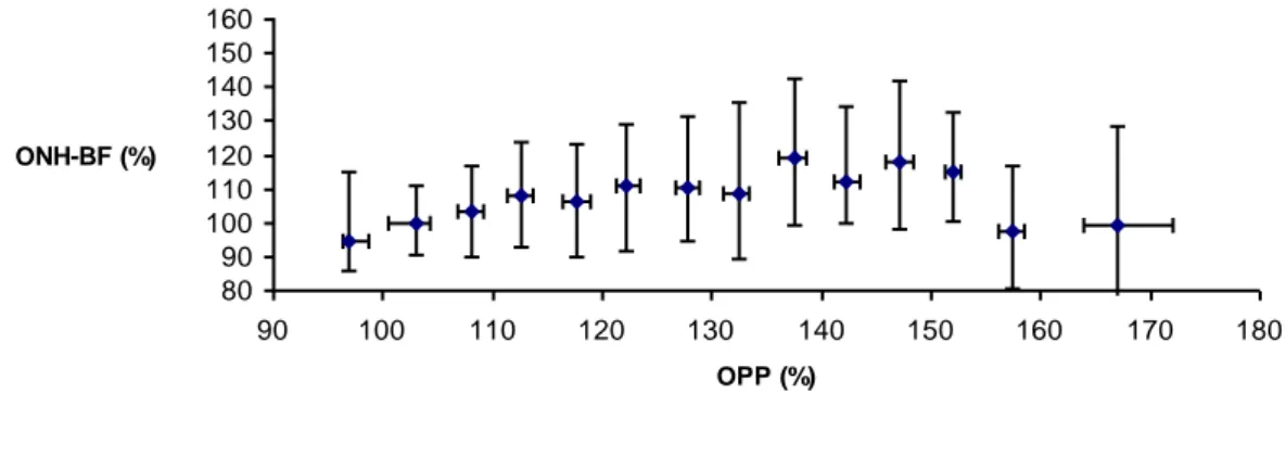
Figure 2 shows the correlation between ONH-BF and increasing OPP intervals. Analysis showed a significant difference between the intervals (p< 0.0001). For a 50% increase in OPP, flow increased by only 15%. Regression of ONH-BF versus OPP values shows that flow increased significantly when OPP increased from 100 to 145% (p=0.0002).
80 90 100 110 120 130 140 150 160 90 100 110 120 130 140 150 160 170 180 OPP (%) ONH-BF (%)
Figure 2: Change in ONH blood flow versus OPP in 15 healthy subjects over 2 min of handgripping. Each data point represents a median of successive flow values of the percentage change in OPP (data presented ± 25 and 75% quartiles)
In all subjects, the increase in vascular resistance versus OPP during handgripping showed a significant linear relationship as a function of the OPP (p=0.0013, Figure 3). For instance, a 50% increase in OPP was associated with a 30% increase in resistance. At the end of the exercise, the mean vascular resistance was increased by as much as 125±2%.
80 100 120 140 160 180 200 220 90 100 110 120 130 140 150 160 170 180 OPP (%) Resistance (%)
Figure 3: Change in resistance versus OPP in 15 healthy subjects over 2 min of handgripping. Each data point represents a median of successive resistance values of the percentage change in OPP. (data presented ± 25 and 75% quartiles)
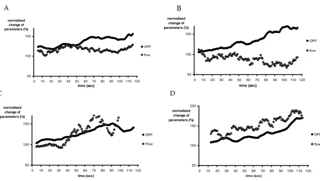
We could roughly identify four particular response patterns of ONH-BF to increase in OPP in this experiment, as illustrates figures 4.
50 100 150 0 10 20 30 40 50 60 70 80 90 100 110 120 time (sec) normalized change of parameters (%) OPP flow 50 100 150 0 10 20 30 40 50 60 70 80 90 100 110 120 time (sec) normalized change of parameters (%) OPP flow 50 100 150 0 10 20 30 40 50 60 70 80 90 100 110 120 time (sec) normalized change of parameters (%) OPP Flow 50 100 150 200 0 10 20 30 40 50 60 70 80 90 100 110 120 tim e (sec) normalized change of parameters (%) OPP flow
Figure 4: Examples of ONH blood flow and MAP in four subjects (#20, 1, 3,
B
C D
and 12) during handgripping (Data presented as mean of the moving average)
In the first case (subject #20, figure 4A), handgripping induced a 155% increase in OPP, whereas ONH-BF remained relatively constant during the 2 min of exercise. For the second case (#1, figure 4B), in spite of a continual increase in OPP (165%), flow tended to decrease during handgripping to −8% from baseline at the end of the exercise. Subject #3 (figure 4C) showed a BF response with two distinct phases during OPP increases, a phase of stability in flow followed by increases in ONH-BF to 177% (C). Finally, subject #12 (figure 4D) showed an almost parallel increase in both parameters during handgripping; OPP and BF increased 170%, at the end of the exercise.
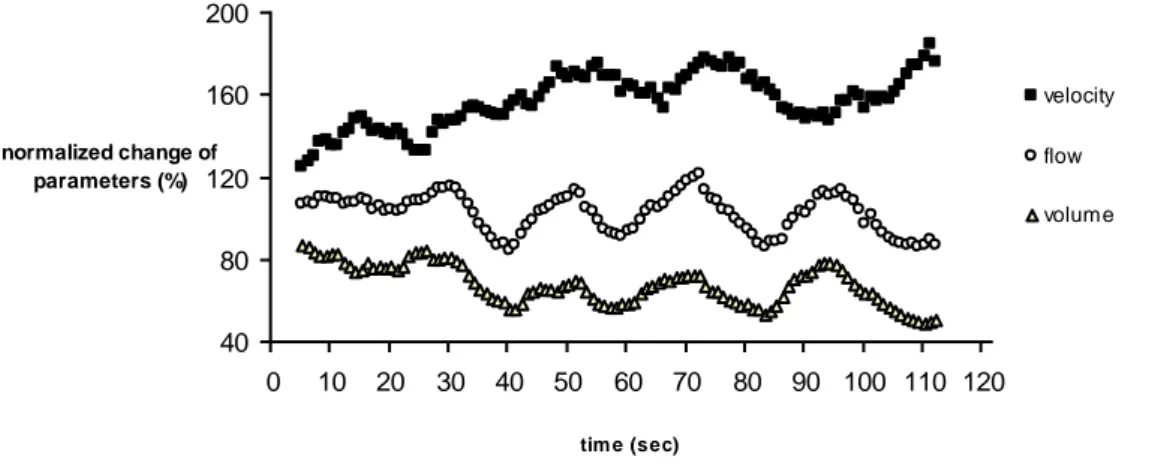
In some subjects, the analysis of blood flow parameters across time suggested oscillations in ONH parameters during the exercise (see example in Figure 5). Oscillation in ONH-BF with a period of approximately 15–20 s (frequency, 0.06–0.05 Hz), synchronized to volume fluctuations. In 75% of the subjects, a phase shift of approximately 180° between volume and velocity changes was found.
40 80 120 160 200 0 10 20 30 40 50 60 70 80 90 100 110 120 tim e (sec) normalized change of parameters (%) velocity flow volume
Figure 5: ONH blood flow, velocity, and volume in subject #5 during handgripping exercise (data presented as mean of the moving average)
Figure 6 shows the spectral Fourier analysis of velocity and ONH-BF in three of the subjects. Oscillations lasting 1 s and 3 s corresponding to frequencies of 60 cycles/min (1 Hz) and 20 cycles/min (0.3 Hz), respectively, predominated in this analysis. The lower frequencies shown in Figure 5 did not appear due to the short duration of the handgripping experiment (2 min).
Figure 6: Power spectrum of the LDF recording for velocity and ONH-BF of three subjects
DISCUSSION
This experimental study in healthy humans provides new insight into the relationship between ONH-BF and BP, i.e., a constant increase in vascular resistance associated with the continuous increase in BP. These new data illustrating the autoregulation process of BF in the ONH suggest that the increase in vascular resistance does not fully counterbalance the increase in OPP over the 2 min of isometric exercise.
An increase in arterial pressure can be obtained after intravenous injection of drugs in humans and animals, such as adrenaline,[29] norepinephrine, angiotensin II,[30] kallikrein, bradykinin, and angiotensin I and II, [31] or using occluders on the thoracic aorta and inferior vena cava in animals.[32] A noninvasive alternative in humans is to increase BP during isometric[4, 33] or dynamic exercise.[34-36] Handgripping is an isometric exercise that causes a greater increase in BP than dynamic exercise[34-36] principally by stimulating the sympathetic nervous system.[37, 38] This is a reproducible and noninvasive test of sympathetic function with a well-defined reflex pathway,[39] leading to an increase in muscle sympathetic nerve activity and plasma catecholamines. Isometric exercise also increases heart rate, arterial BP, cardiac output, left ventricular contractility, and systemic vascular resistance.[28, 40-42] The cardiovascular effect depends mainly on the intensity of the isometric contraction and the duration of the isometric exercise,[43] irrespective of muscle mass.[44] In this experiment, we selected healthy subjects who obtained a regular and significant increase in BP in order to obtain a homogeneous group. The variability in the response can be due to a contraction that represents a very low percentage of the true maximal
voluntary strength.[28] The approach used herein is also original since all previous studies examined the relationship between BP and blood flow in a discontinuous manner, with measurements every 30 s or every minute [2-5, 20,
45]
or before and at the end of exercise.[6, 20, 27]
This experimental primary result showed that the vascular resistance increase was regular over time, parallel to the increase in OPP. The increase in vascular resistance reflects the possibility of autoregulation in the ONH. Strictly speaking, autoregulation of flow is the intrinsic ability of a tissue to maintain relatively constant blood flow despite variations in perfusion
pressure, which can be demonstrated in the absence of neural and hormonal influences.[46, 47] The mechanisms underlying autoregulation are based on a balanced contribution of myogenic and metabolic components involving the interaction of factors released by the vascular endothelium, for instance, including nitric oxide.[48]. In the optic nerve head, the effect of sympathetic activation can be excluded due to the absence of ortho- or parasympathetic innervation in humans.[49, 50]
The existence of autoregulation at the ONH site was demonstrated in healthy subjects by Movaffaghy in 1998[8] when OPP was increased during squatting, and in 1997 by Riva[51], when OPP was decreased after a rapid rise in IOP. These results also suggested that the autoregulation mechanism was limited to 30–60% of OPP above baseline.[22]
In contrast with previous studies using discontinuous measurements, the continuous ONH-BF and BP measurements used in the present investigation allowed studying the
dynamic autoregulation[13] as previously described in nonhuman primates. The present results suggest constant blood flow-regulating kinetics (with regular increase in vascular resistance) allowing stability of blood flow for low
levels of BP increase and a limited autoregulation capacity with higher levels of BP increase. As suggested in Figures 3 and 4, some variability exists among healthy subjects with autoregulatory mechanisms leading to a stability of ONH-BF during increase in OPP in 10 out of 15 healthy subjects.
Whereas one previous study combining real-time measurement of BP and choroidal blood flow (ChBF) in healthy subjects during biking (dynamic exercise) showed stable ChBF during the increase in OPP, a close linear model of the change in vascular resistance versus OPP was demonstrated (y = 1.025x −7.478, r=0.96).[2]
The first study reporting the use of LDF for ONH-BF measurement during isometric exercise[8] also suggested a linear
relationship between vascular resistance and OPP (r=0.92) in two healthy subjects: ONH-BF started to increase when the OPP increase was greater than 30%. However, the data reported herein strongly suggest that the range of regulation is progressively overtaken with time.
In healthy subjects, the ONH-BF increase was associated with a proportional increase in velocity. This suggests that vasoconstriction, parallel to the rise of BP, takes place outside of the sampled volume, probably in the arterioles proximal to the ONH capillary bed of the neuroretinal rim, supplied by retinal arterioles and sometimes the posterior ciliary artery for the surface nerve fiber layer, and the fine centripetal branches from the peripapillary choroid for the prelaminar region.[52] Other blood flow modifications before the eye may also contribute to this regulation. Velocity in the ophthalmic artery has been shown to increase by approximately 30% during dynamic[19] or isometric exercise.[4] The parallel rise between the ophthalmic artery pressure and the brachial artery pressure indicates that regulation is
achieved in the central retinal artery or in the posterior ciliary arteries.[5, 19] Autoregulation observed in the ONH is close to that reported for retinal blood flow. Continuous recording of retinal blood flow has not been reported to date. In response to an acute increase in BP, retinal BF remains stable with an up to 141% increase in BP. Autoregulatory mechanisms of the retinal and ONH vasculature probably share certain metabolic and myogenic
mechanisms. Retinal vasoconstriction during isometric exercise is modified by ET-A receptor blockade, indicating that endothelin-1 is involved in this process.[53] On the other hand, high levels of circulating noradrenaline have little impact on retinal blood flow.[54]
The pressure–flow relationship and the kinetics of vascular resistance described in this study should be evaluated in further experiments in order to study the mechanisms involved, using pharmacological interventions on nitric oxide (after administration of the nitric oxide synthase inhibitor LNAME) or the endothelin system (ETA receptor antagonist inhibitor BQ-123).[55] For
instance, ET receptor blockade has been reported to blunt retinal vessel response during isometric exercise, suggesting that endothelin-1 is involved in retinal blood flow regulation when OPP increases.[53]
The analysis of ONH-BF variations during exercise showed rhythmic changes in BF parameters during exercise, previously described in rest conditions in animals [56-59] and humans.[60, 61] The recurrent pattern of variations at 1 Hz are synchronous with the heart rate and represent oscillatory changes of arteriolar diameter induced by pulsatile flow of the cardiac cycle. The 0.3-Hz frequency is synchronous with respiration. The slower oscillatory patterns (0.06 Hz) already described in the ONH[60] in basal
conditions but also in exercise conditions[62] may correspond to vasomotor frequency or α waves, representing intrinsic myogenic activity of the smooth muscle cells in resistance vessels. According to Stefanovska,[63] this oscillation is associated with blood pressure regulation. The smooth muscle cells in the vessel walls continuously respond to changes in intravascular pressure, known as the myogenic response. At the cellular level, vasomotion is associated with oscillations in intracellular calcium concentrations and membrane potential.[64]
As illustrated in Figure 5, the fluctuations in volume and velocity were out of phase by 180°, as previously described in the minipig [56], possibly the result of a change in venule diameter, predominantly present at the surface of the optic nerve head.
In the present study, the amplitude variation of fluctuations during exercise as compared with baseline data remains to be determined. Our analysis was limited by the duration of baseline measurements, too short for a rhythmic analysis of low frequencies.
With regard to the new LDF device, this technique has a high temporal and spatial resolution (M. Geiser and al. EVER meeting 2008, e5315, New
optical device for functional studies of the optic nerve head). However,
current limitations are the measurement of BF in arbitrary units, the limited penetration of the laser beam estimated at 300 m [65],and the need for good ocular fixation.
In conclusion, the combination of real-time measurement of ONH blood flow using LDF and BP during isometric exercise provides a
noninvasive method precisely defining the blood flow–pressure relationship. The approach in patients with optic nerve vascular disease, such as normal tension glaucoma[7] or nonarteritic ischemic optic nerve neuropathy, could also give insight into possible impaired autoregulation. Applications in patients should take into account the current limitations of the LDF system, however, such as optical considerations (refractive errors of the eye), the variability in ONH-BF response, and the requirement of stable ocular fixation.
REFERENCES
1. Delaey, C. and J. Van De Voorde, Regulatory mechanisms in the
retinal and choroidal circulation. Ophthalmic Res, 2000. 32(6): p.
249-56.
2. Lovasik, J.V., et al., Choroidal blood flow during exercise-induced
changes in the ocular perfusion pressure. Invest Ophthalmol Vis Sci,
2003. 44(5): p. 2126-32.
3. Riva, C.E., et al., Choroidal blood flow during isometric exercises. Invest Ophthalmol Vis Sci, 1997. 38(11): p. 2338-43.
4. Kiss, B., et al., Ocular hemodynamics during isometric exercise. Microvasc Res, 2001. 61(1): p. 1-13.
5. Robinson, F., et al., Retinal blood flow autoregulation in response to
an acute increase in blood pressure. Invest Ophthalmol Vis Sci, 1986.
27(5): p. 722-6.
6. Dumskyj, M.J., et al., Autoregulation in the human retinal circulation:
assessment using isometric exercise, laser Doppler velocimetry, and computer-assisted image analysis. Microvasc Res, 1996. 51(3): p.
378-92.
7. Pournaras, C.J., et al., Regulation of optic nerve head blood flow in
normal tension glaucoma patients. Eur J Ophthalmol, 2004. 14(3): p.
226-35.
8. Movaffaghy, A., et al., Blood flow in the human optic nerve head
9. Kiel, J.W., Modulation of choroidal autoregulation in the rabbit. Exp Eye Res, 1999. 69(4): p. 413-29.
10. Kiel, J.W. and W.A. van Heuven, Ocular perfusion pressure and
choroidal blood flow in the rabbit. Invest Ophthalmol Vis Sci, 1995.
36(3): p. 579-85.
11. Alm, A. and A. Bill, Ocular and optic nerve blood flow at normal and
increased intraocular pressures in monkeys (Macaca irus): a study with radioactively labelled microspheres including flow determinations in brain and some other tissues. Exp Eye Res, 1973. 15(1): p. 15-29.
12. Kiel, J.W., Choroidal myogenic autoregulation and intraocular
pressure. Exp Eye Res, 1994. 58(5): p. 529-43.
13. Liang, Y., et al., Quantification of dynamic blood flow autoregulation in
optic nerve head of rhesus monkeys. Exp Eye Res, 2010. 90(2): p.
203-9.
14. Gugleta, K., et al., Choroidal vascular reaction to hand-grip stress in
subjects with vasospasm and its relevance in glaucoma. Invest
Ophthalmol Vis Sci, 2003. 44(4): p. 1573-80.
15. Movaffaghy, A., et al., Effect of isometric exercise on choroidal blood
flow in type I diabetic patients. Klin Monbl Augenheilkd, 2002. 219(4):
p. 299-301.
16. Tittl, M., et al., Choroidal hemodynamic changes during isometric
exercise in patients with inactive central serous chorioretinopathy.
Invest Ophthalmol Vis Sci, 2005. 46(12): p. 4717-21.
17. Movaffaghy, A., et al., [Effect of squatting on sub-foveal blood flow
defect in pseudophakic eyes operated by cerclage]. Klin Monbl
18. Wimpissinger, B., et al., Effects of isometric exercise on subfoveal
choroidal blood flow in smokers and nonsmokers. Invest Ophthalmol
Vis Sci, 2003. 44(11): p. 4859-63.
19. Kozobolis, V.P., et al., Retrobulbar blood flow and ophthalmic
perfusion in maximum dynamic exercise. Clin Experiment Ophthalmol,
2008. 36(2): p. 123-9.
20. Okuno, T., et al., Ocular blood flow changes after dynamic exercise in
humans. Eye (Lond), 2006. 20(7): p. 796-800.
21. Iester, M., et al., Retinal blood flow autoregulation after dynamic
exercise in healthy young subjects. Ophthalmologica, 2007. 221(3): p.
180-5.
22. Luksch, A., et al., Role of NO in choroidal blood flow regulation during
isometric exercise in healthy humans. Invest Ophthalmol Vis Sci,
2003. 44(2): p. 734-9.
23. Fuchsjager-Mayrl, G., et al., Role of endothelin-1 in choroidal blood
flow regulation during isometric exercise in healthy humans. Invest
Ophthalmol Vis Sci, 2003. 44(2): p. 728-33.
24. Weigert, G., et al., Effects of topical clonidine versus brimonidine on
choroidal blood flow and intraocular pressure during squatting. Invest
Ophthalmol Vis Sci, 2007. 48(9): p. 4220-5.
25. Eeftinck Schattenkerk, D.W., et al., Nexfin noninvasive continuous
blood pressure validated against Riva-Rocci/Korotkoff. Am J
Hypertens, 2009. 22(4): p. 378-83.
26. Farrar, S.M., et al., An evaluation of the Tono-Pen for the
measurement of diurnal intraocular pressure. Am J Ophthalmol, 1989.
27. Longo, A., M.H. Geiser, and C.E. Riva, Posture changes and
subfoveal choroidal blood flow. Invest Ophthalmol Vis Sci, 2004.
45(2): p. 546-51.
28. Lind, A.R., et al., The Circulatiory Effects of Sustained Voluntary
Muscle Contraction. Clin Sci, 1964. 27: p. 229-44.
29. Pournaras, C.J., et al., [Regulation of papillary pO2]. Klin Monbl Augenheilkd, 1992. 200(5): p. 517-8.
30. Gherezghiher, T., J.A. Hey, and M.C. Koss, Systemic blood pressure
and intraocular pressure relationship. J Ocul Pharmacol, 1988. 4(4): p.
291-301.
31. Funk, R., J.W. Rohen, and K. Skolasinska, Intraocular pressure and
systemic blood pressure after administration of vasoactive substances in hypertensive and normal rats. Graefes Arch Clin Exp Ophthalmol,
1985. 223(3): p. 145-9.
32. Kiel, J.W., The effect of arterial pressure on the ocular
pressure-volume relationship in the rabbit. Exp Eye Res, 1995. 60(3): p. 267-78.
33. Jandik, J., et al., Cardiovascular response to hand-grip isometric
exercise in healthy men. Noninvasive study. Physiol Bohemoslov,
1985. 34(6): p. 534-42.
34. Arimoto, M., A. Kijima, and S. Muramatsu, Cardiorespiratory response
to dynamic and static leg press exercise in humans. J Physiol
Anthropol Appl Human Sci, 2005. 24(4): p. 277-83.
35. Asmussen, E., Similarities and dissimilarities between static and
36. Whipp, B.J. and E.E. Phillips, Jr., Cardiopulmonary and metabolic
responses to sustained isometric exercise. Arch Phys Med Rehabil,
1970. 51(7): p. 398-402.
37. Lanigan, L.P., C.V. Clark, and D.W. Hill, Intraocular pressure
responses to systemic autonomic stimulation. Eye (Lond), 1989. 3 ( Pt
4): p. 477-83.
38. Castejon, H., et al., Effect of acute increase in blood pressure on
intraocular pressure in pigs and humans. Invest Ophthalmol Vis Sci,
2010. 51(3): p. 1599-605.
39. Khurana, R.K. and A. Setty, The value of the isometric hand-grip
test--studies in various autonomic disorders. Clin Auton Res, 1996. 6(4): p.
211-8.
40. Rowland, T. and B. Fernhall, Cardiovascular responses to static
exercise: a re-appraisal. Int J Sports Med, 2007. 28(11): p. 905-8.
41. Astrand, I., A. Guharay, and J. Wahren, Circulatory responses to arm
exercise with different arm positions. J Appl Physiol, 1968. 25(5): p.
528-32.
42. Mitchell, J.H., et al., Response of arterial blood pressure to static
exercise in relation to muscle mass, force development, and electromyographic activity. Circ Res, 1981. 48(6 Pt 2): p. I70-5.
43. Mitchell, J.H. and K. Wildenthal, Static (isometric) exercise and the
heart: physiological and clinical considerations. Annu Rev Med, 1974.
25: p. 369-81.
44. Mitchell, J.H., et al., The role of muscle mass in the cardiovascular
45. Michelson, G., M. Groh, and A. Grundler, Regulation of ocular blood
flow during increases of arterial blood pressure. Br J Ophthalmol,
1994. 78(6): p. 461-5.
46. Riva C. E. , A.A., Pornaras C. J. , Ocular Circulation, in Kaufmann. 2010. p. 243-273.
47. Guyton, A.C., Jones, C.E., Coleman, T.G, Circulatory Physiology. Cardiac Output and its Regulation. 1973, Philadelphia: W.B. Saunders Company. 323–352.
48. Okuno, T., et al., Evidence that nitric oxide is involved in
autoregulation in optic nerve head of rabbits. Invest Ophthalmol Vis
Sci, 2002. 43(3): p. 784-9.
49. Mackenzie, P.J. and G.A. Cioffi, Vascular anatomy of the optic nerve
head. Can J Ophthalmol, 2008. 43(3): p. 308-12.
50. Funk, R.H., Blood supply of the retina. Ophthalmic Res, 1997. 29(5): p. 320-5.
51. Riva, C.E., et al., Autoregulation of human optic nerve head blood flow
in response to acute changes in ocular perfusion pressure. Graefes
Arch Clin Exp Ophthalmol, 1997. 235(10): p. 618-26.
52. Hayreh, S.S., Blood flow in the optic nerve head and factors that may
influence it. Prog Retin Eye Res, 2001. 20(5): p. 595-624.
53. Luksch, A., et al., ETa-receptor blockade, but not ACE inhibition,
blunts retinal vessel response during isometric exercise. Am J Physiol
Heart Circ Physiol, 2006. 290(4): p. H1693-8.
54. Jandrasits, K., et al., Effect of noradrenaline on retinal blood flow in
55. Polska, E., et al., Effects of adenosine on intraocular pressure, optic
nerve head blood flow, and choroidal blood flow in healthy humans.
Invest Ophthalmol Vis Sci, 2003. 44(7): p. 3110-4.
56. Riva, C.E., et al., Rhythmic changes in velocity, volume, and flow of
blood in the optic nerve head tissue. Microvasc Res, 1990. 40(1): p.
36-45.
57. Buerk, D.G. and C.E. Riva, Vasomotion and spontaneous
low-frequency oscillations in blood flow and nitric oxide in cat optic nerve head. Microvasc Res, 1998. 55(1): p. 103-12.
58. Braun, R.D., R.A. Linsenmeier, and C.M. Yancey, Spontaneous
fluctuations in oxygen tension in the cat retina. Microvasc Res, 1992.
44(1): p. 73-84.
59. Colantuoni, A., S. Bertuglia, and M. Intaglietta, Microvascular
vasomotion: origin of laser Doppler flux motion. Int J Microcirc Clin
Exp, 1994. 14(3): p. 151-8.
60. Osusky, R., A. Schoetzau, and J. Flammer, Variations in the blood
flow of the human optic nerve head. Eur J Ophthalmol, 1997. 7(4): p.
364-9.
61. Bongard, O. and B. Fagrell, Variations in laser Doppler flux and flow
motion patterns in the dorsal skin of the human foot. Microvasc Res,
1990. 39(2): p. 212-22.
62. Kvernmo, H.D., et al., Spectral analysis of the laser Doppler perfusion
signal in human skin before and after exercise. Microvasc Res, 1998.
63. Stefanovska, A., M. Bracic, and H.D. Kvernmo, Wavelet analysis of
oscillations in the peripheral blood circulation measured by laser Doppler technique. IEEE Trans Biomed Eng, 1999. 46(10): p. 1230-9.
64. Parthimos, D., et al., Dynamics of a three-variable nonlinear model of
vasomotion: comparison of theory and experiment. Biophys J, 2007.
93(5): p. 1534-56.
65. Riva, C.E., et al., Laser Doppler flowmetry in the optic nerve. Exp Eye Res, 1992. 55(3): p. 499-506.
SERMENT D’HIPPOCRATE
En présence des Maîtres de cette Faculté, de mes chers condisciplines et devant l’effigie d’HIPPOCRATE,
Je promets et je jure d’être fidèle aux lois de l’honneur et de la probité dans l’exercice de la Médecine.
Je donnerais mes soins gratuitement à l’indigent et n’exigerai jamais un salaire au dessus de mon travail. Je ne participerai à aucun partage clandestin d’honoraires.
Admis dans l’intimité des maisons, mes yeux n’y verront pas ce q ui s’y passe ; ma langue taira les secrets qui me seront confiés et mon état ne servira pas à corrompre les mœurs, ni à favoriser le crime.
Je ne permettrai pas que des considérations de religion, de nation, de race, de parti ou de classe sociale viennent s’interposer entre mon devoir et mon patient.
Je garderai le respect absolu de la vie humaine.
Même sous la menace, je n’admettrai pas de faire usage de mes connaissances médicales contre les lois de l’humanité.
Respectueux et reconnaissant envers mes Maît res, je rendrai à leurs enfants l’instruction que j’ai reçue de leurs pères.
Que les hommes m’accordent leur estime si je suis fidèle à mes promesses. Que je sois couvert d’opprobre et méprisé de mes confrères si j’y manque.