Cloning, sequencing and overexpression in Escherichia coli of the
alginate-lyase-encoding aly gene of Pseudomonas alginovora: identification of three
classes of alginate lyases
**Frederic Chavagnat*†, Colette Duez†, Micheline Guinand*, Philippe Potin‡, Tristan Barbeyron‡, Bernard Henrissat§, Jean Wallach* and Jean-Marie Ghuysen†
*Laboratoire de Biochimie Analytique et Synthèse Bioorganique, Université Claude Bernard, 43 Boulevard du 11 Novembre 1918, F-69622 Villeurbanne, France, †Centre d'Ingénierie des Protéines, Université de Liège, Sart Tilman, Belgium, ‡Centre d'Etudes d'Océanographie et de Biologie Marine, Station Biologique, Roscoff, France, and §Centre de Recherches sur les Macromolécules Végétales, CNRS, Grenoble, France
Abstract
A gene of Pseudomonas alginovora, called aly, has been cloned in Escherichia coli using a battery of PCR techniques and sequenced. It encodes a 210-amino-acid alginate lyase (EC 4.2.2.3), Aly, in the form of a 233-amino-acid precursor. P. alginovora Aly has been overproduced in E. coli with a His-tag sequence fused at the C-terminal end under conditions in which the formation of inclusion bodies is avoided. His-tagged P. alginovora Aly has the same enzymic properties as the wild-type enzyme and has the specificity of a mannuronate lyase. It can be purified in a one-step procedure by affinity chromatography on Ni2+-nitriloacetate resin. The yield is of 5 mg of enzyme per litre of culture. The amplification factor is 12.5 compared with the level of production by wild-type P. alginovora. The six alginate lyases of known primary structure fall into three distinct classes, one of which comprises the pair P. alginovora Aly and Klebsiella pneumoniae Aly.
Abbreviations used : Aly-[His-tag], His-tagged P. alginovora alginate lyase Aly; HCA, hydrophobic cluster
analysis; NTA, nitriloacetate.
INTRODUCTION
Alginates are linear uronic acid polymers in which β-D-mannuronate and α-L-guluronate residues are linked to form blocks of polymannuronate, polyguluronate and random sequences [1]. Alginates are synthesized as cell-wall polysaccharides by brown seaweeds and as exopolysaccharides by bacteria. Bacterial alginates are mainly of the polymannuronate type, with O-acetyl groups at the 2- and/or 3-position of D-mannuronate [2]. Alginate facilitates the attachment of mucoid Pseudomonas aeruginosa strains to the tracheal epithelium and respiratory mucins, protects the bacteria from phagocytes and prevents antibiotic uptake [3-5]. Consequently it is a major pathogenic factor in cystic fibrosis patients [1,6].
Alginate lyases have been isolated from various sources [7,8]. They catalyse the breakdown of alginate by a β-elimination mechanism, causing formation of a double bond between C-4 and C-5, β-elimination of the 4-O-glycosidic bond and production of 4-deoxy-L-erythro-hex-4-ene pyranosyluronate at the non-reducing end of the resulting oligosaccharide [7,9]. The primary structures of five alginate lyases are known. A1-I of a
Sphingomonas sp. (converted into A1-II and A1-III by a self-processing reaction) depolymerizes non-acetylated
and acetylated alginates [10,11]. AlxM of the marine bacterium A.T.C.C. 433367 [12], ALY of Pseudomonas sp. OS-ALG-9 [13] and AlgL of P. aeruginosa [14,15] are each mannuronate lyases. Aly of Klebsiella pneumoniae subsp. aerogenes is a guluronate lyase [16]. Three alginate lyases have been overproduced [16-18]. The
previously described AlxM was cleaved between two cysteine residues, Cys-169 and Cys-183, themselves linked by a disulphide bridge [12]. It has been overproduced in Escherichia coli as an unnicked polypeptide chain referred to as AlxMB [18].
** This paper is dedicated to the memory of Professor Georges Michel.
The nucleotide sequence of P. alginovora aly is deposited in the EMBL/GenBank/DDBJ Nucleotide Sequence Databases under accession no. X83679.
As reported previously [19], Pseudomonas alginovora is an alginate lyase producer. The present paper describes the purification of a P. alginovora alginate lyase, Aly, and the cloning and sequencing of the encoding gene, aly. It defines the conditions under which the enzyme can be overproduced in E. coli and purified in a one-step procedure. It also identifies three distinct classes of alginate lyases.
MATERIALS AND METHODS
Bacterial strains, plasmids, oligonucleotides and culture conditions
P. alginovora strain XO17, CIP 102941, was maintained at 25 °C on sea water nutrient agar plates (Difco
Laboratories). It was grown at 25 °C in Zobell Medium as described by Boyen et al. [19].
E. coli Max Efficiency DH5α (Gibco BRL, Life Technologies, Inc.), E. coli Top 10 F' (Invitrogen, Leek, The
Netherlands), lysogenic E. coli BL21 (DE3) and E. coli HMS174 (DE3) (Novagen) were used as hosts for recombinant plasmids. The E. coli transformants were grown at temperatures ranging from 18 to 37 °C in Luria-Bertani (LB), Terrific Broth (TB) [20] and M9H [21] media containing 50 µg of ampicillin and 30 µg of chloramphenicol/ml. pUC18 (Gibco BRL) was used to subclone DNA fragments and to prepare double-stranded DNA for sequencing. pET-22b (for the expression of recombinant proteins and conferring ampicillin resistance) and plysS (for the stabilization of target genes and conferring chloramphenicol resistance) were from Novagen. Oligonucleotides were synthesized by Eurogentec, Liège, Belgium.
Alginate lyase plate assay
Alginate lyase-producing E. coli strains were identified by growing them at 28 °C on 'black medium' plates containing 0.5% agar, 0.5% yeast extract (Difco), 1 % high-viscosity alginate of Macrocystis pyrifera (Sigma, St. Quentin Fallavier, France), 0.5% active charcoal (Serva, Heidelberg, Germany), 50 µg/ml ampicillin and 30
µg/ml chloramphenicol [22].
Alginate lyase activity
The substrates were low-viscosity, acetyl-group-free, alginates from Macrocystis pyrifera (60% mannuronate; Sigma) and Laminaria hyperborea (44% mannuronate; Algocean, Landernau, France) and low-molecular-mass mannuronate and guluronate polymers, each containing 15-20 saccharide units (gifts from Dr. A. Heyraud, CERMAV, Grenoble, France). Enzyme solutions (400 µl) containing 0.3 % substrate in 225 mM Tris/HCl, pH 7.5, or 75 mM sodium phosphate (pH 7.5)/ 450 mM NaCl were incubated at 25 °C for 5 min. The unsaturated non-reducing groups produced by β-elimination were measured using 3-deoxy-D-manno-octulosonic acid (up to 50 nmol) as standard. The conditions described in Karkhanis et al. [23] were modified as in Malissard et al. [18]. One unit of enzyme activity produced 1 µmol of non-reducing, unsaturated, terminal group per min.
Production, purification, trypsin degradation and amino acid sequencing of wild-type P. alginovora Aly
The enzyme was produced and purified as described by Boyen et al. [19]. P. alginovorα XO17 was grown in 5 litres of Zobell medium supplemented with 0.1% sodium alginate from Lαminariα digitαtα (Sobalg). The enzyme present in the culture supernatant was concentrated by ultrafiltration through 10 kDa cut-off membranes and purified by ammonium sulphate precipitation, gel filtration and anion-exchange chromatography.
SDS/PAGE (12% gels) of P. αlginovorα Aly was carried out according to Laemmli and Favre [24]. Gels were stained with PAGE Blue 83 or electroblotted for 2 h at 0.8 mA/cm2 on an Immobilon membrane (Millipore), using a Pharmacia LKB Novablot apparatus [25]. Membrane, with electrotransferred Aly, was inserted into the sequencing chamber of an Applied Biosystems 473A amino acid microsequencer. Acrylamide slices containing about 40-50 µg of SDS/PAGE-purified Aly (stained with 0.003 % Amido Black) were submitted to proteolysis with 1 µg of trypsin in 150 µl of 0.1 M Tris/HCl (pH 8.8)/0.01 % Tween 20 for 5 h at 37 °C. Trypsin-degraded Aly was submitted to reverse-phase HPLC on a 250 mm × 4.6 mm µBondapak C18 column. Elution, carried out with an acetonitrile gradient from 2to45 % in 0.1 % trifluoroacetic acid at a flow rate of 0.2 ml/min, allowed many peptides to be separated. Among them, peptides P1 and P2 were eluted at 22 and 24 % acetonitrile respectively. They each were submitted to amino acid microsequencing in the 473A microsequencer. The amount of phenylthiohydantoin derivative of the first amino acid residue was about 210 pmol for both peptides. The N-terminal sequences of Aly and P2 were used to synthesize nucleotides O1 and O2 respectively (see the Results and discussion section).
DNA preparation and nucleotide sequencing
Genomic DNA from P. alginovorα was isolated as in Hopwood et al. [26]. Plasmids were extracted from E. coli cells by the alkaline lysis method [27]. Recombinant plasmids were constructed and agarose gel electrophoresis was performed according to Sambrook et al. [20]. Restriction enzymes (Gibco BRL; Eurogentec) and T4 DNA ligase (Boehringer, Mannheim, Germany) were used with the buffers provided and under the conditions recommended by the suppliers. DNA segments cloned into pUC18 were sequenced by the dideoxynucleotide chain termination method [28] using M13 universal and reverse oligonucleotides or synthetic oligonucleotides as primers. Denaturation of double-stranded DNA was performed according to Zhang et al. [29]. Sequencing reactions were carried out by using the T7 sequencing kit (Pharmacia LKB Biotechnology, Uppsala, Sweden) with [35S]dATP labelling (ICN Biomedicals) or the Autoread sequencing kit (Pharmacia LKB) with fluorescent primers or incorporation of fluorescent dUTP. In this latter case, electrophoresis was done with an A.L.F. DNA sequencer.
PCR amplification
Mixtures (100 µl) containing DNA template (1-2 µ g), deoxynucleotide triphosphates (20 nmol each) and oligonucleotide primers (0.5-1 µg each) were treated with Vent® DNA polymerase (2 units) in Vent buffer (New England Biolabs, Leusden, The Netherlands) or with Goldstar® DNA polymerase (0.4-1 unit) in Goldstar buffer (Eurogentec)/4 mM MgSO4. The reactions were carried out in a DNA Thermal cycler (Biometra ; Trio
Thermoblock) for 30 cycles, each with a 1 min denaturation at 94 °C, a 1 min annealing at Tm-5 °C (where Tm is
the melting temperature of the primers) and a 150 s extension at 72 °C. The final elongation step at 72 °C was for 10 min. The Tm values of the primers were calculated using Primer Analysis Software OLIGO version 3.4
(National Biosciences, Hamel, MN, U.S.A.). The PCR products were purified using the QIA quick-spin PCR purification kit (Qiagen, Westburg, Leusden, The Netherlands).
Purification of His-tagged P. alginovora alginate lyase Aly (Aly-[His-tag])
E. coli HMS8 cells were grown at 18 °C in 1 litre of TB medium containing 50 /tg/ml ampicillin and 30 µg/ml
chloramphenicol, collected by centrifugation, suspended in 100 ml of 50 mM sodium phosphate (pH 8)/500 mM NaCl, frozen and thawed (allowing the resident T7 lysozyme to perform cell lysis), and then the preparation was incubated with 1.7 units/ml DNAse I (EC 3.1.21.1 ; Sigma) for 20 min at 20 °C. The lysate, containing 387 mg of total protein (specific enzyme activity 1.35 units/mg of protein in 225 mM Tris/HCl, pH 7.5) was collected by centrifugation at 40000 g for 20 min and loaded on a 1.12 cm × 10 cm Ni2+-nitriloacetate (NTA) resin column (Qiagen) equilibrated with 50 mM sodium phosphate (pH 7.0)/500 mM NaCl. Inactive proteins were eliminated by washing the column first with the same buffer and then with 50 mM sodium phosphate (pH 6.0)/500 mM NaCl. The adsorbed alginate lyase was eluted at about 180 mM imidazole in the last buffer using a two-step linear gradient of imidazole from 0 to 150 mM (4.5 bed vols.) and from 150 to 500 mM (0.5 bed vol.) at a flow rate of 0.5 ml/min. The active fractions were stored at -20 °C. The proteins were estimated according to Bradford [30], using BSA as standard.
Anti-(alginate lyase) polyclonal antibodies and immunoblot assays
Polyclonal antibodies, raised against P. alginovorα Aly or AlxMB [18] alginate lyases, were prepared by
injecting 60-100 µg of each enzyme into rabbits (two pairs), first in complete Freund's adjuvant and then in incomplete Freund's adjuvant, at 21-day intervals. Sera were collected 57 days after the first injection,
decomplemented at 56 °C for 30 min and filter-sterilized (0.45 µm). To purify the antibodies, the alginate lyases (100 µg) were each submitted to SDS/PAGE (12%) and electrotransferred. Membrane strips, stained with Ponceau S, were incubated at 4 °C for 16 h with 5 ml of the relevant antiserum and washed successively with 20 mM Tris/HCl (pH 7.5)/500 mM NaCl (TBS), with 3 % gelatin in TBS containing 0.05 % Tween 20 (TTBS) to remove non-specifically bound antibodies, and with 5 ml of 0.1 M glycine, pH2.5, to dissociate the bound antibodies. The glycine buffer was neutralized immediately with 1 M NaOH (approx. 200 µl), and the purified antibodies were stored at -20 °C.
ELISA assays (in duplicate) were performed essentially as described by Bourdenet et al. [31]. The microtitre plates (Micro ELISA Dynatech) were coated with 0.1 ml of a solution of 5 µg/ml alginate lyase. The ELISA titres were calculated by multiplying the reciprocal of a serum dilution within the linear portion of the absorbance curve (A490 0.7-1.2) by the corresponding A490 value.
Alginate lyases (0.2 µg) were submitted to SDS/PAGE (12%) in the presence of prestained SDS/PAGE standards (Bio-Rad Laboratories, Ivry-sur-Seine, France) and transblotted on to Immobilon membranes. The membranes were treated with 3 % gelatin in TBS to block their protein-binding capacity, washed with TTBS and treated with purified anti-AlxMB antibodies (100-fold dilution) and anti-Aly antibodies (1000-fold dilution).
Complexes were detected as purple-red bands using the Bio-Rad immunoassay kit, a goat anti-(rabbit IgG)-alkaline phosphatase conjugate (1:3000 dilution) and the reagents p-Nitro Blue Tetrazolium chloride and 5-bromo-4-chloro-3-indolyl phosphate, toluidine salt.
pi values, homology searches and hydrophobic cluster analysis (HCA)
Experimental pI values were measured by agarose gel slab electrofocusing using Pharmalytes 3-10 (Pharmacia) under the conditions recommended by the manufacturer. Theoretical pI values were computed using GCG software [32]. A homology search through the PIR protein sequence data bank (version 32) was made using FASTA and TFASTA [33]. For HCA, sequences taken from various data banks were converted into HCA plots using the HCA-PLOT program from Doriane (Le Chesnay, France). In these plots the amino acid residues are represented by their standard one-letter code, except for proline, glycine, serine and threonine which are represented by , ♦, and □ respectively. HCA scores were computed as described by Gaboriaud et al. [34] and Lemesle-Varloot et al. [35].
Table 1 Amino acid sequencing data of P. alginovora Aly. Amino acid residues are numbered according to the
sequence shown in Figure 2.
Peptide N-terminal amino acid sequence Amino acid positions Wild-type Aly GSFNDISWTLENEDN 1-15
Tryptic peptide P1 IWSSSIEVGGEE 168-179 Tryptic peptide P2 IGAYQLTGGEGEFHV 185-199
RESULTS AND DISCUSSION
Gene cloning and sequencing: amino acid sequence of P. alginovora Aly
Wild-type Aly produced by P. alginovora was purified to homogeneity (yield 0.4 mg per litre of culture) and the trypsin-degraded polypeptides P1 and P2 were isolated as described in the Materials and methods section. Table 1 gives the N-terminal amino acid sequences of Aly and of polypeptides P1 and P2.
The strategy of gene cloning involved the generation of the PCR products X, Y, Z and W, as illustrated in Figure 1. The nucleotide sequence of aly and the translated amino acid sequence of P. alginovora Aly are shown in Figure 2.
Product X
The genomic P. alginovora DNA was used as template, and oligonucleotide O1 (encoding the heptapeptide F3NDISWT9 of Aly; Table 1) and oligonucleotide O2 [complementary to the E194GEFH(V)199-encoding
sequence of peptide P2; Table 1] were used as primers. The reaction product X, identified by gel electrophoresis, was about 600 bp long. It was subcloned into pUC18 at SmaI and sequenced using the M13 universal and reverse primers and oligonucleotides O3 (nucleotides 752-774) and O4 (complementary to nucleotides 822-843). Product X encoded a 197-amino-acid polypeptide, the sequence of which was the Phe3-Val199 sequence shown in Figure 2.
Product Y
The HindIII, DraI, ClaI and EcoRV genomic libraries were used as templates in an inverse PCR [36] with oligonucleotide O5 (nucleotides 1046-1068) and oligonucleotide 06 (complementary to nucleotides 527-548) as primers. (In an inverse PCR, a recircularized DNA fragment is used as template to amplify a gene from an internal position, and the sense of the amplification from the primers is opposite to that of a direct PCR.) The reaction product Y (about 2000 bp long), obtained with the HindIII library, was subcloned into pUC18 at SmaI and sequenced with the M13 universal primer and oligonucleotides O7 (complementary to nucleotides 285-306)
and O8 (nucleotides 1-22) (assay no. 1), and with the M13 reverse primer and oligonucleotide O9
(complementary to nucleotides 1519-1541) (assay no. 2). Assay no. 1 gave the nt 1-nt 548 sequence shown in Figure 2. The nt 422-nt 535 sequence encoded a 23-amino-acid signal peptide (Met-23 to Ala-1) fused to the Gly1 -Asn15 sequence of the wild-type enzyme (Table 1). Assay no. 2, however, revealed that a fragment was deleted from nt 1046 as a result of the subcloning into pUC18.
Figure 1 Strategy used for the cloning and sequencing of the P. alginovora alginate-lyase-encoding gene aly.
Primers used for PCR amplification are indicated by → and ← ; sequences initiated with the M13 universal or reverse primers are indicated by ● ; and sequences initiated with synthetic nucleotides are indicated by O. The arrows indicate the orientation and length of the sequenced DNA segments. The unshaded box represents the signal-peptide-encoding sequence; the shaded box represents the mature alginate lyase-encoding gene. The hatched area in Y is a deleted segment (see the text).
Figure 2 Nucleotide sequence of P. alginovora aly and deduced amino acid sequence of its product Aly. The
N-terminal amino acid sequences of the mature enzyme and the tryptic peptides P1 and P2 are underlined. Arrows indicate the putative stem-loop area. RBS, ribosome-binding site.
Product Z
Product Z (about 600 bp long) was obtained with oligonucleotides 03 (as above) and O10 (complementary to nucleotides 1289-1309 with a PstI site fused at the 5' end). Product Z was subcloned into pUC18 between SmaI and PstI and sequenced in both directions with the M13 universal and reverse primers and oligonucleotide O5. It contained the nt 1046-nt 1170 segment that was missing from product Y. It also contained a TAA stop codon
starting at position 1121.
Product W
Products Y, X and Z arranged in an ordered sequence contained a 699-nucleotide open reading frame that started with a methionine-encoding ATG codon at position 422 and terminated with the TAA codon at position 1121. To check the accuracy of the sequence, the Goldstar and Vent DNA polymerases were each used to amplify the entire gene with oligonucleotide O10 (as above) and oligonucleotide O11 (nucleotides 422-449, with the Asp718 and NdeI sites fused in tandem at the 5' end). The two polymerases each generated the same product, W (approx. 900 bp long). Subcloning into pUC18 at the Asp718 and PstI sites and sequencing confirmed the nucleotide sequence shown in Figure 2. Segments located upstream and downstream of P. alginovora aly had similarities with known E. coli consensus sequences. A putative GAGGA Shine-Dalgarno sequence occurred 5 bp upstream from the ATG initiation codon. The TATatT and TTGcCt sequences located 130 bp upstream from this Shine-Dalgarno sequence might correspond to the '-10' and '-35' regions of a promoter [37]. A putative stem-loop area located 18 bp downstream from the TAA stop codon might be a transcription termination site [38]. Moreover, this hairpin structure was followed by a TTTTT sequence, reminiscent of a Rho-inde-pendent termination site.
P. alginovora aly translated into a 210-amino-acid (Gly1-Lys210) protein preceded by a 23-amino-acid (Met-23 -Ala-1) signal peptide (Figure 2). The theoretical molecular mass, 23 570 Da, of the signal-peptide-free protein coincided perfectly with the experimental value of 24 kDa derived from the migration of wild-type P. alginovora Aly on SDS/ PAGE. The theoretical (4.69) and experimental (5.20) pI values were similar. The tryptic peptides P1 and P2 occurred near the C-terminal end of the protein (Figure 2).
Overproduction, purification and properties of Aly-[His-tag]
To overexpress aly and facilitate purification of the overproduced enzyme, NdeI and XhoI sites were created by PCR at the 5' and 3' ends of aly respectively using, as primers, the sense oligonucleotide O11 (as above) and the antisense oligonucleotide O12 (complementary to nt 1098-1120, with a XhoI site fused at the 5' end). The 719 bp PCR product was cloned into pET-22b between NdeI and XhoI sites. The resulting plasmid, pET-aly, contained
P. alginovora aly with its own Met-23-Ala-1 signal-peptide-encoding sequence and the His-tag (i.e.
LEHHHHHH)-encoding sequence fused at the 3' end. The gene was under the control of the T7/lac operator. In consequence, its expression was inducible by isopropyl β-D-thiogalactoside.
pET-aly was used to transform E. coli Top 10 F' as described by Sambrook et al. [20]. The pair 'supercoiled pET-aly/plysS' was used to transform E. coli BL21 (DE3), yielding E. coli BL34, and E. coli HMS174 (DE3), yielding E. coli HMS8. The production of Aly-[His-tag] by the (ampicillin- and chloramphenicol-resistant) transformants was monitored by varying the growth conditions: the medium, the temperature, the time and the duration of the induction. The produced protein underwent aggregation into inclusion bodies at growth temperatures of 24 °C or higher. The optimal conditions for the production of the enzyme in a water-soluble form were: a temperature of 18 °C; initiation of induction by adding 1 mM isopropyl β-D-thiogalactoside at A600
= 1.3 (in the case of E. coli BL34 grown in M9H medium) or at A600 = 0.9 (in the case of E. coli HMS8 grown in
TB medium); and a duration of induction of 7 h (E. coli BL34) or 17 h (E. coli HMS8).
As described in the Materials and methods section, E. coli HMS8 was grown in 1 litre of TB medium under optimal conditions, the cells were lysed by freezing and thawing and the enzyme solution (387 mg of protein; specific activity 1.35 units/mgin 225 mM Tris/HCl, pH 7.5) was purified by chromatography on a Ni2+-NTA resin column (yield 5 mg of protein; specific activity 71 units/mg in 225 mM Tris/HCl, pH 7.5). The
amplification factor of 12.5 (by reference to the yield of enzyme production by the wild-type strain) was lower than that obtained with other recombinant alginate lyases [16-18]. This relatively low amplification factor may be attributable to the low temperature at which the E. coli transformants must be grown to avoid the formation of inclusion bodies.
Aly-[His-tag] had the same N-terminal amino acid sequence (GSFNDISW) as P. alginovora wild-type Aly, showing that cleavage of the signal peptide occurred during synthesis in E. coli. As shown by SDS/PAGE (Figure 3A), Aly-[His-tag] was more than 90 % pure, and its apparent molecular mass was slightly greater than that of P. alginovora Aly (because of the presence of the additional His-tag octapeptide). The theoretical (5.35) and experimental (5.8) pI values coincided well.
Figure 3 SDS/PAGE (12% gels) of AlxMB (lanes 1), P. alginovora Aly-[His-tag] (lanes 2) and P. alginovora Aly
(lanes 3). Coomassie Blue staining is shown in (A). Western immunoblots are also shown using antisera directed to AlxMB (B) and Aly (C). All samples were solubilized in SDS/PAGE sample buffer at 100 °C for 5 min.
Prestained proteins of standard molecular masses (kDa) are shown.
In 225 mM Tris/HCl, pH 7.5, buffer (I 0.18) and at 25 °C, P. alginovora Aly and Aly-[His-tag] had identical degrading activities towards the alginate substrates tested (see the Materials and methods section) : 97 units/mg with a mannuronate polymer (15-20 units), 75 units/mg with M. pyrifera alginate (60 % mannuronate), 20 units/mg with L. hyperborea alginate (44 % mannuronate) and 7 units/mg with a guluronate polymer (15-20 units). At higher ionic strength (I 0.66), and in 75 mM sodium phosphate (pH 7.5)/450 mM NaCl buffer, the enzyme activity was decreased by 50 %. The conclusion that P. alginovora Aly is a mannuronate lyase and not a guluronate lyase is at variance with that proposed previously [19]. P. alginovora might produce a guluronate lyase in addition to Aly.
P. alginovora Aly as a member of a particular class of alginate lyases
The six alginate lyases of known primary structure (including P. alginovora Aly) have various molecular masses. The polypeptide chains (including the signal peptide) contain 413 residues (Sphingomonas A1-III), 398 residues (Pseudomonas ALY), 368 residues (P. aeruginosa AlgL), 307 residues (K. pneumoniae Aly), 285 residues (marine bacterium sp. AlxM) and 233 residues (P. alginovora Aly). Hence one important conclusion derived from the present study is that an ~200-amino-acid polypeptide can adopt the required folding topology for breaking glycosidic linkages by a β-elimination reaction.
Homology searches carried out by Baron et al. [16] using methods based on single amino acid properties revealed statistically insignificant or marginal similarities between ALY, AlgL, AlxM and K. pneumoniae Aly, except for the presence of a 9-amino-acid motif (YFKAGVYNQ) at positions 257-265 in AlxM and 274-282 in
K. pneumoniae Aly, close to the C-terminal end of the polypeptide chains.
These four alginate lyases were re-examined by HCA, and the same method was applied to A1-III and P.
alginovora Aly. HCA is a powerful method for comparing proteins that are weakly related in primary structure.
It rests upon a duplicated representation of the amino acid sequences on an α-helical two-dimensional pattern in which the hydrophobic residues tend to form clusters. As the six amino acid residues adjacent to residue i are i-4,
i-3, i-1, i+1, i+3 and i+4, distant information becomes visible more readily. Hydrophobic clusters of similar
shapes, sizes and relative positions are associated with definite secondary structures. The extent of similarity between two amino acid sequences is expressed by a HCA score, as defined in [34,35]. HCA scores of 65 % are found among proteins having the same overall folding (but with significant structural differences), and values of 80% or more are found among proteins of super-imposable three-dimensional structures.
Table 2 Amino acid sequence classes of alginate lyases. The amino acid numbering includes the signal peptide.
The numbers of amino acid identities and similarities were derived from the alignments shown in Figure 4. ∑ indicates the number of identities plus similarities, as a percentage. Pairwise HCA scores have been computed from the HCA plots, as described by Gaboriaud et al. [34].
Alginate lyase Sequence Amino acid residues No. of identities No. of similarities ∑ (%) HCA score Class 1
Sphingomonas sp. A1-III Gln59-Ala407 349
P. aeruginosa AlgL Gln26-Ser368 343 91 80 49
Sphingomonas sp. A1-III Tyr136-Ala330 195
P. aeruginosa AlgL Me92-Ala290 199 60 53 56.8 69.9
Class 2
Pseudomonas sp. ALY Met1-Lys183 183
Photobacterium sp.* AlxM Met1-Lys190 190 35 55 47.4 67.4
Class 3
K. pneumoniae Aly Phe29-Glu221 193
P. alginovora Aly Phe26-Glu219 194 40 48 45.4 71.7
* A tentative assignment to the genus Photobacterium has been proposed for the marine bacterium A.T.C.C. 433367 [39].
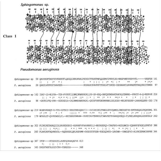
The HCA plots of the alginate lyases and the amino acid sequence alignments derived from these plots are shown in Figure 4. The results of the analysis are summarized in Table 2. The following observations were made, (a) The extent of similarity between A1-III and AlgL is largely above the cut-off point on the basis of which one may safely assume similarity in the three-dimensional structures. These two alginate lyases fall into class 1. One may note, however, that the highest level of similarity resides within the ~200-amino-acid
sequences that extend from Tyr136 to Ala330 in A1-III and from He92 to Ala290 in AlgL. (b) ALY, AlxM and the K.
pneumoniae and P. alginovora Alys each contain a polypeptide segment also about 200 amino acids long which,
upon pairwise comparison, allows two additional amino acid sequence classes to be defined. ALY and AlxM are of class 2, and the K. pneumoniae and P. alginovora Alys are of class 3. The HCA scores are about 70 %, which implies that the pairs of polypeptides share related folding topology. (c) Inter-class similarity is almost non-existent. As shown by Western blotting (Figures 3B and 3C), P. alginovora Aly does not react with anti-AlxM antibodies (titre 300; dilution 1:100), AlxM does not react with anti-(P. alginovora Aly) antibodies (titre 3000; dilution 1:1000), but P. alginovora Aly and its derivative Aly-[His-tag] react with the anti-Aly antibodies. (d) A conserved aspartic acid residue occurs at positions 179 in A1-III and 135 in AlgL, at positions 58 in ALY and 67 in AlxM, and at positions 67 and 70 in P. alginovora and K. pneumoniae Alys respectively. (e) With the
exception of P. alginovora Aly, which is 233 amino acids long, the ~200-amino-acid polypeptide that appears to define the 'core' of the five other alginate lyases represents only part (sometimes less than half) of the amino acid sequence. The mannuronate lyase AlxM and the K. pneumoniae guluronate lyase Aly each have a conserved 9-amino-acid segment close to the C-terminal end of the protein. These observations suggest that, like many glycosyl hydrolases [40], the alginate lyases may be of modular design. (f) The proposed class grouping does not correlate with the substrate specificity of the alginate lyases. An increasing number of proteins are known that have similar folds but insignificant sequence similarities [41]. The alginate lyases of classes 1, 2 and 3 and, perhaps, the pectate lyases (which also cleave glycosidic linkages by a β-elimination reaction) may belong to the same superfamily and may have a common fold signature. These questions are still open. At the present time, only the pectate lyases PelC and PelE of Erwinia chrysanthema [42] and Pel of Bacillus subtilis [43] are of known three-dimensional structure. They each adopt the same fold topology of parallel β-strands wound into a large right-handed coil [44].
Figure 4 HCA plots and deduced amino acid alignments of alginate lyases. In HCA plots, conserved
hydrophobic amino acids and clusters are shaded in dark grey; conserved hydrophilic amino acids are shaded in light grey. Vertical lines show the proposed correspondences between the sequences. In the alignments, amino acid identities are indicated by vertical lines and similarities by asterisks. Sequences were taken from: PRF 2009330A (Sphingomonas sp. Al), SwissProt Q06749 (P. aeruginosa), DDBJ D10336 (Pseudomonas sp. OS-ALG-9), SwissProt P39049 (Photobacterium ATCC 433367), the present study (P. alginovora), and GenBank L19657 (K. pneumoniae).
Acknowledgement
The work in Belgium was supported in part by the Belgian programme on Interuniversity Poles of Attraction initiated by the Belgian State, Prime Minister's Office, Services Fédéraux des Affaires Scientifiques, Techniques et Culturelles (PAI no. 19) and the Fonds de la Recherche Scientifique Médicale (contract no. 3.4531.92). C.D.
is Chercheur Qualifié of the Fonds National de la Recherche Scientifique. The work in France was supported by the Centre National de la Recherche Scientifique (GDR 1002), the Ministère Recherche et Technologie (EA 1659), the Région Rhône-Alpes and the Programme Tournesol 1994: Echanges Scientifiques France-Communauté Française de Belgique.
REFERENCES
1 Gacesa, P. and Russel, N. J. (1990) in Pseudomonas Infection and Alginates: Biochemistry, Genetics and Pathology (Gacesa, P. and Russel, N. J., eds.), pp. 29-49, Chapman and Hall, London
2 Skjäk-Braek, G., Grasdalen, H. and Larsen, B. (1986) Carbohydr. Res. 154, 239-250 3 Ramphal, R. and Pier, G. B. (1985) Infect. Immun. 47, 1-4
4 Simpson, J. A., Smith, S. E. and Dean, R. T. (1988) J. Gen. Microbiol. 134, 29-36
5 Nichols, W. W., Evans, M. J., Slack, M. P. E. and Walmsley, H. L. (1989) J. Gen. Microbiol. 135, 1291-1303 6 Pedersen, S. S. (1992) Acta Pathol. Microbiol. Immunol. Scand. 100 (Suppl. 28), 1-79
7 Gacesa, P. (1992) Int. J. Biochem. 24, 545-552
8 Sutherland, I. W. (1995) FEMS Microbiol. Rev. 16, 323-347
9 Murata, K., Inose, T., Hisano, T., Abe, S., Yonemoto, Y., Yamashita, T., Takagi, M., Sakaguchi, K., Kimura, A. and Imanaka, T. (1993) J. Ferment. Bioeng. 76, 427-437
10 Hisano, T., Yonemoto, Y., Yamashita, T., Fukuda, Y., Kimura, A. and Murata, K. (1995) J. Ferment. Bioeng. 79, 538-544
11 Yonemoto, Y., Tanaka, H., Hisano, T., Sakaguchi, K., Abe, S., Yamashita, T., Kimura, A. and Murata, K. (1993) J. Ferment. Bioeng. 75, 336-342
12 Malissard, M., Duez, C., Guinand, M., Vacheron, M. J., Michel, G., Marty, N., Joris, B., Thamm, I. and Ghuysen, J.-M. (1993) FEMS Microbiol. Lett. 110, 101-106
13 Maki, H., Mori, A., Fujiyama, K., Kinoshita, S. and Yoshida, T. (1993) J. Gen. Microbiol. 139, 987-993 14 Schiller, N. L., Monday, S. R., Boyd, C. M., Keen, N. T. and Ohman, D. E. (1993) J. Bacteriol. 175, 4780-4789 15 Boyd, A., Ghosh, M., May, T. B., Shinabarger, D., Keogh, R. and Chakrabarty, A. M. (1993) Gene 131, 1-8 16 Baron, A. J., Wong, T. Y., Hicks, S. J., Gacesa, P., Willcock, D. and McPherson, M. J. (1994) Gene 143, 61-66
17 Hisano, T., Nishimura, M., Yamashita, T., Sakaguchi, K., Takagi, M., Imanaka, T., Kimura, A. and Murata, K. (1994) J. Ferment. Bioeng. 78, 79-83
18 Malissard, M., Chavagnat, F., Duez, C., Vacheron, M. J., Guinand, M., Michel, G. and Ghuysen, J.-M. (1995) FEMS Microbiol. Lett. 126, 105-112
19 Boyen, C., Bertheau, Y., Barbeyron, T. and Kloareg, B. (1990) Enzyme Microb. Technol. 12, 885-890
20 Sambrook, J., Fritsch, E. F. and Maniatis, T. (1989) Molecular Cloning: A Laboratory Manual, 2nd edn., Cold Spring Harbor Laboratory Press, Cold Spring Harbor
21 Kamura, O., Ikeda, K., Fujiwara, K. and Motokawa, Y. (1992) J. Biol. Chem. 267, 18284-18290 22 Von Riesen, V. L. (1980) Appl. Environ. Microbiol. 39, 92-96
23 Karkhanis, Y. D., Zeltner, J. Y, Jackson, J. J. and Carlo, D. J. (1978) Anal. Biochem. 85, 595-601 24 Laemmli, U. K. and Favre, M. (1973) J. Mol. Biol. 80, 575-599
25 Matsudaira, P. (1987) J. Biol. Chem. 262, 10035-10038
26 Hopwood, D. A., Bibb, M. J., Chater, K. F., Kieser, T, Bruton, C. J., Kieser, H. M„ Lydiate, D. J., Smith, C. P., Ward, J. M. and Schrempf, H. (1985) Genetic Manipulation of Streptomyces: A Laboratory Manual, The John Innes Foundation, Norwich
27 Birnboim, H. C. and Doly, J. (1979) Nucleic Acids Res. 7, 1513-1523
28 Sanger, F., Nicklen, S. and Coulson, A. R. (1977) Proc. Natl. Acad. Sci. U.S.A. 74, 5463-5467 29 Zhang, H., Scholl, R., Browse, J. and Somerville, C. (1988) Nucleic Acids Res. 16, 1220 30 Bradford, M. M. (1976) Anal. Biochem. 72, 248-254
31 Bourdenet, S., Vacheron, M. J., Guinand, M., Michel, G. and Arminjon, F. (1990) Eur. J. Biochem. 192, 379-385 32 Devereux, J., Haeberli, P. and Smithies, O. (1984) Nucleic Acids Res. 12, 387-395
33 Pearson, W. R. and Lipman, D. J. (1988) Proc. Natl. Acad. Sci. U.S.A. 85, 2444-2448 34 Gaboriaud, C., Bissery, V., Benchetrit, T. and Mornon, J. P. (1987) FEBS Lett. 224, 149-155
35 Lemesle-Varloot, L., Henrissat, B., Gaboriaud, C., Bissery, V., Morgat, A. and Mornon, J. P. (1990) Biochimie 72, 555-574 36 Silver, J. (1991) in PCR: A Practical Approach (McPherson, M. J., Quirke, P. and Taylor, G. R., eds.), pp. 137-146, IRL Press at Oxford University Press, Oxford
37 Harley, C. B. and Reynolds, R. P. (1987) Nucleic Acids Res. 15, 2343-2360 38 Rosenberg, M. and Court, D. (1979) Annu. Rev. Genet. 13, 319-353 39 Romeo, T. and Preston, J. F. (1986) Biochemistry 25, 8385-8391
40 Gilkes, N. R., Henrissat, B., Kilburn, D. G., Miller, R. C. and Warren, R. A. J. (1991) Microbiol. Rev. 55, 303-315 41 Dobson, C. M. (1995) Struct. Biol. 2, 513-517
42 Yoder, M. D., Lietzke, S. E. and Jurnak, F. (1993) Structure 1, 241-251
43 Pickersgill, R., Jenkins, J., Harris, G., Nasser, W. and Robert-Baudouy, J. (1994) Nature Struct. Biol. 1, 717-723 44 Henrissat, B., Heffron, S. E., Yoder, M. D., Lietzke, S. E. and Jurnak, F. (1995) Plant Physiol. 107, 963-976


![Figure 3 SDS/PAGE (12% gels) of AlxM B (lanes 1), P. alginovora Aly-[His-tag] (lanes 2) and P](https://thumb-eu.123doks.com/thumbv2/123doknet/6205568.160266/8.892.112.561.188.405/figure-sds-page-gels-alxm-lanes-alginovora-lanes.webp)