Genetics of amyotrophic lateral sclerosis
par
Véronique Valérie Belzil
Département de Physiologie, Programme en Sciences Neurologiques Faculté des Études Supérieures et Postdoctorales
Thèse présentée à la Faculté des Études Supérieures et Postdoctorales en vue de l’obtention du grade de Ph.D.
en Sciences Neurologiques
Février, 2012
Faculté des Études Supérieures et Postdoctorales
Cette thèse intitulée:
Genetics of amyotrophic lateral sclerosis
Présentée par : Véronique Valérie Belzil
a été évaluée par un jury composé des personnes suivantes :
Vincent Castellucci, président-rapporteur Guy A. Rouleau, directeur de recherche
Patrick A. Dion, co-directeur Richard Robitaille, membre du jury Eric Shoubridge, examinateur externe Maja Krajinovic, représentante du doyen de la FES
Résumé
La sclérose latérale amyotrophique (SLA) est la maladie des neurones moteurs la plus fréquente, affectant 4-6 individus par 100,000 habitants à l’échelle mondiale. La maladie se caractérise par une faiblesse et une atrophie musculaire suite à la dégénérescence des neurones du cortex moteur, tronc cérébral et moelle épinière. Les personnes atteintes développent les premiers symptômes à l’âge adulte et la maladie progresse sur une période de trois à cinq ans. Il a été répertorié qu’environ 10% des patients ont une histoire familiale de SLA; 90% des gens affectés le sont donc de façon sporadique. La découverte il y a 19 ans de mutations dans le gène zinc/copper superoxide dismutase (SOD1), présentes dans 15-20% des cas familiaux de SLA et environ 2% du total des individus affectés, a été l’événement déclencheur pour la découverte de variations génétiques responsables de la maladie. La recherche sur la génétique de la SLA a connu une progression rapide ces quatre dernières années avec l’identification de mutations dans de nouveaux gènes. Toutefois, même si certains de ces gènes ont été démontrés comme réellement liés à la maladie, la contribution d’autres gènes demeure incertaine puisque les résultats publiés de ceux-ci n’ont pas, à ce jour, été répliqués. Une portion substantielle de cas reste cependant à être génétiquement expliquée, et aucun traitement à ce jour n’a été démontré comme étant efficace pour remédier, atténuer ou prévenir la maladie.
Le but du projet de recherche de doctorat était d’identifier de nouveaux gènes mutés dans la SLA, tout en évaluant la contribution de gènes nouvellement identifiés chez une importante cohorte multiethnique de cas familiaux et sporadiques. Les résultats présentés sont organisés en trois sections différentes. Dans un premier temps, la contribution de mutations présentes dans le gène FUS est évaluée chez les patients familiaux, sporadiques et juvéniles de SLA. Précisément, de nouvelles mutations sont rapportées et la proportion de mutations retrouvées chez les cas familiaux et sporadiques de SLA est évaluée. De plus,
une nouvelle mutation est rapportée dans un cas juvénile de SLA; cette étude de cas est discutée. Dans un deuxième temps, de nouvelles avenues génétiques sont explorées concernant le gène SOD1. En effet, une nouvelle mutation complexe est rapportée chez une famille française de SLA. De plus, la possibilité qu’une mutation présente dans un autre gène impliqué dans la SLA ait un impact sur l’épissage du gène SOD1 est évaluée. Finalement, la dernière section explique la contribution de nouveaux gènes candidats chez les patients atteints de SLA. Spécifiquement, le rôle des gènes OPTN, SIGMAR1 et SORT1 dans le phénotype de SLA est évalué.
Il est souhaité que nos résultats combinés avec les récents développements en génétique et biologie moléculaire permettent une meilleure compréhension du mécanisme pathologique responsable de cette terrible maladie tout en guidant le déploiement de thérapies suite à l’identification des cibles appropriées.
Mots-clés : Sclérose latérale amyotrophique, maladie des neurones moteurs, dégénération
neuronale, génétique humaine, mutations rares, séquençage de gènes candidats, SOD1, TARDBP, FUS, OPTN, SIGMAR1, SORT1.
Abstract
Amyotrophic lateral sclerosis (ALS) is the most common of motor neuron diseases, affecting 4-6 individuals per 100,000 individuals worldwide. ALS is characterized by muscle weakness and atrophy caused by the degeneration of neurons located in the motor cortex, brain stem and spinal cord. This fatal disease generally has an adult onset and progresses over a three to five year period. While 10% of patients affected have a family history of the disease, 90% of cases do not and are considered sporadic. The finding of mutations in the zinc/copper superoxide dismutase gene (SOD1) gene 19 years ago in about 15-20% of familial ALS (FALS) patients and approximately 2% of overall cases developed the interest of identifying rare genetics variants causing the disease. The ALS research field experienced a rapid progression during the last four years as mutations in new genes have been identified. While mutations in some of those new genes have been clearly linked to ALS, the role of others is still questionable and so far has not been positively replicated in other populations. Importantly, a significant portion of cases still need to be genetically explained and, unfortunately, there is still no effective treatment to cure, attenuate or prevent the disease.
The aim of this Ph.D research project was to identify new ALS mutated genes while analysing the causative role of other newly identified genes in a large familial and sporadic ALS cohort of different origins. The results presented here are categorized into three different sections. First, the contribution of FUS mutations to familial, sporadic and juvenile ALS is analysed. Specifically, new FUS mutations are reported in ALS cases and the proportions of variants present in the tested familial and sporadic ALS cohorts are assessed. In addition, a new mutation is reported in a juvenile ALS patient, and this interesting case is discussed. Second, new genetic avenues are explored for the SOD1 gene. Precisely, a new and complex SOD1 mutation is reported in a French ALS family.
Moreover, the possibility that other ALS mutated genes influence SOD1 splicing events is evaluated. Third, the contribution of new candidate genes is evaluated. Precisely, the contribution of OPTN, SIGMAR1 and SORT1 genes to the ALS phenotype is assessed.
Hopefully, our different findings combined with recent developments in genetics and molecular biology will permit a better understanding of the pathological mechanisms involved in the disease and will lead to the identification of the right targets in order to develop appropriate therapeutics for ALS patients.
Keywords : Amyotrophic lateral sclerosis, motor neuron disease, neurodegeneration,
human genetics, rare mutations, candidate genes sequencing, SOD1, TARDBP, FUS, OPTN, SIGMAR1 et SORT1.
Table of contents
Résumé ... i
Abstract ... iii
Table of contents ... v
List of Tables ... xi
List of figures ... xii
List of symbols ... xiii
Acknowledgments ... xx
Chapter 1 : Introduction ... 1
1.1 Physiology, clinical manifestations, and epidemiology of amyotrophic lateral sclerosis ... 1
1.1.1 Physiology ... 1
1.1.2 Clinical manifestations ... 3
1.1.3 Epidemiology ... 5
1.2 Etiology of amyotrophic lateral sclerosis ... 7
1.2.1 Genetic component to ALS: familial versus sporadic ALS ... 7
1.2.2 Genetic component to ALS: loci and genes identified ... 9
1.2.2.1 ALS1: SOD1 ... 9 1.2.2.2 ALS2: ALSIN ... 11 1.2.2.3 ALS3: chromosome 18q21 ... 11 1.2.2.4 ALS4: SETX ... 11 1.2.2.5 ALS5: SPG11 ... 12 1.2.2.6 ALS6: FUS ... 13 1.2.2.7 ALS7: chromosome 20p13 ... 14 1.2.2.8 ALS8: VAPB ... 14 1.2.2.9 ALS9: ANG ... 15 1.2.2.10 ALS10: TARDBP ... 15 1.2.2.11 ALS11: FIG4 ... 16
1.2.2.12 ALS12: OPTN ... 17 1.2.2.13 ALS13: ATXN2 ... 17 1.2.2.14 ALS14: VCP ... 18 1.2.2.15 ALS15: UBQLN2 ... 18 1.2.2.16 ALS-FTD: CHMP2B ... 19 1.2.2.17 ALS-FTD: chromosome 9q21-q22 ... 20 1.2.2.18 ALS-FTD: C9ORF72 ... 20
1.2.3 Association studies in ALS ... 23
1.2.4 Environmental component to ALS... 24
1.2.4.1 Toxicity and ALS ... 25
1.2.4.2 Environmental interactions, genes and epigenetics ... 29
Chapter 2 : Contribution of FUS mutations to ALS ... 32
2.1 Mutations in FUS cause FALS and SALS in French and French Canadian populations ... 33
2.1.1 Rationale ... 34
2.1.2 Contribution of authors ... 35
2.1.3 Abstract ... 35
2.1.4 Introduction ... 36
2.1.5 Materials and Methods ... 37
2.1.5.1 Standard Protocol Approvals, Registrations, and Patient Consents ... 37
2.1.5.2 Subjects ... 38
2.1.5.3 Gene Screening ... 38
2.1.5.4 Protein sequence alignment... 38
2.1.5.5 Phosphorylation sites prediction ... 39
2.1.6 Results ... 39
2.1.7 Discussion ... 40
2.1.8 Acknowledgments ... 41
2.1.9 Tables and Figures ... 42
2.2 Identification of novel FUS mutations in sporadic cases of amyotrophic lateral sclerosis ... 48 2.2.1 Rationale ... 49 2.2.2 Contribution of authors ... 50 2.2.3 Abstract ... 50 2.2.4 Introduction ... 51
2.2.5 Materials and Methods ... 52
2.2.5.1 Standard Protocol Approvals, Registrations, and Patient Consents ... 52
2.2.5.2 Subjects ... 52
2.2.5.3 Gene Screening ... 52
2.2.5.4 Protein sequence alignment... 53
2.2.6 Results ... 53
2.2.7 Discussion ... 55
2.2.8 Acknowledgments ... 56
2.2.9 Tables and Figures ... 57
2.2.10 Supplemental material ... 62
2.3 Identification of a FUS splicing mutation in a large family with amyotrophic lateral sclerosis ... 64 2.3.1 Rationale ... 65 2.3.2 Contribution of authors ... 66 2.3.3 Abstract ... 66 2.3.4 Short communication ... 67 2.3.5 Acknowledgments ... 70
2.3.6 Tables and Figures ... 71
2.4. Novel FUS deletion in a patient with juvenile amyotrophic lateral sclerosis ... 73
2.4.1 Rationale ... 74
2.4.2 Contribution of authors ... 76
2.4.3 Abstract ... 76
2.4.5 Case presentation ... 78
2.4.6 Methods ... 80
2.4.7 Results ... 80
2.4.8 Comments ... 81
2.4.9 Acknowledgments ... 83
2.4.10 Tables and Figures ... 84
Chapter 3 : New genetic avenues for the SOD1 gene ... 85
3.1 A mutation that creates a pseudoexon in SOD1 causes familial ALS ... 86
3.1.1 Rationale ... 87
3.1.2 Contribution of authors ... 88
3.1.3 Abstract ... 88
3.1.4 Introduction ... 89
3.1.5 Materials and Methods ... 90
3.1.6 Results ... 91
3.1.7 Discussion ... 93
3.1.8 Acknowledgments ... 94
3.1.9 Tables and Figures ... 95
3.2 No effect on SOD1 splicing by TARDBP or FUS mutations ... 98
3.2.1 Rationale ... 99 3.2.2 Contribution of authors ... 100 3.2.3 Abstract ... 100 3.2.4 Introduction ... 100 3.2.5 Methods ... 101 3.2.6 Results ... 102 3.2.7 Comments ... 102 3.2.8 Acknowledgments ... 102
3.2.9 Tables and Figures ... 103
Chapter 4 : Contribution of mutations in new candidate genes to ALS... 104
4.1.1 Rationale ... 106
4.1.2 Contribution of authors ... 107
4.1.3 Abstract ... 107
4.1.4 Introduction ... 107
4.1.5 Results and Discussion ... 108
4.1.6 Supplementary material ... 110
4.2 Genetic analysis of SIGMAR1 as a cause of familial ALS with dementia ... 117
4.2.1 Rationale ... 118
4.2.2 Contribution of authors ... 119
4.2.3 Abstract ... 120
4.2.4 Introduction ... 120
4.2.5 Materials and Methods ... 122
4.2.5.1 Standard Protocol Approvals, Registrations, and Patient Consents. ... 122
4.2.5.2 Subjects ... 122
4.2.5.3 Gene Screening. ... 122
4.2.5.4 Hexanucleotide repeat analysis ... 123
4.2.6 Results and Discussion ... 123
4.2.7 Acknowledgments ... 125
4.3 Analysis of the SORT1 gene in familial amyotrophic lateral sclerosis ... 126
4.3.1 Rationale ... 127 4.3.2 Contribution of authors ... 128 4.3.3 Abstract ... 128 4.3.4 Introduction ... 129 4.3.5 Methods ... 130 4.3.6 Results ... 130 4.3.7 Discussion ... 131 4.3.8 Supplementary Material ... 132
4.3.9 Tables and Figures ... 139
5.1 Discussion ... 141
5.2 Conclusions and future perspectives ... 152
References ... 154
List of Tables
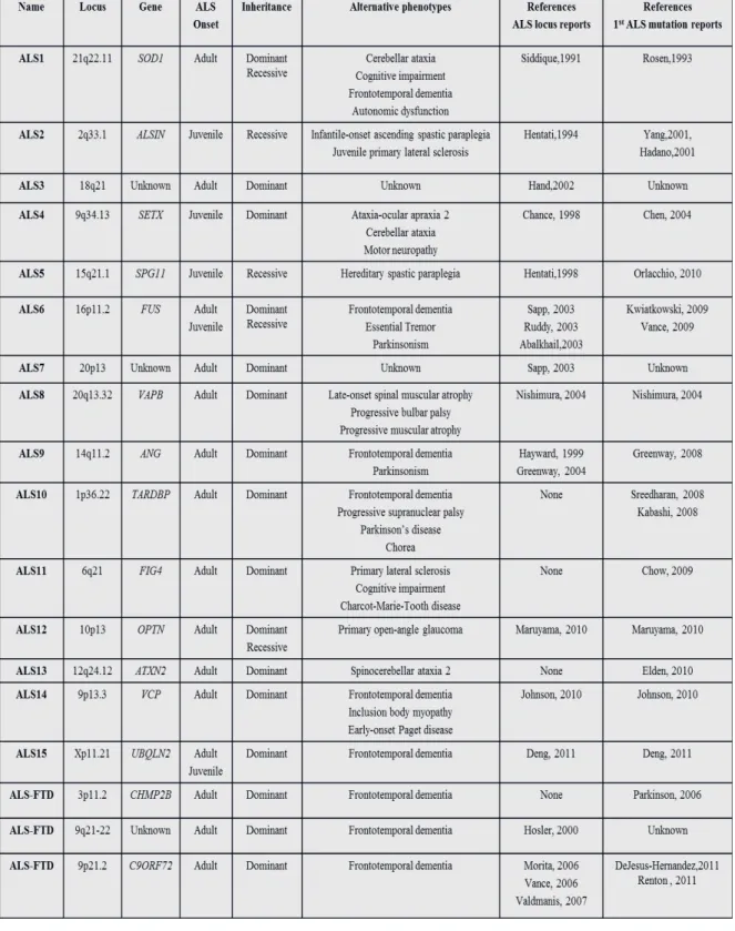
Table I Summary of ALS associated loci ... 22
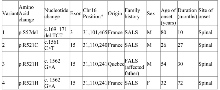
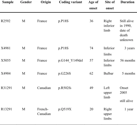
Table II Clinical and genetic profile of ALS patients with mutations in the FUS gene ... 42
Table III Primers and conditions for FUS ... 44
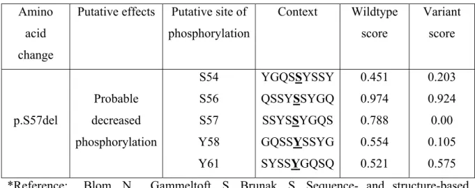
Table IV Phosphorylation site prediction scores of deletion in FUS ... 45
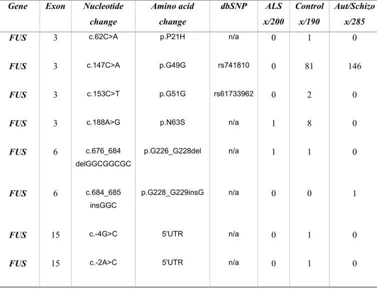
Table V FUS variants found in controls and aut/schizo patients ... 46
Table VI Description of coding genetic variations in FUS found in SALS patients and/or control participants ... 57
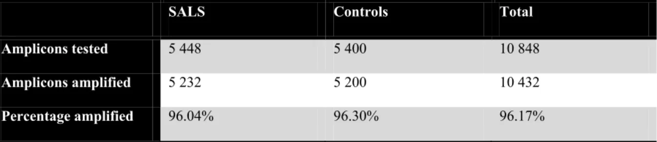
Table VII Summary of amplicons sequenced and analyzed in FUS for SALS and control samples ... 62
Table VIII Clinical profile of SALS patients with novel coding variations in the FUS gene ... 63
Table IX LOD scores for markers surrounding SOD1 on chromosome 21 ... 95
Table X Description of genetic variations in OPTN found in FALS/SALS patients and control participants ... 116
List of figures
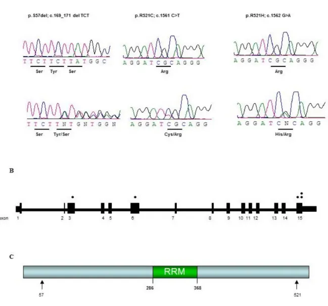
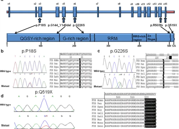
Figure 1 Sequence traces and position of mutations in FUS ... 43 Figure 2 Protein sequence alignment of FUS in different species ... 47 Figure 3. Position of the P18S, G226S and Q519X mutations in FUS gene, sequence traces
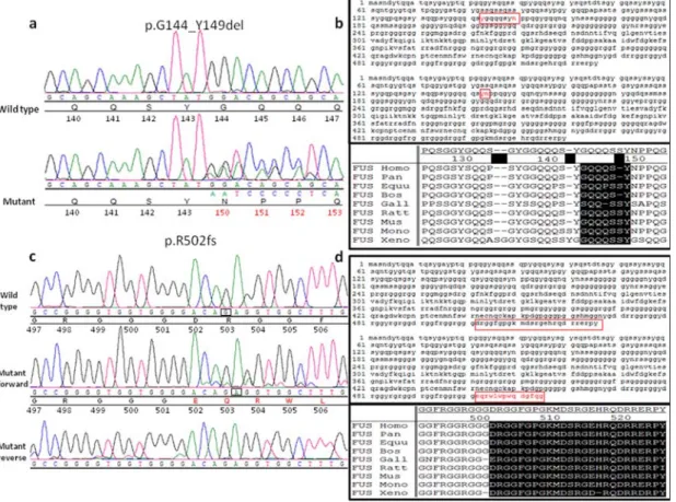
and across species conservation ... 59 Figure 4 Sequence trace and protein sequence of the G144_Y149del and R502fs mutations
in FUS ... 60 Figure 5 Pedigree of the family with the c.1542-2A>C variant in FUS ... 71 Figure 6 Splicing mutation in FUS: agarose gel, chromatograms and schematic
representation ... 72 Figure 7 Chromatograms, protein sequence and amino acid conservation in JALS FUS
mutated sample... 84 Figure 8 Pedigree of the family with the haplotype on chromosome 21 ... 96 Figure 9 Novel pseudoexon in SOD1: agarose gel and schematic representation ... 97 Figure 10 Agarose gel electrophoresis of SOD1 mRNA in TARDBP and FUS mutated
samples ... 103 Figure 11 Agarose gel electrophoresis of SORT1 mRNA and chromatograms ... 140 Figure 12 Genes and factors contributing to ALS and other associated phenotypes ... 151
List of symbols
Nucleotides: A Adenine G Guanine T Thymine C Cytosine Amino acids: A Alanine C Cysteine D Aspartic acid E Glutamine acid F Phenylalanine G Glycine H Histidine I Isoleucine K Lysine L Leucine M Methionine N Asparagine P Proline Q Glutamine R Arginine S Serine T Threonine V Valine W Tryptophan Y Tyrosine X StopAbbreviations :
aa amino acids
AAS Anabolic/androgenic steroids AD Alzheimer’s Disease
AFM Association Française contre les Myopathies ALS Amyotrophic Lateral Sclerosis
ANG Angiogenin
AOA2 Ataxia-Ocular Apraxia-2
ASSP Alternative Splice Site Predictor
ATXN2 Ataxin 2
Aut/schizo Autistic/schizophrenic
BDGP Berkeley Drosophila Genome Project BMAA Beta-N-Methylamino-L-alanine
bp base pair
c. coding
C9ORF72 Chromosome 9 Open Reading Frame 72 cDNA complementary deoxyribonucleic acid
CFTR Cystic Fibrosis Transmembrane conductance Regulator CHMP2B Chromatin-Modifying Protein 2B
CHOP DNA damage-inducible transcript 3
chr. chromosome
CIHR Canadian Institute of Health Research
cM centiMorgan
CSF Cerebrospinal Fluid
CMAP Compound Muscle Action Potential CRH Corticotropin Releasing Hormone c-terminal carboxyl-terminal
dbSNP Single nucleotide polymorphism database DCTN Dynactin
del deletion
DNA Deoxyribonucleic acid DPP6 Dipeptidyl-Peptidase 6 EEG Electroencephalography
ELF-MF Extremely Low Frequency Magnetic Fields ELP3 Elongation Protein 3
EMF Electromagnetic Fields EMG Electromyography
ER Endoplasmic Reticulum F Female
FALS Familial Amyotrophic Lateral Sclerosis FGGY FGGY carbohydrate kinase domain FIG4 SAC1 lipid phosphatase domain FRDA Friedreich Ataxia
FRSQ Fonds de la Recherche en Santé du Québec FTD Frontotemporal Dementia
FTLD Frontotemporal Lobar Degeneration FUS Fused in Sarcoma
FUS/TLS Fused in Sarcoma/Translocated in Liposarcoma
GRN Granulin
GWAS Whole Genome Association Studies HD Huntington’s Disease
HGMD Human Gene Mutation Database
hnRNPs Heterogeneous ribonucleoproteins HPA Hypothalamic-Pituitary-Adrenal HRE Hypoxia-Response Element HSP Hereditary Spastic Paraplegia
IAHSP Infantile onset Ascending Hereditary Spastic Paraplegia IBMPFD Inclusion Body Myopathy with Paget’s Disease of Bone
ins insertion
ITPR2 Inositol 1,4,5-triphosphate receptor type 2 JALS Juvenile Amyotrophic Lateral Sclerosis JPLS Juvenile Primary Lateral Sclerosis
Kg Kilogram
LMN Lower Motor Neurons LOD Logarithm of odds LOH Loss-of-heterozygosity M Male
Mb Megabase
MND Motor Neuron Disease
MRI Magnetic Resonance Imaging mRNA messenger Ribonucleic Acid n. number
NCBI National Center of Biotechnology Information Nd1-L Actin-stabilizing protein
n-terminal amino-terminal
NTG Normal Tension Glaucoma
OP Organophosphate
OPTN Optineurin p. protein
PBP Progressive Bulbar Palsy PCR Polymerase Chain Reaction PD Parkinson’s Disease PDB Paget’s Disease of Bone
PDC Parkinsonism-Dementia Complex PGRN Progranulin
PLS Primary Lateral Sclerosis PMA Progressive Muscular Atrophy
POAG Primary Open-Angle Glaucoma RefSeq Reference Sequence
RNA Ribonucleic Acid ROS Reactive Oxygen Species RRM RNA-Recognition Motif
SALS Sporadic Amyotrophic Lateral Sclerosis SBMA Spinal Bulbar Muscular Atrophy
SE Selenium
SETX Senataxin
SIFT Sorting Intolerant From Tolerant
SIGMAR1 Sigma Nonopioid Intracellular Receptor 1 SLA Sclérose Latérale Amyotrophique
SMA Spinal Muscular Atrophy SNAP Sensory Nerve Action Potential SNP Single Nucleotide Polymorphism SOD1 Superoxide Dismutase 1
SORT1 Sortilin Transcription Factor Binding Site 1 SPG11 Spastic Paraplegia 11
TARDBP TAR DNA Binding Protein TDP-43 TAR DNA Binding Protein 43 UBQLN2 Ubiquilin 2
UCSC Universiy of California at Santa Cruz
UK United Kingdom
UMN Upper Motor Neurons UNC13 Unc-13 homolog A US United States UTR Untranslated Region
VAPB VAMP-associated protein type B VCP Valosin-Containing Protein
VEGF Vascular Endothelial Growth Factor A
I dedicate this work to my two little angels, Victoria and Angelic, and to my precious husband Luc. Thank you for your understanding, patience, love and support along this demanding journey. I love you with all my heart.
“On the mountains of truth you can never climb in vain: either you will reach a point higher up today, or you will be training your powers so that you will be able to climb higher tomorrow."
Acknowledgments
I would very much like to acknowledge the support and supervision I received from my director Guy Rouleau and my co-director Patrick Dion. Their expertise, impressive knowledge in the field and dedication to their students definitely contributed to my growth as a researcher as well as a person. I absolutely appreciated all the constructive comments and advices, and I thank you both for transmitting me your inspiration and passion for medical research.
In addition, I would like to thank all the Rouleau lab members who supported me in different ways during the last five years. Especially, I would like to acknowledge the dedicated work, essential advices and explanations as well as learning sessions offered by Daniel Rochefort and Pascale Hince. You are precious knowledgeable assets of the Rouleau lab, and I am extremely grateful for the time you have given me. Thank you to Claude Marineau for essential administrative support and advices. I would also like to offer a special thank you to Judith St-Onge, Isabelle Bachand, Annie Levert and Catherine André-Guimont, whose technical support was essential to the results I obtained during the course of my Ph.D. I have learned a lot from you all in different ways. I would like to thank my predecessor, Paul Valdmanis, who was also my mentor. Thank you for your time, patience, teaching, and friendship. I have learned so much from you, and hope we will be able to work again together in the future. Thank you to past students and fellows who definitely participated to my development and knowledge in genetics: Inge Meijer, Dominique Verlaan, Amélie Piton and Jean-Baptiste Rivière. Thank you to Alex Dick, a former summer student, who took the time to show me all the basic genetic techniques when I started in the lab. Thank you to current students and fellows, Nancy Merner, Anne Noreau, Simon Girard, Valérie Lavastre, Hélène Catoire, Cynthia Bourassa, and Shawn Stochmanski who have shared generously their expertise with me at different times. Thank
you for the bioinformatics support provided by Dan Spiegelman, Édouard Henrion and Ousmane Diallo. I would also like to recognize the technical support of Annie Raymond, Pascale Thibodeau, Sylvia Dobrzeniecka, and Karine Lachapelle. I would finally like to acknowledge the coordination work of Julie Gauthier and Claudia Gaspar, and to thank Anne Dejarlais, Anna Szuto and Pierre Provencher for sample DNA collection and clinical information organization.
I was helped and inspired by different principal investigators or clinicians, who took the time to personally offer me their advices, encouragements, and support; for this, I am extremely grateful. Specifically, I would very much like to thank Christine Vande Velde and Nicolas Dupré, as well as the members of my thesis committee, Pierre Drapeau, Alex Parker and Nathalie Arbour. I would also like to acknowledge the financial support received from the Rouleau lab during my first two years as a Ph.D student, as well as the doctoral research award received by the Canadian Institute of Health Research (CIHR) for the last three years of my doctorate studies.
Last but not least, I would like to thank my parents and brothers for their support and encouragements during my studies, my precious friends for understanding that I was not able to offer them much time during the last five years, my two daughters Victoria and Angelic for being my ray of light each and every single day, and my husband Luc for his unconditioned love and support for the last fourteen years.
Chapter 1 : Introduction
1.1 Physiology, clinical manifestations, and epidemiology of
amyotrophic lateral sclerosis
1.1.1 Physiology
Amyotrophic lateral sclerosis, commonly named Lou Gehrig’s disease or Charcot’s disease is also called motor neuron disease (MND) in the United States, which actually refers to a larger spectrum of heterogeneous diseases affecting the motor neurons. Specifically, motor neuron diseases include classic amyotrophic lateral sclerosis (ALS), progressive bulbar palsy (PBP), spinal bulbar muscular atrophy (SBMA), progressive muscular atrophy (PMA), spinal muscular atrophy (SMA), hereditary spastic paraplegia (HSP) and primary lateral sclerosis (PLS). These seven disorders are all characterized by a progressive degeneration of motor neurons but differ in terms of where the neuronal death takes place. PBP, SMA, SBMA, and PMA result from the death of lower motor neurons (LMN) while HSP and PLS are explained predominantly by the degeneration of upper motor neurons (UMN). ALS affects both the UMN and LMN of the corticospinal tract located in the motor cortex, brainstem and anterior horn of the spinal cord. Specifically, neuronal degeneration in the cortex deprived the LMN located in the brain stem and spinal cord to receive executive command from the brain, while neuronal death in the anterior horn of the spinal cord causes denervation of skeletal muscles. The term amyotrophy in ALS actually refers to the atrophy of the denervated muscles. As the motor neurons located in the anterior and lateral corticospinal tract degenerate in patients, they are replaced by gliosis. The terms lateral sclerosis refer to the hardening of this region because of gliosis.1
The first symptoms of motor neuron degeneration in ALS patients are usually experienced during mid-adulthood and progresses very rapidly, usually on a 3 to 5 years period. Affected individuals finally deceased following the denervation of respiratory muscles.
The cause of neuronal death in ALS is currently unknown, and no effective treatment to prevent, slow down or stop neurodegeneration exists to date. Riluzole is the only treatment that brings a small beneficial effect, slightly extending the life of patients of two to three months.2 Significant efforts have been deployed for decades worldwide to better understand the pathological mechanisms involved in ALS and eventually develop therapeutics. Noteworthy, findings about the pathology of other neurodegenerative diseases contributed to the identification of common molecular events between different but somehow related neuronal diseases. Specifically, various neurodegenerative disorders have been associated to the pathological accumulation of misfolded proteins in neurons and glial cells, specific aggregation protein components being a characteristic of different neurological conditions. Importantly, aggregations of TDP-43, FUS or SOD1 proteins are detected immunohistochemically in neurons of ALS patients. Aggregations containing one of these proteins are found exclusively without the presence of the other two, dividing the molecular pathology of ALS into three distinctive groups.3-10 A common view is that
misfolded endogenous proteins form inclusions then aggregates, this way initiating the disease and contributing to its progression by the acquisition of a toxic gain of function. Specifically, these new acquired properties include increasing hydrophobicity and/or sequestration of important cellular components into the aggregates, inhibiting proteasomes, generating oxidative species, and/or influencing other pathways.11 Another possibility is that the functional non-misfolded portion of the protein is not sufficient to efficiently perform its role in the neuronal cell, the misfolded protein not being able to perform its function. This loss of function also prevents other proteins to perform their roles after being recruited into the aggregates. One last possibility is that toxicity of aggregates do not
initiate the disease per say but is actually a defensive response from the cell to protect itself following another unknown toxic event.12
1.1.2 Clinical manifestations
Patients experiencing UMN degeneration develop muscle weakness and spasticity, hyperreflexia and pseudobulbar palsy, while LMN death is characterized by muscle weakness and atrophy, cramps, hyporeflexia and fasciculations.13 A progressive spreading of symptoms must be observed within the same and/or other regions of the body, and the ALS diagnosis is made only after the certitude that the UMN are involved along with LMN. However, early diagnosis could be difficult since patients often have symptoms overlapping with other MND at the first stages of the disease.14 Also, in some patients, either UMN or LMN are predominantly involved throughout the disease progression, making the diagnosis even more difficult. Patients are usually classified into four different categories using the El Escorial criteria: suspected, possible, probable, or definite ALS.15
ALS diagnosis is usually made by excluding all other neurological disorders after obtaining evidence from electrophysiological, imaging, cerebrospinal fluid, or serological studies16. Specifically, patients undergo complete neurological, motor, cranial nerves, sensory and cerebellar examinations, cervical and spinal magnetic resonance imaging (MRI), electromyography (EMG), lumbar puncture, and toxicology/biochemical blood screenings. Indeed, cortical morphology analyses revealed specific cortical thinning in the precentral gyrus of ALS patients in addition to relative thinning in the temporal regions of individuals experiencing a rapid progression of the disease.17 Consistent white matter reduction is also recorded in the corpus callosum while grey matter reduction is specifically observed in primary and supplementary motor areas, as well as in the anterior cingulate and temporal lobe regions of ALS patients.18 Electrophysiologic studies evaluate muscles
denervation and motor nerve conduction using compound muscle action potential (CMAP) and sensory conduction velocities using sensory nerve action potential (SNAP). ALS patients show muscles denervation while their sensory functions are mainly intact. A complete physical examination is needed to evaluate the atrophy, spasticity and weakness of muscles in limbs asymmetrically/unilaterally or symmetrically/bilaterally. Precisely, muscle weakness is first experienced in the lower or upper limbs in about 70-75% of affected individuals, making spinal onset the most common form of ALS onset. About 20-25% of cases develop the first symptoms in the bulbar region, while approximately 3% of patients experience a respiratory onset. Presence of hyperreflexes is also investigated, as well as the existence of fasciculations, clonus and Babinski signs, and absence of Hoffman signs. Considering that up to 50% of ALS cases have some cognitive deficits,19 the cognitive functions of patients are also assessed by an extended neuropsychological examination. The progression of symptoms is regularly evaluated by clinicians, monitoring drastic weight loss because of muscular atrophy, and difficulty eating after the development of dysphagia and dysarthria.
Detailed information is collected concerning the health status of family members constituting previous, current and following generations. It is estimated that 90% of ALS patients do not report any previous familial history of motor neuron symptoms, the disease mainly affecting people in a sporadic way. The remaining 10% of patients do have additional family members with similar symptoms or with other related neurodegenerative diseases. While ALS is an adult-onset disease, some ALS families have been reported with other affected members developing the disease before 25 years of age, which is considered a juvenile form of amyotrophic lateral sclerosis (JALS).20 Also, other families were reported with members affected with dementia such as Alzheimer’s disease (AD) or frontotemporal dementia (FTD),21 or members developing Parkinsonism or Parkinson’s diseases (PD)22. While some studies reported that neurodegenerative disease aggregates within ALS kindred,22, 23 it was recently demonstrated after a large prospective
population-based study in the Netherlands that familial aggregation of ALS, dementia and PD is significantly lower than previously assumed.24
Familial and sporadic ALS patients are almost undistinguishable based on clinical manifestations.25 The only two variations that exist concern the age of onset and the sex distribution. Familial amyotrophic lateral sclerosis (FALS) tends to develop ten years earlier than sporadic amyotrophic lateral sclerosis (SALS), with a mean age of onset around 45 years. Also, sporadically affected males tend to be slightly more predominant, with a ratio of 1.3-1.6:1 female,25 while males and females are equally affected in ALS families.
1.1.3 Epidemiology
ALS is the most common of MND worldwide. It newly affects about 1-2 individuals per 100,000 inhabitants each year, approximating its prevalence to 4-6 cases per 100,000 individuals.26, 27 While it is perceived that the incidence of ALS tend to increase
and major fluctuations have been reported both in men and women, no consistent trend was reported through a decade.28 In addition, it was demonstrated that the apparent incidence increase is explained by growing ascertainment cases because of better ALS diagnosis.29, 30 Change in the reported incidence could also be explained by the increasing age of the general population because of longer lifespan expectation, this way increasing the mortality frequency from ALS, especially in women.31, 32 While the mean age of onset is 45 years of age for the familial cases and 55 years of age for the sporadic cases, a study using a large sample of affected individuals confirmed an increased incidence in the 60 to 69 year age group.30 Precisely, ALS is uncommon in individuals under 30 years old, but significantly more common among people in their 50s, with a sharp increase until the seventh decade. Considering that the mean age of onset peaks in mid-adulthood but very young or very old
onsets have been reported, it cannot be concluded that ALS is an age or aging related disorder.
The struggle to diagnose ALS was explained in the previous section, raising the difficulty to accurately estimate incidence, specifically in certain age group. It is particularly true for the elderly developing motor neuron symptoms, considering the difficulty to differentiate ALS from other various comorbidities affecting muscle strength, motion and cognitive functions often seen in this age group. Indeed, musculoskeletal pain and fatigue are the most frequently reported symptoms by older adults,33 and ALS tends to be more frequently misdiagnosed among patients over 60 years old, especially those living in large cities.34 Importantly, elderly are also less likely to encounter neurological services, and delivery of routine medical services is not optimal for this age group.35
Another changing issue is the male to female ratio of affected individuals. Past reports estimated the ratio to be 2.6:1 in the 60s and 70s but recently decreased to 1.1:1 in the 90s.30, 36, 37 This can be explained by the higher incidence of women seeking medical
advice and receiving diagnosis38 or by the higher lifespan expectancy of women, which increases their probability to eventually develop the disease. Another possibility is the changing lifestyle of women because of the socioeconomic modifications of the last century, which has become more comparable to men’s lifestyle. Women are increasingly exposed to occupational and environmental risk factors, which may explain the decrease in the male to female ratio. Another interesting report described an increased risk for ALS among smokers,39 especially females,40 underlying another important contributing factor.
While it is commonly stated that the mean disease duration range between three to five years, precisely 70-80% of cases decease within five years.41 The remaining 20-30% experiences an extremely fast or an unusually slow progression of symptoms. A marked variation in terms of disease progression during the first three years after onset has been reported, suggesting a wider progression spectrum of the disease. Again, problems with the differential diagnosis between ALS and other motor neuron disorders might in part explain this variation. An important contributing factor is the site of onset of the first symptoms, with bulbar and respiratory onsets usually ending in shorter disease duration since respiratory muscles are affected more quickly. Another explanation is the contribution of modifier genes, influencing the resulting phenotype in each patient by accelerating or reducing its progression rate. Finally, certain essential motifs in a given protein known to cause ALS might be prone to accelerate neuronal degeneration when mutated if the genetic change is translated into a non-functional protein. It is also possible that other mutations located in the same gene might only partially deprive the cell of the protein function, resulting in a slower disease progression. The only variables that have been clearly shown to independently predict ALS outcome are age at the onset,29, 42-46 site of onset,42, 45-49 and
speed of symptoms progression.47, 50 In summary, increased survival was predicted by a younger age of onset, a spinal onset, less severe symptoms at the time of the first visit, and a body mass index loss lower than 5%.46 Another study predicted a longer survival for
patients with the predominant involvement of upper motor neuron.51
1.2 Etiology of amyotrophic lateral sclerosis
1.2.1 Genetic component to ALS: familial versus sporadic ALS
Familial cases of ALS represent 10% of overall cases. 25 This estimation is based on the analysis of a few large pedigrees with a Mendelian autosomal dominant inheritance and a complete penetrance. The remaining portion of patients is considered to be affected
sporadically, with no previous reports of motor neuron symptoms in other family members. This classification is indeed made by default, and some proposed to name sporadic ALS isolated ALS. In fact, it could be difficult to obtain accurate or complete information about other kindred who are sometimes affected with another neurodegenerative disorder which is often seen in ALS families. Apparent sporadic onset can be found in very small ALS families, the number of chances to develop the disease for other members is this way reduced by the family size. Also sometimes a patient has lost contact with some family members and is not aware of the health status of those members. Also, some siblings of apparent sporadic cases are reluctant to report symptoms, are sometimes misdiagnosed, or deceased before developing the first ALS signs, the sporadic patient actually being part of a familial syndrome. Adoption or illegitimacy could also shuffle the cards. Recessive form of the disease, ALS onset in children before the onset of symptoms in one of their parent who is a mutation carrier, or incomplete penetrance in families can also lead to a sporadic categorization of cases.52
This being said, the active investigation of genealogies actually evaluated the prevalence of FALS to 17-23%.53-56 Considering that FALS cases actually represent about 20% of overall cases, that the clinical manifestation of SALS and FALS is almost undistinguishable, and that mutations identified in FALS have also been found in SALS cases this way confirming the contribution of genetics to sporadic ALS, it is clear that the identification of FALS genes would extend our understanding of the ALS pathology affecting both sporadic and familial cases. Moreover, recently, one group assessed the relative risk for ALS in families counting more than 6,000 Swedish individuals who have been first classified as sporadic ALS patients. They also evaluated the concurrence of ALS in more than 86,000 Swedish twin pairs. They reported a significant higher risk for siblings or children of ALS patients to develop the disease, and concluded that a major genetic role contributes to familial ALS.57 Based on these assumptions, it is reasonable to claim that most, if not all ALS cases can be explained by genetic predispositions. Consequently,
significant progress in the genetics of ALS has been made during the last two decades. Precisely, mutations in 13 different ALS genes have been identified among 15 different ALS loci, and two different ALS-FTD genes have been identified among three different ALS-FTD loci (see table I). These are described thoroughly in the next section.
1.2.2 Genetic component to ALS: loci and genes identified
1.2.2.1 ALS1: SOD1
Section of the editorial entitled: SOD1 mutations : more to learn. To be published in the Canadian Journal of Neurological Sciences, March 2012,39 :2.
In 1989, genetic analysis using 150 families with classical ALS helped identify two regions of possible linkage on chromosomes 11 and 21.58 Further evidence of linkage on chromosome 21q22.1-q22.2 was published in 1991, this way identifying the first ALS locus currently known as ALS1.59 In 1993, an international consortium reported 11 different SOD1 missense mutations in 13 out of 18 dominantly inherited ALS families.60 Since then, about 168 disease-causing mutations (Human Gene Mutation Database: http://www.hgmd.org),61 87% of which are nucleotide substitutions, have been identified among the 153 amino acids of this five-exon gene. The remaining 13% of mutations are deletions, nonsense or splicing mutations which affect the length of the protein. 61-64 Even if most SOD1 mutations are transmitted in an autosomal dominant manner, a few families have been reported with a reduced penetrance or a recessive transmission among members.65 Moreover, compound heterozygotes have also been reported, with two different heterozygote SOD1 mutations in the same patient.66 An intriguing report recently described a patient affected with both familial ALS and cerebellar ataxia and an SOD1 mutation.67 Rare SOD1 mutated cases with frontotemporal dementia (FTD), cognitive impairment, and
autonomic dysfunction have also been reported.68-70 Interestingly, some substitutions affecting particular SOD1 amino acids are associated with a slow disease progression, while other amino acid substitutions sometimes located in the same region of the gene, are found in patients with a very fast progression.64, 71, 72 The effect on rate of progression is thought to be related to the fact that some mutants lead to a stable protein while others are highly unstable.55, 64, 73-75 Approximately 42 mutations have been reported in SALS cases, representing 25% of the total SOD1 variation and about 1% of SALS cases. Precisely, 15-20% of familial cases result from SOD1 mutations, hence variations in this gene explain approximately 1-2% of overall cases.60
The superoxide dismutase 1 (SOD1) protein is ubiquitously expressed and is mainly located in the cytosol of cells, catalyzing the reduction of the superoxide anion to O2 and
H2O. Most mutations reduce dismutation,76 but some have normal or only slightly reduced
dismutase activity.73, 74 Based on the dominant inheritance and the fact that SOD1 knockout mice have no motor neuron phenotype while overexpression of mutant SOD1 does,77, 78 it is agreed that mutant SOD1 acquires a novel cytotoxic function which promotes neurodegeneration. This toxic gain-of-function has been proposed to involve different mechanisms including protein aggregation and misfolding, oxidative stress, mitochondrial dysfunction, microglia activation, glutamate excitotoxicity, and defects in axonal transport.79 Specifically, the presence of protein misfolding and aggregation is a recurrent observation in cells of ALS patients, which may inactivate or impair normal processes such as proteasomal degradation or chaperone function.80 Some observations also suggested that SOD1 might be involved in RNA processing after the finding that mutant SOD1 impaired the post-transcriptional processing of VEGF mRNA, which encodes an important neuroprotective factor, this way provoking a significant decline in VEGF expression.81 In addition, SOD1 toxicity has been found to modify wild type (wt) SOD1 by inducing it to misfold.82 Moreover, it was demonstrated that proximity to mutant SOD1 in non-neuronal cells such as microglia and astrocytes is necessary for the toxicity of neighboring motor
neurons.83 Though extensive research has been conducted to understand the specific pathways involved, it is still unclear how mutant SOD1 leads to the ALS phenotype.
1.2.2.2 ALS2: ALSIN
In 1994, a locus on chromosome 2q33-q35 was reported after a linkage analysis using a large ALS family from Tunisia.84 In 2001, two different homozygous deletions in the ALS2 gene causing a loss-of-function of its encoded protein were identified in one amyotrophic lateral sclerosis and two primary lateral sclerosis autosomal recessive families with members experiencing a juvenile onset of the diseases with a slow progression.85, 86 While no ALS2 mutations were reported in adult-onset typical ALS, variants were observed in infantile–onset ascending spastic paralysis (IAHSP).87 The 34-exon ALS2 gene encodes the GTPase regulator alsin, which plays a role in intracellular endosomal trafficking. 88
1.2.2.3 ALS3: chromosome 18q21
A locus on chromosome 18q21 was identified in 2002 after performing a genome scan using a large European family with 20 members affected with classical ALS.89 The
disease was transmitted in an autosomal dominant fashion, and all affected members developed typical ALS. While a maximum lod score of 4.5 was obtained, no causative mutations in the ALS3 region have been identified to date.
1.2.2.4 ALS4: SETX
A locus on chromosome 9q34 was identified in 1998 and was named ALS4.90 After testing 19 genes in the region, SETX autosomal dominant mutations have been identified in juvenile FALS and SALS patients experiencing a slow progression of the disease without
the involvement of bulbar and respiratory muscles.91 Interestingly, autosomal recessive mutations have also been identified in patients with spinocerebellar ataxia, specifically with ataxia-ocular apraxia 2 (AOA2), ataxia with elevated levels of alpha-fetoprotein, distal amyotrophy, and peripheral neuropathy.92, 93 In fact, it is believed that this mutated type of recessive ALS is an intermediate form of motor neuron disease, standing between ALS and spastic paraplegia, while involving lower limbs and excluding the bulbar region. No mutations were reported in adult-onset ALS. The SETX gene encodes for the senataxin protein that contains a DNA/RNA helicase domain in its c-terminal, suggesting a role in DNA repair and RNA processing.91 It was also suggested that the gene play a role in the coordination of transcriptional events.94
1.2.2.5 ALS5: SPG11
Mapping of the ALS5 locus to chromosome 15q15.1-q21.1 was obtained in 1998 using five families from Europe and North Africa with affected members developing typical ALS at an earlier age of onset. This form of juvenile ALS was believed to be the most prevalent form of recessive ALS.95 SPG11 (spatacsin) mutations were first identified
in 2010 in juvenile ALS patients characterized by a long-term survival.96 Compound heterozygous deletions were recently identified by our group after the whole exome sequencing of two affected family members with a recessively inherited juvenile motor neuron disease.73 Spatacsin is a transmembrane protein ubiquitously expressed in the nervous system which is phosphorylated upon DNA damage. While mutations in the SPG11 gene were previously involved in spastic paraplegia,97 it is interesting to note that, in this study, one affected family member displayed atypical juvenile ALS and the other developed classical hereditary spastic paraplegia (HSP), this intra-familial phenotypic heterogeneity reinforcing the idea that motor neuron diseases are part of a continuum.
1.2.2.6 ALS6: FUS
In 2003, three ALS families linked to chromosome 16q12 were published by three different groups. 98-100 The discovery of mutations in a DNA/RNA binding protein in 2008101, 102 prompted geneticists to look for mutations in genes encoding proteins having similar functions. Consequently in 2009, two groups reported mutations in the FUS gene located on chromosome 16p11.2 which encodes another DNA/RNA binding protein, this way elucidating the ALS6 locus.6, 103 This discovery was made after the identification of a family of Cape Verdean origin with a possible recessive inheritance pattern. A cluster was identified on chromosome 16, and sequencing revealed a homozygous missense mutation (H517Q) in exon 15 of the FUS gene in all affected members of this family. Additional screening identified 13 other dominant mutations in 24 different families. No FUS mutations were found in sporadic ALS cases, but later reports established the frequency of FUS mutations to be present in about 4% of familial cases, 1% of sporadic patients, and less than 5% of overall cases.62, 104 More than 52 different mutations have been identified so far in this 15 exons gene (Human Gene Mutation Database: http://www.hgmd.org).61 Interestingly, almost all mutations are clustered in the c-terminal of the protein, mostly lying in the final 17 amino acids of FUS. While the associated phenotype is typical ALS, mutations in juvenile ALS patients as well as in FTD and essential tremor cases have been reported.64, 71, 104, 105 ALS patients with FUS mutations seem to develop the first symptoms
earlier, have a higher rate of bulbar onset, and experience a more rapid progression when compared to patients with SOD1 mutations.71
The fused in sarcoma (FUS) protein is ubiquitously expressed and is predominantly located in the nucleus of cells. However, FUS immunoreactive inclusions have been detected in the neuronal and glial nuclei and cytoplasms of patients affected not only with ALS, but also with FTLD as well as Huntington’s, Alzheimer’s, and Parkinson’s disease,106
this observation defining a new proteinopathy in neurodegeneration. The spectrum of FUS RNA targets still has to be defined in order to establish the normal function of the protein.
1.2.2.7 ALS7: chromosome 20p13
Linkage to chromosome 20 was established in 2003 using one typical ALS family with a dominant mode of inheritance from the Boston area.100 The best LOD score was obtained using markers on the distal short arm of the chromosome. However, the linkage was not reproduced with any other ALS families, and was obtained after genotyping only two affected individuals of the same generation. The ALS7 locus was claimed to be probable but less secure at the time of publication, and the gene have not yet been identified.
1.2.2.8 ALS8: VAPB
A locus on chromosome 20q13, now known as ALS8, was identified using eight different families from Brazil.75 Founder studies demonstrated a common Portuguese ancestor to all families. The dominant P56S missense mutation in the VAPB gene was identified in all affected members,76 which has been demonstrated to induce the formation of insoluble cytoplasmic aggregates of the mutant protein. Interestingly, the same mutation gave rise not only to ALS, but also alternate phenotypes including late-onset spinal muscular atrophy, progressive bulbar palsy, and progressive muscular atrophy. 76 The same mutation has also been identified in other ALS patients of different origins including German, Japanese and American, but surprisingly has not been found in Portuguese.62, 79,
107, 108 Only one other mutation in the gene has been reported to date to cause ALS. 78 The
VAMP-associated protein type B participates in intracellular transport and is mainly located in the endoplasmic reticulum.
1.2.2.9 ALS9: ANG
Linkage analysis using Scottish and Irish families permitted the identification of a region on chromosome 14q11.2, making this locus the ninth to be discovered in ALS.83, 109 The finding that mutant SOD1 binds to VEGF and alters its expression prompted the screening of candidate genes located in the ALS9 locus sharing the same metabolic pathway. Indeed, to date, about 21 variants have been identified in the ANG gene (Human Gene Mutation Database: http://www.hgmd.org).61 However, only one dominant variant was actually shown to cosegregate with the disease in a unique Dutch family with one member affected not exclusively with ALS, but also with Parkinsonism and FTD.81 The same variant was identified in French, North American, Irish, Scottish and Swedish patients affected with classical ALS,62, 94, 110, 111 making it the most common ANG mutation reported to date. Functional expression studies demonstrated a loss of angiogenic function of the mutant protein.110
1.2.2.10 ALS10: TARDBP
Mutations in the TARDBP gene101, 102 were first reported after the discovery that its encoded protein TDP-43 is the principal constituent of neuronal cytoplasmic inclusions in ALS and FTD patients,112 this way identifying the ALS10 locus on chromosome 1p36.22.
More than 49 dominant mutations have been identified so far in adult onset ALS (Human Gene Mutation Database: http://www.hgmd.org),61 mostly lying in the c-terminal portion of the protein. Mutations have been reported in about 5% of familial ALS cases, 0.5-2% of sporadic patients, and approximately 5% of overall cases.101, 102, 113, 114 While most TARDBP mutated patients are affected with typical ALS, some develop alternate phenotype including FTD, progressive supranuclear palsy, Parkinson’s disease and chorea.67, 93
The tar DNA-binding protein (TARDBP) gene encodes a nucleic DNA/RNA binding protein that is redistributed to the cytoplasm of neurons and glial cells when mutated. TDP-43 is involved in DNA/RNA processing, a common function that later guided the screening of the FUS gene located in the ALS6 locus. 68-70 TDP-43 immunoreactive inclusions have been observed both in the nucleus and cytoplasm of neurons and glial cells, defining a unique proteinopathy, distinct from the one observed in patients with FUS inclusions.115 Postranslational alterations of TDP-43 such as hyperphosphorylation, ubiquitination and cleavage have been reported to modify its interaction with other proteins involved in RNA metabolism,96, 106 hence influencing pre-mRNA splicing, RNA stability and axonal transport.116 Some of these interactions were found to dependent on TDP-43 RNA-binding, whereas others are RNA-independent. 117 Specifically, some TDP-43 interacting proteins cluster into two different interaction networks: a nuclear or splicing cluster and a cytoplasmic or translation cluster.117 This suggests that TDP-43 assumes different roles in RNA metabolism, and acts in the nucleus as well as the cytoplasm. Additional TDP-43 RNA targets need to be identified in order to better understand the pathway in which they are involved, as well as to determine their precise role in neurodegeneration.
1.2.2.11 ALS11: FIG4
Mutations in the FIG4 gene located on chromosome 6q21 have been first identified in 2007 in severe cases of Charcot-Marie-Tooth disease characterized by an early onset and involving both sensory and motor neurons.118 The finding of mutations in patients affected with a disease involving the motor neurons prompted the screening of FIG4 in ALS and PLS patients. Nonsynonymous variants were found in nine patients out of 473, mutations being present in about 2% of the tested cohort. Among them, seven patients were diagnosed with classical ALS and two had PLS. Six of the dominant variants identified were shown to be deleterious.119 No other FIG4 mutations reports have been published since then and
further screening in other populations is needed to properly establish the genetic contribution of FIG4 to the ALS pathogenesis. FIG4 is a phosphatidylinositol 3,5-bisphosphate (PtdIns(3,5)P2) that might play a role in autophagy in the nervous system, while mutations have been proposed to contribute to inclusion body diseases.120
1.2.2.12 ALS12: OPTN
Homozygosity mapping using six ALS patients from Japanese consanguineous families helped define in 2010 the ALS12 locus on chromosome 10p13. After sequencing 17 candidate genes in the region, variants were identified in OPTN,121 a gene in which mutations were previously associated with primary open-angle glaucoma (POAG).122 Additional mutations were identified by the same group in familial and sporadic classical ALS cases causing both recessive and dominant traits. Subsequent screening of OPTN in patients of European origin did not confirm the genetic implication of optineurin in ALS, at least for this specific population,123, 124 and more reports are needed. Mutated OPTN cytoplasmic distribution has been however demonstrated to differ from wt OPTN, and OPTN-immunoreactive cytoplasmic inclusions have been observed. OPTN have also been demonstrated to be recruited in TDP-43 or SOD1 inclusions.121
1.2.2.13 ALS13: ATXN2
An extended (CAG)n repeat in ATXN2 was first reported in 1996 in patients affected with spinocerebellar ataxia-2.125 While normal chromosomes contain 14 to 31 repeats interrupted by one to three CAA repeats, chromosomes of patients contain a pure stretch of 34 to 57 CAG repeats. The CAG is highly unstable during transmission, and its size is negatively correlated with the age of symptoms onset in 50% of cases.125 The ALS13 locus on chromosome 12q24.12 was attributed to the ATXN2 gene in 2010 when it was
discovered that an intermediate repeat length ranging from 27 to 33 glutamines was significantly associated with ALS.126 Additional reports confirmed the same association, and it was also shown that intermediate repeats were CAA interrupted.127-130 It was also demonstrated that polyQ expansions in ATXN2 enhance its interaction with TDP-43, and that both ATXN2 and TDP-43 relocalize to stress granules after oxidative stress. 126 Ataxin-2 is primarily located in the Golgi apparatus. While expression of full-length extended ataxin-2 disrupted the normal morphology of the Golgi complex,131 it is still unclear how intermediate repeat length influences the risk to develop ALS.
1.2.2.14 ALS14: VCP
In 2004, missense mutations were found in the VCP gene located on chromosome 9p13.3 in patients affected with FTD or inclusion body myopathy with Paget disease of bone (IBMPFD).132 The valosin-containing protein (VCP) is associated with a spectrum of essential cell protein pathways including cell cycle, homotypic membrane fusion, nuclear envelope reconstruction, postmitotic Golgi reassembly, DNA damage response, suppression of apoptosis, and ubiquitin-dependent protein degradation.132 In 2010, exome
sequencing revealed a heterozygous mutation as a cause of adult-onset ALS with or without FTD in an Italian family,133 this way identifying the 14th locus for the disease. While a few
additional mutations were reported in FALS and SALS cases,133-136 none were found in the
Australian ALS population.137 It can be concluded that mutations in VCP is not a common cause of ALS.
1.2.2.15 ALS15: UBQLN2
Linkage analysis using a large ALS family composed of 19 affected members with either a juvenile or an adult disease onset permitted the identification of the ALS15 locus
on chromosome Xp11.21.20 The disease was transmitted in a dominant fashion among the family members, with a reduced penetrance in females and no evidence of male-to-male transmission. Therefore, 41 candidate genes out of 191 coding genes in the region were sequenced, and a single cosegregating mutation was identified in the UBQLN2 gene. Additional FALS or FALS-FTD cases were screened for mutation in this intronless gene, and four novel mutations were identified. The five missenses found by this group all affected proline residues located in a single PXX repeat region. Ubiquilin-2 was shown to be present not only in inclusions located in the hippocampus and pyramidal neurons, but also to be involved in inclusion formation in X-linked ALS.20 These observations are critical, considering that protein inclusions are a persistent landmark in neurodegenerative diseases, and especially in ALS and FTD. Further mutation reports are however needed.
1.2.2.16 ALS-FTD: CHMP2B
Linkage to chromosome 3p11.2 was established using a large Danish family with autosomal dominant FTD.138, 139 In 2005, the mutation responsible for the disease in this family was reported to be located in the CHMP2B gene. The chromatin modifying protein 2B (CHMP2B) is part of an endosomal secretory complex which is believe to participate in endosomal trafficking.140 Another variant in the same gene was additionally found in an
unrelated individual with unspecific dementia.141 While two different groups did not find
any CHMP2B causative mutations in their tested FTD cohort,142, 143 two other groups found one truncated mutation in one autosomal dominant FTD patient and one missense mutation in an ALS patient.144, 145 Mutations in the CHMP2B gene are found rarely in ALS or FTD patients, and its causative role in neurodegeneration is questionable.
1.2.2.17 ALS-FTD: chromosome 9q21-q22
Genomic screening was first conducted using 16 families from the Boston data set with members affected with either dominant ALS or ALS-FTD, which permitted the identification of a locus on chromosome 9q21-22. Subsequent analysis was performed to independently confirm the region involved by genotyping four other families from the Chicago data set with members affected with ALS and/or FTD.146 The mutated gene causing the two phenotypes in these families still needs to be identified.
1.2.2.18 ALS-FTD: C9ORF72
Three different groups contributed to the identification of a major locus on chromosome 9 by reporting three overlapping loci after conducting linkage analysis using different autosomal dominant families with members affected with ALS and/or FTD along with TDP-43 proteinopathy.21, 147, 148 Additional reports helped refine the minimum linkage region to 3.7 Mb, containing only five known genes.149-153 The common coding and non-coding region was extensively sequenced by many for several years, but no causative mutations were identified. Several association studies conducted in the ALS and FTD sporadic population confirmed the implication of the chromosome 9p locus in both diseases.154-157 Rigorous efforts finally permitted the identification of important
heterozygote hexanucleotide repeat expansions in the chromosome 9 open reading frame 72 (C9ORF72) gene in three of the reported families linked to the chromosome 9p.21 locus.
158, 159 The gene has three identified transcripts and the expansions are either located in the
first intron or the promoter region of the gene. Specifically, the maximum size of the hexanucleotide repeat in control participants was 23 units, while it ranged approximately between 700 and 1600 in patients. The same noncoding but highly conserved GGGGCC expansions were further found in a significant proportion of familial and sporadic ALS and/or FTD cases. Precisely, 11.7% of familial FTD and 23.5% of familial ALS patients were found with the repeat, making this variant the most common genetic abnormality
identified to date for those two familial forms of diseases.158 In addition, repeat expansions were present in almost one-half of Finnish familial ALS cases and in about one-third of familial ALS cases of European descent.159 While the C9ORF72 protein is uncharacterized and its domains are unknown, it is expressed in a variety of tissues and is mainly a cytoplasmic and synaptic protein in neurons.159 Using lymphoblastoid cell lines, it was demonstrated that the hexanucleotide repeat expansions in C9ORF72 are associated with a decreased expression of C9ORF72 mRNA.158 The same group observed RNA foci in neuronal tissues of ALS and FTD patients. It is suggested that these expansions interfere with the normal expression of the protein, causing the loss of one alternatively spliced C9ORF72 transcript and the formation of nuclear RNA foci. It is also suggested that variations in the repeat length influence the resulting expressed phenotype, 158 explaining the development of ALS and FTD in some familial cases, while other family members develop either ALS or FTD. Finally, anticipation might be present in C9ORF72 extended FALS patients, considering that younger members of Finnish ALS-FTD families experienced a very early age of onset. More reports are needed to establish more precisely the proportion of C9ORF72 extended patients.
1.2.3 Association studies in ALS
Whole genome association studies (GWAS) have been proven to be a powerful tool in genetics after the results obtained from research on different complex diseases. In GWAS, thousands of single nucleotide polymorphisms (SNPs) are genotyped across the genome in a large number of samples without any preconceived assumption about diseases. Many association studies have been conducted in different populations worldwide using sporadic cases of ALS, but not all have been conclusive. The first report failed to significantly identify hits but contributed to future studies by making the SNP data freely available on the internet. 160 Those data were used as a replication set in a follow-up study identifying FGGY as a candidate gene for ALS.160 Another GWAS was conducted by a group from the Netherlands, reporting an association with the ITPR2 gene.161 This study also combined the results obtained with the data available on the internet after the first GWAS, and failed to re-associate the ITPR2 gene with sporadic ALS. A novel association with the DPP6 gene was however identified,162 this way replicating an already published
Irish study.163 However, the Nertherlands and Irish groups included data from the same cohorts, which explains the replicated association of the DPP6 gene. One subsequent study replicated this association in an Italian cohort,164 while data obtained using samples from Poland, France and Canada did not.165, 166 Another study attempted to replicate the associations with hits obtained from previous GWAS, but was unable to confirm any conclusive association.167 Association of the gene ELP3 to ALS was also reported after performing a GWAS using three different populations.168 Two susceptibility loci were also identified without replicating previous associated genes or SNPs, including one within the boundaries of the UNC13A gene and another on chromosome 9p21.2.157 The identification of the susceptibility region on chromosome 9p21.2 was the first conclusive data obtained from a GWAS in ALS research, considering that a major ALS-FTD locus was known to exist in the same region after several linkage studies. Subsequently, two additional groups reported an association between sporadic ALS and a region on chromosome 9p21 in the Finnish and British populations.154, 155
Unfortunately, GWAS findings in one population are rarely replicated in others, and no genes have been concretely associated to ALS. Intriguingly, genes identified using candidate-based approaches have not been the top hits in GWAS. Indeed, the published associations to chromosome 9p were the only replicated and conclusive GWAS in ALS research. The availability of several thousand cases with definite diagnosis is a key element for GWAS to identify genetic factors responsible for a disease. The high degree of allelic and non-allelic heterogeneity in SALS cases is a limiting factor for the identification of causative genes, considering that different disease-causing alleles may exist within the same gene. Moreover, the identification of rare highly penetrant mutations in genes such as TARDBP or FUS in SALS patients is problematic given that GWAS can only detect common low penetrant variants. The whole genome or exome sequencing approaches seem to be more suitable for the identification of such novel rare causative variants in ALS, and the results that will be obtained from such approaches in the next few years will hopefully highlight unknown genetic contributions to familial and sporadic ALS.
1.2.4 Environmental component to ALS
The first association of an environmental factor contributing to the emergence of ALS was with the Chamorro indigenous people of Guam, who presented an extremely high incidence of the disease, who consumed a lot of flying fox and consequently accumulated cycad neurotoxins in their organism including beta-N-methylamino-L-alanine (BMAA) shown to be produced across the cyanobacterial order.169, 170 Later, a report of an ALS cluster near Lake Mascoma associated the increased disease incidence to the presence of cyanobacteria in the surrounding water area.171 Many in vivo studies using mice, rats, monkeys and chicks demonstrated that exposure to BMAA induce neurodegenerative symptoms.170 Results obtained from zebrafish also demonstrated a disruption in neuronal
development resulting from BMAA exposure. In vitro studies using rodents also concluded that BMAA predominantly acts on motor neurons by increasing the generation of reactive oxygen species (ROS) and Ca(2+) influx along with disrupting mitochondrial activity. Consequently, neuronal death is believed to result from excitotoxic mechanisms.170 No other robust association have been reported since then, and the search for other causative evidence connecting environmental risk factors or neurotoxic chemicals to sporadic ALS has not been fruitful. Nevertheless, some epidemiologic trends suggested that several environmental influences might be linked to the ALS etiology. Several environmental toxic exposures are believed to induce the liberation of free radicals, which might lead to oxidative stress. Precisely, free radicals are ROS produced normally in biological systems. Human cells have developed a complex system of defence mechanisms to eliminate excessive ROS accumulation which otherwise has the ability to damage lipids, proteins, and DNA. When cellular antioxidant defences are unable to keep the ROS levels below a toxic threshold because they are overwhelmed with free radicals or they are encoded in insufficient amount, cells experience oxidative stress.172 A few environmental factors have been proven to increase ROS levels and hence the risk to develop ALS. These include lead, mercury, and selenium exposure, contact with pesticides and insecticides, intense physical activities, head injuries, electromagnetic fields (EMF) exposure and tobacco smoking.173
These studies are briefly explored in the next section.
1.2.4.1 Toxicity and ALS
Since most ALS cases are sporadic, many studies have been devoted in the last decades to the possible contribution of neurotoxic chemicals to ALS phenotype. Accumulated evidence confirmed the potency of metals to induce toxicity to cells and consequently cause a number of pathologies. Precisely, many metals are able to catalyse the formation of ROS and indeed damage key proteins, leading to protein denaturation and