Université de Montréal
Impact of (pro)renin receptor deficiency in adipose tissue using
a genetically engineered mouse model
par Basma Ahmed
Département du Physiologie, Université du Montréal Faculté de Médecine
Mémoire présentée à la Faculté de médecine en vue de l’obtention du grade de maîtrise
en physiologie
[Décembre, 2012]
© Basma Ahmed, 2013 Université de Montréal
Université de Montréal
Faculté des études supérieures et postdoctorales
Cette mémoire intitulée:
Impact of (pro)renin receptor deficiency in adipose tissue using a genetically engineered mouse model
Présentée par : Basma Ahmed
a été évaluée par un jury composé des personnes suivantes :
Remi Rabasa-Lhoret, président-rapporteur Julie L. Lavoie, directeur de recherche
Ondrej Seda, co-directeur Ashok Srivastava, membre du jury
Résumé
La stimulation du récepteur de la rénine/prorénine [(P) RR], un membre récemment découvert du système rénine-angiotensine (SRA), augmente l'activité du SRA et des voies de signalisation angiotensine II-indépendante. Pour étudier l'impact potentiel du (P)RR dans le développement de l`obésité, nous avons émis l'hypothèse que les souris déficientes en (P)RR uniquement dans le tissus adipeux (KO) auront une diminution du poids corporel en ciblant le métabolisme du tissu adipeux, l'activité locomoteur et/ou la prise alimentaire. Ainsi, des souris KO ont été générées en utilisant la technologie Cre/Lox. Le gain de poids et la prise alimentaire ont été évalués hebdomadairement dans les mâles et femelles KO et de type sauvage (WT) pendant 4 semaines alors qu’ils étaient maintenu sur une diète normal. De plus, un groupe de femelles a été placé pour 6 semaines sur une diète riche en gras et en glucides (HF/HC). La composition corporelle et l'activité ambulatoire ont été évaluées par l’EchoMRI et à l’aide de cages Physioscan, respectivement. Les tissus adipeux ont été prélevés et pesés. De plus, les gras péri-gonadaux ont été utilisés pour le microarray. Finalement, le niveaux d'expression d'ARNm du (P)RR ont été évalués.
Comme le gène du (P)RR est situé sur le chromosome X, les mâles étaient des KOs complets et les femelles étaient des KOs partielles. Les souris KO avaient un poids corporel significativement plus petit par rapport à WT, les différences étant plus prononcées chez les mâles. De plus, les femelles KOs étaient résistantes à l'obésité lorsqu'elles ont été placées sur la diète HF/HC et donc elles avaient significativement moins de masse grasse par rapport aux WTs. L’analyse histologique des gras péri-gonadaux des KOs nous ont dévoilés qu’il avait une réduction du nombre d'adipocytes mais de plus grande taille. Bien qu'il n'y ait eu aucun changement dans la consommation alimentaire, une augmentation de près de 3 fois de l'activité ambulatoire a été détectée chez les mâles. De plus, nous avons observé que leurs tibias étaient de longueur réduite ce qui suggère fortement l'affection de leur développement. Les gras péri-gonadaux des souris KO avaient une expression réduite de l`ABLIM2 (Actin binding LIM protein family, member 2) qui est associé avec le diabète de type II chez l'humain. Ainsi, les données recueillies suggèrent fortement que le (P)RR est impliquée dans la régulation du poids corporelle.
Mots-clés: Le récepteur de la rénine/prorénine, tissue adipeux, obésité, le système rénine-angiotensine, Souris KO
Abstract
Stimulation of the (pro)renin receptor [(P)RR], a recently discovered member of the renin-angiotensin system (RAS), increases the activity of the RAS and stimulates renin-angiotensin II-independent signaling pathways. To investigate the possible impact of the (P)RR on obesity development, we hypothesized that mice deficient in the (P)RR specifically in their adipose tissue (KO) would have a decrease in body weight by targeting adipose tissue metabolism, locomotor activity and/or food intake. As such, KO mice were generated using the Cre/Lox technology. Weekly weight gain and food intake were assessed in both male and female KO and wild-type (WT) littermates for 4 weeks on a normal diet. A group of females were also placed for an additional 6 weeks on a high-fat/high-carbohydrate diet (HF/HC). Body composition and physical activity were evaluated using EchoMRI and Physioscan cages, respectively. Adipose tissues were collected and weighed at sacrifice. Moreover, perigonadal fat was used for Gene assay and histological analysis. (P)RR mRNA expression levels were evaluated using real-time PCR.
Different circulating metabolites and proteinuria were measured by ELISA kits.
As the (P)RR gene is located on the X chromosome, males were complete KOs and females were partial KOs. KO body weights were significantly lower compared to WTs, the differences being more pronounced in males. Female KOs were resistant to obesity development when placed on a HF/HC diet and as such, had significantly smaller fat mass as well as lower circulating leptin levels compared to WTs. All KO perigonadal fat had a reduced number of adipocytes but of bigger size. Although there were no changes in food intake, an almost 3-fold increase in activity was detected in males. Moreover, they presented with shorter tibial length which strongly suggests that they may have developmental issues. Gonadal fat of KO mice showed a reduced expression of ABLIM2 gene (Actin binding LIM protein family, member 2) which is associated with type II diabetes in humans. Conversely, no obvious changes in glycemia were detected while tendencies for lower proteinuria could be observed.
The data collected thus strongly suggests that the (P)RR is implicated in body weight
regulation.
Keywords: (Pro)renin receptor, Adipose tissue, Renin-Angiotensin system, Obesity, Knocked out mice
Table of contents
Résumé ... iii
Abstract ... iv
Table of contents ... v
List of tables ... vii
List of figures ... viii
List of abbreviations ... x
Dedication ... xii
Acknowledgment ... xiii
Chapter 1 : Introduction ... 1
1.1 The renin-angiotensin system (RAS) cascade ... 1
1.2 Renin and prorenin ... 4
1.3 Renin binding proteins (RnBPs) ... 5
Chapter 2 : The (pro)renin receptor ... 6
2.1 Discovery ... 6
2.2 Structure ... 6
2.3 Distribution ... 7
2.4 Signaling pathways of the (P)RR ... 8
2.5 Alternate molecular forms of the (P)RR ... 12
2.6 Animal models of the (P)RR ... 16
Chapter 3 : Obesity ... 18
3.1 Epidemiology and definition ... 18
3.2 Obesity treatment ... 20
3.3 White adipose tissue ... 20
3.4 Brown adipose tissue ... 25
Chapter 4 : The renin-angiotensin system and obesity ... 26
4.2 Animal studies ... 27
Chapter 5 : Adipose tissue renin-angiotensin system ... 31
5.1 Local tissue RAS ... 31
5.2 Adipose tissue RAS and obesity ... 32
5.3 Adipose tissue RAS and insulin sensitivity ... 33
Chapter 6 : Hypothesis ... 35
Chapter 7 : Materials and Methods ... 36
7.1 Animals ... 36
7.2 Genotyping ... 38
7.3 Body composition analysis ... 40
7.4 Locomotor activity ... 40
7.5 Tissue collection ... 41
7.6 Histology and adipose tissue cellularity ... 42
7.7 Real-time PCR for (P)RR expression ... 42
7.8 Plasma metabolites ... 44
7.9 Proteinuria ... 45
7.10 Gene Affymetrix transcriptome ... 46
7.11 Statistical analysis ... 47
Chapter 8 : Results ... 48
Chapter 9 : Discussion ... 76
Chapter 10 : Conclusion and perspectives ... 82
List of tables
Table 1: Primers for Ap2-Cre recombinase and (P)RR loxP genes ... 39
Table 2: Weights and lengths of different tissues in male mice ... 56
Table 3: Weights of different heart chambers in male mice ... 56
Table 4: Weights and lengths of different tissues in female mice on normal diet ... 69
Table 5: Weights of different heart chambers in female mice on normal diet. ... 69
Table 6: Weights and lengths of different tissues in female mice on HF/HC diet. ... 70
List of figures
Figure 1: The renin-angiotensin system (RAS) cascade ... 3
Figure 2: Mechanism of action of (P)rorenin receptor ... 10
Figure 3: (P)RR and Wnt signaling ... 15
Figure 4: White adipose tissue ... 21
Figure 5: White and brown adipocytes’ differentiation ... 23
Figure 6: Expression of the RAS ... 31
Figure 7: Genotype strategy for AP2-KO mice. ... 36
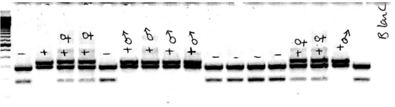
Figure 8: A gel image for the Ap2-Cre recombinase genotyping PCR ... 38
Figure 9: A gel image for the (P)RR loxP genotyping PCR. ... 40
Figure 10: Physioscan cage ... 41
Figure 11: (P)RR expression in different adipose tissues of male mice. ... 48
Figure 12: (P)RR expression in different adipose tissue pads of female mice. ... 49
Figure 13: (P)RR expression in different tissues of male mice. ... 49
Figure 14: (P)RR expression in different organs of female mice. ... 50
Figure 15: Body weight of male mice. ... 51
Figure 16: Male mice. ... 51
Figure 17: Food intake of male mice ... 52
Figure 18: EchoMRI data analysis for male mice. ... 54
Figure 19: Weights of different fat pads. ... 55
Figure 20: Locomotor activity of male mice. ... 57
Figure 21: Body weight of female mice on normal diet. ... 58
Figure 22: Body weight and weight gain of female mice on HF/HC diet. ... 59
Figure 23: Female mice on HF/HC diet: ... 60
Figure 24: Food intake of female mice on normal diet. ... 61
Figure 25: Food intake of female mice on HF/HC diet. ... 62
Figure 26: EchoMRI analysis for female mice on normal diet. ... 64
Figure 27: EchoMRI analysis for female mice on HF/HC diet. ... 65
Figure 28: Weights of different fat pads for female mice on normal diet. ... 66
Figure 30: Locomotor activity of female mice. ... 71 Figure 31: Plasma leptin level of female mice on HF/HC diet. ... 71 Figure 32: White adipose tissue histology. ... 73
List of abbreviations
1. ACE: Angiotensin-converting enzyme 2. ACEi: ACE inhibitor
3. AGT: Angiotensinogen 4. Ang I: Angiotensin I 5. Ang II: Angiotensin II
6. AP2-Cre: AP2-Cre recombinase mice
7. AT1R and AT2R: Ang II type 1 or type 2 receptors
8. ATP6AP2: ATPase, H+ transporting, lysosomal accessory protein 2 9. BAT: Brown adipose tissue
10. BMI: Body mass index 11. COX-2: Cyclooxygenase-2
12. DEXA: Dual energy X- ray absorptiometry 13. EC domain: Extracellular domain
14. EDTA: Ethylendiaminetetra acetic acid
15. ERK1/2: Extracellular signal regulated kinases 1 and 2 16. FAS: Fatty acid synthase
17. HDL: High density lipoprotein
18. HEK-293T cells: HEK cells expressing the large T-antigen of simian virus 19. HF/HC: High-fat/high-carbohydrate diet
20. HSP 27: Heat shock protein 27 21. IC domain: Intracellular domain
22. IGF2R: Mannose-6-phosphate/insulin-like growth factor II receptor 23. IPITT: Intra-peritoneal insulin tolerance test
24. JNK: c-jun N-terminal kinase 25. KO: Knock out
26. LDL: Low density lipoprotein
27. M 8-9: 8.9 kDa membrane-sector fragment 28. MAPK: Mitogen-Activated Protein Kinase
29. NAFLD: Non-alcoholic fatty liver disease 30. NEP: Neutral endopeptidase 24.11
31. OGTT: Oral Glucose Tolerance Test 32. PAI-1: Plasminogen activator inhibitor-1 33. PCP: Planar cell polarity
34. PCR: Polymerase chain reaction 35. PEP: Prolyl-endopeptidase 36. PGF: Perigonadal fat
37. PI3K: Phosphotidylnositol-3 kinase 38. PLZF: Promyelocytic Zinc Finger
39. PPARγ: Peroxisome proliferator-activated receptor γ 40. PRF: Perirenal fat
41. PRRB: (P)RR blocker
42. (P)RR: renin/prorenin receptor 43. (Pro)renin: Renin and prorenin 44. RAS: Renin-angiotensin system 45. RnBP: Renin binding protein
46. s(P)RR: Soluble form of the renin/prorenin receptor 47. SCF: Subcutaneous fat
48. (TGF)-ß1: Fibrogenic cytokine transforming growth factor 49. UCP-1: Uncoupling protein 1
50. V-ATPase: Vacuolar H+-ATPase 51. VLDL: Very low density lipoprotein 52. WAT: White adipose tissue
53. WHO: World health organization 54. WT: Wild-type
Dedication
To my mother, father, husband, big and small family for their
continuous encouragement to complete that work.
Acknowledgment
It would have been impossible for this work to be accomplished without the help given by all direct or indirect participants. I am heartily thankful to my supervisor, Dr. Julie L. Lavoie, who gave me the chance to show my research capabilities in her laboratory. Also, I thank her for her encouragement, guidance and support from the first day in her laboratory. Dr. Lavoie enabled me to develop a great understanding of the project and helped me to acquire much scientific and technical knowledge. Over two years with Dr. Lavoie, she was always available and ready for any discussion with absolute patience that any student need to. I also offer my regards and blessings to my co-supervisor, Dr. Ondrej Seda for all of his support to the project and continuous encouragement and kind words.
I ran my project on a precious mouse model that needed great care at the animal facility. For that I would thank Catherine Michel for taking care of the mice and for her help with the organization of this work.
I would like to thank all of those who supported me in any respect during the completion of the project. Thanks for the members of the evaluation committee during my study; Dr. Ashok Srivastava and Dr. Stephanie Fulton for their continuous support and helpful comments. I thank people at the IRIC and Dr. Louis Gaboury for their help on the histological study. I would also thank all my colleagues and friends at the Technopole Angus where I ran all the studies for the project.
I gratefully thank my family (parents, sisters and brother) who helped me to be where I am now. My parents always pushed me to do my best throughout my studies. The fact that I ran my studies away from them didn’t prevent their continuous support in the moments of stress and their sharing
in the moments of happiness and success. Thank you my parents for believing in me and being proud of me all the time and supporting me wherever I am.
I also owe sincere and earnest thankfulness to my husband and my daughter who accompanied me every day during my studies for their patience, great support and full understanding of my needs. Both I and my husband have always dreamed of continuing our post-graduate studies internationally, with his positive inspirations and consistent work, we were able to make our dreams true. Thanks for my newly arrived little cute son, who accompanied me while writing this report.
Thanks to my friend Marwa Seif, the most reliable friend one could have. Her friendship is one of the best things happened to me in the new country. She was always there to provide me with a sincere help and good time over last two years. I can never forget her encouraging and supporting words.
Thanks to my new friend Yousra Albasyoni and to her marvelous personality. Throughout this study, she always tried to relieve my stress and support me with loyal advices.
Thanks for you all. Basma Ahmed
Chapter 1 : Introduction
1.1 The renin-angiotensin system (RAS) cascade
The renin-angiotensin system (RAS) has an important role in controlling blood pressure, fluid homeostasis and salt balance 1, 2. The reaction cascade typically starts in the juxtaglomerular (JG)
cells of the kidney (Figure 1). There, the inactive prorenin (the renin proenzyme) is synthesized to be converted into active renin (an aspartyl protease) mainly in response to a decrease in blood pressure or increase in sympathetic activity. Mature renin is stored in granules in the JG cells and then released into the circulation while a smaller percentage remains in the kidneys to exert different intra-renal actions. Renin has been found to be synthesized in other sites such as brain, adrenal gland and visceral adipose tissue 3
Renin, in turn, works on the N-terminal of a liver plasma protein of type globulin called angiotensinogen (AGT), its only known substrate, to release a decapeptide, angiotensin I (Ang I). Ang I has mild vasoconstrictor effects but not sufficient to affect blood pressure
.
3
Further cleavage at the C-terminal of Ang I occurs via the angiotensin-converting enzyme (ACE), from the blood vessel endothelium in the lung, to form an 8 amino acids peptide, angiotensin II (Ang II), the most physiologically active component of the system
.
3
Ang II acts through cell surface receptors, Ang II type 1 or type 2 receptors (AT1R and AT2R), which are classified as G protein-coupled receptors
.
4. They have a wide distribution and are
expressed by many cell types such as in the lung, liver, brain, kidneys and adrenals 2,5
Ang II has been shown to act mainly via AT1R to produce most of its known physiological effects. For example, it elevates the arterial blood pressure by producing a potent arteriolar vasoconstriction as well as by stimulating thirst.
In addition, it decreases renal sodium and water excretion as it stimulates the secretion of aldosterone from the cortex of adrenal glands 1,2, 6. Ang II also plays a role in the regulation of cell
growth, inflammatory responses and oxidative stress mainly via AT1R. On the other hand, AT2R has been found to mainly oppose AT1R action in many aspects, for example, it causes vasodilatation, inhibits cell differentiation, cell growth and apoptosis 7, 8. This receptor is highly expressed in foetal tissues and its expression levels decrease after birth 9
Recently, it was shown that other metabolites of Angs could have different biological activities. For example, Ang III is derived from Ang II by removal of one amino acid from its N- terminal. This has been shown to occur in the brain as well as the kidneys by the action of aminopeptidase A where it has been demonstrated to have a role in the regulation of blood pressure by acting through AT1R. Further degradation of Ang III by aminopeptidase N reveals Ang IV
.
10,2
Ang (1-9) can be produced from Ang I by the action ACE 2. Ang (1-7) can then be produced from Ang (1-9) by the ACE or directly from Ang II through the activity of ACE 2 at its C-terminal
.
11, 12. In contrast to ACE, ACE 2 does not convert Ang I into Ang II and is unaffected by ACE
inhibitors (ACEi). Ang (1-7) acts via a specific receptor (Mas receptor) to produce vasodilatation, natriuresis in addition to its cardioprotective effects 10.
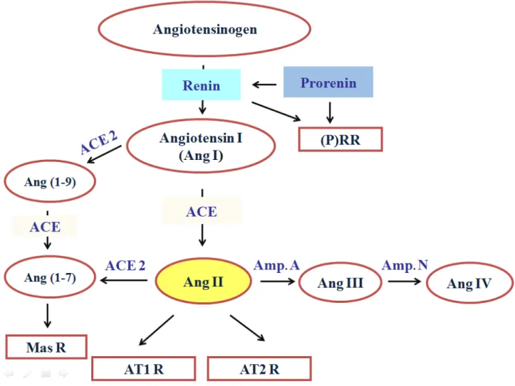
Figure 1: The renin-angiotensin system (RAS) cascade
Classical RAS cascade starts by the production of Ang I from Angiotensinogen which further be converted into Ang II that works via two receptors; AT1R and ATR2. Other Angiotensins can be also produced such as Ang III, Ang IV, Ang (1-9) and Ang (1-7) that works through the Mas receptor. The (pro)renin receptor ((P)RR) was added recently to the cascade. Ang I, Angiotensin I; Ang II, Angiotensin II; Ang III, Angiotensin III; Ang IV, Angiotensin IV; Ang (1-7), Ang (1-7); Ang (1-9), Ang (1-9); ACE, Angiotensin converting enzyme; ACE 2, Angiotensin converting enzyme 2; Mas R, Mas receptor; AT1R, Angiotensin receptor type 1; AT2R, Angiotensin receptor type 2; Amp. A, Aminopeptidase A; Amp. N, Aminopeptidase N.
1.2 Renin and prorenin
As mentioned before, prorenin is the inactive renin precursor. Structurally, renin has 2 lobes with a cleft in-between carrying its active site with two aspartyl residues. Prorenin has 43 amino acid residues at its N-terminal called the prosegment that overlies the renin active site cleft and thus hinders the access to AGT 13
Prorenin can be activated by two mechanisms: 1) Irreversible proteolytic, by cleavage of the prosegment which occurs via both proconvertase 1 and cathepsin B, in vivo, mainly in the juxtaglomerular cells of the kidney
.
14, 15; 2) Reversible nonproteolytic, by unfolding the
prosegment from the active site cleftvia low pH or partial activation by low temperatures which have never been demonstrated in vivo 16, 17
Many tissues such as the eye, adrenal gland, adipose tissue, placenta and brain secrete inactive prorenin, but not active renin, into surrounding tissues and plasma
.
18-21. Of note, the kidney is the
only known tissue to secrete active renin and as such, only prorenin can be detected in the circulation after bilateral removal of the kidneys 22. However, local RAS activity has been found in
non-renal sites which suggests that there may be a way by which prorenin gets activated locally 23.
Moreover, under physiological conditions, plasma prorenin levels are about 10 folds higher than renin 24. These are even higher, for undetermined reason, in certain physiological states such as in
pregnancy and some pathological conditions as the diabetic nephropathy and retinopathy where plasma prorenin is used as a marker for microvascular complications 19, 24, 25
On the other hand, other non-renal tissues such as the heart and vascular wall are unable to synthesize prorenin and rely on the circulation as their major source of prorenin which would then be responsible for the local activation of the RAS
.
In subsequent studies, tissue prorenin was found to be activated in these organs and could lead to local production of angiotensin peptides 21, 28 although there were originally no known mechanisms
for its activation in non-renal tissues. In addition, Ang II-independent actions of renin per se were found in cultured human mesangial cells 29
Taken together, this suggests that there is a mechanism by which prorenin in different tissues can be activated locally. Consequently, many have proposed a renin/prorenin (which we will collectively refer to as (pro)renin) binding protein (RnBP) or receptor to be implicated.
.
1.3 Renin binding proteins (RnBPs)
Several RnBPs have been demonstrated. One of these was found to be similar to the enzyme N-acyl-D-glucosamine 2 epimerase which can inhibit renin in vitro. However, its role in regulating renin activity in vivo was not determined 30. Another RnBP, the mannose-6-phosphate/insulin-like
growth factor II receptor (IGF2R) was proposed. However, that receptor is now considered to be mainly a clearance mechanism for (pro)renin since binding was found to produce rapid internalization of the (pro)renin/receptor complex, followed by rapid prorenin cleavage to active renin which is then slowly degraded 31. Moreover, no extracellular or intracellular Ang production
Chapter 2 : The (pro)renin receptor
2.1 Discovery
Studies in the last 5 years have reported the presence of a new promising (pro)renin receptor [(P)RR] which has been shown to activate the RAS as well as Ang II-independent signaling pathways 29, 33, 34. Using radio-labeled renin, Nguyen’s group identified a receptor that could
specifically bind to both renin and prorenin with very high affinity in cultured human mesangial cells 29
In addition, an increase in
. In contrast to the IGF2R, no renin internalization or degradation followed the binding.
3H-thymidine incorporation without any effect on the cell count and a
marked increase in plasminogen activator inhibitor-1 (PAI-1) antigen were observed subsequent to the binding. They suggested that renin produced mesangial cell activation which was characterized by a change in fibrinolytic capacity of the cells. Furthermore, they demonstrated that these effects were Ang II-independent as they were unaffected by either ACE inhibition or AT1R and AT2R blockade 33. Subsequently, they cloned the receptor using an adult human kidney library and transfected the (P)RR cDNA into cells which lack the ability to bind renin 34
2.2 Structure
.
The (P)RR is a 45-kDa membrane protein that specifically binds (pro)renin although prorenin has a higher affinity for the receptor compared to renin 35, 36. Indeed, Nabi et al. showed that both rat
prorenin and renin can bind to rat (P)RR that was expressed by baculovirus expression system with a dissociation constant (Kd) values of 8 nm and 20 nm respectively 36. Structurally, (P)RR is
a 350 amino-acids protein which has a small 20 amino acids intracellular domain (IC) and a single transmembrane domain (TM)34 as well as an extracellular domain (EC) where (pro)renin binds 37.
As mentioned earlier, the prorenin has a peptide sequence near its N-terminal called the prosegment which covers the renin active site. It was found that the prorenin can bind to its receptor via a small amino acid sequence of the prosegment called the handle region 37-39.
However, renin, which lacks the prosegment, can bind to its receptor although the mechanism is still unclear. Nabi et al. demonstrated that a new segment present in both renin and prorenin, called the hinge region, could bind to the (P)RR but with less affinity than that of the handle region 38.
Later, it was discovered that the (P)RR is the full form of a smaller protein associated with the vacuolar H+-ATPase (V-ATPase) 40. Consequently, the name of the gene coding for the (P)RR is
the ATPase, H+ transporting, lysosomal accessory protein 2 (ATP6AP2). The gene is located on the short arm of the X chromosome (Xp11.4) and encodes for a unique protein that gets further processed intracellularly 41
2.3 Distribution
.In humans, (P)RR mRNA has been detected with high levels in the heart, placenta and brain while lower levels are present in the kidney and liver in addition to barely detectable levels in the lung and skeletal muscles 34. In the human brain, it has been shown that the (P)RR mRNA is highly
expressed specifically in the frontal lobe as well as in the pituitary. Moreover, cells of the human anterior pituitary paraventricular and supraoptic nuclei were positively stained for the receptor by immunohistochemistry and were found to co-localize with arginine vasopressin and oxytocin 42. The receptor has also been localized in the mesangium of renal glomeruli as well as in the sub-endothelium of renal and coronary arteries of normal human kidney and heart 34
Interestingly, the (P)RR gene has been detected in isolated human adipocytes by Engeli et al. .
Furthermore, the same group detected the (P)RR mRNA in abdominal subcutaneous adipose tissue extracts of obese menopausal women43. However, since the tissue extract is rich in blood vessels
which also highly express the (P)RR mRNA, Achard V et al. later did further studies on the stromal areas and isolated stromal cells of human visceral as well as subcutaneous adipose tissues. They were thus able to demonstrate specific synthesis of the functional (P)RR in human adipose tissue 44
2.4 Signaling pathways of the (P)RR
.2.4.1 Ang I production
When renin binds to the (P)RR, it displays a four-fold increase in its catalytic efficiency to convert AGT into Ang I while prorenin shows a non-proteolytic activation 34. Thus, the (P)RR could have
a very important role in the local production of Ang II in many tissues given that renin has been proposed to be the rate limiting step of the RAS (Figure 2) 34
The mechanism implicated in the non-proteolytic activation of prorenin observed with the binding to the (P)RR could be due to conformational changes
.
17, 35, 45, 46. Furthermore, renin activation has
been proposed to be due to the proximity of the different components of the RAS, that is, Ang I produced by membrane-bound renin may be more easily converted to Ang II by membrane-bound ACE. However, a recent study investigating the presence of different forms of AGT proposes an alternate mechanism. The authors demonstrated that in both human and mice, AGT is present in two different forms, oxidized and reduced. The reduced form was found to be less easily transformed to Ang I as the cleavage site is buried within the structure of the molecule. Furthermore, the oxidized form was more susceptible to enzymatic cleavage by renin bound to the (P)RR which resulted in a 4-fold increase in Ang II production 47.
2.4.2 Ang II-independent signaling
2.4.2.1 Mitogen-Activated Protein Kinase (MAPK)
It has been reported that binding of (pro)renin to its receptor triggers intracellular signaling which is Ang II-independent since it occurs in the presence of renin and ACEi as well as AT1R blockers. Ang II-independent pathways include rapid phosphorylation of the serine and tyrosine residues of the (P)RR and phosphorylation as well as activation of mitogen-activated protein kinase (MAPK) pathways such as extracellular signal regulated kinases 1 and 2 (ERK1/2), P38 and c-jun N-terminal kinase (JNK), as well as heat shock protein 27 (HSP 27) (Figure 2) 48. The prorenin activation of the latter pathway has been shown to lead to changes in actin filament dynamics which are known to keep the integrity of the cell architecture and growth. Of note, no changes in intracellular Ca+2 or cAMP have been observed with (P)RR stimulation 34. Following ERK1/2
activation via the (P)RR, an increase in the fibrogenic cytokine transforming growth factor (TGF)-ß1 has been observed (Figure 2) 49. This in turn activates profibrotic molecules such as, PAI-1, fibronectin protein and collagen I, independently of Ang II 49, 50. Furthermore, these effects were
found to be blocked by TGF-ß1 antibody administration 50. It has recently been suggested that this
occurs through an increased expression of Nox4 with a subsequent increase in superoxide anion production 51
2.4.2.2 Promyelocytic Zinc Finger (PLZF)
.
Another (P)RR signal transduction pathway which has been reported in vitro involves direct interaction between the C-terminal domain of the (P)RR and the promyelocytic zinc finger (PLZF) transcription factor 52. Following renin stimulation, the transcription of the p85α subunit of the
This in turn inhibits the transcription of the (P)RR, creating a short negative feedback loop (Figure 2). Thus, high levels of (pro)renin could inhibit (P)RR expression and prevent excessive receptor activation 53.
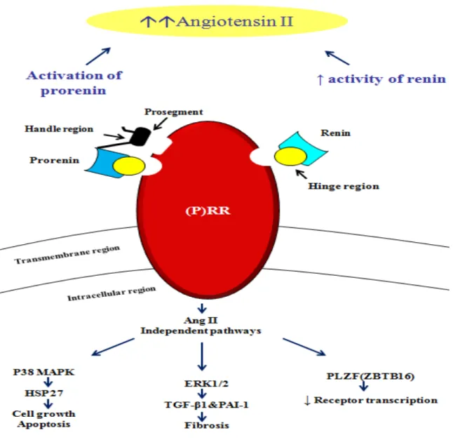
Figure 2: Mechanism of action of (P)rorenin receptor
Binding of the (pro)renin to the (P)RR leads to increased enzymatic activity of renin and renders prorenin non-proteolytically active which, taken together, increases the production of Ang II. At the same time, this binding leads to simultaneous activation of Ang II-independent intracellular signaling cascades. Binding sites for renin and prorenin to the receptor, hinge (yellow circles) and handle region (black rectangle), are also shown. ERK1/2, extracellular signal regulated kinases 1 and 2; HSP 27, heat shock protein 27; p38 MAPK, mitogen-activated protein kinase p38; PAI-1, plasminogen activator inhibitor-1; PLZF, promyelocytic zinc finger; TGF-ß1, transforming growth factor ß1. Modified from Ahmed B. and Lavoie J.L. 48
2.4.2.3 Wnt/β-catenin signaling
The implication of the (P)RR in the Wnt/β-catenin signaling pathway (Figure 3) has recently been reported. Wnt proteins have been shown to be essential for normal embryogenesis and regulate many cellular aspects in adult tissues. For example, dysregulation of Wnt signaling has been associated with cancer development 48,54
In a study to understand the mechanism by which the Wnt receptors get activated, Cruciat et al. showed that the (P)RR was required for the activation of the Wnt-canonical pathway. Using a genome wide siRNA screen in HEK-293T cells (HEK cells expressing the large T-antigen of simian virus), they found that three siRNA directed towards the (P)RR resulted in inhibition of Wnt3 signaling at the level of, or upstream to the LRP 5/6 receptor
.
55. Moreover, they
demonstrated that the (P)RR could bind to the components of the Wnt receptor complex, Frizzled and LRP 5/6, via it’s EC domain. They also found that the (P)RR acts as an adaptor between the LRP 5/6 receptor and the V-ATPase which induces LRP 5/6 phosphorylation and as such, activates the β-catenin pathway 55
On the other hand, (P)RR was also found to be involved in the Wnt non-canonical pathway; the Wnt/PCP (planar cell polarity)
.
56, 57. Indeed, two groups have confirmed that the (P)RR can
regulate PCP in Drosophila as well as the convergent-extension movements in Xenopus gastrulae. Interestingly, these events are independent of renin as neither the Drosophila nor Xenopus expresses renin 56, 57.
2.5 Alternate molecular forms of the (P)RR
As mentioned previously, the receptor has 3 main domains: the TM, EC and IC domains. Previous studies investigating the receptor have mainly focused on the role of the full length (P)RR. Lately, it has become obvious that other forms of the receptor might also be playing important roles.
2.5.1 (P)RR and V-ATPases
Indeed, Ludwig et al. discovered by chance in 1998 a truncated form of the receptor composed of only the TM and the IC domains 58. They described the C-terminal part of the (P)RR protein as a
8.9 kDa membrane-sector fragment (M 8-9) that associates with the V-ATPase present in chromaffin cell membranes (Figure 3). (P)RR and M 8-9 protein seem to be derived from the same (P)RR gene transcript. Accordingly, the (P)RR gene was named; ATP6AP2 58. V-ATPases are
ATP-dependent proton pumps. Structurally, they are composed of a peripheral V1 and an integral V0 domain that are formed of 8 and 6 different subunits respectively. The V1 domain is responsible for ATP hydrolysis while the V0 domain translocates protons. Thus, the main roles of V-ATPases are the acidification of intracellular compartments, such as lysosomes and secretory vesicles, and proton pumping in different cell types such as in osteoclasts, macrophages and renal cells 59
Later, another functional link between the (P)RR and the V-ATPase was demonstrated by Advani
et al. as they first reported a high expression of the (P)RR on the apical membrane of rat collecting
ducts intercalated A cells as well as in the convoluted and distal tubules of human kidneys .
60. They
further determined the co-localization of (P)RR with the V-ATPase B 1/2 in this subtype of intercalated cells 60. Moreover, the implication of the (P)RR/V-ATPase interaction in the (P)RR
They demonstrated that the (P)RR ERK1/2 activation was attenuated when they pre-treated Madin Darby canine kidney cells, a collecting duct/ distal tubule lineage, with bafilomycin; a selective V-ATPase inhibitor 60
Lately, additional data supporting the (P)RR/V-ATPase relation has been reported. For example, it was found that mice deficient in the (P)RR specifically in cardiomyocytes, developed severe heart failure and died by 3 weeks of age, although no cardiac anomalies could be detected at birth. Interestingly, cardiomyocytes from these knock-out mice showed depleted levels of the V0 domain without any effect on the V1 domain of the V-ATPase. This could be reproduced in cultured embryonic fibroblasts with specific ablation of the (P)RR gene
.
40. As a result, (P)RR loss in
cultured cells impaired vesicular acidification. In addition, cultured cardiomyocytes treated with bafilomycin showed similar phenotype to what was observed in isolated cardiomyocytes from knock-out animals 40. Similarly, two groups reported that the (P)RR/V-ATPase is crucial for
podocyte function and survival using mice with specific deletion of the (P)RR in podocytes 61, 62.
Knock-out mice were born without any detectable kidney abnormalities but developed a disruption of the glomerular filtration barrier that lead to a nephrotic syndrome which produced sever renal failure. As such, mice died as early as 2 to 4 weeks of age and their podocytes showed marked foot process effacement, alteration of their actin cytoskeleton, reduction of the slit diaphragm proteins and further numerous autophagic vacuoles. Interestingly, and in line with what was noticed with cardiac specific deletion of the (P)RR, (P)RR deletion in podocytes was found to be associated with a suppression of the V0 domain of the V-ATPase. This produced V-ATPase dysfunction that resulted in deacidification of intracellular vesicles. Moreover, previous abnormalities could also be reproduced by treating cultured podocytes with bafilomycin 61, 62.
Taken together, we can conclude that the (P)RR plays an important role in regulating V-ATPase activity and that the V-ATPase is implicated in (P)RR Ang II-independent signaling.
2.5.2 Soluble form of the (P)RR
Recently, a truncated soluble form of the (P)RR [s(P)RR] of 28 kDa that contains only the EC domain has been described. It is present in both plasma and urine and retains the ability to bind renin and prorenin (Figure 3) 63, 64. Similarly to the full length (P)RR, the s(P)RR has been shown
to activate prorenin and thus stimulate the production of Ang I 64-66 although there is no data
concerning its impact on renin activity. Studies have suggested the possible implication of several enzymes such as furin or metalloproteases in the production of the s(P)RR 63, 65. If we take into
consideration the data showing that oxidized AGT is more readily cleaved by renin bound to the (P)RR, we can conclude that the s(P)RR may constitute a novel mechanism for the development of hypertension as well as other RAS-associated diseases.
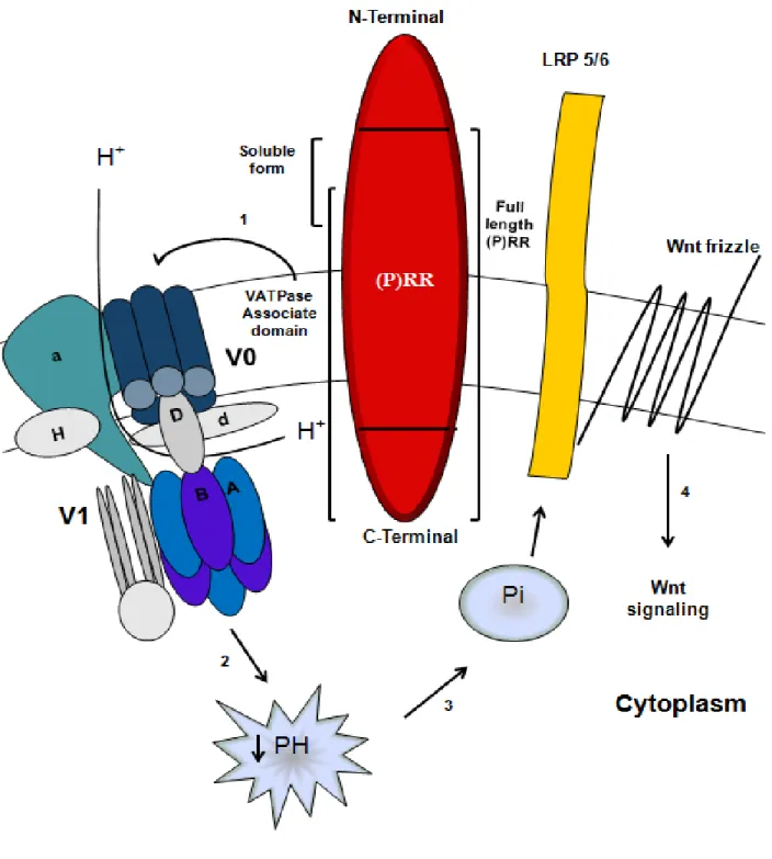
Figure 3: (P)RR and Wnt signaling
(P)RR acts as an adaptor between LRP 5/6 and V-ATPase (1) the later generates a proton gradient (2) that is essential for the phosphorylation and activation of LRP 5/6 (3) which in turn activates β-catenin signaling. LRP 5/6, low-density lipoprotein receptor-related protein 5/6; (P)RR, (Pro)renin receptor; Pi, phosphate ion. Modified from Ahmed B. and Lavoie J.L.48.
2.6 Animal models of the (P)RR
2.6.1 Deletion of the (P)RR
Generating a knock-out model of the (P)RR is an extremely interesting tool to study the receptor’s physiological roles. In fact, it has been found that ubiquitous deletion of the receptor is lethal, which reflects the importance and necessity of the (P)RR gene in embryonic development 41
For instance, mutation of the (P)RR gene by gene trap in zebrafish embryos has been shown to be lethal before the end of embryogenesis and was associated with developmental abnormalities including neuronal cell death as well as body and eye hypopigmentation
.
41. As mentioned earlier in
the text, the (P)RR was found to be implicated in the Wnt signaling pathways which could explain these results. In contrast, tissue specific deletion of the receptor is not developmentally lethal although animals develop lethal defects after birth. For instance, knocking down the (P)RR in mice cardiomyocytes produced fatal heart failure at 3 weeks of age 40 while specific deletion of the
(P)RR in podocytes resulted in severe renal failure and animal death by 2 to 4 weeks of age 61, 62
2.6.2 Overexpression of the (P)RR
.
Transgenic animals which overexpress the (P)RR gene are an interesting alternative that has been employed to define the pathogenic effects and evaluate the in vivo relevance of the receptor. For instance, rats overexpressing the human (P)RR ubiquitously presented with slow progressive nephropathy produced mainly by Ang II-independent signaling. Indeed, these rats showed significant proteinuria with aging and glomerulosclerosis at 28 weeks of age as well as enhanced renal TGF-ß1 expression without any elevation in renal Ang II, blood glucose or blood pressure 67.
In contrast to imidapril, an ACEi, complete prevention of all mentioned symptoms could be achieved by chronic administration of a (P)RR blocker (PRRB), also known as the handle region peptide 67. Moreover, renal enhancement of ERK, p38, and JNK immunostaining was observed.
The ERK and p38 activation were also completely inhibited by the PRRB while JNK activation was only attenuated but all three were unaffected by ACEi 67. By using the same model, it was
shown that transgenic rats had no changes in their blood pressure however their renal cortical cyclooxygenase-2 (COX-2) mRNA and protein levels were significantly increased. In addition, urine sodium excretion was found to be also increased which could be explained by activation of both RAS dependent and independent mechanisms as a result of increased PRR expression. Interestingly, COX-2 inhibition produced a significant decrease in renal cortical blood flow specifically in the transgenic animals 68
On the other hand, rats overexpressing human (P)RR specifically in vascular smooth muscle developed progressive cardiovascular symptoms. Rats developed normally but exhibited spontaneous cardiovascular phenotypes at the age of 6 months such as marked increase in systolic blood pressure and an unexpected elevation in heart rate
. Given that cortical COX-2 has been suggested to be implicated in diabetic hyperfiltration, these results suggest that the (P)RR may be involved in this effect.
69. These symptoms were aggravated with
age and earlier appearance of these symptoms could be observed in rats with higher transgene expression. In addition, this was accompanied by an increased plasma aldosterone levels as well as a significant increase in plasma aldosterone/renin ratio 69. This demonstrates that aldosterone
production was enhanced via the increased adrenal Ang II as a result of (P)RR overexpression without changing the renin levels.
Chapter 3 : Obesity
3.1 Epidemiology and definition
The dramatic increase in the prevalence of obesity among Canadians over the past 30 years has been deemed to constitute an “epidemic.” In 2011, 18.3 %, approximately 4.5 million, of Canadian adults were reported to be obese 70. In 2010, the IASO/IOTF (International Obesity Taskforce)
analysis revealed that approximately 1.0 billion adults are overweight and a further 475 million are obese worldwide. Globally, 150 million school-aged children were found to be overweight and 50 million were obese 71
WHO defines obesity as an abnormal or excessive fat accumulation that presents a risk to health. WHO classifies obesity using the body mass index (BMI) which is calculated by dividing the body weight by the square of the height (Kg/m
. Indeed, the World Health Organization (WHO) refers to the escalating global epidemic of obesity as “globesity,”
2). According to WHO, people with BMI equal to or
greater than 25 are considered overweight while obese individuals are characterized by a BMI equal to or greater than 30. Obesity is a multifactorial problem in regards to its etiology. Many theories have been proposed in trials to find a defined etiology such as gene-environmental and gene-behavioral interactions. However, the corner stone for occurrence of obesity remains the increased energy intake in the form of caloric consumption over the energy expenditure 72. Lately,
many factors were found to be implicated in increased caloric intake such as increased portion size, ready-made food and sweetened beverages as well as attractive advertising to promote them 72.
The continuous change in life style towards a more sedentary behavior leads to a decrease in energy expenditure 72.
Several studies have reported that an increase in BMI is positively correlated with higher mortality rates 73, 74 as a result of the increased the incidence of many health problems 75. For instance,
obesity is strongly associated with the development of type II diabetes in men and women of all ethnic groups 76-78 by causing insulin resistance. One of the proposed mechanisms for this effect is
the increased production of the adipokines (elements that are produced by the adipose tissue) that enhance insulin resistance, such as resistin, and the decrease in those that stimulate the insulin sensitivity, such as adiponectin 79. Another theory is that the increased secretion by adipose tissue
of inflammatory chemokines enhances macrophage activation and infiltration into the adipose tissue. Activated macrophages in turn secrete different cytokines that affects the insulin sensitivity
80. As obesity is associated with an increase in circulatory and visceral free fatty acids, excess fatty
acids also promote the development of insulin resistance in the liver as well as in skeletal muscles
79. Obesity is also associated with a disturbed lipid profile (dyslipidemia) in the form of decreased
levels of circulatory high density lipoprotein (HDL) cholesterol while increased levels of circulatory triglycerides, cholesterol, low density lipoprotein (LDL) cholesterol, very low density lipoprotein (VLDL) cholesterol and apolipoprotein B 75. Moreover, it has been reported that obesity is associated with elevated risk for cerebrovascular strokes 75. For example, a study
reported that a BMI above 30 Kg/m2 was associated with increased the risk for total and ischemic
but not for hemorrhagic strokes 81. Also, obese individuals have been reported to be more prone to
develop heart diseases 75. For instance, the risk of developing heart failure was found to be
increased by x-fold in obese subjects compared to those with a normal BMI82. Obesity is also a major risk factor for some respiratory diseases such as obstructive sleep apnea 75. In addition,
pregnant obese women are at higher risk of developing gestational diabetes and preeclampsia as well as more subject to caesarean sections 83. Moreover, the incidence of other health problems
was found to be increased with obesity such as the osteoarthritis, psychological disorders, and even some types of cancers such as breast, colon, esophagus and kidney 72
3.2 Obesity treatment
.
Many treatments for obesity have been proposed such as lifestyle modifications including dieting and exercise, psychological therapies as well as bariatric surgeries including Roux-en-Y bypass or gastric banding 84. In addition, several anti- obesity drugs have been tested to control weight gain
but have been stopped due to the increased risk of developing cardiovascular disorders, such as desoxyephedrine, phentermine and diethylpropion (amphetamine derivatives) as well as fenfluramine and dexfenfluramine (serotonin-releasing agents) 84. Sibutramine (noradrenaline and
serotonin-reuptake inhibitor) was believed to reduce weight gain by decreasing food intake and increasing energy expenditure, but it was reported to have serious side effects on the cardiovascular system. For example, long term use of Sibutramine in subjects with high cardiovascular risk was found to increasing the risk of nonfatal myocardial infarction and nonfatal stroke 85. Conversely, Orlistat has been approved as an anti-obesity drug that decreases fat
absorption by inhibiting the pancreatic lipases 86
3.3 White adipose tissue
. Apart from associated gastrointestinal symptoms such as bloating and diarrhea, Orlistat doesn’t seem to have any serious side effects and as such, is a more promising treatment option.
White adipose tissue (WAT) (Figure 4) is formed of adipocytes and a stromal vascular compartment or fraction where the preadipocytes are located 87. Almost all adipose tissue in the
adult human body is WAT with a very wide distribution with the exception of certain tissues such as the eyelids, the penis and the scrotum. WAT can be divided according to the pattern of
distribution into subcutaneous and internal fat. Subcutaneous fat lies between the skin dermis and the outer layer of the muscles (fasciae). Internal fat includes visceral fat and non-visceral fat such as bone marrow. Visceral fat includes all intra-abdominal and pelvic fat such as omental, peri-renal, mesenteric fat as well as ectopic fat sites including heart, liver and muscles. The distribution and density of adipose deposits depends on the age, sex and ethnic group 88, 89. For instance, the subcutaneous fat of women is noticed to be located mainly in the buttocks, thigh (gluteofemoral) and breast while around the abdomen (lumbosacral), neck and upper arm for men and this distribution becomes more obvious with age 89. Of note, mature adipocytes count no more than
half of the total cell content since WAT also contains other cell types such as fibroblasts, endothelial cells, preadipocytes, and macrophages 90.
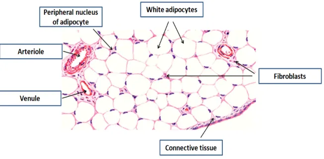
Figure 4: White adipose tissue
Hematoxylin and eosin stained section of mesenteric fat showing unilocular white adipocytes. Fat dissolved during the staining and cells appeared as empty spaces with small rim of cytoplasm and peripheral nuclei. Cells are separated by connective tissue that entangles fibroblasts and blood vessels. Modified from DiFiore’s Atlas of histology 91.
Adipose tissue cells are derived initially from the mesenchymal stem cell (Figure 5). As can be seen, a series of differentiation cascades that end by the production of mature adipocytes are required and are under the control of several transcription factors 89. The peroxisome
proliferator-activated receptor γ (PPARγ) is one of the key transcription factors. It is a very important nuclear receptor that has been found to induce adipogenesis92 and plays a role in determining WAT
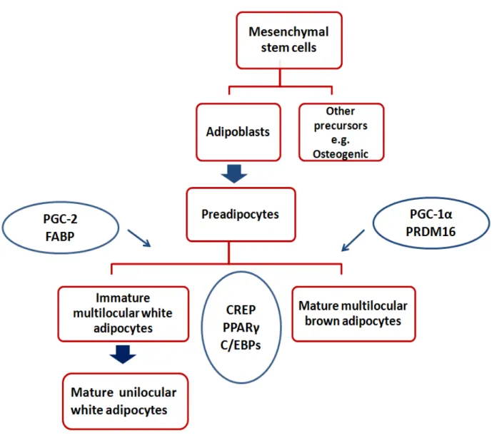
Figure 5: White and brown adipocytes’ differentiation
The differentiation cascade of adipocytes from mesenchymal stem cells which is enhanced by several transcription factors is presented in this figure. CREB: (cAMP-response-element)-binding protein; PPARγ: peroxisome proliferator-activated receptor-gamma; C/EBPs: CCAAT/enhancer-binding proteins; PGC-2: peroxisome proliferator-activated receptor-gamma co-activator-2; FABP: fatty-acid binding protein; PGC-1α: peroxisome proliferator-activated receptor-gamma co-activator-1alpha; PRDM16: PR domain containing 16. Modified from obesity science to practice
The main function of WAT seems to be energy storage that could easily be released during periods of negative energy balance and supply the different organs with fatty acids to be oxidized 95. The
WAT mass is determined by both the number and the size of adipocytes. It was proposed that during states of positive energy balance, adipocytes first become hypertrophic (increase in size) and then hyperplasia follows (increase in number). As such, adipocyte hyperplasia occurs only when adipocytes attain their maximal capacity of expansion as mentioned above. This capacity seems to be genetically determined 96
However, WAT has other roles; for example, it acts as a heat insulator as well as it protects and supports internal organs
.
97. It was also found that WAT plays an important role in several
homoeostatic mechanisms such as the regulation of blood cell precursors, immunity and insulin sensitivity 97. Moreover, it has been found in the last decade that adipocytes are able to release
many factors, named adipokines, that play an important role not only in the regulation of energy homoeostasis but also on other physiological processes 98. For instance, it has been found that
WAT secretes a 16-kDa protein called leptin 99 which was found to be a product of the Ob gene.
Mutation of this gene results in severe obesity as can be seen in the Ob/Ob mice 100, 101. In addition, plasma leptin levels correlate directly with adipocyte size and body fat mass thus it increases markedly with the development of obesity in both human and rodents 99. Indeed, leptin informs
the brain about the energy status of the body. For example, in positive energy balance state, leptin crosses the blood brain barrier to stimulate its receptor in the brain which is highly expressed in the hypothalamic neurons 102, 103. Thus it inhibits the orexigenic and enhances the anorexiogenic pathways of the hypothalamus to strongly suppress food intake and increase the energy expenditure. However, as leptin levels usually increase with the development of human and rodent obesity, this strongly suggests the development of leptin resistance in this condition 102, 103.
Leptin also has other metabolic functions. For example, it was found that leptin can suppress the expression of the insulin gene as well as insulin secretion in human pancreas 104
Another adipokine called adiponectin is also secreted by white adipocytes. Conversely to what is found with leptin, adiponectin levels were found to be decreased with the development of obesity and to be increased by food deprivation
.
105. Adiponectin acts through its receptors, AdipoR1 and
AdipoR2, which are widely distributed in skeletal muscles and liver respectively. In skeletal muscles, adiponectin increases fatty acid oxidation and glucose uptake 105. Decreased adiponectin
levels in humans and total ablation of adiponectin in rodents were associated with the development of hepatic insulin resistance, inflammation as well as vascular injuries thus it also has an insulin sensitizing action 105
3.4 Brown adipose tissue
.Brown adipose tissue (BAT) in mammals plays a role in the regulation of both basal and inducible energy expenditure through thermogenesis. BAT cells have a lot of mitochondria that express a specific type of protein called the uncoupling protein 1 (UCP-1) at their inner membranes. UCP-1 allows the dispersion of the electro-chemical gradient that is generated through the respiratory chain in the form of heat rather than ATP, a process called uncoupling of the oxidative phosphorylation 106. In rodents, BAT is found to be abundant in newborns especially in their
inter-scapular areas 107. Of note, brown adipocytes can also be found in other WAT sites especially in the inguinal, retroperitoneal and peri-ovarian region of reproductive fat. Interestingly, the number of brown adipocytes as well as the expression of UCP-1 was found to be increased in these sites after cold exposure 108. In humans, it has been thought for a long time that BAT is found only in
Chapter 4 : The renin-angiotensin system and obesity
4.1 Human studies
A study reported that, plasma AGT concentrations are significantly and positively correlated with BMI as well as plasma leptin levels in normotensive non-obese young men suggesting a relation between adipose mass and circulating AGT levels 109. Many reports showed the positive
correlation of circulatory AGT and obesity 110-113. It was also found that the expression of the AGT gene was increased in subcutaneous adipose tissue of obese men in correlation to the body weight
114. In addition, the expression level in subcutaneous and omental fat was positively correlated with
an increase in waist circumference 115. Another group observed an increase in circulating RAS
activity with a decrease in adipose tissue AGT gene expression in obese menopausal women compared to their lean counterparts 43. However, although AGT expression has sometimes been found to be decreased, it is thought that the increase in adipose mass produces an overall increase in AGT adipose tissue content which would contribute to the increase in circulating AGT observed with obesity. Interestingly, in this study a significant decrease in RAS activity and further reduction in adipose tissue AGT expression were noticed after only 5 % reduction in body weight
43. In line with these results, a study using Telmisartan, an AT1R antagonist, demonstrated the
implication of the RAS on the distribution of body fat in patients with metabolic syndrome. Indeed, Telmisartan lead to significant and selective decrease in visceral but not subcutaneous fat which was associated with an improvement in insulin sensitivity116. Similar results were obtained
by another group also using Telmisartan when administered to Japanese hypertensive patients which was associated with an increase in serum adiponectin levels 117.
It was also found that the use of Enalapril (an ACEi) for 16 weeks in moderately hypertensive patients was associated with a significant reduction of their body weights 118
4.2 Animal studies
.
Many in vivo animal studies have found that the RAS might play a role in the pathogenesis and occurrence of obesity. For instance, renal ACE activity was found to be increased in association with obesity development in mice 119. Moreover, it was noticed that mice with ubiquitous AGT
deficiency were resistant to weight gain in response to both a normal and high fat diet which may be as a result of their significant increase in locomotor activity 120. No changes in the dietary fat excretion were detected while an altered white adipose tissue development in the form of adipocyte hypotrophy and a decrease in their triglyceride content with an associated decrease in fatty acid synthase (FAS) activity were shown 120. It was reported that AT1Ra/AT1Rb null mice showed
slower rate of body weight gain after birth which was mainly as a result of a decrease in fat mass
121. Another study showed that mice lacking AT2R were also resistant to diet induced weight gain
and obesity as well as obesity related glucose intolerance 122. They also showed increased levels of
lipid oxidation. As a result, adipose tissue displayed adipocyte hyperplasia and hypotrophy. The authors reported that the small sized adipocytes could be explained by the associated decrease in the activity of the FAS in addition to decreased gene expression of PPARγ, the lipoprotein lipase (LPL) as well as the fatty acid transporters CD36 and fatty acid protein (aP2) which stimulate the fatty acid uptake and storage 122. Conversely, the authors couldn’t find the exact mechanism
implicated in the adipocyte hyperplasia since Ang II has been reported to have a stimulatory effect on preadipocytes differentiation via AT2R 123.
However, they suggested that Ang II may have an inhibitory effect on preadipocytes proliferation
in vivo via the AT2R as what was reported in smooth muscles and endothelial cells 122
Moreover, it was observed that knocking down the AT1Ra in mice attenuated the high-fat diet induced obesity, weight gain and adiposity as a result of the increased energy expenditure and sympathetic activity without any changes in food intake. In addition, these mice were protected from some components of the metabolic syndrome as they had low blood pressure and showed an attenuation of the impaired glucose tolerance and insulin resistance as compared to their wild type littermates
.
124
On the other hand, in vitro studies revealed conflicting data about the role of AT1R and AT2R in adipocytes. For instance, AT1R but not AT2R protein was detected throughout the differentiation process of 3T3-L1 preadipocytes
. Thus it could be concluded that AT1R and AT2R may have synergistic effects in regards to adipose tissue development in vivo.
125 while a hypertrophic effect on 3T3-L1 and human adipocytes
in primary culture via AT2R was also reported 126
Of note, an inhibitory effect of Ang II through the AT1Ra on human preadipocytes conversion in primary culture was reported
.
127, 128 while other group showed Ang II stimulatory effect on Ob1771
mouse adipocytes differentiation through AT2R 123
In another study, mice lacking the renin gene presented with lighter body weights compared to their wild-type littermates and these differences were accentuated by the administration of a high-fat diet. This was associated with an increase in metabolic rate as well as a decrease in intestinal dietary fat absorption
. The discrepancy of the results might be explained by the variability in adipocyte sources and /or the experimental duration.
129. In addition, no changes in food intake or in locomotor activity were
Similarly, mice deficient in the ACE gene were leaner as they presented with a reduced body weight and showed less fat depots. This was associated with an increase in their total and resting energy expenditure with no changes in their food consumption and locomotor activity although their fecal fat excretion was not affected 130. The controversy in regards to fat excretion data may
be due to the different methods used. For instance, chloroform: methanol solution was used for ACE null mice versus petroleum benzene for AGT knocked out mice 130, 120 while fecal acid
steatocrit was measured in mice lacking the renin gene 129
As such, it was hypothesized that the administration of RAS blockers might show similar results to what had been observed with the deletion of the different RAS genes. Indeed, Sprague–Dawley rats receiving a normal diet that were treated with Perindopril, an ACEi, had a reduced weight gain as well as a decreased plasma leptin levels compared to the untreated group. This was not associated with any changes in their food intake and was considered to be mainly as a result of a reduction in their body fat mass as measured by Dual energy X- ray absorptiometry (DEXA) with no changes in their lean or bone mass
.
131. Authors suggested that these effects may be due to a
reduction in both plasma and tissue angiotensin which is known to promote adipocytes differentiation and adipogenesis although angiotensin levels were not assessed. Similar results were observed in Long Evans rats which received another ACEi, captopril, on either normal or high-fat diet but with noticeable changes in food intake 132. Administration of the perindopril in the
drinking water of Sprague–Dawley rats since birth was also associated with a decrease in body weight and food intake 133. It has been suggested that the discrepancy in the food intake results might be explained by the difference between each ACEi to access and affect the brain nuclei at different doses 132. Indeed, it has been shown that chronic treatment, over ten months, with
syndrome, was beneficial in preventing obesity and hyperinsulinemia while reducing hypertension without changing food intake 134. Authors proposed that blocking the RAS might increase insulin
secretion and thus enhance catabolism as suggested by the increased plasma urea levels with normal kidney functions in the captopril treated group. Based on these studies, we can conclude that the RAS has a role in the pathogenesis of obesity in rodents.
Chapter 5 : Adipose tissue renin-angiotensin system
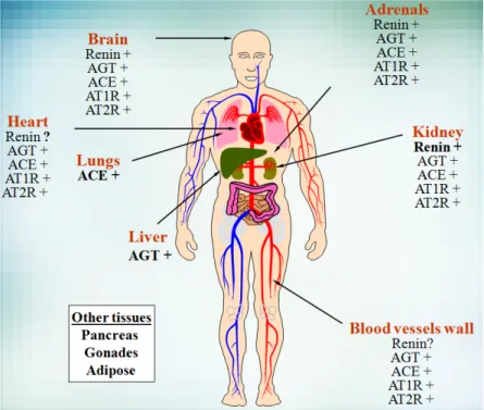
5.1 Local tissue RAS
It was demonstrated that the RAS is not only active systemically but that there are also complete RAS in many tissues (Figure 6). Indeed, critical components of the RAS have been described in several organs such as the brain, kidney, gonads, heart, pancreas and adipose tissue (Figure 6) 2.
This strongly suggests a potential autocrine and paracrine role of the RAS 135. For example, brain Ang II has been found to act as a neurotransmitter 136. Moreover, local RAS could be involved in
different pathologies. For instance, brain and kidney RASs have been demonstrated to contribute to the development of hypertension 2.
Figure 6: Expression of the RAS
Sites of expression of the different components of the RAS are shown. Classical sites of synthesis for the endocrine RAS are in bold. AGT, Angiotensinogen; ACE, Angiotensin-converting enzyme; AT1R, Angiotensin receptor type 1; AT2R, Angiotensin receptor type 2. Modified from Lavoie J.L. and Sigmund C.D. 2.
5.2 Adipose tissue RAS and obesity
Local RAS in adipose tissue has recently been a rich research area. In an early report, transgenic mice which overexpressed human AGT under the control of its own promoter showed high expression of the gene mRNA in their white as well as brown adipose tissue with stronger expression in male mice compared to females 137. In another report, the AGT mRNA expression
was found to be 4 times higher in visceral fat compared to subcutaneous fat of human subjects with a BMI of less than 30 kg/m2. In obese subjects, AGT gene expression was significantly higher than
leaner subjects in subcutaneous adipose without noticeable changes in visceral fat 138. In line with these results, another group found that AGT gene expression and protein secretion were significantly higher in visceral as compared to subcutaneous isolated adipocytes from normal male rats. Interestingly, a 50% reduction in the AGT secretion from different adipose tissue depots occurred after castration and was reversed by the administration of testosterone 139. Furthermore,
several studies have shown that the high expression of AGT in adipose tissues and its constitutive excretion from mature adipocytes was present in rodents as well as in humans 140. Indeed, adipose
tissue was found to be responsible for up to 30% of the circulating levels of AGT in rodents 141.
Moreover, targeted AGT overexpression in mice adipose tissue was found to be associated with obesity development and hypertension 141. It is important to know that adipose tissue also
expresses other components of the RAS such as renin, ACE and ACE2. As such, this contributes to the synthesis of Ang II and other angiotensin peptides in the adipose tissue 123, 142. Hence, adipose
tissue not only contributes to Ang II production but it also allows for its autocrine and paracrine action since AT1R, AT2R and MasR are expressed in both adipocytes and periadipocytes 143. In
with the development of both genetic and diet induced obesity not only in rodents 144, 145 but also in
humans 114
Ang II enhances the lipogenesis in adipocytes by increasing the activity .
146 and the gene
transcription of the adipocyteFAS through the AT2 receptors 126, 147. It also stimulates the activity
of other lipogenic enzymes such as glycerol-3-phosphate dehydrogenase 146. Through the same receptor, Ang II was also found to work as a lipogenic hormone since it promotes the accumulation of triglycerides in murine adipocytes and in human adipose cells in primary culture which promotes adipocyte growth 126. Another group found that Ang II had a dose dependent
anti-lipolytic effect on human subcutaneous adipose tissue and skeletal muscle in both normal weight and obese subjects 148. Later, the same authors found that this effect occurred mainly via AT1R 149. Furthermore, an enhanced lipolytic activity with a subsequent increase in plasma free fatty acids and significant decrease in epididymal fat weight were noticed in fasted mice treated with valsartan, an AT1R antagonist 150. Of note, Ang II was found to enhance the differentiation of
preadipocytes into mature adipocytes by different mechanisms via both AT1R 142 and AT2R 123, 151
5.3
Adipose tissue RAS and insulin sensitivity
. Overall, Ang II seems to play an evident role in enhancing adipocyte lipid storage and expanding white adipose tissue by different mechanisms.
Generally, acute Ang II administration is associated with an improved insulin sensitivity and adipocyte glucose consumption in rodents as well as in type 2 diabetic subjects. Conversely, chronic Ang II infusion in rats was found to induce significant insulin resistance and impaired insulin signaling such as, reduced adipocyte and skeletal muscle glucose uptake due to increased oxidative stress 152.
Locally, overexpression of AGT in mice adipose tissue was found to be associated with increased insulin resistance in parallel to obesity development 153.
Chapter 6
: Hypothesis
As mentioned above, RAS activation is associated with obesity development. As a new member of the RAS, the (P)RR was found to increase renin enzymatic activity and thus stimulates the production of Ang I, and as a result, Ang II. In addition, the (P)RR was found to have specific Ang II-independent signaling pathways as mentioned earlier. Therefore, we expect that the (P)RR might also have a role in regulating body weight and obesity development although little is known about this role.
We have recently demonstrated in our laboratory that the (P)RR mRNA levels are increased specifically in white adipose tissue depots in a mouse model of diet-induced obesity and that systemic administration of a (P)RR blocker reduces weight gain and appetite 154. Furthermore, this
is accompanied by a normalization of many circulating metabolites such as free fatty acids, triglycerides and glucose although the exact mechanism is unknown. Hence, to determine the implication of adipose tissue in this novel role of the (P)RR, mice deficient for this receptor (AP2-KO) specifically in their white adipose tissue were studied. We hypothesize that knocking down the receptor in white adipose tissue will decrease weight gain and prevent the development of obesity and its related complications, such as type II diabetes.
Chapter 7 : Materials and Methods
7.1 Animals
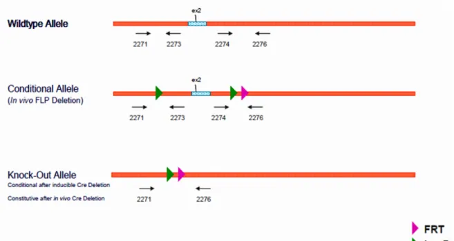
Mice deficient for the (P)RR in their white adipose tissue were generated using the Cre/Lox technology. (P)RR floxed mice (a generous gift from Merck Frosst Canada) in which exon 2 of the ATP6AP2 gene was flanked between loxP sites, were bred with AP2-Cre recombinase mice (AP2-Cre mice; Jackson Laboratories) that express the (AP2-Cre-recombinase enzyme under the control of a white adipocyte promoter (Figure 7).
Figure 7: Genotype strategy for AP2-KO mice.