Université de Montréal
A mitochondrial perspective on striated muscle physiopathology: insights from sepsis, denervation, and dystrophinopathies.
Par Richard Godin
Sous la tutelle du Dr. Yan Burelle, faculté de pharmacie Université de Montréal
Thèse présentée à la faculté des etudes supérieures et postdoctorales en vue de l’obtention du grade de doctorat
Résumé
La mitochondrie est de plus en plus reconnue pour sa contribution à la dégénerescence musculaire. Les dysfonctions mitochondriales, en plus de causer une défaillance énergétique, contribuent à la signalisation apoptotique, stimule la production de ROS et peuvent induire une surcharge calcique. Ces caractéristiques sont tous reliées à certains types de myopathies. Cette thèse met en lumières comment certaines dysfonctions mitochondriales peuvent intervenir dans la pathogenèse de diverses myopathies. Nous démontrons que les dysfonctions mitochondriales sont impliqués dans l’atrophie dû à la perte d’innervation. Par contre, la désensabilisation de l’ouverture du pore mitochondrial de transition de perméabilité, via ablation génétique de cyclophiline-‐D, ne prévient ni la signalisation apoptotique mitochondrial ni l’atrophie. Nous avons aussi observé des dysfonctions mitochondriales dans le muscle atteint de dystrophie musculaire de Duchenne qui furent améliorés suite à une transfection de PGC1-‐α, laquelle résulta aussi en une amélioration de la pathologie. Finalement, nous démontrons que le recyclage de mitochondrie par les voies de mitophagies et de contrôles de la qualité impliquant Parkin et possiblement d’autres voies de signalisation inconnues sont cruciales au recouvrement cardiaqe lors d’un choc septique.
Mots clés: mitochondrie, ROS, mPTP, respiration, signalisation apoptotique, atrophie,
muscle squelettique, dénervation, dystrophie musculaire de duchenne, PGC1-‐ α, biogenèse, parkin, mitophagie, septicémie, métabolisme cardiaque.
Summary
Mitochondria are increasingly being recognized for their role in contributing in cellular damage. Mitochondrial dysfunctions, in addition to causing energy failure, contribute to apoptotic signaling, stimulate ROS production and calcium overload. These are all features of various types of myopathies. This thesis sheds light on how mitochondrial dyfunctions may contribute to the pathogenesis in certain myopathies that have been found to show mitochondrial abnormalities. Specifically, we found that although mitochondrial dysfunctions are involved in denervation-‐associated atrophy, desensitizing mitochondrial permeability transition pore opening through genetic ablation of CyclophilinD does not prevent mitochondrial apoptotic signaling nor atrophy in this model of chronic inactivity. We also observed mitochondrial dysfunctions in the Duchenne dystrophic muscle that were improved after PGC1-‐α transfection, which also resulted in an amelioration of the disease presentation. Finally, we found that mitochondrial recycling, led by Parkin and alternate mitophagy pathways a crucial component of cardiac recovery in sepsis.
Keywords: mitochondria, ROS, mPTP, respiration, apoptotic signaling, atrophy, skeletal
muscle, denervation, duchenne muscular dystrophy, PGC1-‐α, biogenesis, Parkin,
TABLE OF CONTENT
Résumé………..………….……….. i
Summary……….…….………ii
List of abbreviations………..vi
List of figures………..…..vii
1 FOREWORD ... 1
1.1 Introduction to the articles: ... 2
1.1.1 Study 1: ... 2
1.1.2 Study 2: ... 3
1.1.3 Study 3: ... 4
1.2 Introduction to the literature review: ... 6
2 REVIEW OF LITERATURE ... 8
2.1 Mitochondrial properties and functions and their implication in striated muscle pathology ... 9
2.1.1 The mitochondrial structure ... 9
2.1.2 Tissue oxidative capacity and the process of oxidative phosphorylation ... 11
2.1.2.1 The electron transport chain ... 11
2.1.2.2 Mitochondrial volume ... 14
2.1.2.3 Mitochondrial biogenesis & PGC-‐1 α ... 17
2.1.2.4 Mitochondrial respiratory controls ... 21
2.1.3 Generation of reactive oxygen species (ROS) by the respiratory chain ... 22
2.1.3.1 Reactive Oxygen Species ... 22
2.1.3.2 Regulation of ROS production ... 27
2.1.3.3 ROS signaling ... 28
2.1.3.4 Oxidative stress ... 29
2.1.4 Mitochondrial permeation, induction of cell death and calcium homeostasis ... 31
2.1.4.1 Inner mitochondrial membrane permeabilization ... 31
2.1.4.2 Outer mitochondrial membrane permeabilization ... 33
2.1.4.3 Downstream effects of mitochondrial membrane permeabilization ... 34
2.1.4.4 Regulating PTP opening ... 38
2.2 Mitophagy: An Important Mechanism of Organellar Quality Control ... 39
2.2.1 Molecular quality control ... 40
2.2.2 Aggregation ... 41
2.2.3 Autophagy ... 43
2.2.3.1 Autophagosome formation ... 43
2.2.4 Mitophagy ... 48
2.2.5 Physiological and pathophysiological autophagy/mitophagy ... 51
2.3 Mitochondrial dysfunction and their overall contribution to selected myopathies/cardiopathies ... 53
2.3.1 Denervation and other models of muscle disuse ... 53
2.3.1.1 Oxidative capacity in muscle disuse ... 54
2.3.1.2 ROS production in muscle disuse ... 55
2.3.1.3 Mitochondrial permeabilization in muscle disuse ... 57
2.3.1.4 Autophagy/mitophagy in muscle disuse ... 59
2.3.2 Duchenne muscular dystrophy ... 61
2.3.2.1 Oxidative capacity in DMD ... 61
2.3.2.3 Oxidative stress and DMD ... 63
2.3.2.4 Mitochondrial permeabilization in DMD ... 64
2.3.2.5 Autophagy/mitophagy in DMD ... 66
2.3.3 Sepsis ... 68
2.3.3.1 Myocardial depression ... 69
2.2.3.2 Oxidative phosphorylation dysfunction in septic cardiomyopathy ... 72
2.2.3.3 NOS hyper reactivity ... 74
2.2.3.4 Oxidative stress in septic cardiomyopathy ... 75
2.2.3.5 Mitochondrial permeabilization in septic cardiomyopathy ... 77
2.2.3.6 Mitophagy in sepsis-‐induced cardiac dysfunction ... 79
2.4 References ... 82
3 EXPERIMENTAL STUDIES ... 115
3.1 Study No 1: Cyclophilin-‐D is dispensable for atrophy and mitochondrial apoptotic signaling in denervated muscle. ... 116
3.1.1 Non-‐technical summary ... 117
3.1.2 ABSTRACT ... 117
3.1.3 INTRODUCTION ... 118
3.1.4 METHODS ... 120
3.1.4.1 Ethics approval and animal care: ... 120
3.1.4.2 Mitochondrial functional assays in permeabilized muscle fibres: ... 120
3.1.4.3 Calcium retention capacity: ... 120
3.1.4.4 Caspase activity assay: ... 121
3.1.4.5 Immunoblotting : ... 121
3.1.4.6 Statistical analyses: ... 121
3.1.5 RESULTS ... 122
3.1.5.1 Morphometric data: ... 122
3.1.5.2 Mitochondrial sensitivity to PTP opening in permeabilized muscle fibres: ... 122
3.1.5.3 Cell death signaling in whole muscle: ... 122
3.1.6 DISCUSSION ... 126
3.1.6.1 Atrophy and mitochondrial apoptotic signalling in denervated muscle: ... 126
3.1.6.2 Susceptibility to PTP opening in vitro: ... 128
3.1.7 3.1.7 Conclusion: ... 129
3.1.9 References ... 130
3.2 Study No 2: PGC1α gene transfer during muscle regeneration restores mitochondrial biomass and improves mitochondrial calcium handling in post-‐necrotic mdx skeletal muscle.133 3.2.1 ABSTRACT ... 134
3.2.2 INTRODUCTION ... 134
3.2.3 METHODS ... 137
3.2.3.1 Animal care ... 137
3.2.3.2 Mitochondrial functional assays in permeabilized muscle fibres ... 137
3.2.3.3 Mitochondrial respiration ... 137
3.2.3.4 Enzyme activities ... 138
3.2.3.5 Mitochondrial H2O2 release ... 138
3.2.3.6 Mitochondrial H2O2 scavenging ... 139
3.2.3.7 Calcium retention capacity ... 139
3.2.3.8 PGC1α gene transfer in vivo ... 140
3.2.3.9 Immunoblotting ... 140
3.2.3.10 Histology and Immunostaining ... 141
3.2.3.11 Statistical analyses ... 142
3.2.4 RESULTS ... 142
3.2.4.2 Mitochondrial content and respiratory function in normal and mdx mice ... 144
3.2.4.3 Mitochondrial ROS metabolism and susceptibility to PTP opening ... 144
3.2.4.4 Effect of forced expression of PGC1α on mitochondrial properties and muscle biochemistry ... 149
3.2.5 DISCUSSION ... 153
3.2.5.1 Alteration of the mitochondrial functional phenotype in dystrophin-‐deficient muscle ... 153
3.2.5.2 Effects of PGC1α gene transfer on mitochondrial function in dystrophin-‐deficient muscle ... 156
3.2.6 Conclusion ... 157
3.2.7 REFERENCES ... 159
3.3 Study No 3: Protective role of Parkin in sepsis-‐induced cardiac contractile and mitochondrial dysfunction………. . 164
3.3.1 ABSTRACT: ... 165
3.3.2 INTRODUCTION ... 166
3.3.3 RESULTS ... 167
3.3.3.1 Absence of a distinct cardiac phenotype despite mild mitochondrial dysfunction in Parkin-‐/-‐ mice: ... 167
3.3.3.2 Parkin is involved in recovery of cardiac and mitochondrial function following sepsis: ... 172
3.3.3.3 Parkin deficiency does not impair mitophagy and enhances macro-‐autophagy in the heart: ... 176
3.3.3.4 Parkin deficiency does not affect mitochondrial biogenesis signalling and OPA1 levels in response to sepsis: ... 177
3.3.4 DISCUSSION ... 183
3.3.4.1 Sepsis-‐induced mitochondrial dysfunction and QC: ... 183
3.3.4.2 Mechanisms of autophagic mitochondrial clearance during sepsis: ... 186
3.3.5 METHODS ... 191
3.3.5.1 Animal care: ... 191
3.3.5.2 Cardiac function: ... 191
3.3.5.3 Mitochondrial functional assays in permeabilized muscle fibers: ... 192
3.3.5.4 Mitochondrial respiration: ... 192
3.3.5.5 Mitochondrial H2O2 release: ... 192
3.3.5.6 Calcium retention capacity: ... 193
3.3.5.7 Enzyme activity: ... 193
3.3.5.8 Preparation of isolated mitochondria: ... 193
3.3.5.9 Transmission electron microscopy: ... 194
3.3.5.10 Immunoblotting: ... 194 3.3.5.11 Autophagy flux: ... 195 3.3.5.12 Quantitative real-‐time PCR: ... 196 3.3.5.13 Statistical analysis: ... 196 3.3.6 References ... 199 4 CONCLUSION ... 204 4.1 References ... 212
List of abbreviation
Ca2+: Calcium
ROS: Reactive Oxygen Species PTP: Permeability Transition Pore CypD: Cyclophilin D
WT:Wild-‐type
DMD: Duchenne muscular Dystrophy
PGC1-‐α : Peroxisome proliferator-‐activated receptor gamma coactivator 1-‐alpha QC: Quality Control
LPS: Lipopolysaccharide
mtDNA: mitochondrial Deoxyribonucleic Acid OMM: Outer mitochondrial Membrane
TOM/TIM: Translocase Outer/Inner Membrane VDAC: Voltage-‐Dependant Anion Channel ETC: Electron Transport Chain
ATP/ADP: Adenosine Tri/Diphoshate NRF: Nuclear Respiratory Factor
TFAM: Mitochondrial Transcription Factor A AMPK: Adenosine Monophosphate activated Kinase
SIRT1: NAD-‐dependent deacetylase silent mating type information regulation 2 homolog NADH: Nicotinamide Adenine DInucleotide
FADH: Flavin Adenine Dinucleotide SOD: Superoxide Dismutase
ANT: Adenine Nucleotide Translocase CYPD: Cyclophilin D
BH: Bax Oligomer HSP: Heat Shock Protein ER: Endoplasmic Reticulum LC3: Light-‐Chain
ATG: Autophagy-‐related Genes
mTOR: mammalian Target Of Rapamycin Drp: Dynamin-‐Related Protein
Mfn: Mitofusin
TUNEL: Terminal deoxynucleotidyl transferase dUTP nick end labeling OXPHOS: Oxidative Phosphorylation
TLR: Toll-‐Like Receptor
List of tables and figures:
Figure 1: The Electron Transport Chain: ... 13
Table 1: Mitochondrial fractional density in human striated muscle ... 16
Figure 2: Metabolic demands regulate mitochondrial volume ... 20
Figure 3: Mitochondrial scavenging of ROS ... 26
Figure 4: Mitochondrial permeabilization and apoptosis ... 37
Figure 5: Outlining the aggresome formation process ... 42
Figure 6: Autophagy from induction to digestion ... 47
Figure 7: Fragmentation of mitochondria for mitophagy ... 50
Figure 8: Cardiac metabolism during sepsis ... 71
Figure 9: Effect of denervation in Ca2+ induced PTP opening in vitro ... 124
Figure 10: Effect of denervation on mitochondrial apoptotic signaling ... 125
Figure 11: Muscle histology, mitochondrial density and respiratory function in normal and dystrophin-‐deficient muscle ... 143
Figure 12: Mitochondrial reactive oxygen species dynamics and oxidative damage markers in normal and dystrophin-‐deficient muscle ... 146
Figure 13: Susceptibility to Ca2+ induced PTP opening and protease activities in dystrophin-‐deficient muscle ... 148
Figure 14: PGC1-‐α expression levels in TA muscle from dystrophin-‐deficient muscle transfected with PGC1-‐α and control plasmid ... 151
Figure 15: Effect of PGC1-‐α gene transfer on mitochondrial oxidative capacity, Ca2+ handling, and protease activities in dystrophin-‐deficient muscle ... 152
Figure 16: Morphometric and LV-‐function parameters in WT and Parkin -‐/-‐ mice ... 170
Table 2: Baseline mitochondrial function in permeabilized endocardial fibers from WT and Parkin -‐/-‐ mice ... 171
Figure 17: Baseline mitochondrial morphology of hearts from WT and Parkin -‐/-‐ mice ... 174
Figure 18: Baseline cardiac function of hearts from WT and Parkin -‐/-‐ mice and in response to LPS……… ..170
Figure 19: Effect of LPS on mitochondrial function in hearts from WT and Parkin mice ... 175
Figure 20: Morphological evidence of mitophagy in hearts from WT and Parkin -‐/-‐ mice in response to LPS ... 179
Figure 21: Mitophagy signaling in cardiac mitochondria from WT and Parking -‐/-‐ mice ... 180
Figure 22: Effects of LPS and Parkin-‐deficiency on autophagy ... 181
Figure 23: Effects of LPS and Parkin-‐deficiency on mitochondrial biogenesis ... 182
Figure 24: Normal baseline cardiac histology in hearts from WT and Parkin -‐/-‐ mice ... 197
Figure 25: Normal baseline cardiac ultrastructure in hearts from WT and Parkin -‐/-‐ mice ……….………..198
1 Foreword
This thesis was completed under the supervision of Yan Burelle whose research program focuses on the role of mitochondrial (dys)functions in normal physiology and their implication in the pathogenesis of various diseases in striated muscle. Over the years, his laboratory has developed a unique expertise in the assessment of multiple facets of mitochondrial function (respiration, ROS dynamics, Ca2+ regulation, cell death signalling, biogenesis and mitophagy) using various
approaches in vivo, in situ and in vitro. These approaches have been used not only in basic studies using animal models (Picard, 2008, Am J Physiol), but also in tissues from patients in the context of translational research (Picard, 2008, Am J Respir Crit Care Med). The research program in the laboratory is articulated around two main axes. The first research axis is focused on mitochondrial functional abnormalities
associated with genetic and acquired cardiomyopathies, on their role in disease progression, and on testing novel therapeutic strategies targeted to these organelles. The second research axis is focused on the implication of mitochondria in various pathologies affecting skeletal muscle including disuse and muscular dystrophies. This broad scope of interest is well reflected in the present thesis, which includes three articles exploring diverse but inter-‐related questions on the role of mitochondrial dysfunction in cardiac and skeletal muscle pathogenesis.
1.1 Introduction to the articles:
1.1.1 Study 1:
The first article, which was published in The Journal of Physiology in 2011, focused on the implication of mitochondrial dysfunction in muscle atrophy and degeneration following loss of innervation. Prior to this work, several reports (Rasola, 2007, Apoptosis / Halestrap, 2005, Nature), including ours (Csukly, 2006, J. Physiol 2006), provided evidence that mitochondria contributed to the activation of muscle proteolysis and apoptosis through the permeabilization of their membranes and the subsequent release of pro-‐apoptotic factors. However, the mechanisms
involved in mitochondrial membrane permeabilization, in particular the role of the permeability transition pore (PTP), was not clearly delineated. In this first study we therefore used transgenic mice harboring a ubiquitous knockout of the PTP regulating protein cyclophilin-‐D (CypD) to determine whether CypD-‐dependent opening of the PTP played a role in muscle apoptotic signaling and atrophy. We observed that denervation in wild type (WT) mice induced muscle atrophy, a strong increase in the propensity of mitochondria to opening of the PTP in vitro, and activation of mitochondria-‐dependent apoptotic proteolytic signaling. Moreover, although lack of CypD decreased the overall propensity to opening of the PTP of the normal muscle in vitro, it did not prevent denervation-‐induced sensitization to permeability transition, muscle atrophy or the activation of apoptotic signaling. Our study therefore provided direct evidence that CypD, and by extension opening of the PTP, was dispensable for atrophy and activation of apoptotic proteolytic signalling induced by denervation
1.1.2 Study 2:
After completion of this first study, we turned our attention to Duchenne Muscular Dystrophy (DMD), the most frequent form of dystrophy in humans. Although DMD is primarily caused by mutation of the cytoskeletal protein dystrophin, it is suggested by our laboratory (Ascah, 2011, Am J Physiol Heart Circ Physiol / Burelle, 2010, J Mol Cell Cardiol) and others (Reutenauer, 2008, Br J Pharmacol), that mitochondrial dysfunction, a presumed secondary consequence of dystrophin deficiency, could play a role in the pathogenesis of myocyte necrosis. However, the extent to which the multiple facets of mitochondrial function are altered in DMD remained uncertain due to the lack of detailed assessment. Importantly, at the time this study was initiated, the group of Bruce Spielgman had shown that crossing of mdx mice (the murine model of DMD) with mice overexpressing PGC1α, a master regulator of mitochondrial biogenesis, improved skeletal muscle pathology (Handschin, 2007, Genes Dev). However, the underlying mechanisms were not explored in details. In the second study presented in this
thesis, which was published in the Journal of Physiology in 2012 we therefore performed a detailed assessment of mitochondrial function in skeletal muscle from young mdx mice. Short-‐term overexpression of the transcriptional co-‐activator PGC1α, achieved by in vivo plasmid transfection, was then performed to determine whether this intervention could prevent mitochondrial impairment and mitigate associated biochemical abnormalities after the onset of necrosis. Our results showed the presence of multiple mitochondrial abnormalities in dystrophin-‐deficient muscle including a lower mitochondrial biomass and oxidative capacity, a greater ROS buffering capabilities, and an increased vulnerability to Ca2+-‐induced opening of
the mitochondrial permeability transition pore complex (PTP). Importantly, we showed that PGC1α gene transfer restored mitochondrial biomass, normalized the susceptibility to PTP opening, increased the capacity of mitochondria to buffer Ca2+
and reduced the activity levels of the Ca2+-‐dependent protease calpain as well as
caspases 3 and 9. Our results thus provided novel information on the nature of mitochondrial dysfunction in DMD as well as new mechanistic information regarding the beneficial effects of PGC1α overexpression upon dystrophic muscles.
1.1.3 Study 3:
Over the course of this Ph.D. thesis, several studies, mostly in the field of cell physiology have contributed to advance our knowledge on mitochondrial quality control (QC) mechanisms, which, together with mitochondrial biogenesis, is responsible for the maintenance of optimal function of the mitochondrial pool within cells. Among the mitochondrial QC mechanisms, mitophagy, a process whereby defective mitochondria are selectively enclosed in autophagosomes for subsequent degradation into lysosomes, has attracted major interest. This stemmed in part from the discovery of Richard Youle and others that the E3-‐ligase Parkin played an important role in identifying defective mitochondria for autophagic degradation (Narendra, 2009, Autophagy).
The molecular mechanisms regulating mitophagy as well as its role in muscle physiopathology currently remains undefined. However, considering the centrality of this process in the life cycle of mitochondria, we reasoned that mitophagy may be important for the maintenance of optimal mitochondrial and muscle function. In the third study presented in this thesis, which is under review in the journal Autophagy, we tested the hypothesis that Parkin is important for the maintenance of normal mitochondrial function in the heart and adequate response to mitochondrial injury under stress conditions. To this end, the cardiac and mitochondrial phenotype of wild type mice and mice harbouring a deletion of exon 3 of the Parkin gene was assessed under normal conditions, and in response to sepsis induced by E. Coli lipopolysaccharide (LPS). This clinically relevant stress model was used because it has previously been associated with mitochondrial dysfunctions (Supinsky, 2009, Am J Physiol Regul Integr Comp), compensatory mitochondrial biogenesis (Suliman, 2004, Cardiovasc Res) and activation of autophagy (Hickson-‐Bick, 2006, Shock), three features suggesting an important implication of mitochondrial QC in the cardiac response to sepsis. Our results showed a lack of cardiac pathology in young Parkin-‐deficient mice under normal baseline conditions, despite the presence of multiple but mild mitochondrial abnormalities. However, when challenged with LPS, Parkin-‐deficient mice displayed impaired recovery of mitochondrial function and hemodynamics compared to WT mice. Importantly, we observed that LPS induced the recruitment of the autophagy machinery to mitochondria even in the absence of Parkin, likely through compensatory mechanisms involving Bnip3 and Nix, two BH3-‐only protein of the Bcl-‐2 family with a dual role in apoptosis and mitophagy. We also found that non-‐selective macroautophagy was enhanced in parkin-‐deficient compared to controls both at baseline and in response to LPS presumably to compensate for a mitophagy deficit. Overall, this study provided several novel information in this emerging field of research namely that i) mitophagy is strongly induced as part of the normal cardiac response to sepsis, ii) Parkin is important for the maintenance of normal mitochondrial functions in the heart and iii) in absence of Parkin, the alternate mechanisms to recruit the autophagy machinery to mitochondria are present but are likely less optimal as they associate with impaired
mitochondrial and cardiac recovery from sepsis. Of note, we have obtained convincing experimental evidence suggesting similarities between the heart and skeletal muscle with respect to the role of Parkin. However, the results were judged too preliminary to be included in the body of this thesis.
1.2 Introduction to the literature review:
Considering that the three studies included in this thesis are revolving around the role of mitochondrial dysfunctions but are not focused on a single type of muscle (heart vs skeletal muscle) or physio-‐pathological condition (denervation, DMD, sepsis), we have opted for an introduction divided in two sections. In the first section we provide an overview of key mitochondrial properties and functions that are central to myocyte homeostasis and for understanding of the work presented in this thesis. This includes a general description of oxidative phosphorylation, generation and buffering of reactive oxygen species, and regulation of cell death through mitochondrial membrane permeabilization, which have been investigated in all of our three studies. We also describe the process of mitochondrial biogenesis on which we have focused more particularly in our study investigating the effect of PGC1α in the mdx mouse muscle. Finally, we provide an introduction to the mechanisms of mitochondrial quality control (QC), which are emerging as important in the normal turnover of mitochondria and maintenance of optimal mitochondrial function in cells. We specifically focus on autophagy and mitophagy, two processes that we have investigated in the third study on sepsis-‐induced mitochondrial and cardiac dysfunction as well as in other collaborative studies performed during the course of this Ph.D. program but that were not included here (Pauly, 2012, Am J Pathol / Mofarrahi, 2012, Plos one)
In the second part of the introduction, we provide an analysis of how alterations of these various mitochondrial functions may contribute to pathogenesis of the three pathological conditions investigated by this thesis namely, denervation
disorders, muscular dystrophies and sepsis-‐induced dysfunction. In doing so we also highlight the rationale underlying our work.
2 Review of Literature
2.1 Mitochondrial properties and functions and their implication in striated muscle pathology
Mitochondria sit at the intersection of metabolism and cellular signaling for it is the primary site of energy production in the cell and for it emits signals that may alter the metabolism, modify gene expression, regulate the activity of different proteases or even trigger cellular death (Di Lisa, 2009, Pharmacol Rep). Since these processes are intimately linked to skeletal muscle (Dirks, 2006, Ageing Res Rev) and myocardial remodeling (Gustafsson, 2008, Cardiovasc Rev) in both health and disease, elucidating how mitochondria may be integrated in various pathophysiological models has, and continues to be an important research focus. In this section of the thesis, we present key mitochondrial properties and functions that have been classically investigated in heart and skeletal muscle in this context. These include: 1) tissue oxidative capacity and the process of oxidative phosphorylation, 2) the generation of reactive oxygen species (ROS) by the respiratory chain, 3) mitochondrial permeability transition and induction of various forms of cell death including necrosis, apoptosis, and autophagy, and, 4) regulation of cellular calcium homeostasis. We next provide an overview of how alterations of these mitochondrial properties/functions are considered to contribute to select skeletal and cardiac muscle pathologies, emphasizing mainly on those that were studied in the present thesis (i.e. denervation, DMD and sepsis).
2.1.1 The mitochondrial structure
Mitochondria are doubled-‐membrane organelle believed to have evolved from the endosymbiotic inclusion of bacteria specialized in energy production into a proto-‐eukaryotic cell 1.2 billion years ago (Sagan, 1993, J NIH Res). There are different levels of evidence supporting this notion. Firstly, the double membrane of the mitochondria speaks to this possible evolution from bacteria that would have been phagocytized. Secondly, the circular mitochondrial DNA with motifs reminiscent of those observed in certain bacteria also support this hypothesis on the origin of mammalian cells. The outer membrane (OMM) plays an important part
in controlling mitochondrial permeability. It contains numerous transport proteins (TOM, VDAC, CPT, …) that allow a coordinated transport of cytosolic constituents into the mitochondria that are necessary to maintain proper mitochondrial functions. The OMM also consitutes a site of physical interaction with other cellular organelles through protein-‐protein interactions including the cytoskeleton, the endoplasmic/sarcoplasmic reticulum and adjacent mitochondria.
In the intermembrane space, energy transfer players, such as creatine kinase, or important signaling molecules can be found. The two membranes are sometimes linked directly with the formation of complex polypeptide structures that span both layers and optimize the coordination of bilayer transport by linking outer and inner translocating proteins (TOMs and TIMs) as well as the lipid transporters carnitine palmitoyl transferase (CPT-‐I and CPT-‐II).
The inner membrane appears to be more impermeable than the outer membrane, this facilitates the formation of an electrochemical gradient and maintenance of the integrity of the matrix where the Krebs cycle and β-‐oxidation take place and where the fragile mtDNA also lays. The inner membrane is characterized by the presence of cristae, which increases total surface area and maximizes the density of the electron transport chain enchased in the membrane. These cristae are invaginated and give rise to internal compartments that may be built around ATP synthase, giving rise to focal proton gradients and a more efficient energy production system (Mannella, 2006, Biochim Biophys Acta). Cristae junctions are channels that allow exchanges between these isolated compartments and may impact mitochondrial functions. Precisely, they may facilitate ADP diffusion when its absence becomes a limiting factor to respiration, or may even facilitate cytochrome c mobilization and efflux during apoptosis signaling (Mannella, 2013, J Mol Cell Cardiol). Hackenbrock and colleagues observed two main IMM conformations, condensed or orthodox, and may easily go through interconversion in response to different stimuli, with the help of fusion-‐fission machinery (Hackenbrock, 1966, J Cell Biol / Frezza, 2006, Cell). It is now well accepted that
changes in IMM topology as well as overall mitochondrial morphology also impact other mitochondrial functions such as ROS production and calcium handling (Picard, 2013, Am J Physiol).
2.1.2 Tissue oxidative capacity and the process of oxidative phosphorylation 2.1.2.1 The electron transport chain
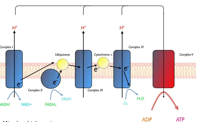
Within each mitochondrion, the energy currency of the cell, ATP, is generated through oxidative phosphorylation, a process whereby redox energy contained within nutrients is extracted through a series of oxido-‐reduction reactions and subsequently used to drive the synthesis of ATP. Thanks to the pioneering work of Hans Krebs in the 1930s, we now know that the carbon-‐based substrates, lipids and carbohydrates, are sent to the mitochondria for oxidation. There, the dehydrogenase enzymes of the Krebs cycle and β-‐oxidation will oxidize these substrates into reducing equivalents (NADH and FADH2) that will be used to feed electrons into the
electron transport chain (ETC) (Krebs, 1937, Biochem J). The electrons provided by NADH will enter the ETC at complex I (NADH dehydrogenase) (See Figure 1). Within complex I, the flavin mononucleotide is the first recipient of the electron, which is then transferred onto ubiquinone (Q), via Fe-‐S clusters, to form the reduced ubiquinol (QH2). The free energy released by the passing of the electron to a lower
energy level is used in moving protons against their natural gradient from the mitochondrial matrix towards the intermembrane space. The Q-‐cycle loop is closed when ubiquinol (QH2) is recycled back into the oxidized ubiquinone (Q) by giving off
its electron to complex III. Inside the complex, also known as cytochrome b-‐c1 reductase, another Fe-‐S cluster awaits the arrival of the electron so that it can be passed onto cytochrome c. Once again, the free energy released from electron transfers in the complex is harnessed to pump protons into the intermembrane space. Cytochrome c, much like ubiquinone does between complex I and III, serves as an electron shuttle between complex III and IV. When cytochrome c reaches complex IV (cytochrome c oxidase), it is oxidized by a copper-‐containing
subcomplex. Next, the electron travels through different hemoproteins until it reaches the ultimate electron acceptor, oxygen. This measurable consumption of oxygen is termed mitochondrial respiration (Chance, 1972, FEBS Lett). Accordingly, complex IV is the third and final site where protons are pumped into the intermembrane space. The electrochemical gradient resulting from the accumulation of H+ in the intermembrane space creates a membrane potential (Δψ)
across the inner mitochondrial membrane. This energy is used to produce a motive force within complex V (ATP synthase) to rephosphorylate ADP into ATP, creating a situation where the higher membrane potential, the higher the phosphorylation capacity. Peter Mitchell first described this phenomenon in his chemosmotic theory, work for which he received the Nobel Prize in the late 1970s (Mitchell, 1961, Nature). Electrons snatched from FADH2, a substrate heavily produced by the lipid
oxidizing β-‐oxidation, follow a nearly identical path to NADH in the ETC. One discerning aspect of FADH2 however, which can also be seen in Figure 1 is its
entrance via complex II (succinate dehydrogenase). The bypass of complex I renders FADH2 a less efficient substrate, as it cannot contribute to complex I proton
pumping although complex III and IV pumping remains intact. Therefore, it can be estimated that for any given level of O2 reduction, FADH2 can only contribute about
two-‐thirds of the normal proton pumping capacity of NADH. Therefore, it is said that the P/O ratio, where the Phosphorylating capacity (P), or the number of ATP molecules that can be formed for each molecule of oxygen that is consumed (O) of NADH and FADH2 is 3 and 2 respectively. The well-‐respected Peter Hinkle suggests
however slight deviations from these generally acceped values (Hinkle, 2005, BBA bioenergetics).
Figure 1: The Electron Transport Chain
Figure 1: The Electron Transport Chain
The chain of oxido-‐reduction exchanges taking place in the electron transport chain allows the creation of an electrochemical gradient across the inner mitochondrial membrane, which is then used to create a motive force at ComplexV, linking ATP production and mitochondrial respiration.
2.1.2.2 Mitochondrial volume
Mitochondria are the most important contributor to aerobic energy production. Accordingly, tissue oxidative capacity is largely determined by mitochondrial volume density (Larsen, 2012, J Physiol). With the inner membrane relatively saturated with ETC, the most effective way to increase oxidative capacity is by increasing mitochondrial volume via biogenesis. The gold standard to assess mitochondrial volume density is measurement of the mitochondrial fractional area using electron microscopy images. Nowadays, 3D reconstruction of the mitochondrial network using fluorescent dyes also offers an interesting perspective to appreciate the cellular distribution of the organelle (Picard, 2013, J Appl Physiol). Popular indirect indexes of mitochondrial density that have shown a good correlation to mitochondrial fractional area, include tissue content of the inner membrane phospholipid, cardiolipin (Larsen, 2012, J Physiol), the enzymatic activity of mitochondrial enzymes involved in the krebs cycle (i.e. citrate synthase), or in the electron transport chain (i.e. cytochrome oxidase). These indexes consistently indicate that mitochondrial volume has an important degree of plasticity. In the face of heightened contractile activity, muscle mitochondrial volume increases (Tonkonogi, 2002, Exerc Sport Sci Rev); whereas chronic inactivity causes a loss in the organellar biomass (Adhihetty, 2007, J Appl physiol). Indeed, in the human vastus lateralis, it is not unusual to observe a 50% increase in mitochondrial density after a few weeks of exercise training (Hoppeler, Pflugers Arch, 1973). Conversely, Adhihetty and colleagues witnessed a 50% decrease in mitochondrial density after 6 weeks of denervation in rat skeletal muscle (Adhihetty, 2007, J Appl Physiol). We observed an even more abrupt reduction in the diaphragm, where a decrease in mitochondrial volume of similar magnitude occurred in a matter of hours after human patients were put on mechanical ventilation (Picard, 2011, Am J Respir Crit Care Med). Although the scale of these changes in response to varying metabolic demands may lead one to believe that environmental factors are the main determinant to mitochondrial volume in muscles, the reality is that mitochondrial tissue content is mostly range-‐bound by pre-‐determined genetic and epigenetic
factors. Indeed, it appears that early during development, muscles become differentiated according to their metabolic profile and this largely determines mitochondrial content (Tiivel, 2000, Mol Cell Biochem). Correspondingly, as seen in Figure 2, in highly oxidative tissues, such as the heart, where oxidative energy demand is great, mitochondria may represent up to 35% of total cell volume (Laguens, J Cell Biol, 1971). On the other hand, in highly glycolytic tissues, such as the gastrocnemius, where aerobic energy production is nearly dispensable, the total volume mitochondria occupy in the cell may be as low as 1%. The range of mitochondrial content in muscles of varying metabolic profile is outlined in Figure 2.
Table 1: Mitochondrial fractional density in striated muscle
Muscle Range of mitochondrial
fractional density (%) Reference
Gastrocnemius 1-‐3 Macdougall, 1979, Med
Sci Sports
Vastus Lateralis 2-‐8 Hoppeler, 1973,
Pflugers Arch
Soleus 8-‐12 Hoppeler, 1987, J
Physiol
Diaphragm 18 Weibel, 1984, Harv Uni
Press
Heart 30-‐38 Laguens, 1971, J Cell
Biol
Table 1: Mitochondrial fractional density in striated muscle
Range of mitochondrial fractional density, determined by electron microscopy across different muscles reported in various studies.
2.1.2.3 Mitochondrial biogenesis & PGC-‐1 α
Peroxisome proliferator activated receptor gamma coactivator-‐1-‐alpha (PGC-‐1α), long considered the master regulator of mitochondrial biogenesis, is overexpressed in periods of mitochondrial volume expansion but also decreases in periods of attrition (Adhihetty, 2007, J Appl Physiol), confirming its role in dictating oxidative capacity. PGC-‐1α directly coactivates a variety of transcription factors including PPARs, thyroid hormone receptors, glucocorticoid receptors, estrogen and estrogen related receptors (ERRs), myocyte enhancing factor-‐2 (MEF-‐2) as well as the forkhead O-‐box (FOXO) (Canto, 2009, Curr Opin Lipidol). Thus, the activation of PGC-‐1α results into a transcriptional program that characterizes the entire metabolic profile of the cell. To this effect, ectopic overexpression of PGC-‐1α in the muscle of mice has been shown to effectively transform fast twitch glycolytic muscles into slow twitch oxidative muscles (Lin, 2002, Nature); making PGC-‐1α the coordinator of metabolic plasticity in muscle (Hood, 2006, J Exp Biol).
PGC-‐1α coordinates mitochondrial biogenesis by acting as a coactivator in the transcription of nuclear-‐encoded proteins that will be incorporated into the mitochondria. The vast majority of these gene products are under the transcriptional control of nuclear respiratory factors (NRFs) and represent the bulk of the proteins necessary to synthesize new mitochondria, but other important transcription factors regulated by PGC-‐1α, such as YY1, are also involved (Scarpulla, 2012, BBA). Before mitochondria can become fully functional entities, these proteins need to be imported through multisubunit membrane complexes and assembled into complex polypeptides with the help of various molecular chaperones (Scarpulla, 2011, Biochim Biophys Acta). However, out of the 37 proteins encoded in the mtDNA, 13 are integral parts of the ETC complexes and are absolutely required for normal biochemical functions of the ETC (Wallace, 2005, Ann Rev Genet). Four of the five ETC complexes require bi-‐genomic expression, leaving succinate dehydrogenase as the sole complex that is completely derived from nuclear DNA. Additionally, PGC-‐1α also serves to increase mitochondrial protein synthesis.
Indeed, amongst the nuclear proteins under the transcriptional control of PGC-‐1α, are factors such as TFAM, TFBs as well as an RNA polymerase, all used in the transcription of the mitochondrial genome (Scarpulla, 2012, Trends endocrin metab). Consequently, it is thought that the transcription of these two distinct compartments is coordinated by PGC-‐1α. Surprisingly, Leick and colleagues revealed that PGC-‐1α was dispensable to mitochondrial biogenesis although its absence somewhat hinders the process (Leick, 2008, Am J Physiol Endocrinol Metab). One potential explanation is that PGC-‐1β, which has been shown to share a high degree of overlap in transcriptional targets with PGC-‐1α, may alleviate these blockades in the absence of PGC-‐1α (Lin, 2002, J Biol Chem). Although they are both necessary to attain normal physiological function (Lai, 2008, Genes Dev), single knockout of either activators will only cause mild metabolic disorders in baseline animals but the deficits become aberrant in the face of mounting stress. Meanwhile a complete deficit of both PGC-‐1α/β is lethal The maintenance of this redundance through evolution may be be explained by PGC-‐1β having upstream regulators distinct from PGC-‐1α. Nonetheless, this may be confounding in the interpretation of the true individual functions of both coactivators (Scarpulla, 2012, Trends endocrin metab). As indicated by the differences in mitochondrial volume density in different muscles, the need for aerobically derived ATP varies a great deal across tissues. Interestingly, PGC-‐1α and β content seems to vary across different muscles in a pattern similar to distributions of mitochondrial content, confirming their role in maintaining higher levels of oxidative capacity (Lin, 2002, Nature).
The transcriptional activity of PGC-‐1α is intensively regulated by post-‐ transcriptional modifications, including phosphorylation, methylation and acetylation (Rodgers, 2008, FEBS lett). Given, the role of PGC-‐1α in determining cellular oxidative functions, it is not surprising to note that energy sensors such as AMP-‐activated protein kinase (AMPK) and Sirtuins play an important role in this mode of regulation. AMPK and Sirtuins are respectively activated by high AMP/ATP and NAD/NADH ratios. These energy and stress sensors are important contributors to homeostasis and are known to intervene in a plethora of signaling pathways with
impacts on physiological processes as varied as ROS production, cell proliferation, ATP production, autophagy and cell death (Canto, 2011 Physiol Bethesda). It has also been shown that AMPK and Sirtuins play a key role in the improved mitochondrial metabolism observed in models of caloric restriction (Figure 3) (Chen, 2008, Genes Dev) and in response to aerobic training (Baar, 2002, FASEB). Concordingly, the repression of these pathways in models of substrate oversupply has also been linked to poor health (Banks, 2008, Cell Metab). The link between energy supply and cellular health in addition to the varied functions of Sirtuins and AMPK have made them potential therapeutic targets in conditions as varied as diabetes, aging, cancer, and neuromuscular disorders (Canto, 2011 Physiol Bethesda). As shown in figure 3, AMPK activation results in higher PGC-‐1α activity via phosphorylation whereas SIRT1 will do so via deacetylation (Rodgers, 2005, Nature / Jager, 2007, Proc Natl Acad Sci).
Figure 2: Metabolic demands regulate mitochondrial volume
Figure 2: Metabolic demands regulate mitochondrial volume
Periods of high AMP/ATP ratios (fasting or high demand) activate AMPK. In periods of high energy demands (exercise), the increased NAD/NADH ratio leads to Sirt1 activation. Both AMPK and Sirt1 increase the transcriptional activity of PGC1, which is tied to mitochondrial biogenesis.