Varicella-Zoster Virus Gene 63 Encodes an Immediate-Early Protein
That Is Abundantly Expressed during Latency
Debrus S[1], Sadzot-Delvaux C[1], Nikkels A F[2], Piette J[1], Rentier B[1]
[1]
Laboratory of Fundamental Virology,Institute of Pathology, University of Liège, B-4000 Liège, Belgium
[2]Laboratory of Dermatopathology Institute of Pathology, University of Liège, B-4000 Liège, Belgium
Abstract : Varicella-zoster virus (VZV) gene 63 encodes a protein with a predicted molecular mass of 30.5 kDa which has amino acid similarities with the immediate-early (IE) protein 22 (ICP-22) of herpes simplex virus type 1. In order to study the expression of this protein during lytic and latent infection, gene 63 was cloned in frame and downstream from the glutathione-S-transferase gene, expressed as a fusion protein, and purified. In VZV-infected Vero cells, antibodies directed against this protein detect two polypeptides of 45 and 38 kDa which are localized both in the cytoplasm and in the nucleus. Using a sequential combination of transcription and protein synthesis inhibitors (actinomycin D and cycloheximide, respectively), we demonstrated the immediate-early nature of this protein, which can thus be named IE63. Using a rat model of VZV latency, we showed that IE63 is heavily expressed, essentially in neurons, during latency. IE63 can also be detected in the skin of patients showing early herpes zoster symptoms.
Varicella-zoster virus (VZV) is an alphaherpesvirus and the causative agent of two human diseases: chicken pox, upon primary infection, and shingles, after reactivation of the latent virus from the dorsal root ganglia. The biology of VZV and the mechanisms involved in latency and reactivation are not well documented, mainly because of the poor growth of the virus in vitro, where it remains highly cell associated. Consequently, a part of our knowledge of VZV is derived from the determination of its nucleotide sequence (6) and from structural and functional comparisons with herpes simplex virus type 1 (HSV-1), another intensively studied alphaherpesvirus (5, 7). Tran-sient-cell-transfection assays with plasmids containing VZV open reading frames (ORFs) were particularly useful for the assignment of potential functions to some of the VZV proteins (2, 8, 9, 17, 24, 28).
The genome of VZV is composed of 71 ORFs encoding 68 proteins that seem to be regulated in a manner similar to those of proteins of other alphaherpesviruses (11, 12). Particularly, the HSV-1 infectious cycle, which is generally used as a reference, is characterized by regulation of gene expression in a cascade fashion (15). The viral genes are subdivided into three classes on the basis of their expression kinetics. Immediate-early (IE or α) genes are transcribed just after virus penetration in the cell, without any de novo viral protein synthesis (1, 3), and encode proteins playing an important role in viral gene regulation (8, 10, 28, 35). Early (E or β) genes encode proteins necessary for DNA synthesis (31), whereas late (L or γ) genes are expressed at the end of the infectious cycle, after DNA replication, and encode structural proteins.
On the basis of the DNA sequence, the ORF-4-, -61-, -62-, and -63-encoded proteins are considered homologous to HSV-1 ICP-27, -0, -4, and -22, respectively (6, 23, 27). However, in spite of their overall sequence homologies, functional similarities between VZV and HSV-1 proteins seem to be limited (22), and until now, only ORF-62 has been shown to encode an immediate-early protein (13). The protein encoded by ORF-63 has homology with ICP-22 of HSV-1, whose functions are not very well characterized (29, 30, 34). The regulatory functions of the ORF-63 protein in VZV have begun to be clarified, showing evidence for down regulative capacities of IE62 expression (14,18). Important are the presence, during latency, of different transcripts, which could include ORF-63 transcripts in human sensory ganglia (4, 16), and the detection of abundant ORF-63 transcripts in ex-planted and cultured human ganglia (39) as well as adult rat dorsal root ganglia studied as a VZV latency model (21, 32). These observations plead for an important role of this polypeptide, probably both in the control of the infectious cycle and in the maintenance of latency. We have thus focused on this important protein encoded by ORF-63 in order to determine its kinetic class and to visualize it in latently infected cells of dorsal root ganglia. Moreover, this protein is shown to be expressed early in human tissue during VZV reactivation.
Construction of recombinant pGex3X-ORF-63, expression of a GST-ORF-63-encoded protein, and preparation of a rabbit antiserum.
ORF-63 lies in the left repeat flanking the short unique segment of the VZV genome from nucleotide 110581 to 111415 (6); an identical copy (ORF-70) is in the opposite orientation in the terminal short repeat. We have chosen to express the ORF-63-encoded protein as a fusion polypeptide with glutathione-S-transferase (GST) in Escherichia coli (37). The coding sequence of VZV gene 63 was obtained by cutting the VZV EcoRI A fragment (21) first with restriction enzymes SpeI and SspI to obtain a 4,027-bp fragment and then with restriction enzyme StyI to generate an 834-bp fragment containing the ATG codon, to which a partially double-stranded oligonucleotide was added by ligation to restore the 3' end. This fragment was introduced into the HindII site of pUC19 to obtain plasmid pUC19-IE63 (P. Jacobs, University of Brussels, Nivelles, Belgium). The coding sequence of gene 63 was then cut out by digesting plasmid pUC19-IE63 with PstI and HindIII, filling in the 3' and 5' termini with bacteriophage T4 DNA polymerase, and introducing the gene 63 coding sequence into the
SmaI site of pGex-3X (Pharmacia-LKB, Uppsala, Sweden) to obtain plasmid pGex-3X/ORF-63. Both
the junction of the GST gene and ORF-63 and the beginning of ORF-63 were sequenced. Bacteria transformed with plasmid pGex-3X/ ORF-63 were induced by 1 mM IPTG (isopropyl-β-D-thioga-lactopyranoside) for 3 h at 37°C. The bacteria were then sonicated in phosphate-buffered saline (PBS)– Triton X-100 (1%, wt/vol) after centrifugation at 15,000 X g (4°C), and the GST-63 fusion protein was released at a high concentration into the supernatant. This fusion protein was clearly visible as a 66-kDa band electrophoresed on sodium dodecyl sulfate– 10% (wt/vol) polyacrylamide gel (SDS-10% PAGE) stained with Coomassie blue compared with the crude extract of cells expressing the 27-kDa GST protein alone (Fig. 1A). This supernatant was used for affinity chromatography on glutathione-Sepharose-4B (Pharmacia-LKB, Uppsala, Sweden). The fusion protein GST-ORF-63 was eluted with reduced glutathione (10 mM) (38), and an aliquot was loaded onto SDS-10% PAGE gels to ensure the purity of the fusion protein (Fig. 1B). The GST-63 polypeptide was used, without any further
purification, for subsequent antibody production.
Antiserum against the GST-63 fusion protein was prepared by immunizing two rabbits with the purified fusion protein. The first immunization was done subcutaneously with 0.2 mg of fusion protein in 0.5 ml that had been emulsified with an equal volume of Freund’s complete adjuvant. The rabbits were given booster immunizations on days 15, 30, 45, 60, and 75 with 0.2 mg of fusion protein in Freund’s incomplete adjuvant. The antiserum was tested by enzyme-linked immunosorbent assay against GST-63 and GST alone (data not shown) before being collected on day 90.
The antiserum was tested first by Western blotting (immu-noblotting) of extracts of VZV-infected and noninfected Vero cells. The antiserum was able to detect two polypeptides of 45 and 38 kDa in infected-cell extracts. No protein was evidenced in the noninfected-cell extracts (Fig. 1C). These two molecular masses are far above the 30.5 kDa predicted by DNA sequence determination (6) for the ORF-63-encoded protein, but this as well as the detection of two different polypeptides could be explained by the presence of numerous potential phosphorylation sites. No antigens were detected in our extracts by the anti-GST antibodies.
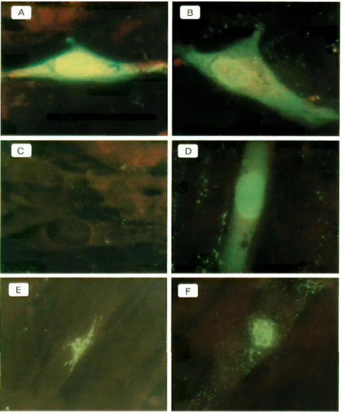
The antiserum (anti-63) was then tested by immunostaining of infected and noninfected Vero cells fixed for 10 min in acetone. After fixation, the nonspecific sites were saturated with PBS-milk (15 g/liter) before being treated with the anti-63 antibodies and revealed by fluorescein-isothiocyanate-conju-gated swine immunoglobulins to rabbit immunoglobulins. The cells were then observed by fluorescence microscopy. The antiserum was able to detect the ORF-63-encoded protein both in the cytoplasms and in the nuclei of the VZV-infected cells (Fig. 2A, B, and D).
The ORF-63-encoded protein is expressed as an immediate-early protein during the VZV infectious cycle.
To analyze the expression and localization of the ORF-63-encoded protein during the viral infectious cycle, cell-free virus was produced by shaking infected cells with 0.5-mm-diameter glass beads. Infected Vero cells (at 80% cytopathic effect) were scraped into SPGA medium (218 mM sucrose, 3.8 mM KH2PO4, 7.2 mM K2HPO4, 4.9 mM sodium glutamate, 1% bovine albumin, and 10% fetal calf serum) (33). Infected cells and glass beads were shaken for 10 s in a Mini Bead-Beater (Biospec Products, Bartlesville, Okla.) and then centrifuged twice at 3,000 rpm at 20°C in an Eppendorf 5415C centrifuge (Hamburg, Germany). Supernatants were used to infect MRC5 cells at confluency with a multiplicity of infection of 5 X 10⁻⁴. Unadsorbed viruses were washed after 1 h, and infected MRC5
cells were fixed for 10 min in acetone at various times postinfection (p.i.) (3, 6, 9, 12, 16, 20, and 24 h).
FIG. 1. (A) SDS-PAGE separation of proteins of 3X-transformed bacteria and of pGEX-3X/GST-63-transformed bacteria. Lanes a and c, pellet; lanes b and d, supernatant. Ten micrograms of each sample was loaded in duplicate. Molecular mass markers are shown in the rightmost lane. (B) SDS-PAGE of the affinity-purified GST-63 fusion protein. (C) Western blotting of extracts of infected and noninfected Vero cells, revealed with the anti-63 serum (diluted 1/500).
The cells were studied by immunostaining both with the anti-63 serum and with a monoclonal antibody developed in our laboratory (VL8) and directed against glycoprotein I (gE), a late VZV protein (25). Uninfected cells were subjected to the same treatment and tested with the two antibodies as negative controls.
Immunofluorescence visualization of the protein encoded by ORF-63 revealed staining in the
cytoplasms and nuclei of infected cells 6 h p.i. (Fig. 2A). This strong staining was also detected with a major localization in the nuclei of the infected cells fixed from 9 h p.i. (Fig. 2B) to 24 h p.i. (data not shown). gE was detected only in the perinuclear endoplasmic reticula of infected cells at 20 h p.i. (Fig. 2E) and was localized in the endoplasmic reticula, and with a punctated form in all the cytoplasms at 24 h p.i. (Fig. 2F).
FIG. 2. Immunofluorescence of MRC5 cells infected with VZV and treated (C and D) or not treated (A, B, E, and F) with the inhibitors (Fig. 3) and revealed with the anti-63 serum (A to D) and the VL8 monoclonal antibody (E and F). Cells were fixed at 6 h (A and C), 9 h (B and D), 20 h (E), and 24 h p.i. (F).
Detection of the ORF-63-encoded protein as early as 6 h p.i. indicates its involvement at the very beginning of the viral cycle and suggests the importance of its regulatory properties (14, 18). The presence of abundant ORF-63-encoded protein at 6 h p.i., like that of IE62 (13), which is a well-defined early protein, and the results of two earlier studies which identified an immediate-early protein of 43 kDa (20, 36), which is consistent with the molecular mass of the ORF-63-encoded protein, suggest strongly that this protein belongs to the immediate-early class.
infected with cell-free VZV was treated with metabolic inhibitors (13), first with cycloheximide (CHX), which blocks protein synthesis, and second with actinomycin D (ActD), a transcription inhibitor.
FIG. 3. (A and B) Immunohistochemical detection of IE63 in lumbar dorsal root ganglia (A) and root (B) sections of a VZV-infected rat. (Inset) Five-hundred-fold magnification of lumbar dorsal root ganglia IE63-positive neurons. Most of the neurons were labelled, and the protein was cytoplasmic, nuclear, or both. Some axons were also positive. Magnifications, X250 (A) and X500 (B). (C) Immunohistochemical detection of IE63 on a cutaneous biopsy of an erythematous lesion of a patient showing early signs of herpes zoster. Nuclear, perinuclear, and cytoplasmic staining was detected in isolated keratinocytes and in a small spongiotic area (magnification, X500).
After adsorption of the virus for 1 h in the presence of CHX (100 μg/ml), the cells were washed three times and incubated with CHX (100 μg/ml) for 5 h. After three washes, ActD-containing medium (10 μg/ml) was added to the cells for 18 h to block transcription. From 3 to 24 h p.i., the cells were fixed
for 10 min in acetone and studied by immunostaining with both the an-ti-63 serum and the anti-gE monoclonal antibody. The infected cells treated with CHX and ActD did not present any staining at 6 h p.i. (Fig. 2C); however, at 9 h p.i. (3 h after removal of CHX), intense staining appeared in the
cytoplasms and nuclei, similar to the staining detected in the untreated cells (Fig. 2D). This staining remained constant in cells fixed at 12, 16, 20, and 24 h p.i. (data not shown). When the cells were tested with anti-gE, no signal was detected in infected cells treated with the inhibitors (data not shown). The use of a sequential combination of inhibitors allows the demonstration of the immediate-early nature of a protein (transcription without de novo viral protein synthesis) (1, 3, 15). The first metabolic inhibitor, CHX, blocked protein synthesis for a period of 6 h, and only the transcripts of the immediate-early proteins could be synthesized without any viral translation. After removal of CHX, ActD was added to block transcription, allowing the translation of preexisting transcripts. The lack of synthesis of gE, which is considered a late protein, under these conditions demonstrated the efficiency of the inhibitor procedure. In the infected cells treated with the inhibitors, the ORF-63-encoded protein appeared 3 h after removal of CHX (9 h p.i.) and could still be detected at 24 h p.i. Since the synthesis of the protein was not blocked by this combination of inhibitors, we can conclude that the ORF-63-encoded protein is an immediate-early protein and can rightfully be named IE63.
After having characterized the expression of the IE63 protein and demonstrated its immediate-early nature, we have attempted to study the cellular localization of the IE63 protein in dorsal root ganglia of latently infected rats and during virus reactivation in humans during shingles episodes.
IE63 localization in latently infected rat ganglia.
Abundant transcripts corresponding to IE63 have been detected in dorsal root ganglia of latently infected adult rats (21, 32). We have thus searched for the expression of the protein in sections of latently infected adult rat dorsal root ganglia. Four million VZV-infected Vero cells (80% cytopathic effect) were inoculated into each adult rat’s footpad. The lumbar ganglia (L4, L5, and L6)
corresponding to the inoculation area were dissected 1 month after inoculation and then fixed in formalin and embedded in paraffin. Five-micrometer sections were deparaf-finized and treated for immunohistochemical staining. The an-ti-63 (diluted 1/250) and anti-gE (diluted 1/20) antibodies were incubated for 30 min at 20°C. Slides were washed and incubated, first with biotinylated antibodies and finally with alkaline phosphatase-conjugated streptavidin (Dako, Glostrup, Denmark), according to the manufacturer’s instructions. The revelation was performed in the presence of new fuchsin as the chromogen, and the slides were counterstained with Mayer’s hemalun. The IE63 protein was detected essentially in neurons, indicated by the red precipitate, and the staining was either cytoplasmic, nuclear, or both (Fig. 3A). Fifty to 80% of the neurons, depending on the section, were positive. Importantly, some nonneuronal cells and several axons located at the beginning of the sciatic nerve were also labelled (Fig. 3B). It should be mentioned that gE was never detected in these sections; thoracic ganglia of infected rats and ganglia from noninfected animals were negative for the two antibodies (data not shown). IE63 is thus strongly expressed during the latent phase of the infection and probably plays an important role in the establishment or maintenance of latency.
IE63 expression in human skin during VZV reactivation.
Because IE63 is detected during latency and at the very beginning of a productive viral cycle in vitro, we have looked for its expression during reactivation in the skin of 49 patients suffering from herpes zoster symptoms. Skin specimens were obtained from these patients after local anesthesia was administered. To prove the presence of VZV in these lesions, in situ hybridization was performed, allowing the detection of VZV nucleic acids (26). The samples were formalin fixed, paraffin
embedded, and treated as described for the dorsal root ganglia. Positive IE63 immunostaining occurred in the nuclei and cytoplasms of epidermal and infundibular keratinocytes (Fig. 3C). In 13 early nonvesicular VZV lesions, the IE63 protein was already expressed in keratinocytes, while gE was not yet detectable. In later stages of cutaneous zoster, i.e., in vesicles, the anti-IE63 and anti-gE antibodies yielded comparable results (data not shown). No staining was found in normal skin or herpes simplex lesions (data not shown).
Our results show that the ORF-63-encoded protein is expressed at the onset of the viral cycle and can thus be classified as an immediate-early protein. We have also demonstrated the presence of the IE63 protein in latently infected cells of rat dorsal root ganglia and the early expression of the IE63 protein in human skin during reactivation. These results demonstrate the importance of the IE63 protein during
the viral productive cycle as well as during latency.
On the basis of the promoter structure and function, it has been suggested that the IE62 protein is the only immediate-early protein (14), with other genes like ORF-4, -29, and -63 being transcribed later (8,18). However, just as α-TIF does for HSV-1, the VZV IE62 protein, brought into the cell in large quantities by the virus (19), transactivates a group of genes, including ORF-63 and ORF-63 (9), both expressed without viral de novo synthesis.
A study of the time-dependent expression of the IE63 protein in latently infected cells and its
regulatory effects on various VZV gene promoters would help to elucidate the role of this protein in the viral latent and productive cycles.
While performing this work, S.D. was a grantee of the I.R.S.I.A., Belgium, and C.S.-D. and J.P. were, respectively, Senior Research Assistant and Research Director of the National Fund for Scientific Research (NFSR), Belgium. This work was supported by grants from the NFSR, the Belgian National Lottery, and the VZV Research Foundation, New York, N.Y.
References
[1]. Batterson, W., and B. Roizman. 1983. Characterization of the herpes simplex virion-associated factor responsible for the induction of α genes. J. Virol. 46:371-377.
[2]. Cabirac, G. F., R. Mahalingam, M. Wellish, and D. H. Gilden. 1990. Trans-activation of viral TK promoters by proteins encoded by varicella zoster virus open reading frames 61 and 62. Virus Res. 15:57-68.
[3]. Campbell, M. E. M., J. W. Palfreyman, and C. M. Preston. 1984. Identification of herpes simplex virus DNA sequences which encode a trans-acting polypeptide responsible for stimulation of
immediate-early transcription. J. Mol. Biol. 180:1-19.
[4]. Croen, K. D., J. M. Ostrove, J. Dragovic, and S. E. Straus. 1988. Patterns of gene expression and sites of latency in human nerve ganglia are different for varicella-zoster and herpes simplex viruses. Proc. Natl. Acad. Sci. USA 85:9773-9777.
[5]. Davison, A. J., and D. J. McGeoch. 1986. Evolutionary comparisons of the S segments in the genomes of herpes simplex virus type 1 and varicella-zoster virus. J. Gen. Virol. 67:1759-1816. [6]. Davison, A. J., and J. E. Scott. 1986. The complete DNA sequence of varicella-zoster virus. J. Gen. Virol. 67:1759-1816.
[7]. Davison, A. J., and N. M. Wilkie. 1983. Location and orientation of homologous sequences in the genome of five herpesviruses. J. Gen. Virol. 64:1927-1942.
[8]. Defechereux, P., L. Melen, L. Baudoux, M.-P. Merville-Louis, B. Rentier, and J. Piette. 1993. Characterization of the regulatory functions of varicella-zoster virus open reading frame 4 gene product. J. Virol. 67:4379–4385.
[9]. Disney, G. H., T. A. McKee, C. M. Preston, and R. D. Everett. 1990. The product of varicella-zoster virus gene 62 autoregulates its own promoter. J. Gen. Virol. 71:2999-3003.
[10]. Everett, R D. 1987. The regulation of transcription of viral and cellular genes by herpesvirus immediate-early gene products. Anticancer Res. 7:589-604.
[11]. Felser, J. M., P. R Kinchington, G. Inchauspe, S. E. Straus, and J. M. Ostrove. 1988. Cell lines containing varicella-zoster virus open reading frame 62 and expressing the ‘‘IE’’175 protein complement ICP4 mutants of herpes simplex virus type 1. J. Virol. 62:2076-2082.
[12]. Felser, J. M., S. E. Straus, and J. M. Ostrove. 1987. Varicella-zoster virus complements herpes simplex virus type 1 temperature-sensitive mutants. J. Virol. 61:225-228.
[13]. Forghani, B., R Mahalingam, A. Vafai, J. W. Hurst, and K. W. Dupuis. 1990.
Monoclonal antibody to the immediate-early protein encoded by varicella-zoster virus gene 62. Virus Res. 16:195–210.
[14]. Hay, J., and W. T. Ruyechan. 1994. Varicella-zoster virus—a different kind of herpesvirus latency? Semin. Virol. 5:241–247.
[15]. Honess, R. W., and B. Roizman. 1974. Regulation of herpesvirus macromo-lecular synthesis. I. Cascade regulation of the synthesis of three groups of viral proteins. J. Virol. 14:8–19.
[16]. Hyman, R. W., J. R. Ecker, and R. B. Tenser. 1983. Varicella-zoster virus in human trigeminal ganglia. Lancet ii:814–816.
[17]. Inchauspe, G., and J. M. Ostrove. 1989. Differential regulation by varicella-zoster virus (VZV) and herpes simplex virus-1 trans-activating genes. Virology 173:710–714.
[18]. Jackers, P., P. Defechereux, L. Baudoux, C. Lambert, M. Massaer, M.-P. Merville-Louis, B. Rentier, and J. Piette. 1992. Characterization of regulatory functions of the varicella-zoster virus gene 63-encoded protein. J. Virol. 66:3899–3903.
[19]. Kinchington, P. R., J. K. Hougland, A. M. Arvin, W. T. Ruyechan, and J. Hay. 1992. The varicella-zoster virus immediate-early protein IE62 is a major component of virus particles. J. Virol. 66:359–366.
[20]. Lopetegui, P., H. Campo-Vera, and K. Yamanishi. 1985. Varicella-zoster virus (VZV)-specific polypeptides detected in cells treated with metabolic inhibitors. Microbiol. Immunol. 29:569–575. [21]. Merville-Louis, M.-P., C. Sadzot-Delvaux, P. Delrée, J. Piette, G. Moonen, and B. Rentier. 1989. Varicella-zoster virus infection of adult rat sensory neurons in vitro. J. Virol. 63:3155–3160.
[22]. Moriuchi, H., M. Moriuchi, H. A. Smith, and J. I. Cohen. 1994. Varicella-zoster virus open reading frame 4 protein is functionally distinct from and does not complement its herpes simplex virus type 1 homolog, ICP27. J. Virol. 68:1987–1992.
[23]. Moriuchi, H., M. Moriuchi, H. A. Smith, S. E. Straus, and J. I. Cohen. 1992. Varicella-zoster virus open reading frame 61 protein is functionally homologous to herpes simplex virus type 1 ICP0. J. Virol. 66:7303–7308.
[24]. Nagpal, S., and J. M. Ostrove. 1991. Characterization of a potent varicella-zoster virus-encoded
trans-repressor. J. Virol. 65:5289–5296.
[25]. Nikkels, A. F., S. Debrus, C. Sadzot-Delvaux, J. Piette, P. Delvenne, B. Rentier, and G. E. Piérard. 1993. Comparative immunohistochemical study of herpes simplex and varicella-zoster infections. Virchows Arch. A 422:121– 126.
[26]. Nikkels, A. F., P. Delvenne, S. Debrus, C. Sadzot-Delvaux, J. Piette, B. Rentier, and G. E. Piérard. Distribution of varicella-zoster virus gpI and gpII and corresponding genome sequence in the skin. J. Med. Virol., in press.
[27]. Ostrove, J. M. 1990. Molecular biology of varicella-zoster virus. Adv. Virus Res. 38:45-99. [28]. Perera, L. P., J. D. Mosca, W. T. Ruyechan, and J. Hay. 1992. Regulation of varicella-zoster virus gene expression in human T lymphocytes. J. Virol. 66:5298-5304.
[29]. Post, L. E., and B. Roizman. 1981. A generalized technique for deletion of specific genes in large genomes: α gene 22 of herpes simplex virus 1 is not essential for growth. Cell 25:227-232.
[30]. Purves, F. C, W. O. Ogle, and B. Roizman. 1993. Processing of the herpes simplex virus
regulatory protein α22 mediated by the UL13 protein kinase determines the accumulation of a subset of α and γ mRNAs and proteins in infected cells. Proc. Natl. Acad. Sci. USA 90:67016705.
[31]. Ruyechan, W. T., and A. C. Weir. 1984. Interaction with nucleic acids and stimulation of the viral DNA polymerase by the herpes simplex virus type 1 major DNA-binding protein. J. Virol. 52:727-733.
[32]. Sadzot-Delvaux, C, M. P. Merville-Louis, P. Delree, P. Marc, J. Piette, G. Moonen, and B. Rentier. 1990. An in vivo model of varicella-zoster virus latent infection of dorsal root ganglia. J. Neurosci. Res. 26:83-89.
[33]. Saito, M., C. Haruyama, H. Ohba, S. Makino, and M. Matumoto. 1981. Recovery of cell-free varicella-zoster virus from Vero cells. Arch. Virol. 68:59-63.
[34]. Sears, A. E., I. W. Halliburton, B. Meignier, S. Silver, and B. Roizman. 1985. Herpes simplex virus 1 mutant deleted in the α22 gene: growth and gene expression in permissive and restrictive cells and establishment of latency in mice. J. Virol. 55:338-346.
[35]. Sekulovitch, R. E., K. Leary, and R. M. Sandri-Goldin. 1988. The herpes simplex virus type 1 α protein ICP27 can act as a trans-repressor or a trans-activator in combination with ICP4 and ICP0. J. Virol. 62:4510-4522.
[36]. Shiraki, K., and R. W. Hyman. 1987. The immediate-early proteins of vari-cella-zoster virus. Virology 156:423-426.
[37]. Smith, D. B., K. M. Davern, P. G. Board, W. U. Tiu, E. G. Garcia, and G. F. Mitchell. 1986. Mr 26,000 antigen of Schistosoma japonicum recognized by resistant WEHI129/J mice is a parasite glutathione S-transferase. Proc. Natl. Acad. Sci. USA 83:8703-8707.
[38]. Smith, D. B., and K. S. Johnson. 1988. Single-step purification of polypeptides expressed in Escherichia coli as fusions with glutathione S-transferase. Gene 67:31-40.
[39]. Vafai, A., R. S. Murray, M. Wellish, M. Devlin, and D. H. Gilden. 1988. Expression of varicella-zoster virus in trigeminal ganglia. Proc. Natl. Acad. Sci. USA 85:2362-2366.