UNIVERSITE DE SHERBROOKE Faculte de genie
Departement de genie chimique et de genie biotechnologique
DEVELOPPEMENT ET VALIDATION DE SURFACES
ET D ’ECHAFAUDAGES PROPICES AU
DEVELOPPEMENT ET AU MAINTIEN DES
FONCTIONS DE CELLULES PANCREATIQUES
BETA
TOWARDS THE DEVELOPMENT AND
VALIDATION OF BIOMATERIAL SURFACES AND
SCAFFOLDS SUITABLE FOR PANCREATIC BETA
CELL DEVELOPMENT AND FUNCTION
These de doctoratSpecialite: genie chimique
Evan Aiozie DUBIEL
Jury : Patrick VERMETTE (directeur) Jonathan LAKEY
Steven PARASKEVAS Marie-Josee BOUCHER Marcel LACROIX (rapporteur)
1+1
Library and Archives Canada Published Heritage Branch Bibliotheque et Archives Canada Direction du Patrimoine de I'edition 395 Wellington Street Ottawa ON K 1A0N 4 Canada 395, rue Wellington Ottawa ON K1A 0N4 CanadaYour file Votre reference ISBN: 978-0-494-93268-1 Our file Notre reference ISBN: 978-0-494-93268-1
NOTICE:
The author has granted a non
exclusive license allowing Library and Archives Canada to reproduce, publish, archive, preserve, conserve, communicate to the public by
telecomm unication or on the Internet, loan, distrbute and sell theses
worldwide, for commercial or non commercial purposes, in microform, paper, electronic and/or any other formats.
AVIS:
L'auteur a accorde une licence non exclusive permettant a la Bibliotheque et Archives Canada de reproduire, publier, archiver, sauvegarder, conserver, transmettre au public par telecomm unication ou par I'lnternet, preter, distribuer et vendre des theses partout dans le monde, a des fins com merciales ou autres, sur support microforme, papier, electronique et/ou autres formats.
The author retains copyright ownership and moral rights in this thesis. Neither the thesis nor substantial extracts from it may be printed or otherwise reproduced without the author's permission.
L'auteur conserve la propriete du droit d'auteur et des droits moraux qui protege cette these. Ni la these ni des extraits substantiels de celle-ci ne doivent etre imprimes ou autrement
reproduits sans son autorisation.
In compliance with the Canadian Privacy A ct some supporting forms may have been removed from this thesis.
W hile these forms may be included in the document page count, their removal does not represent any loss of content from the thesis.
Conform em ent a la loi canadienne sur la protection de la vie privee, quelques
form ulaires secondaires ont ete enleves de cette these.
Bien que ces form ulaires aient inclus dans la pagination, il n'y aura aucun contenu manquant.
RESUME
Le diabete mellitus de type I est une maladie de plus en plus abondante. Cette demiere est caracterisee par la destruction auto-immunitaire des ilots de Langerhans incluant les cellules de type p qui produisent de l’insuline dans le pancreas endocrinien. Une option de traitement pour les patients atteints de cette maladie est notamment une greffe des ilots de Langerhans. Ce traitement est limits du au nombre restreint de donneurs d’organes et aussi a la perte de fonctionnalite des ilots suite a la greffe. Les etudes effectuees tout au long de cette these ont pour optique d’adresser ces contraintes par le biais de la science des biomateriaux. La these debute avec un survol details des concepts de base et des complexites associes aux interactions de type cellules et surfaces trouvees dans la litterature. II s’agit specifiquement des interactions physiques et chimiques, des systemes experimentaux ainsi que des caracterisations et modifications associes aux interactions entre cellules et surfaces. La premiere etude de nature experimentale examine la morphogenese des cellules progenitrices ductales (PANC-1 cell line) vers des ilots qui produisent des agregats semblables a des ilots (ILA). Le tout est fait sur des surfaces de carboxymethyl dextrane (CMD) sur lesquelles le RGD est greffe via un lien covalent. L’expression des marqueurs d ’lLAs (cytokeratin-19, Ki67, et E-cadherin) qui peuvent etre associes k un changement de phenotype de ces cellules a ete evaluee ainsi que la secretion et 1’expression de l’insuline. La seconde etude de nature experimentale a pour optique 1* immobilisation de la fibronectine (FN) sur les memes surfaces de CMD mentionnees auparavant sur lesquelles des cellules ayant un phenotype fi (INS-1 cell line) ont prolifere. Lors du processus d’immobilisation, plusieurs solutions ont ete etudiees. L’immobilisation de la fibronectine sur des surfaces de CMD a ete validee par la spectrometrie de photoelectrons induits par rayons X. Le mecanisme d’immobilisation a ete determine par imagerie et mesures de force par microscopie a force atomique, la spectroscopie de dichroisme circulaire ainsi que par la diffusion dynamique de la lumiere. De plus, la croissance des cellules de type INS-1 et la secretion d’insuline ont ete evaluees. La demiere etude de nature experimentale visait l ’etude de la coculture des cellules endotheliales et des ilots de pore dans un gel de fibrine. L ’effet de la presence des cellules endotheliales sur la production d’insuline des ilots a ete evalue. De plus, l’apoptose cellulaire en coculture a ete evaluee et comparee aux cultures simples.
Mots cl6s: biomateriaux; forces de surface et intermoleculaires; modifications de surface;
surfaces anti-adherentes; adsorption de proteines; diabete; ilots de Langerhans; secretion d ’insuline
ABSTRACT
Type I diabetes mellitus is a disease o f increasing abundance, characterized by the auto-immune destruction o f insulin producing p-cells comprising islets o f Langerhans in the endocrine pancreas. A treatment option for patients with severe type I diabetes mellitus is the surgical transplantation of islets of Langerghans. This treatment is limited because o f pancreas donor scarcity and the progressive loss of islet graft function after both islet isolation from the donor pancreas and transplantation. The present studies within this thesis aim to address these issues by means of biomaterials science. It begins with a detailed broad review of general concepts and complexities associated with cell - surface interaction studies. More specifically physical & chemical interactions, experimental systems, and surface modification & characterization associated with cell - surface interactions are discussed. The first experimental work studies the morphogenesis of ductal-like progenitor cells (i.e., PANC-1 cell line) to insulin expressing and producing islet-like aggregates (ILA) on well-defined low-fouling carboxymethyl-dextran (CMD) surfaces upon which the ligand arginine-glycine-asparic acid is covalently grafted. ILA expression patterns of markers (i.e., cytokeratin-19, Ki67, and E-cadherin) typically associated with a change PANC-1 phenotype were assessed along with insulin expression and secretion. The second experimental study involves the immobilization of the protein fibronectin (FN) onto low-fouling CMD surfaces upon where cells of a P-cell-like phenotype (i.e., INS-1 cell line) were cultured. Various solutions were used during the immobilization process. The successful immobilization of FN on CMD was assessed via X-ray photoelectron spectroscopy and the
m echanisms of immobilization were assessed via atomic force microscopy imaging & force measurements, circular dichroism spectroscopy, and photon correlation spectroscopy. INS-1 cell growth and insulin secretion were also assessed. The final experimental work studied the effect o f the co-culture of endothelial cells with isolated porcine islets in fibrin gel. The effect endothelial cells had on the capacity of islets to secrete insulin was accessed. In addition, cellular apoptosis was accessed with co-cultures and compared to cultures without endothelial cells.
Keywords: Biomaterials; Surface and Intermolecular Forces; Surface Modification; Low-
ACKNOWLEGEMENTS
First and foremost, I would like to thank my mentor and research director Patrick Vermette for the opportunity to perform the boundless scientific research that enlivens me. The past years have been a pleasure and I look forward to our future work together.
I would also like to thank Dr. Rennian Wang and her research group at the University o f Western Ontario for providing me with their laboratory and knowledge that gave me the training necessary to perform much o f my lab work.
I am also grateful to Dr. Steven Paraskevas and Craig Hasilo at McGill University for providing me with a generous quantity of isolated islets of Langerhans that was used to access the feasibility of my final experimental study.
A very special thank you goes to Dr. Jonathan Lakey, Morgan Lamb, and their entire laboratory for their continuous and unrestrictive supply of islets throughout my final experimental study. Such work would not have been performed without such generosity.
I would also like to thank Mathew Evans for his assistance in the French language.
Finally, I would like to thank all my colleagues over the years at the Universite de Sherbrooke for providing a pleasant work environment and members o f the jury (i.e., Dr. Jonathan Lakey, Dr. Steven Paraskevas, Dr. Marie-Josee Boucher, and Dr. Marcel Lacroix) for providing their insights and for their longevity in reading a particularly lengthy thesis.
TABLE OF CONTENTS
RESUME... i
ABSTRACT... ii
ACKNOWLEGEMENTS...iii
LIST OF FIGURES... viii
LIST OF TABLES... xii
LIST OF SYMBOLS...xiii
LIST OF ACKRONYMS...xv
Chapter 1 INTRODUCTION... 1
Chapter 2 Bridging the Gap Between Physicochemistry and Interpretation Prevalent in Cell - Surface Interactions... 5
Foreword... 6
2.1 Abstract... 8
2.2 Introduction... 9
2.3 Cellular Adhesion to Surfaces... 13
2.3.1 Cell Surface... 13
2.3.2 Cellular Adhesion...15
2.3.3 Protein Mediation between Cells and Surfaces... 17
2.4 Interactions in Cellular Adhesion... 21
2.4.1 Chemical Interactions... 22
2.4.2 Physical Interactions... 23
2.4.3 Interaction Measurements... 33
2.5 Systems for Studying Cell-Material Interactions... 34
2.5.1 In Vivo Systems... 34
2.5.2 Cells in a Three-Dimensional Environment...35
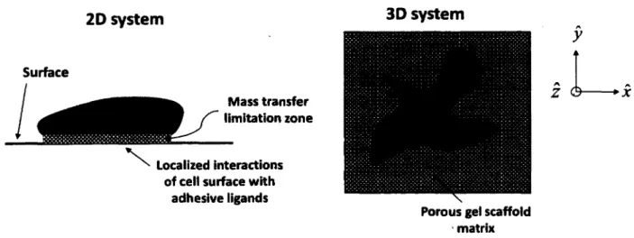
2.5.3 Differences between Two-Dimensional and Three-Dimensional Cell Culture Systems 37 2.5.4 Properties of Cell Culture Systems... 40
2.5.4.1 Mechanical Properties... 40
2.5.4.2 Biochemical Properties... 45
2.5.5 Materials for Three-Dimensional Cell Culture Systems...49
2.5.5.1 Natural Materials...49
2.5.5.2 Synthetic Materials... 51
2.5.6 Micropattemed Cell Cultures... 52
2.5.7 Current State and Future Direction of Three-Dimensional Cell Culture Systems.. 54
2.6 Modification, Properties, and Analysis of Surfaces... 55
2.6.1 Modified Surfaces in Biomaterials Science...55
2.6.2 Surface Fabrication... 56
2.6.3 Self-Assembled Monolayers (SAMs)...59
2.6.4 Plasma Polymerization...62
2.6.5 Properties and Analysis of Modified Surfaces in Cell Science...65
2.6.6 Contact Angle in Correlating Cell Responses toward Biomaterial Surfaces 67 2.6.6.1 Hydrophobic Interactions... 69
2.6.6.2 Contact Angle Measurements... 71
2.6.6.3 Contact Angle Hysteresis... 72
2.6.6.4 Meaning and Relevance o f Contact Angle Measurements... 73
2.6.7 Low-Fouling Surfaces... 76
2.7 Conclusions... 83
2.8 Acknowledgment...88
Chapter 3 In Vitro Morphogenesis of PANC-1 Cells into Islet-Like Aggregates Using RGD- Covered Dextran Derivative Surfaces... 89
Foreword...90
3.1 Abstract... 92
3.2 Introduction... 93
3.3 Materials and Methods... 95
3.3.1 Surface Preparation... 95
3.3.2 X-ray Photoelectron Spectroscopy... 97
3.3.3 Cell Culture...97
3.3.4 Immunofluorescence... 98
3.3.5 Morphometric Analysis... 99
3.3.7 Insulin Secretion...100
3.3.8 Proliferation Assay... 100
3.3.9 Glycosaminoglycan Staining...100
3.3.10 Data and Statistical A nalysis...101
3.4 Results...101
3.4.1 X-ray Photoelectron Spectroscopy... 101
3.4.2 RGD-CMD Surfaces Promote Islet-Like Aggregate (ILAs) Formation...102
3.4.3 RGD-CMD Surfaces Result in Insulin-Secreting ILAs...105
3.4.4 RGD-CMD Surfaces Promote Cell Adhesion and Increase Cell Proliferation.... 107
3.4.5 Integrins as and a vP3 are Expressed in RGD-CMD IL A s... 108
3.5 Discussion... 108
3.6 Conclusion... 112
3.7 Acknowledgment... 112
Chapter 4 Solution Composition Impacts Fibronectin Immobilization on Carboxymethyl-Dextran Surfaces and INS-1 Insulin Secretion... 113
Foreword...114
4.1 Abstract...116
4.2 Introduction...117
4.3 Materials and Methods...119
4.3.1 Surface Preparation... 119
4.3.2 X-ray Photoelectron Spectroscopy...120
4.3.3 Atomic Force Microscopy...120
4.3.4 Circular Dichroism Spectroscopy... 121
4.3.5 Photon Correlation Spectroscopy... 122
4.3.6 Cell Culture... 122
4.3.7 Cell Number...122
4.3.8 Glucose-Stimulated Insulin Secretion... 123
4.3.9 Statistical Analysis... 123
4.4 Results and Discussion... 124
4.4.1 Fibronectin Grafting onto CMD Surfaces Occurs Only with Solutions o f Low Salt Concentration...124
4.4.2 Solution Composition Does Not Significantly Alter Fibronectin Conformation/Size 125
4.4.3 Topography of Fibronectin Surfaces Made in CaCl2 Solution Differed Subtly From
Those Made in MgCl2 and MnCl2... 127
4.4.4 Solution Composition During Fibronectin Immobilization Does Not Affect INS-1 Cell Growth But Does Affect Insulin Secretion... 130
4.5 Conclusion... 134
4.6 Acknowledgment... 135
Chapter 5 Co-Culture o f Young Porcine Islets with Liver Microvascular Endothelial Cells in Fibrin Improves Insulin Secretion and Reduces Apoptosis...136
Foreword... 137
5.1 Abstract...139
5.2 Introduction... 140
5.3 Materials and Methods...141
5.3.1 Islet Isolation and Maturation of Porcine Islets...141
5.3.2 Cell Culture, Islet Culture, Fibrin Gels, and Time Lapse Microscopy...142
5.3.3 Glucose-Stimulated Insulin Secretion... 143
5.3.4 Immunohistological Staining and Morphometric Analysis... 144
5.4 Results and Discussion...144
5.4.1 Time Lapse Microscopy... 144
5.4.2 Insulin Content in Media and Response to Glucose Stimulation... 145
5.4.3 Porcine Islet Insulin Expression and Cellular Apoptosis...149
5.5 Conclusion...153 5.6 Acknowledgements... 153 Chapter 6 CONCLUSIONS...154 LIST OF REFERENCES...161 APPENDIX A ... 207 APPENDIX B ... 211 APPENDIX C ... 214
LIST OF FIGURES
Figure 2.1: (A) Traditional cell-surface experimental setup. (B) Cross section of generic
eukaryotic cell surface...10
Figure 2.2: Structure and dynamics o f cellular adhesion to surfaces...17
Figure 2.3: Primary structure of fibronectin...19
Figure 2.4: Double-layer like interaction between a modified substrate bearing a charge and a cell surface with charge bearing receptors...27
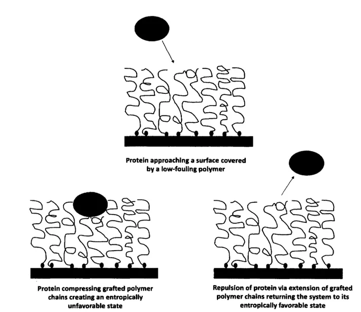
Figure 2.5: Steric repulsion of proteins from a polymer-grafted surface... 31
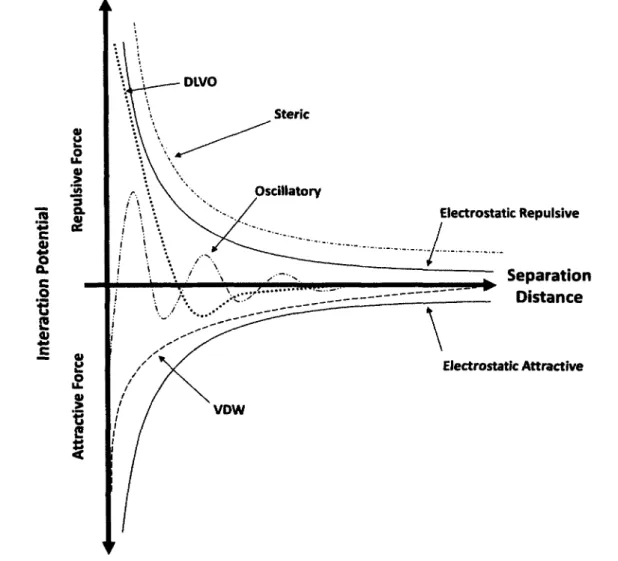
Figure 2.6: Interaction potentials o f surface and intermolecular forces in biological systems 32 Figure 2.7: Key conceptual differences between 2D and 3D culture systems... 36
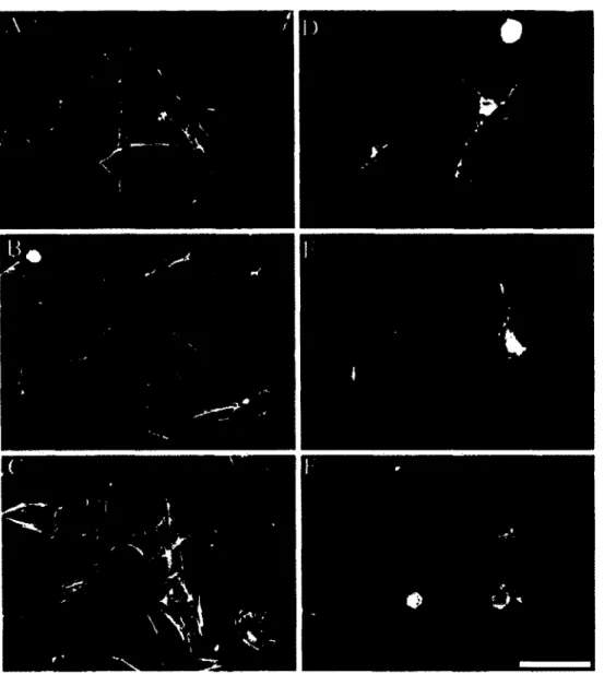
Figure 2.8: Human fibroblasts project a dendritic network o f extensions in collagen matrices but not on collagen-coated cover slips... 38
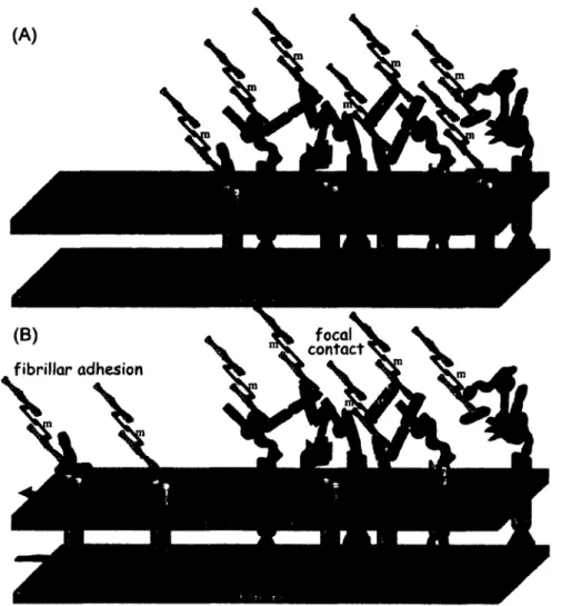
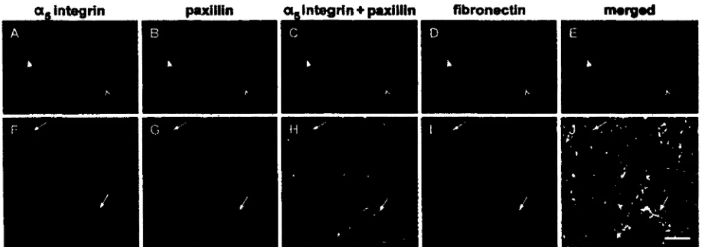
Figure 2.9: In vivo 3D matrix adhesions differ from focal or fibrillar adhesions on 2D substrates. ... 46
Figure 2.10: Self-assembled monolayer exposed to proteins and cells... 59
Figure 2.11: Surface immobilized polymer conformations... 77
Figure 2.12 Dependence o f CMD conformation on experimental conditions and its effect on cellular resistance... 80
Figure 2.13: Primary and secondary adsorption of proteins on low-fouling surfaces... 82
Figure 3.1: Biomaterial surface preparation scheme...96
Figure 3.2: XPS survey and high resolution C Is spectra... 102
Figure 3.3: Immunofluorescence staining for Ki67 (A-C), CK-19 (D-F), and E-cadherin (E-Cad) (G-I) of PANC-1 cells on selected culture conditions over tim e...104
Figure 3.4: Expression level of Ki67 (A), CK-19 (B), and E-cadherin (E-Cad) (C) o f PANC-1 cells cultured on the different surfaces over tim e...105
Figure 3.5: (A) ILA insulin secretion after 14 days. (B) ILA insulin secretion per million cells after 14 days. (C) ILA insulin and glucagon expression after 14 days for CMD... 106
Figure 3.6: (A) Phase contrast microscopy of PANC-1 aggregation to ILAs and expansion in SFM. (B) Total cellular content measured by the CyQUANT® assay during SFM culture...107 Figure 3.7: Representative ILA as integrin expression after 14 days for TCPS (A). av03 integrin expression after 14 days for RGD-CMD (B )...108
Figure 4.1: (A) Elemental surface composition via XPS survey. (B) High resolution C Is XPS spectra of FN-CMD surfaces and intermediate layers with EDC/NHS activation . (C) High resolution C Is XPS spectra of FN-CMD surfaces and intermediate layers without EDC/NHS activation... 125 Figure 4.2: (A) Circular dichroism spectra o f FN in various aqueous solutions. (B) Mean
hydrodynamic diameter, Dh of lOOpg/mL FN in various aqueous solutions measured via PCS. 126 Figure 4.3: (A-C) 750nm x 750nm AFM images in liquid (i.e., lx PBS) using Tapping™ mode o f FN-CMD grafted in the presence o f lOpM CaCL, IOjiM MgCL, and lOpM MnCL,
respectively. (D) RMS roughness of FN-CMD surfaces, CMD surfaces in various solutions, and HApp-modified and clean glass substrates...128 Figure 4.4: (A) INS-1 cell numbers on surfaces measured via CyQUANT®. (B) INS-1 cell insulin secretion following glucose stimulation after 7 days o f culture. (C) Stimulation index for INS-1 cells from 2.8mM Glucose to 28mM Glucose... 131
Figure 5.1 : Time lapse microscopy of: (A) PLMEC+PI(in Fibrin) - porcine islets co-cultured with liver microvascular endothelial cells embedded in fibrin. (B) PI(in Fibrin) - porcine islets embedded in fibrin. (C) PI-PLMEC monolayer - porcine islets on top of a liver microvascular endothelial cell monolayer...145 Figure 5.2: Insulin content of basal media measured via ELISA ... 146 Figure 5.3: Insulin content and stimulation index o f glucose-stimulated insulin secretion assay for (A) 3D PLMEC+PI(in Fibrin) and PI(in Fibrin) cultures and (B) 2D PI-PLMEC monolayer and PI-TCPS cultures... 148
Figure 5.4: Sample immunofluorescence images of insulin and glucagon expression and immunohistochemistry images o f apoptosis (i.e., TUNEL) o f porcine islets for all culture
condition at 2 days and 7 days... 149 Figure 5.5: (A) Expression levels o f insulin and glucagon and 0 day, 2 days, and 7 days. (B) Percent loss of insulin expressing P-cells from 2 days to 7 days. (C) Expression of apoptotic cells measured via TUNEL... 151
Figure A. 1: Phase contrast microscopy of adherent (RGD-CMD) surfaces and non-adherent (RGE-CMD, CMD, and TCPS) surfaces... 207 Figure A.2: Stained sections for GAG using Alcian Blue of (A) PANC-1 ILAs (14 days) and (B) mouse intestine as a positive control...208 Figure A.3: Immunofluorescence staining o f as integrin o f cells grown on RGD-CMD (A-C), RGE-CMD (D-F), CMD (G-I), and TCPS (J-L)...209 Figure A.4: Immunofluorescence staining o f avp3 integrin o f cells grown on RGD-CMD (A-C),
RGE-CMD (D-F), CMD (G-I), and TCPS (J-L)...210
Figure B.l: High resolution C Is XPS spectra outlining differences between successful (lOpM CaC^) and unsuccessful (lx PBS and 150mM NaCl) FN grafting to CMD surfaces (A) and to CMD surfaces activated (act.) and non-activated (no act.) with (B) CaCh, (C) M gCL, and (D). MnCl2... :... 211 Figure B.2: CMD AFM force - separation distance profiles in various solutions... 212 Figure B.3: 750nm x 750nm AFM images in liquid using Tapping™ mode of (A-B) intermediate surfaces and (C-F) CMD layers in various solutions...213
Figure C.l: Time lapse microscopy of porcine liver microvascular endothelial cells (PLMEC) and porcine islets embedded in fibrin gel over 6 days [i.e., PLMEC+PI(in Fibrin)] and PLMEC embedded in fibrin gel after 24h (i.e., 24h-PLMEC) and 6 days (i.e., 6 Days-PLMEC)...214 Figure C.2: Time lapse microscopy of porcine islets embedded in fibrin gel [i.e., PI(in Fibrin)] over 6 days...215
Figure C.3: Time lapse microscopy of porcine islets seeded over a monolayer o f porcine liver microvascular endothelial cells (i.e., PI-PLMEC monolayer)...216 Figure C.4: Time lapse microscopy of porcine islets seeded over tissue culture polystyrene (i.e., PI-TCPS). Single cells o f fibroblastoid morphology appear early and proliferate throughout the culture period... 217 Figure C.5: Immunofluorescence image o f insulin and glucagon expression at 0 days (left). Immunohistochemistry image o f apoptosis detected via TUNEL at 0 days (right)... 218 Figure C.6: Level of cells expressing glucagon in all culture conditions... 218
LIST OF TABLES
Table 2.1: Cell-surface studies... 12
Table 2.2: Cellular attachment to fibronectin domains...20
Table 2.3 Surface modification substrates and chemicals...57
Table 2.4: Properties of cell culture surfaces...58
Table 2.5: Static water contact angle and cell/protein responses...68
Table 2.6: Advancing, receding, and dynamic water contact angle and cell/protein responses... 69
Table 3.1: The total number and mean diameter of islet-like aggregates (ILAs) obtained from PANC-1 cells cultured on the different surfaces after 7 days on average... 103
LIST OF SYMBOLS
Chapter 2
a Length of monomer comprising polymer chain
Ce s Physical constant (incorporating surface geometry, charge, and solution
conditions) of Double Layer interaction
Cv d w Geometric constant (analogous to Hamaker constant) o f van der Waals interaction
8 Diameter of solvent molecules
D Distance between adsorption points o f polymer on a surface e Dielectric permittivity of interacting medium
e 0 Permittivity o f free space
0 Water contact angle
0a d v Advancing water contact angle
0d y n Dynamic water contact angle
0r e c Receding water contact angle
0stat Static water contact angle
Eo Contact energy
k Debye length
Lo Thickness of polymer adlayer in free form q Magnitude o f electric charge
r Separation distance
Rg Radius o f gyration
a Surface density of adsorbed polymer w(r) Interaction potential
Chapter 3
a Equivalent diameter o f prolate spheroid b Polar diameter of prolate spheroid c Equatorial diameter o f prolate spheroid
Chapter 4
c Molar concentration Dh Hydrodynamic diameter 1 Path length 0 Ellipicity [0] Molar ellipicity xivLIST OF ACKRONYMS
2D Two Dimensional
3D Three Dimensional
Act. Activated with EDC( 1 ethyl3 (3
-dimethylaminoproplyl)carbodiimide) and NHS (N- hydroxysuccinimide)
ADV Advancing
AFM Atomic Force Microscopy
ANOVA Analysis of Variance
Au( 111) Miller Oriented (lmn) Gold Surface
BM Basement Membrane
BSA Bovine Serum Albumin
CAD Computer-Aided Design
CCBD Central Cell Binding Domain
CD Circular Dichroism
CMD Carboxymethyl-Dextran
CMRL-1066 Connaught Medical Research Laboratories medium
CK-19 Cytokeratin-19
DAPI 4\6-diamidino-2-phenylindole, dihydrochloride
DL Double-Layer
DMEM Dulbecco’s Modified Eagle Medium
DYN Dynamic
E-Cad E-Cadherin
ECM Extracellular Matrix
EDC l-ethyl-3-(3-dimethylaminoproplyl)carbodiimide
ELISA Enzyme-Linked Immunosorbent Assay
EMET Epithelial to Mesenchymal to Epithelial Transition
EMT Epithelial to Mesenchymal Transition
ERK Extracellular Signal-Regulated Kinase
FAK Focal Adhesion Kinase
FBS Foetal Bovine Serum
FCCS Fluorescent Cross-Correlation Spectroscopy
FCS Fluorescent Correlation Spectroscopy
FEP Fluorinated Ethylene Propylene
FGF Fibroblast Growth Factor
Fn Fibronectin (Chapter 2)
FN Fibronectin (Chapter 4)
FN-CMD Fibronectin covalently grafted to a Carboxymethyl Dextran surface
FRAP Fluorescence-Recovery After Photobleaching
GAG Glycosaminoglycan
GBD Gelatin Binding Domain
GRGDS Glycine- Arginine-Glycine-Asparic Acid-Serine
GRGES Glycine-Arginine-Glycine-Glutamic Acid- Serine
GSIS Glucose-Stimulated Insulin Secretion
HApp w-heptylamine Plasma Polymerization
HBD Heparin Binding Domain
H-bond Hydrogen Bond
HBSS Hank’s Balanced Salt Solution
HGF Hepatocyte Growth Factor
'H-NMR Hydrogen-1 (Proton) Nuclear Magnetic Resonance
HUVEC Human Umbilical Vein Endothelial Cell
IC Immunochemistry
ICCS Image Cross-Correlation Spectroscopy
ICS Image Correlation Spectroscopy
ILA Islet-Like Aggregate
INS-1 Insulinoma Cell Line
IBMX 3-isobutyl-1 methylxanthine
IEQ Islet Equivalent
LPA Lysophosphatidic Acid
LR Ligand-Receptor
MALDI Matrix-Assisted Laser Desorption/Ionization
NHS N-hydroxysuccinimide
No Act. Not Activated with EDC( 1
-ethyl-3-(3-dimethylaminoproplyl)carbodiimide) and NHS (N- hydroxysuccinimide)
OWLS Optical Waveguide Lightmode Spectroscopy
PANC-1 Pancreatic Carcinoma Cell Line
PBS Phosphate Buffered Saline
PC Phosphatidylcholine
PCR Polymerase Chain Reaction
PCS Photon Correlation Spectroscopy
PDGF Platelet Derived Growth Factor
PEG Poly(Ethylene Glycol)
PEO Poly(Ethylene Oxide)
PFC Perfluorocarbon
PHSRN Proline-Histidine-Serine-Arginine-Asparagine
PI Porcine Islets
PI(in Fibrin) Porcine Islets embedded in Fibrin gel
PI-PLMEC(in Fibrin) Porcine Islets and Porcine Liver Microvascular Endothelial Cells embedded in Fibrin gel
PI-PLMEC monolayer Porcine islets on a Porcine Liver Microvascular Endothelial Cell monolayer
PI-TCPS Porcine islets on Tissue Culture Polystryrene
PLMEC Porcine Liver Microvascular Endothelial Cells
PTFE Polytetrafluoroethylene
QCM Quartz Crystal Microbalance
REC Receeding
RF Radio Frequency
RGD Arginine-Glycine-Asparic Acid
RGD-CMD Arginine-Glycine-Asparic Acid covalently grafted to a Carboxymethyl Dextran surface
RGE Arginine-Glycine-Glutamic Acid
RGE-CMD Arginine-Glycine-Glutamic Acid covalently grafted to a Carboxymethyl Dextran surface
RMS Root Mean Square
RPMI Roswell Park Memorial Institute medium
SAM Self-Assembled Monolayer
SCM Serum Containing Media
S.E.M. Standard Error of the Mean
SFA Surface Force Apparatus
SFM Serum-Free Media
SIMS Secondary Ion Mass Spectrometry
SMC Smooth Muscle Cell
SPR Surface Plasmon Resonance
Sulfo-SMCC Sulfosuccinimidyl4(VMaleimidomethyl)cyclohexane1 -carboxylate
TCPS Tissue Culture Polystyrene
TUNEL Terminal deoxynucleotidyl transferase dUTP Nick End Labelling
UV Ultra-Violet
VDW van der Waals
VEGF Vascular Endothelial Growth Factor
Vn Vitronectin
XPS X-ray Photoelectron Spectroscopy
YIGSR Tyrosine-Isoleucine-Glycine-Serine-Arginine
Chapter 1
Diabetes mellitus largely exists in two forms; type I diabetes characterized by the lack o f insulin production and type II characterized by ineffective insulin use. According to the World Health Organization, approximately 346 million people worldwide have diabetes, 90% being Type II. Symptoms of diabetes include excessive thirst, frequent urination, hunger, and fatigue. Complications o f diabetes include heart disease, stroke, blindness, neuropathy, and kidney failure to name a few. It is uncommon but of increasing occurrence, that cases o f type I and type II diabetes are diagnosed in the same patient [1]. The Canadian Diabetes Association predicts diabetes to cost Canada $16.9 billion in the year 2020 alone.
Type I diabetes results in the auto-immune destruction of insulin producing P-cells that comprise islets of Langerhans in the endocrine pancreas. Treatment of type I diabetes often involves insulin therapy which involves rigorous monitoring of glucose levels in the blood and subsequent exogenous injections of insulin. Avoidance o f the numerous complications associated with type I diabetes requires particular patient compliance to monitor blood glucose levels and inject insulin when necessary. However, despite insulin therapy, for some patients, blood-glucose levels remain uncontrollable. Such extreme cases can require islet transplantation as an option of therapy.
Islet transplantation has shown particular promise [2]. Islet transplantation involves the isolation o f islets from a donor pancreas, typically followed by infusion into the hepatic portal vein of the patient [3]. The primary limitation o f islet transplantation is a severe shortage o f donor islets for transplantation. Another limitation is that islet isolation results in the loss of islet function and eventual cell death [4]. This may contribute to the progressive loss of islet graft function (i.e., insulin secretion) in transplanted patients. The present thesis aims to help address these limitations through two means: through a broad review on the fundamental intermolecular and surface forces involved in cell - surface interactions, and application of such fundamentals to biomaterial surface and scaffold design upon which the response of cells of pancreatic endocrine relevance are assessed.
Three types of cells/tissue of endocrine pancreatic relevance were utilized. First, Pancreatic Carcinoma (PANC-1), a cell line believed to be analogue to pancreatic ductal progenitor cells that arguably differentiate into islets [5]. PANC-1 cell response to biomaterial surfaces was thus accessed with the aim of producing surrogate insulin secreting tissue; an attempt to address the
shortage o f donor pancreases. Second, Insulinoma (INS-1) were utilized, a cell line that expresses and secretes insulin in a glucose responsive manner, analogue to those o f P-cells comprising islets [6]. INS-1 cell response towards biomaterials surfaces was assessed with the aim o f improving insulin secretion to glucose stimulation. This was done with the hope of addressing the reduced functionality o f p-cells comprising islet grafts through the identification o f an additional factor (i.e., solution composition during protein adsorption) typically not considered when designing biomaterial surfaces to improve isolated islet functionality. Specifically the hope was that this would help further improve insulin secretion to glucose stimulation. Finally, islets isolated from porcine pancreases were utilized. Isolated islets were co-cultured with endothelial cells in a biomaterial scaffold with the aim to improve insulin secretion and reduce cell death by apoptosis. The use o f isolated islets attempts to address again, the progressive loss of islet insulin secreting capacity after isolation from the pancreas.
Chapter 2 of this thesis titled, “Bridging the Gap Between Physicochemistry and Interpretation
Prevalent in Cell - Surface Interactions”, consists of a broad review on the fundamental
intermolecular and surface forces that are involved in cell - surface interactions. Such forces are the origin in a series o f actions and reactions that ultimately govern cell behavior (i.e., adhesion, spreading, migration, proliferation, differentiation, and function). Therefore, the review is not only relevant to the application of biomaterials science as a therapeutic strategy to diabetes mellitus, as is done in this thesis, but also to the application of biomaterials science to many other therapeutic strategies. Within the review, the cell surface and cellular adhesion o f cell surface receptors to ligands are discussed. Fundamental interactions such as Coulomb, van der Waals, double-layer, hydrophobic, H-bonding, and steric repulsion along with their relevance to biology are discussed. Also, systems [two-dimensional (2D) and three-dimensional (3D)] for studying cell - material interactions are presented. Finally, surface modification, properties, chemistry, and characterization is presented and discussed. Throughout the entire review, the advantages, disadvantages, and complexities of all topics are discussed.
In Chapter 3 titled, “/« vitro Morphogenesis o f PANC-1 Cells into Islet-Like Aggregates Using
RGD-Covered Dextran Derivative Surfaces”, PANC-1 cells were exposed to biomaterial
surfaces with well-defined properties and their possible transformation into surrogate insulin expressing and producing tissue was assessed. Briefly, low-fouling carboxymethyl dextran
(CMD) surfaces bearing covalently grafted arginine-glycine-asparic acid (RGD) [i.e., RGD- CMD] appear to be the most promising as they not only express insulin (as the controls did as well) but had the most insulin in the culture media.
For Chapter 4 titled “Solution Composition Impacts Fibronectin Immobilization on
Carboxymethyl-Dextran Surfaces and INS-1 Insulin Secretion”, fibronectin was covalently
grafted onto low-fouling CMD surfaces using various solutions. It was found that not only does solution composition play a role in subsequent INS-1 insulin secretion on fibronectin-bearing CMD surfaces, but it also impacts whether or not fibronectin can in actual fact be grafted onto low-fouling CMD surfaces.
Chapter 5 titled, “Co-Culture o f Young Porcine Islets with Liver Microvascular Endothelial
Cells in Fibrin Improves Insulin Secretion and Reduces Apoptosis”, involves studying the effect
endothelial cells have on isolated islet insulin secretion and programmed cell death (i.e., apoptosis). Briefly, it was found that endothelial cells have beneficial effects on insulin secretion whether or not one employs a 2D or 3D cell culture system. Nevertheless, there was a more pronounced improvement in insulin secretion capacity and further reduction in apoptosis when using 3D fibrin cultures in conjunction with endothelial cells.
Finally, Chapter 6, titled “Conclusion” presents general insights into each experiment. In addition, future direction is suggested.
Chapter 2
Bridging the Gap Between Physicochemistry and
Interpretation Prevalent in Cell - Surface Interactions
Foreword
Authors and Affiliation :
Evan A. Dubiel : Ph.D. Candidate, Universite de Sherbrooke, Departement de genie chimique et de genie biotechnologique
Yves Martin : Lecturer, Universite de Sherbrooke, Departement de genie chimique et de genie biotechnologique
Patrick Vermette : Professor, Universite de Sherbrooke, Departement de genie chimique et de genie biotechnologique
Date of Acceptance : October 22, 2010
State of Acceptance : Final Version Published J o u rn a l: Chemical Reviews
Reference :
[Dubiel, E.A. and Vermette, P. (2009) Complexities in Understanding Cellular Interactions to Modified Surfaces, 27th Annual Conference o f the Canadian Biomaterials Society, Laval University, Quebec City, Quebec, Canada]
[Dubiel, E.A., Martin, Y., and Vermette, P. (2011) Bridging the Gap Between Physicochemsitry and Interpretation Prevalent in Cell - Surface Interactions, Chemical Reviews, volume 111, p. 2900-2936]
Contribution :
This article contributes to the literature review component of this thesis. The abstract was presented at the 27 th Annual Conference o f the Canadian Biomaterials Society. The content of the article was published in Chemical Reviews (note that Chemical Reviews does not publish abstracts). Section 2.5 was written by Yves Martin (except Section 2.5.2, 2.5.4.1-2.5.4.2, and 2.5.6 where Evan A. Dubiel also contributed). Section 2.6.6 was written by Patrick Vermette. All other sections were written by Evan A. Dubiel. All work was done under the direction and supervision o f Patrick Vermette.
T itre en fran$ais :
Le rapprochement entre la physico-chimie et 1’interpretation des mecanismes gouvemant les interactions entre les cellules et les surfaces
R6sum 6:
Les interactions entre les cellules et les surfaces modifiees sont d’une grande importance dans le domaine de la medecine. La comprehension de ces demieres implique plusieurs aspects s’etendant dans les domaines de la biologie, de la chimie ainsi que la physique. Le nombre de projets de recherche effectuee dans ce domaine a augmente depuis les dix demieres annees. Malgre ce fait, la comprehension des phenomenes d ’interaction entre cellules et surfaces reste tres mauvaise. Ce manque est du notamment a la variation des parametres lors des experiences. Les interactions sont typiquement deduites suite a une periode d’exposition predeterminee des cellules a une surface modifiee. II existe typiquement trois couches d’interactions dans un systeme cellule - surface : (i) un substrat, (ii) une couche intermediaire, comprenant notamment des proteines ainsi que certaines macromolecules et (iii) la cellule elle-meme. II existe done trois interfaces colloi'dales d’interaction. Lors de l’analyse de la reponse des cellules, les forces surfaciques et intermoleculaires incluant notamment la force Coulomb, les liens van der Waals, la force de double - couche et la force sterique a l ’interface et a l ’interieur de l ’interface sont souvent ignores. Par contre, ces forces se trouvent a l’origine d ’une serie d’actions et de reactions gouvemant le comportement des cellules (l’adhesion, l'etalement, la migration, la proliferation ainsi que la differentiation). Les forces mentionnees auparavant influencent les proprietes du substrat modifie tout en influen?ant radicalement la conformation de la proteine adsorbee. Ces demieres agissent en tant que mediateur dans le processus de l’adhesion cellulaire et elles gouvement ainsi le comportement cellulaire. Malheureusement, du a la complexity des systemes biologiques, il est difficile d’etablir et de correler I’effet de ces forces sur le phenomene de l’adhesion cellulaire et le comportement subsequent de ces demieres. De plus, l’utilisation des monocouches auto assemblies et de la polymerisation par plasma comme outils de modification des proprietes de surface possede plusieurs contraintes nefastes souvent ignorees. Certains de ces effets nefastes proviennent du fait que les forces surfaciques et intermoleculaires sont souvent negligees et du fait que la caracterisation de surface est inappropriee. Ces actions entrainent souvent l’obtention des proprietes de surface inattendues menant k des resultats d’interactions cellulaires mal guidees. En resume, il existe une abondance marquee de publications impliquant des interactions cellulaires avec des surfaces modifiees, mais il existe peu ou aucune comprehension generale du phenomene decrit.
2.1 Abstract
Cellular interactions on modified surfaces have a wide variety o f applications in medicine. Understanding cellular interactions to modified surfaces involves aspects o f biology, chemistry, and physics. The past decade has seen an explosion o f research in the area. However, despite such a growth in research, very little comprehensive understanding on cell interactions to modified surfaces exists. This is partly due to variations in experimental parameters. Typically, cell interactions are assessed after a randomly chosen period o f exposure to a modified surface and culture condition. Generally, there are three layers o f interaction in a cell - surface system: (i) an inert substrate, (ii) a multi-component intermediate layer consisting o f proteins and other macromolecules and, (iii) the cell. Thus there are three colloidal interfaces o f interaction. Surface and intermolecular forces including Coulomb, van der Waals, double - layer, and steric to name a few, at and within each interface, are often overlooked when studying cell response to modified surfaces. However, these forces are in actual fact the origin in a series of actions and reactions that ultimately govern cell behavior (i.e. adhesion, spreading, migration, proliferation, and differentiation). Such forces influence the properties o f the modified substrate dictating the conformation of the adsorbed proteins that mediate cell adhesion thus governing subsequent cell behavior. Unfortunately, the complexity of biological systems makes it difficult to explicitly correlate how these forces effect the dynamics o f cellular adhesion and subsequent cell behavior. Furthermore, the use o f self - assembled monolayers and plasma polymerization to modify the properties of surfaces have complications that are often overlooked. Such complications manifest from neglecting surface and intermolecular forces, and inadequate surface characterization resulting in unintended surface properties leading to misleading results in cellular interactions with surfaces. Thus while there is an abundance o f literature on cellular interactions to modified surfaces, little to no general understanding exists.
2.2 Introduction
Biomaterials science and tissue engineering involve the study and design of materials capable of eliciting a desired and controllable response from cells and biological environments. Research efforts studying the processes at interfaces between engineered synthetic materials and biological environments have exploded in the past two decades [7]. Such research applies to the design of surfaces for medical implantation ranging from dental implants [8-10], contact lenses [11-13], drug delivery [14-16] and the growth of tissue substitutes in scaffolds [17-19]. Particular emphasis is placed on engineering surfaces that would rapidly promote the integration of an implant {e.g., artificial joints) into the host to reduce healing time [20]. In conjunction, many efforts have also focused on developing surfaces for the prevention of inflammation and thrombosis of implanted tissue and foreign materials [21-25]. These studies highlight only some o f the research in biomaterials science and tissue engineering. Within biomaterials science, tissue engineering, and the numerous other sub-disciplines that have emerged over the past two decades are cell-material and cell-surface interactions.
Many studies involving cell-surface and cell-material interactions focus solely on an end result after almost randomly chosen periods of times and culture conditions. Less emphasis is placed on understanding the mechanisms that influence cell behavior on a surface. As a result, there is an abundance of literature on information regarding the behavior and response o f cells towards surfaces but limited understanding of the events and conditions that factor into such behavior. One of the aims of this article is to outline the influence surface and intermolecular forces have on the response and behavior o f cells on surfaces. Particularly, emphasis is placed on the influence of electrostatic and van der Waals forces, hydrophobic interactions, hydrogen bonding, and steric repulsion.
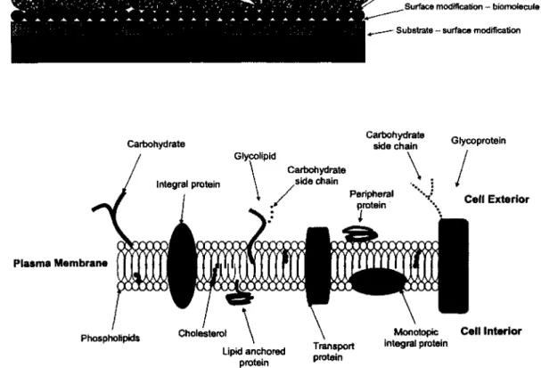
A traditional cell-surface colloidal system generally consists of four interfaces: (i) substrate- surface modification, (ii) the surface modification-biomolecule, (iii) the biomolecule-cell, and (iv) the cell-culture medium. As shown in Figure 2.1, the system is typically engineered in a “bottom-up” fashion. For example, a substrate is modified by means o f self-assembled monolayers (SAM) or plasma polymerization. Subsequently, proteins, extracellular matrix (ECM) components, or other biomolecules, such as growth factors are adsorbed to the surface
via chemisorption or physisorption, followed by cell seeding in culture media. While almost all studies employ such a colloidal system, its relevance to the in vivo reality is a credible issue. As observations suggest, the traditional cell culture approach is not necessarily representative of the architecture, mechanical forces, and mass transport properties o f a physiological environment. As such, large efforts should be made in biomaterials and tissue engineering to design in vitro systems that further represent the physiological in vivo reality. While advances have been made and some studies have in actual fact elicited a desired cell response, much research is still required to truly understand cell-surface and cell-material interactions in order to significantly advance not only molecular biology, but also biomaterials and tissue engineering research.
(A)
Cell - culture medium
Biomolecule - cell
Surface modification - biomofecule
Substrate - surface modification
(B) Carbohydrate Plasma Membrane Glycolipid
V
Carbohydrate side chain Integral protein Peripheral protein Cholesterol Phospholipids Celf Exterior Lipid anchored protein Transport protein Monotopic integral protein Cell InteriorFigure 2.1: (A) Traditional cell-surface experimental setup. (B) Cross section of generic
eukaryotic cell surface (i.e., plasma membrane). Many of the components portray a localized net electric charge. Figures not drawn to scale.
Many studies have been performed on cell adhesion and responses to surfaces using a variety o f phenotypes and surface modifications [26-36]. As shown in Table 2.1, results of previous studies on cell adhesion and responses to surfaces always vary somewhat between subsequent studies. The differences in results can be a consequence of varying phenotypes, surface modification, and/or a variety o f other differences in experimental methods. For example, both Webb et al. [36] and Keselowsky et al. [30] studied osteoblast adhesion to amine (NH2)- and
methyl (CH3)-bearing surfaces. Webb et al. found osteoblast activity (adhesion and migration)
followed CH3 > NH2 while Keselowsky et al. found the opposite. Between the two studies there
are many differences in the experimental processes. Webb et al. [36] modified substrates with organosilanes while Keselowsky et al. [30] used organothiol SAM. Webb et al. [36] did not cover the modified surface with proteins while Keselowsky et al. [30] coated the organothiol- modified surface with fibronectin (Fn). Both conclude that cell response and behavior is dictated by the surface chemistry of the modified substrate. While such a conclusion is logical and correct, due to the differences in experimental methods, it is next to impossible to draw a comprehensive understanding on the surface mechanisms that govern osteoblast response and behavior. One can only draw conclusions based on the specific surface composition employed, as these researchers have done. While these studies suggest a useful strategy for understanding and modulating cell behavior, they do not provide insight into the specific mechanisms that govern such behavior, nor strategies to control and modulate such behavior for biomedical applications. Limitations such as these just described can be extended to nearly all (including the few presented in Table 2.1) biomaterials and tissue engineering related literature. The result is a large quantity of literature with limited basis o f general comprehensive knowledge and understanding.
In a recent keynote address [37], R. Nerem categorized biomaterials and tissue engineering research over the last two decades. The 1990’s were classified as “the go go years” in which it was seen as a potential scientific revolution and interest and research exploded. The years 2000 to 2005 were categorized as “the sobering years” as researchers began to realize the enormous problems and complexities associated with such research. The year 2005 to present is categorized as “back to the future” as some researchers are now focusing on overcoming the many hurdles to retain the potential that research in biomaterials, tissue engineering, and other related disciplines was believed to have in the 1990’s [37]. We agree with R. Nerem’s
categorization and hope this article helps advancing the science to the potential it was initially believed to have.
Table 2.1: Cell-surface studies
Phenotype
fibroblast endothelial osteoblast leukocyteModification
Ref:
organosilane + SM° [27] organothiol + PM* [31] commercial TCPS [28] organosilane + SM [35] commercial TCPS [33] + PM Commercial TCPS [33] + PM organosilane + PM [32] organosilane [29] organosilane [36] organothiol + PM [30] organothiol [26] organothiol + PM [34] organothiol [34]Relative level of adherenCe/behavior
NH2 a COOH > CH3 = OH TCPSc> C H 3>COOH UPSrf > TCPS n h2 > c h3 Primaria™ > TCPS Primaria™ > TCPS NH2 > glass NH2 > C H CH3 > NH2 NH2 > CH3 CH3 > OH > COOH O H > C O O H > C H 3 CH3 > COOH > OH
aSM = serum modification. PM = protein modification. TCPS = tissue culture polystyrene. ^UPS = untreated polystyrene
We begin with presenting biological aspects. The cell surface {i.e., the plasma membrane) is briefly discussed, followed by cellular adhesion of the cell surface to ligands. Physical aspects are then presented. Fundamental interactions including Coulomb, van der Waals, double-layer, hydrophobic, H-bonding, and steric repulsion are introduced and their relevance to biology is discussed. An extensive discussion on systems for studying cell-material interactions follows. Finally, surface chemistry is presented as it is essential to biomaterials science. Particular emphasis is placed on SAM and plasma polymerization modifications to enhance chemical activity, along with their advantages and disadvantages with respect to surface uniformity and stability. The design of low-fouling surfaces is also discussed.
Depending on the scientific background o f the reader, sections of this article may appear trivial. However, the primary aim o f this review is to collectively present literature relevant to cell- material and cell-surface interactions from all three fundamental scientific disciplines in order to present the reader with a wide spectrum o f perspectives o f such literature subsequently directing future research considerations and interpretation in the direction o f general comprehension o f cell behavior on surfaces. We believe that fundamental understanding is necessary to identify and design surfaces, materials, and systems to elicit and control a desired cell response.
This article covers a large timeline of scientific literature, as this was necessary to provide a full picture of the concepts behind cell-material interactions. Most studies directly related to cell- material interactions that are reviewed in our article have largely been published in the last 20 years. At the same time, we also review the fundamental concepts of surface science that are necessary to better understand and appreciate such interactions, some which originated nearly a century ago.
2.3 Cellular Adhesion to Surfaces
2.3.1 Cell Surface
Generally, the cell surface can be depicted as shown in Figure 2.1. The plasma membrane (i.e., cell surface) is selective in the reactions that are allowed to proceed and the molecules that are allowed to penetrate. The plasma membrane mainly consists o f a lipid bilayer composed of phospholipids. Scattered throughout the lipid bilayer are membrane proteins and carbohydrates (generically known as polysaccharides).
The plasma membrane is a non-rigid body. Plasma membrane structure is largely represented using the Fluid-Mosaic model proposed by Singer and Nicolson [38]. The Fluid-Mosaic model utilized many previous studies in its formulation [39] and is continuously being redefined. In the Fluid-Mosaic model, lipids and proteins behave like a fluid and for all intensive purposes are free to move around, in a diffusive manner, within the two-dimensional structure of the plasma membrane [40, 41]. The lipid component of the cell surface gives the cell its fluid-like properties. The three primary classes of lipids composing the cell membrane are phospholipids, glycolipids, and cholesterol. The most abundant lipids are the phospholipids. Glycolipids are
lipids with a carbohydrate side chain. Cholesterols are located within the interior of the plasma membrane. Carboxyl groups close to the polar head o f the phospholipids interact with hydroxyl groups of the cholesterol via H-bonding. The result is increased spatial separation o f adjacent polar head groups of the phospholipids. During interaction between a plasma membrane protein
(i.e., a receptor) with another ligand, there is an abundance o f cholesterol within the vicinity of
the membrane protein relative to the plasma membrane as a whole. This further isolates the membrane protein from the rest o f the plasma membrane aiding in the ligand-receptor interaction. The formed structure is known as a lipid raft.
The two main functions o f membrane proteins are to provide communication between the cell and its external environment (i.e., cell signaling) and to regulate the flow of substances into and out of the cell. A membrane protein (i.e., a cell receptor) interacts with a signaling protein (i.e., a ligand). This is the first in a series o f events that dictate subsequent cell function and behavior. Transport proteins are membrane proteins that regulate the flow o f substances in and out o f the cell. To name a few, substances can include the intake o f nutrients or the removal of waste from the cell.
There are many different types of membrane proteins. Classification is dependent on the structure and the manner in which it is integrated to the plasma membrane. For example, membrane proteins containing carbohydrate side chains are called glycoproteins and those that do not penetrate the plasma membrane are known as peripheral membrane proteins. Peripheral membrane proteins are anchored to the plasma membrane via weak electrostatic interactions and H-bonding. Proteins that are covalently bound to the lipid molecules within the plasma membrane are known as lipid-anchored membrane proteins.
The majority of membrane proteins are categorized as integral membrane proteins. Integrins and cadherins are classified as integral membrane proteins. Integral membrane proteins possess hydrophobic regions that interact with the hydrophobic interior o f the plasma membrane (i.e., the tails of the phospholipids). As such, there is a high affinity o f the protein for the plasma membrane making integral membrane proteins the most difficult to isolate from the plasma membrane. Integral membrane proteins also possess hydrophilic regions by which they interact with the external aqueous environment of the cell. Some integral membrane proteins protrude
from only one side o f the cell, known as integral monotopic proteins while others span the entire thickness o f the plasma membrane, known as transmembrane integral proteins.
This discussion has been overly simplified for the purpose o f brevity. We have only discussed the plasma membrane as a barrier that defines the boundary o f a cell and its external environment as that is the most relevant for this article. However, the plasma membrane also acts as a boundary for the separation of organelles in the intracellular compartment. For detailed discussion on the overall context o f the plasma membrane and more detail into its structure, the reader is referred to widely used biology texts by Lodish et al. [42] and Becker et al. [43]. The interactions (H-bonding and hydrophobic) mentioned here are discussed in detail in Section 2.4.2.
2.3.2 Cellular Adhesion
It is generally believed within the scientific community that cells adhere to surfaces through an intermediate interaction between an extracellular matrix (ECM) protein and a surface. ECM proteins are introduced to the surface by two ways. In one way, cells synthesize and secrete ECM proteins such as fibronectin (Fn) and vitronectin (Vn) to promote adherence [44]. In the other way, proteins are pre-adsorbed to the surface independently prior to the introduction of cells to the surface. Subsequently, when cells are introduced to a protein-coated surface, less protein synthesis via the cells is necessary.
Cellular adhesion is a dynamic process that involves many different proteins [45, 46]. Adherence of cells to surfaces is achieved through an interaction between adhesive ligands on the ECM proteins and receptors on the cell surface. This interaction is mediated by integrins [47]. Integrins are heterodimeric transmembrane glycoproteins that consist o f a and P subunits [48]. While we discuss only integrin cell receptors in this article, we note to the reader that there are other known types of cell receptors. In general, most cell receptor molecules fall into five categories: immunoglobins, integrins, selectins, mucins, and cadherins. For a concise review on the different types of cell receptors, the reader is referred to literature by Pierres et al. [49].
The two main classifications o f cellular adhesions are focal contacts and fibrillar adhesions. Focal adhesion contacts are flat elongated structures usually located at the cell periphery, which
involve the cytoskeletal proteins tensin, vinculin, a-actinin, and talin as well as signaling proteins including focal-adhesion-kinase (FAK) and paxillin. Fibrillar adhesions are more centrally located and involve the cytoskeletal protein tensin [50].
The dynamics o f cellular adhesion are hypothetically illustrated in an informative commentary by Zamir and Geiger [51]. a5pl and its interaction with Fn along with the aVp3 integrin appear to be the most studied complexes in conjunction with cellular adhesion. As shown in Figure 2.2, adhesions initially contain both a5pi and aVp3 integrins. Co-localization of a5/aV subunits appears early during adhesion, then over time, these alpha subunits segregate [52]. Moreover, experiments have shown that P3 integrins remain with the initial adhesions while p i undergoes translocation [53]. A contractile force provided by the protein myosin II translocates the a5pi along with tensin centripetally towards the interior of the cell and forms a fibrillar adhesion in a process known as fibrillogenesis. In the same manner as the a5 and p i integrin subunits, Fn has been shown to translocate during cellular adhesion [54]. Furthermore, covalent linking of Fn to a substrate can impede fibrillogenesis [55]. On the other hand, aVp3 does not translocate even though the same contractile force is present leaving what is referred to as a focal adhesion contact [51, 56]. Depending on the substrate properties and composition, focal contacts alone or both focal contacts and fibillar adhesions may be present in cellular adhesions [55, 57].
It should be mentioned, as was done in the studies just highlighted, that there are likely other integrin and ECM complexes involved in cellular adhesion. Most integrin receptors have the ability to bind to multiple adhesive ligands. In addition, an adhesive ligand can bind with multiple integrin receptors [58]. For a concise review on integin a and p subunits and their associated ligand receptors along with insight into the resultant signaling and cell function, we refer the reader to articles by Hynes [48, 59].
Figure 2.2: Structure and dynamics of cellular adhesion to surfaces. (A) Cellular adhesions initially contain both a5pi/Fn and avp3/Vn integrin-ECM adhesions. (B) A contractile force provided by myosin II translocates the a5pi/Fn centripetally towards the cell center to form a fibrillar adhesion leaving behind a focal contact. Abbreviations; a = actin, a = a-actinin, m = myosin II, p = parvin/actopaxin, pa = paxillin, ta = talin, te = tensin, and vi = vinculin. Reproduced with permission from reference [51]. Copyright 2001 The Company of Biologists.
2.3.3 Protein Mediation between Cells and Surfaces
As already mentioned, receptors on the cell surface interact with the adhesive ligands o f the ECM proteins that are adsorbed to the underlying substrate to form adhesions (i.e., focal and fibrillar structures). The availability of such adhesive ligands for interaction with cells is dependent on interactions between the ECM protein and the underlying substrate. As a result of
such interactions, ECM proteins will undergo conformational and orientation changes upon adsorption to a substrate. The nature of the interaction mechanisms (i.e., surface and intermolecular forces) involved is the next subject of this article.
Proteins are non-rigid bodies. Thus, while the primary structure remains intact upon interaction with a surface, the secondary, tertiary, and quaternary structures may undergo spatial changes from their natural conformation. This is known as a conformational change. An orientation is the axial and azimuthal angles of the protein with respect to an arbitrary axis. The resultant conformation and orientation of a protein due to interactions with a surface has a direct impact on which particular ligands are available for interactions with cell surface receptors, thus directly influencing the behavior of cells on the surface.
To provide a specific example of various ligands influencing cell behavior, let us consider Fn. Fn is a dimer glycoprotein of ~ 450kDa found in the ECM and blood. It reacts with many biomolecules and plays a wide variety o f roles in physiological processes such as embryogenesis, angiogenesis, and wound healing [60-64].
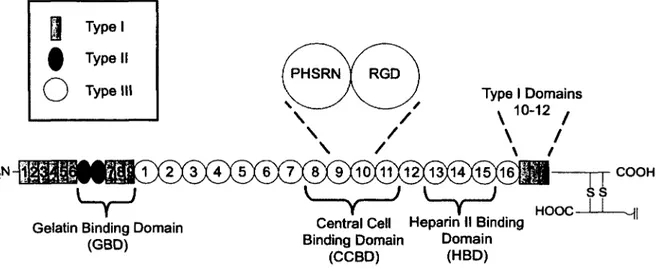
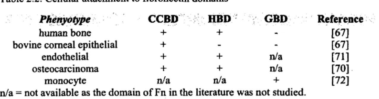
Plasma and cellular Fn are similar but vary slightly. We assume the structure of plasma and cellular Fn as identical for the purpose of brevity. For differences in the structure of plasma and cellular Fn, we refer the reader to Hayashi and Yamada [65]. Fn has a primary structure as shown in Figure 2.3. There are two similar polypeptide chains o f ~ 225kDa each. Each chain has a slight variation in amino acid sequence [66]. There is no absolute general consensus on the specific functions of each of the three domains presented in Figure 2.3 with respect to cellular function. Various phenotypes respond differently to the domains o f Fn, particularly, the gelatin binding domain (GBD) and heparin II binding domain (HBD). Such differences are a result of different phenotypes having different cell surface receptors. While one phenotype may have a cell receptor specific for a particular domain of Fn, another phenotype may lack a receptor with such specificity. For example, as shown in Table 2.2, human bone cells show attachment to the HBD while bovine comeal epithelial cells do not [67]. The central cell binding domain (CCBD) is the primary ligand believed to be responsible for cellular adhesion to Fn. Within this domain (see Figure 2.3) is the 9th-10th type III repeat, which contains the proline-histidine-serine- arginine-asparagine (PHSRN) synergy site and the adhesive tri-peptide RGD sequence [68]. The PHSRN synergy site is not responsible for adhesion alone. Rather in unison with the RGD site,
cellular adhesive affinity is enhanced [69]. In general, the HBD produces a modest (if any) effect on cellular adhesion alone [67]. However, together with the CCBD, cells show enhanced migration and proliferation in the presence o f the HBD [70, 71]. As shown in Table 2.2, the GBD does not contribute to cellular adhesion for both human bone and bovine comeal epithelial cells [67]. However, for monocytes, the GBD domain is believed to play a role in cellular adhesion [72]. We note that there are many other domains o f Fn that we do not include here, again for the purpose o f brevity. The reader is referred to a review by Hynes and Yamada for identification and the biological properties o f such domains [73].
Type I Domains Type II PHSRN RGD
O Typ®1,1
H2N-)1 HOOC\ 1°-12 /
COOHGelatin Binding Domain (GBD)
Central Cell Heparin II Binding Binding Domain Domain
(CCBD) (HBD)
Figure 2.3: Primary structure of fibronectin (Swiss Institute o f Bioinformatics, ExPASy Proteomics Server. http://www.expasy.org/cgi-bin/protparam7P02751, accessed on July 28 , 2010). There are two nearly identical mirror segments of approximately 220-230 kDa joined by two disulfide bonds.
A single ligand has multiple influences on a single phenotype, and varying phenotypes have different responses to single ligands. More importantly, since protein conformation and orientation influences which adhesive ligands are available for interactions with cells, we explicitly see how protein conformation and orientation directly influences cell behavior. Because properties of the substrate influence protein conformation and orientation, the substrate has a direct influence on cell behavior.
Table 2.2: Cellular attachment to fibronectin domains
Phenydtype
CCBD
HBD
GBD
Reference
human bone + + - [67]
bovine comeal epithelial + - - [67]
endothelial + + n/a [71]
osteocarcinoma + + n/a [70]
monocyte n/a n/a 4- [72]
n/a = not available as the domain o f Fn in the literature was not studied.
It is important to note that the information outlined in the previous paragraph has limitations. Many of the studies presented drew conclusions from either utilizing synthetic domain fragments o f Fn or through the blocking o f particular domains via antibodies. The complete absence of certain domains as a result of using domain fragments likely has drastic effects on cell behavior. Cellular function is rarely the result of one ligand on a protein. Rather it is a result o f simultaneous cell receptor interactions with multiple surface ligands. For example, it is believed that GBD stimulates fibrillogenesis in fibroblast adhesion but is not a direct contributor to cell adhesion. Rather fibrillogenesis is stimulated through a cooperative mechanism of the cell surface receptor transglutaminase with both the CCBD and GBD [74]. In conclusion, while synthetic fragments of Fn may give some information regarding the role o f particular ligands, such results should be interpreted with caution as in a physiological environment, such synthetic fragments do not exist. Furthermore, even when not using synthetic Fn fragments, one still likely has Fn fragments and even contaminants within their system. Fn is usually purified from blood plasma using chromatography. Regardless o f purification technique, the end product contains trace amounts of contaminants such as proteolytic fragments, fibrinogen, and plasma gelatinase [75-77]. Thus, all results should be interpreted with caution as some may not be completely physiologically representative.
The orientation and conformation of surface adsorbed Fn determines which particular ligands (CCBD, GBD, and HBD) are available for potential interactions with cell receptors, ultimately eliciting cell function. The conformation of Fn is dependent on parameters such as the type of Fn (e.g., plasma, cellular) and the conditions of the solution environment (i.e., pH, salt presence, etc.) [78], For the case of surface adsorption, the properties of the surface and/or conjugation mode has influence on Fn conformation [79, 80]. Regardless, depending on such parameters, Fn
![Table 2.1: Cell-surface studies Phenotype fibroblast endothelial osteoblast leukocyte Modification Ref:organosilane + SM° [27]organothiol + PM* [31]commercial TCPS [28]organosilane + SM [35]commercial TCPS [33]+ PMCommercial TCPS [33]+ PMorganosilane + PM](https://thumb-eu.123doks.com/thumbv2/123doknet/5429429.127153/35.924.113.776.244.704/endothelial-modification-organosilane-organothiol-commercial-organosilane-pmcommercial-pmorganosilane.webp)