Elucidating the role of the family of
GalNAc-Transferases in aberrant protein O-glycosylation in
the progression of epithelial ovarian cancer
Thèse
Razan Sheta
Doctorat en Médecine Expérimentale
Philosophiae Doctor (Ph.D.)
Québec, Canada
Elucidating the role of the family of
GalNAc-Transferases in aberrant protein O-glycosylation in
the progression of epithelial ovarian cancer
Thèse
Razan Sheta
Sous la direction de:
iii
RÉSUMÉ
Le cancer épithélial de l’ovaire (CEO) est la forme de cancer gynécologique la plus létale. Ainsi, la compréhension des changements moléculaires associés à ce cancer métastatique ovarien peut mener à l’identification de nouvelles cibles thérapeutiques essentielles. La glycosylation, une modification post-traductionnelle, joue un rôle important dans de nombreuses fonctions cellulaires. Cette glycosylation participe à des événements physiopathologiques majeurs durant la progression tumorale. De plus, il a été prouvé que l’expression aberrante des structures glycanes interfère avec des mécanismes cellulaires comme l’adhésion, la migration et la prolifération des cellules.
Dans ce contexte, notre laboratoire a récemment montré que le gène codant pour la protéine N-acétylgalactosaminyltransférase 3 (GALNT3), membre de la famille des GalNAc-Transférases (GalNAc-Ts), est hypométhylé et que la protéine GALNT3 est plus fortement exprimée dans les tumeurs CEO dont la sévérité est de grade élevé (“high-grade (HG) serous”), en comparaison avec des tumeurs à potentiel malin faible (“low malignant potential (LMP) ”) et des tissus ovariens normaux.
Ces observations indiquent un fort potentiel oncogénique pour le gène GALNT3 dans les stades avancés du CEO. Ces premières constatations suggèrent également que la surexpression de GALNT3 peut jouer un rôle important dans la tumorigenèse du CEO en augmentant sa dissémination via une O-glycosylation de type mucine aberrante. Ces glycosylations anormales peuvent donc être impliquées dans la carcinogenèse ovarienne et nécessitent une étude approfondie.
Dans ce projet de recherche, nous proposons d’approfondir les observations déjà obtenues in vitro en utilisant un modèle in vivo chez la souris, afin d’élucider le rôle fonctionnel de la GALNT3 et d’autres membres de cette famille dans la progression du CEO. A partir d’une étude de glycoprotéomique indépendante de la masse, qui a permis d’identifier des glycopeptides intacts ou métaboliquement marqués, ce projet de recherche a rendu possible la définition précise du rôle de GALNT3 dans la O-glycosylation des cibles de type mucine au sein des cellules CEO. Ainsi, via une recherche ciblée dans la base de données « Swiss-Prot » du protéome humain, nous avons trouvé plusieurs centaines de glycoprotéines et glycopeptides uniques, différemment exprimés dans les clones cellulaires dépourvus en
iv
GALNT3 KD. Par la suite, nous avons identifié les gènes codant pour ces glycoprotéines et glycopeptides. Nous avons notamment trouvé, parmi la liste, un groupe de gènes impliqués dans le métabolisme cellulaire dont les modifications post-traductionnelles sont, de manière intéressante, principalement supprimées dans les clones GALNT3 KD.
De plus, nous nous sommes intéressés aux autres membres de la famille des GalNAc-Ts dans le CEO et nous avons montré que de multiples membres et pas uniquement GALNT3 peuvent jouer un rôle important dans la dissémination et la progression du CEO. De plus, une découverte très intéressante fut la redondance possible des rôles joués par certains membres de la famille des GalNAc-Ts dans le CEO. Ainsi, nous avons identifié GALNT6 qui serait, à l’image de GALNT3, impliquée dans la dissémination et la progression du CEO. Cette implication du GALNT6 est supportée par le fait que cette protéine a les mêmes fonctions que GALNT3, suggérant un effet compensatoire de GALNT6 en absence de GALNT3. Pour tester cette hypothèse, nous avons abolie l’expression des deux protéines GALNT3 et GALNT6, in vivo, et nous avons observé une effet significatif sur la formation des tumeurs et la survie des animaux. Pour la suite de ce projet, nous proposons d’analyser la structure glycane des différentes glycoprotéines identifiées dans les cellules cancéreuses, afin de déterminer les altérations des modifications O-glycanes suite à la perte d’expression de GALNT3 et d’autres membres de la famille des GalNAc-Ts. En conclusion, notre étude contribue à comprendre la participation du glycoprotéome dans la tumorigenèse du CEO et à identifier d’autres cibles de type mucine ou des O-glycoprotéines dont l’expression aberrante serait modulée dans le CEO. Ainsi, pris dans son ensemble, ce projet de recherche montre la possibilité de discriminer entre des cellules cancéreuses et des cellules contrôles via les glycosylations de leurs protéines et permet d’entrevoir la glycobiologie comme une voie prometteuse pour l’identifier de nouveaux biomarqueurs pour le diagnostic du CEO.
v
ABSTRACT
Epithelial ovarian cancer (EOC) is the most lethal gynecologic malignancy, thus understanding the molecular changes associated with ovarian cancer metastasis could lead to the identification of essential therapeutic targets. Glycosylation is a post-translational modification (PTM) of proteins playing a major role in various cell properties. Glycosylation participates in major pathophysiology events during tumor progressions, and the aberrant expression of glycan structures was shown to interfere with cell properties such as cell adhesion, migration, and proliferation. The lab has previously identified the polypeptide N-acetylgalactosaminyltransferase 3 (GALNT3) gene, a member of the GalNAc-Transferases (GalNAc-Ts) gene family, as hypomethylated and overexpressed in high-grade (HG) serous EOC tumors, compared to low malignant potential (LMP) EOC tumors and normal ovarian tissues. Taken together, the data obtained were indicative of a strong oncogenic potential of the GALNT3 gene in advanced EOC and suggest that GALNT3 overexpression might contribute to EOC dissemination through aberrant mucin O-glycosylation, thus specifying some of the putative mechanisms of abnormal glycosylation implicated in ovarian carcinogenesis, which warrant further investigation. The current research project focused on expanding the in vitro observations obtained by using animal models to investigate in vivo the functional significance of GALNT3 and other close members of the GalNAc-Ts gene family in serous EOC progression. Moreover, by applying a mass-independent chemical glycoproteomics platform to characterize intact, metabolically labeled glycopeptides, this project more profoundly characterized the role of GALNT3 in aberrant O-glycosylation of mucin-like targets in EOC cells. Isotopically recorded ions were searched against the Swiss-Prot human proteome; and data obtained were indicative of hundreds of unique glycoproteins and glycopeptides that were differentially expressed upon GALNT3 KD. Related gene groups were identified, and interestingly, genes implicated in mechanisms of cellular metabolic functions, and PTMs were found to be predominantly suppressed in GALNT3 KD clones. In accordance, we also investigated the role of other members of the GalNAc-T family in EOC and we showed that multiple members and not only GALNT3 can play an important role in EOC cancer dissemination and progression. One very interesting finding was the redundant role some
vi
members of the GalNAc-T family members play in EOC. We investigated the compensatory functions of GALNT3 and GALNT6, and we were able to demonstrate these two genes can impose that synthetic backup. Furthermore, we found that and their ablation can affect animal survival and tumor formation as observed both in vivo and in vitro. In continuation of this work, this project will focus on analyzing the glycan structures of those differentially expressed glycoproteins, to further examine the specific O-glycans alterations associated with the GALNT3 and other members of the GalNAc-Ts upon gene knockout (KO). Fully elaborated glycopeptides can reveal structural details of the glycoproteome, thus our results could give important information on the glycome in EOC cells, and the identification of other O-glycoproteins/mucin-like targets whose aberrant expression may be modulated by these in EOC. Taken together, the ability to mark differences in the glycosylation of proteins between cancer cells and control cells can emphasize glycobiology as a promising field for potential biomarker identification.
vii
TABLE OF CONTENTS
RÉSUMÉ ... iii
ABSTRACT ... v
TABLE OF CONTENTS ... vii
LIST OF TABLES ... xii
LIST OF FIGURES ... xiii
LIST OF ABBREVIATIONS ... xvi
ACKNOWLEDGMENTS ... xxvi PREFACE ... xxviii PRÉFACE ... xxxi Chapter 1: Introduction ... 1 1.1 Ovarian cancer ... 2 1.1.1 Overview of ovarian cancer ... 2 1.1.2 The different types of ovarian cancer ... 2 1.1.3 The five histopathological subtypes of EOC ... 4 1.1.3.1 High-Grade Serous Carcinoma (HGSC) ... 5 1.1.3.2 Endometrioid carcinoma (EC) ... 6 1.1.3.3 Clear cell carcinoma (CCC) ... 6 1.1.3.4 Low-grade serous carcinoma (LGSC) ... 6 1.1.3.5 Mucinous carcinoma (MC) ... 7 1.1.4 EOC tumor staging and grading ... 7 1.1.5 Carcinogenesis and genetics of EOC ... 11 1.1.6 EOC metastasis ... 14 1.1.7 Risk factors of EOC ... 15 1.1.8 Prevention and detection of EOC ... 17 1.1.9 Treatment methods of EOC ... 18 1.1.10 Experimental models of EOC ... 20 1.1.10.1 EOC cell lines ... 20 1.1.10.2 Genetically engineered animal models ... 22 1.1.10.3 Xenograft models of EOC ... 23
1.2 Glycosylation and cancer ... 24
1.2.1 Role of glycosyaltion in cancer development ... 24 1.2.2 The chemistry and biology of protein glycosylation ... 25 1.2.2.1 N-linked glycosylation ... 25 1.2.2.2 O-linked (mucin) glycosylation ... 26 1.2.2.3 Synthesis of O-glycan structures ... 27 1.2.3 Terminal glycan linkages/modifications and their role in cancer development ... 31
viii
1.2.3.1 Lewis antigens and blood group antigens ... 31
1.2.3.2 Sialylation ... 32
1.2.3.3 Fucosylation ... 34
1.3 The chemistry and biology of mucin glycoproteins and their role in cancer ... 34
1.3.1 The two classes of mucins ... 34 1.3.2 Normal functions of mucins ... 38 1.3.3 The roles of mucins in cancer development ... 39 1.3.3.1 Role of mucin protein alterations in inducing cellular invasion, migration and metastasis in cancer ... 40 1.3.3.2 Role of mucin protein alterations in inflammation and immune suppression ... 41 1.3.3.3 Role of mucins in EOC development (focus on MUC1) ... 42 1.3.4 The role of aberrant O-glycosylation in cancer development ... 43 1.4 GalNAc-Transferases (GalNAc-Ts) ... 46 1.4.1 Regulation of Mucin type O-glycosylation ... 46 1.4.2 The human GalNAc-T gene family ... 47 1.4.3 Peptide and glycopeptide substrate specificities of the GalNAc-Ts family ... 49 1.4.5 In vivo models of GalNAc transferases and the concept of GalNAc-T redundancy ... 51
1.5 Glycoproteomics approaches in studying alterations in glycoproteins expression and structure ... 52 1.5.1 Challenges in studying the process of protein glycosylation ... 52 1.5.2 Glycoproteomic techniques for glycoprotein analysis ... 54 1.5.3 Other technologies for glycan profiling ... 55 1.5.3.1 Affinity metabolic labeling-a new engineering method for the incorporation of unnatural substrates ... 56 1.5.3.2 Chemoselective ligation to metabolically installed unnatural cell surface oligosaccharides ... 59 1.5.4 The new glycoproteomic approach – the isotopic targeted glycoproteomics (IsoTaG) platform ... 60
1.6 Problematic, hypothesis and objectives ... 65
Chapter 2: ... 69
Chapter 2.A: A metabolic labeling approach for glycoproteomic analysis reveals altered glycoprotein expression upon GALNT3 knockdown in ovarian cancer cells ... 69
Chapter 2.B: Proteomic dataset for altered glycoprotein expression upon GALNT3 knockdown in ovarian cancer cells ... 69
2.1 Preface ... 71
2.2 Résumé en français ... 72
2.3 Abstract ... 73
2.4 Introduction ... 74
ix 2.5.1 Cell culture ... 77 2.5.2 Metabolite labeling and protein enrichment ... 77 2.5.3 Mass spectrometry ... 78 2.5.4 Database searching and Label Free Quantification ... 79 2.5.5 Bioinformatic annotation & analysis ... 79 2.6 Results ... 81 2.6.1 Purification and LC/MS analysis of glycosylated proteins from control and GALNT3 KD A2780s cells ... 81 2.6.2 Comparative proteomic analysis of differentially regulated glycoproteins identified between control and GALNT3 KD A2780s EOC cells. ... 82 2.6.3 Cellular classification of differentially regulated proteins identified between control and GALNT3 KD A2780s EOC cells ... 83 2.6.4 Pathways and network analyses following GALNT3 gene KD in EOC cells ... 84 2.7 Discussion ... 86 2.8 Acknowledgements ... 93 2.9 References ... 94
2.10 Figure and table legends ... 100
2.11 Tables ... 103
2.12 Figures ... 104
Chapter 2.B: Proteomic dataset for altered glycoprotein expression upon GALNT3 knockdown in ovarian cancer cells ... 110
2.13 Data ... 112
2.14 Experimental design, Materials and Methods ... 113
2.14.1 Chemical Glycoproteomics Enrichment using Click Chemistry ... 115 2.14.2 Western Blot Analysis ... 116 2.14.3 Database searching and Label Free Quantification ... 116 2.14.4 Bioinformatic annotation & analysis ... 117 2.15 Acknowledgements ... 119 2.16 References ... 120 2.17 Figure legends ... 122 2.18 Figures ... 123
Chapter 3: Altered expression of different GalNAc-Transferases (GalNAc-Ts) is associated with disease progression and poor prognosis in women with high-grade serous ovarian cancer ... 126
3.1 Preface ... 128
3.2 Résumé en français ... 129
x
3.4 Introduction ... 131
3.5 Material and Methods ... 133
3.5.1 Patient cohort ... 133 3.5.2 Cell culture ... 133 3.5.3 Western blotting ... 133 3.5.4 Tissue Micro arrays (TMAs) and immunohistochemistry (IHC) ... 134 3.5.5 Scoring and Statistical analysis ... 135 3.6 Results ... 136 3.6.1 Analyses of the expression profiles of different members of the GalNAc-Ts family in both ovarian HGSC tumors and EOC cell lines ... 136 3.6.2 IHC analysis of the expression patterns of five GalNAc-Ts in numerous EOC tumors using TMAs ... 137 3.6.3 Association of GalNAc-Ts expression with clinicopathological data ... 139 3.7 Discussion ... 140 3.8 Acknowledgements ... 144 3.9 References ... 145
3.10 Figure and table legends ... 151
3.11 Tables ... 153
3.12 Figures ... 156
Chapter 4: Elucidating the role of the polypeptide N-acetylgalactosaminyltransferase 3 (GALNT3) and its closest homolog-GALNT6 in mediating aberrant O-glycosylation associated with ovarian cancer dissemination ... 160
4.1 Preface ... 161 4.2 Résumé en français ... 162 4.3 Abstract ... 163 4.4 Introduction ... 165 4.5 Results ... 168 4.5.1 Overexpression of GALNT6 in GALNT3 KO EOC cells ... 168 4.5.2 GALNT3 and T6 KO reduced cell proliferation rate, migration, invasion and cell cycle in EOC cells ... 169 4.5.3 Evidence of full functional redundancy in protein glycosylation by GALNT6 in GALNT3 KO EOC cells ... 170 4.5.4 Molecular mechanisms of GALNT3 and GALNT6 action in EOC cells ... 171 4.5.5 Double GALNT3 GALNT6 gene KO reduces EOC metastasis in vivo ... 174 4.6 Discussion ... 176
4.7 Materials and methods ... 180
4.7.1 Cell culture ... 180
xi 4.7.3 Short hairpin RNA (shRNA) - mediated GALNT6 knockdown in EOC cells ... 180 4.7.4 Western blotting ... 181 4.7.5 Functional assays ... 181 4.7.6 Semi-quantitative RT-PCR (sqRT-PCR) ... 182 4.7.7 Quantitative PCR (qPCR) ... 182 4.7.8 VVA lectin pull-down assay for O-glycosylated (GalNAc-conjugated) proteins ... 182 4.7.9 Gene expression profiling and data analysis ... 183 4.7.10 Peritoneal tumor formation in mice ... 183 4.8 Acknowledgements ... 185 4.9 References ... 186 4.10 Figure legends ... 193 4.11 Figures ... 197 4.12 Supplemental figures ... 207
Chapter 5: General Discussion ... 209
5.1 Discussion ... 210 5.1.1 The application of glycoproteomics studies in EOC ... 212 5.1.2 Expression level analysis of different GalNAc-Ts in EOC and correlation with disease progression ... 218 5.1.3 Examining the concept of functional redundancy amongst members of the GalNAc-Ts in EOC ... 221
General Conclusions and Perspectives ... 224
Bibliography ... 228 Annex ... 254 Abstract ... 255 Introduction ... 255 Protein glycosylation ... 257 N-linked glycosylation ... 257 O-linked (mucin) glycosylation ... 258 Aberrant glycosylation in cancer ... 259 Altered glycosylation implicated in EOC etiology ... 261 N-linked glycosylation in ovarian cancer ... 261 O-linked (mucin-type) glycosylation in ovarian cancer ... 262 Conclusion and perspectives ... 264 References ... 265 Figure legends ... 271 Figures ... 272
xii
LIST OF TABLES
Chapter 1: Introduction
Table 1.1 FIGO stage grouping for primary carcinoma of the ovary ... 8
Table 1.2. Characteristic mutations found in EOCs ... 13
Table 1.3. Risk factors for EOC ... 16
Table 1.4. List of EOC cell lines ... 21
Table 1.5. Structures of O-glycan cores and antigenic epitopes found in mucins. ... 28
Table 1.6. Chemical reporters and bioorthogonal reactions used in living systems. .... 60
Chapter 2: A metabolic labeling approach for glycoproteomic analysis
reveals altered glycoprotein expression upon GALNT3 knockdown in
ovarian cancer cells
Table 2.1. The five top-scoring genetic networks affected following glycoproteomics analysis of GALNT3 KD in A2780s EOC cells ... 103Chapter 3: Proteomic dataset for altered glycoprotein expression upon
GALNT3 knockdown in ovarian cancer cells
Table 3.1. Detailed patients’ clinicopathological characteristics ... 153Table 3.2. Cox regression analysis to predict progression-free survival (PFS) ... 154
Table 3.3. Dilution and technique used for each antibody in IHC and Western blot analyses. ... 155
Chapter 5: General Discussion
Table 5.1. PubMed search output of proteomics studies and ovarian cancer ... 212xiii
LIST OF FIGURES
Chapter 1: Introduction
Figure 1.1. The origin and type of EOC tumors ... 4
Figure 1.2. The different cellular origins of EOCs ... 5
Figure 1.3. The two major histologic subtypes of EOC and their sub classifications .. 10
Figure 1.4. Model systems from primary human tissues ... 20
Figure 1.5. N- and O-glycosylated proteins ... 27
Figure 1.6. Schematic representation of the biosynthesis and processing of O-linked glycans ... 31
Figure 1.7. Structure of the major transmembrane protein Mucin1 ... 35
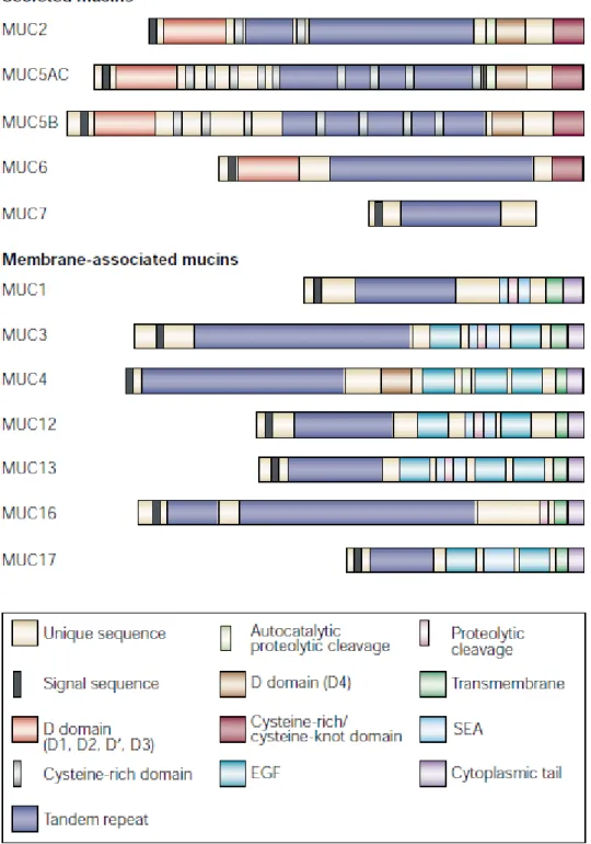
Figure 1.8. The different secreted and membrane bound mucin type proteins. ... 37
Figure 1.9. Role of glycans in the metastatic cascade ... 45
Figure 1.10. Mucin-type O-linked glycosylation and the initiation process that is controlled by a family of 20 enzymes known as GalNAc-Ts ... 47
Figure 1.11. Detailed domain structure of the multiple GalNAc-Ts ... 48
Figure 1.12. Phylogenetic and genomic tree of the human GalNAc-T gene family ... 49
Figure 1.13. General strategy of glycoproteins detection and analysis ... 54
Figure 1.14. The bioorthogonal chemical reporter strategy ... 57
Figure 1.15. Strategy for metabolic labeling of mucin-type O-linked glycoproteins with an azido GalNAc analog (GalNAz) for proteomic analyses ... 58
Figure 1.16. The GlcNAc and GalNAc salvage and O-GlcNAc signaling pathway ... 62
Figure 1.17. Structure of cleavable probe for mass-independent glycoproteomics ... 63
Figure 1.18. The IsoTag approach ... 64
Chapter 2: A metabolic labeling approach for glycoproteomic analysis
reveals altered glycoprotein expression upon GALNT3 knockdown in
ovarian cancer cells
Figure 2.1. Glycoproteinwss predictions ... 104Figure 2.2. Volcano plots illustrate differentially regulated proteins ... 105
Figure 2.3. Cellular component Gene Ontology analysis ... 106
Figure 2.4. Functional pathway analysis ... 107
Figure 2.5. Canonical pathway analysis ... 108
Figure 2.6. Network analysis of dynamic gene expression in A2780s cells based on the 2-fold glycoprotein expression list obtained following GALNT3 KD ... 109
Figure 2.7. Schematic overview of the glycoproteomic workflow used ... 123
Figure 2.8. Western blot analysis of glycoproteins enrichment in control and GALNT3 KD A2780s cells ... 124
Figure 2.9. GO cellular component analysis of significantly enriched proteins found upon GALNT3 KD ... 125
xiv
Chapter 3: Altered expression of different GalNAc-Transferases
(GalNAc-Ts) is associated with disease progression and poor prognosis in
women with high-grade serous ovarian cancer
Figure 3.1 GalNAc-Ts expression in EOC cells and EOC tumors ... 156 Figure 3.2 GalNAc-Ts protein expression in HG samples compared to LMP and non-tumoral ovarian samples ... 157 Figure 3.3. Multiple Correspondence Analyses (MCA) of the relationship of the
GalNAc-Ts expression in the HG ovarian tissue samples, and Western blot analysis of GALNT3, T6, T9 and T14 expression in control (Ctrl) and GALNT3 knockdown (KD) A2780s clones ... 158 Figure 3.4 Kaplan-Meier progression-free survival curves showing the association between GalNAc-Ts expression patterns and prognosis in HG ovarian cancer ... 159
Chapter
4:
Elucidating
the
role
of
the
polypeptide
N-acetylgalactosaminyltransferase 3 (GALNT3) and its closest homolog-
GALNT6 in mediating aberrant O-glycosylation associated with ovarian
cancer dissemination
Figure 4.1. Protein-protein interaction network of GALNT3 and GALNT6 ... 197 Figure 4.2. Western blot protein expression analysis of GALNT3 KO and GALNT3/T6 KO clones ... 198 Figure 4.3. Effect of GALNT3 and GALNT6 double KO on A2780s cell proliferation migration, invasion and cell cycle ... 199 Figure 4.4. Cell-cycle profiling of GALNT3 and GALNT3/T6 double KO clones ... 200 Figure 4.5. Western blot analysis of FN1 and MUC1 in the GALNT3 KO and
GALNT3/T6 KO clones ... 201
Figure 4.6. Canonical pathway analysis for a dataset of differentially expressed genes (≥ 1.5-fold) following GALNT3 KO and GALNT3/T6 KO in the A2780s cells ... 202 Figure 4.7. Functional pathway analysis for a dataset of differentially expressed genes (≥ 1.5-fold) following GALNT3 KO and GALNT3/T6 KO in the A2780s cells ... 203 Figure 4.8. Network analysis of dynamic gene expression in A2780s cells based on the 1.5-fold gene expression list obtained following GALNT3 KO ... 204 Figure 4.9. Network analysis of dynamic gene expression in A2780s cells based on the 1.5-fold gene expression list obtained following GALNT3/T6 double KO ... 205 Figure 4.10. In vivo examination of the effect of GALNT3 and GALNT3/T6 KO in tumor formation and survival in immunodeficient (SCID) mice ... 206
List of supplemental figures of Chapter 4: Elucidating the role of the
polypeptide N-acetylgalactosaminyltransferase 3 (GALNT3) and its
closest homolog-GALNT6 in mediating aberrant O-glycosylation
associated with ovarian cancer dissemination
xv
Supplemental Figure 4.1. Protein analysis of GALNT3 and GALNT6 expression in CaOV3 cell line ... 207 Supplemental Figure 4.2 Gene expression validation analysis of microarray data .... 207 Supplemental Figure 4.3. In vivo analysis of GALNT3 and GALNT3/T6 double KO in SCID mice ... 208
Annex
Figure 1. Schematic depiction of N-linked glycosylation ... 272 Figure 2. Schematic depiction of O-linked-(mucin type) glycosylation ... 273
xvi
LIST OF ABBREVIATIONS
ABH Lewis antigens and blood group
Ac4GalNAc Tetraacetylated N-acetylgalactosamine
Ac4GalNAz Peraceetylated N-azidoacetylgalactosamine
ADC Antibody-drug conjugates
AGC Automatic gain control
AKAP4 A-kinase anchor protein 4
ALSA Antibody-lectin sandwich array
AMPK AMP-activated protein kinase
ANXA4 Annexin A4
ASM Acid sphingomyelinase
ATG5 Autophagy protein 5
B4GALT1 Beta-1,4-Galactosyltransferase 1 BAC
BCAAs
Bacterial artificial chromosome Branched-chain amino acids
BCAT1 Branched chain amino-acid transaminase 1 BCAT2 Branched chain amino-acid transaminase 2
BOT Borderline ovarian tumors
CA-125 Cancer antigen 125
Cas9 CRISPR associated protein 9
CCC Clear cell carcinoma
CCRCC Clear cell renal cell carcinoma
CEO CF
Cancer épithélial de l’ovaire Cystic fibrosis
xx
Con A Concanavalin A
CRC Colorectal cancer
CRISPR CT
Clustered regularly interspaced palindromic repeats Cytoplasmic tail
CuAAC Copper-catalyzed azide-alkyne cycloaddition
DDOST Dolichyl-diphosphooligosaccharide-protein glycosyltransferase
DSB Double strand breaks
EC Endometrioid carcinoma
ECM Extracellular matrix
EGF Epidermal growth factor
EGFR Anti-epidermal growth factor receptor
ELF3 Transcription factor E74-like factor 3
EMT Epithelial–mesenchymal transition
EOC Epithelial ovarian cancer
ER Endoplasmic reticulum
ETD Electron transfer dissociation
FAK Focal adhesion kinase
FDA Food and Drug Administration
FFPE Formalin-fixed paraffin-embedded
FIGO Federation of Gynecology and Obstetrics
FL Follicular lymphoma
FOLR Folate receptor
FTC Familial tumoral calcinosis
FTE Fallopian tube epithelial cells
GALE UDP-galactose 4′-epimerase
GalNAc N-acetylgalactosamine
GalNAc-Ts Galnac transferases
xxi
GALNT3 N-acetylgalactosaminyltransferase 3
GCA Gastric adenocarcinoma
GEMMs Genetically engineered mouse models
GlcNAc N-acetylglucosamine
GlcNAz N-azidoacetylglucosamine
GO Gene ontology
GOLPH3 Golgi phosphoprotein 3
gRNA Guide RNA
GT1 Catalytic luminal domain
HCC Hepatocellular carcinoma
HDL High-density lipoprotein
HE-4 Human epididymis protein 4
HGSC High-grade serous carcinoma
HHS Hyperostosis syndrome
Hic-5 HILIC
Hydrogen peroxide-inducible clone-5
Hydrophilic interaction liquid chromatography
HOSE Human ovarian surface epithelial
IHC Immunohistochemistry
ILK Integrin-linked kinase
IMS Imaging mass spectrometry
IP Intraperitonial
IPA Ingenuity pathway analysis
IsoTaG Isotopic targeted glycoproteomics
IV Intravenous
KD Knockdown
KO Knockout
LC/MS Liquid chromatography/mass spectrometry
xxii
LGSC Low grade serous carcinoma
LMP Low-malignant potential
LSCC Laryngeal squamous cell carcinoma
LTQ Linear trap quadrupole
LWAC Lectin weak affinity chromatography
MALDI Matrix-assisted laser desorption/ionization
MAP1B Microtubule-associated protein 1B
MAPK Mitogen-activated protein kinases
MC Mucinous carcinoma
MCA Multiple correspondence analyses
MD Moderately differentiated
MET Mesenchymal-epithelial transition
MnSOD Manganese superoxide dismutase
MSI Mass spectrometry imaging
MSP Methylation-specific PCR
MTDH Metadherin
MTHFD2 Mitochondrial folate-coupled dehydrogenase
MUC1 Mucin-1
NACT Neoadjuvant chemotherapy
nanoLC Nanoscale capillary liquid chromatography
NB Neuroblastoma
NEOC Non-epithelial ovarian cancers
NF-Kappa B Nuclear factor kappa-light-chain-enhancer of activated B cells
NHEJ Non-homologous end joining
NID1 Nidogen-1
NK Natural killer
NSCLC Non-small cell lung cancers
xxiii
ORF Open reading frame
OSCC Oral squamous cell carcinoma
OSE Ovarian surface epithelial cells
OST Oligosaccharyltransferases
OST Oligosaccharyltransferases
PAM PARP
Protospacer adjacent motif Poly ADP ribose polymerase
PD Poorly differentiated
PDAC Pancreatic ductal adenocarcinoma
PFS Progression-free survival
PI3K Phosphoinositide 3-kinase complex
PKA Protein kinase A
PLA Proximity ligation assay
PTM Post-translational modifications
PTMs Ost-translational modifications
RCC Renal cell carcinoma
ROCK The rhoa Kinase
RRMP2 Ribonucleotide reductase M2
RRSO Risk-reducing salpingo-oophorectomy
SC Subcutaneous
SCC Squamous cell carcinoma
shRNA Short hairpin RNA
Sia Sialic acid
SILAC Stable isotope labeling with amino acids in cell culture
siRNA Small interfering RNA
SMPD1 Sphingomyelin phosphodiesterase 1
SNO Superficial scrapings from normal ovaries
xxiv
SOD2 Magnesium superoxide dismutase
SPEG Solid-phase extraction of N-linked glycopeptides
SSM Superficial spreading melanoma
ST6GalNAc1 ST6 N-Acetylgalactosaminide Alpha-2,6-Sialyltransferase 1
STT3B STT3B, Catalytic Subunit Of The Oligosaccharyltransferase Complex
SV40 Simian virus 40
TAA Tumor associated antigen
TALENs As transcription activator-like effector nuclease
TGFβ1 Transforming growth factor beta
TL Transcription activator-like
TM Transmembrane domain
TMA Tissue microarrays
TMAs Tissue microarrays
TOF Time of flight
TUBB3 Tubulin beta 3
UDP-GalNAc UDP-N-acetylgalactosamine
UDP-GlcNAc UDP-N-acetylglucosamine
VNTR Variable number of tandem repeats
VVA Vicia villosa agglutinin
WGA Wheat germ agglutinin
xxv
“Take pride in how far you have come and have faith in how far you can go.”
xxvi
ACKNOWLEDGMENTS
This thesis represents not only the written work put together in this manuscript, but it is a milestone of the hard work and achievements I have accomplished in the past few years. I have been given some unique and challenging opportunities that have allowed me to create tools for performing many of the complex research presented here. This thesis presents the many lessons I have learned whether they were technical or personal. This work is also a presentation of the work of dozens of people who I wish to thank and acknowledge.
First and foremost I wish to thank my supervisor, professor Dimcho Bachvarov. He has been supportive form the beginning. Dr. Bachvarov has supported me not only by providing assistance in this research project; he has also guided me all throughout the years. Dr. Bachvarov’s support and mentorship has really helped me become more confident in myself, and my scientific abilities. I also highly appreciate the opportunities given by Dr. Bachvarov to present my scientific findings at multiple Oncology and Ovarian cancer meetings. Finally, during the difficult times when writing this thesis, Dr. Bachvarov gave me the moral support and the freedom I needed to move on.
I would also like to thank my friend and colleague Adnen Faddaoui for his tremendous help in introducing me to all of the experimental techniques used in the lab, also for his intellectual scientific input in many parts of my project. I also would like to acknowledge Mrs. Magdalena Bachvarova who has assisted in many of the complex experiments presented in this thesis, and for making the workplace a comfortable place to work.
I would also thank all members of our collaboration teams, for without their help, scientific experience and knowledge this thesis project wouldn't have come together. Ms. Florence Roux-Dalavi has provided me with tremendous help and support when faced with very challenging concepts of mass-spectrometry and complex data acquisition. I would also like to specially thank Dr. Christina Woo for introducing us to the IsoTag approach and teaching us the methodology needed to initiate our project. I would like to thank her for hosting me in her laboratory at Harvard Medical School, for which she has provided me with an opportunity to examine the challenging work environment of glycoproteomics. I would like to thank the gynecologic oncology surgeons for providing us with patient specimens as well as insight into the clinical aspects of ovarian cancer. We are also
xxvii
extremely grateful to the women with ovarian cancer who generously donated their tissue samples to support our research.
I would also like to thank my thesis committee who has helped in guiding me through my pre-doc exam, which has prepared me my thesis exam. Thank you Dr. Stéphane Gobeil for being a great advisor who helped in the conception and application of several of the approaches employed in this study.
I would like to thank organizations at Laval University Cancer Research Centre for providing my with Bourse d’excellence du CRC. In addition special thanks to the Glyconet society, for providing me with the opportunity to travel to Boston and work under the supervision of Dr. Christina Woo.
Lastly I would like to thank my family starting from my biggest supporter and role model, my father Ammar Sheta. My father is my best friend, he has given me the greatest chance to travel and study abroad, which has opened many doors that got me to where I am right now. This thesis was impossible without his support and encouragement. I would also like to thank my sister Reeman Sheta for always knowing how to provide me with the source of energy to keep going. She has supported me whenever I needed it.
I would like to give special thanks to my husband Abid Oueslati; he has been one of my biggest supporters. He has helped my immensely with his scientific input and guidance. He provided me with ideas that have helped me progress well in my research work. I would also like to thank my daughter Reema Oueslati for being understanding during the long days and nights spent away from home.
xxviii
PREFACE
The scientific findings and data presented in this thesis is the product of three years of work as a PhD graduate student in the laboratory of Dr. Dimcho Bachvarov. I have contributed as the first author to five manuscripts and 1 review.
Chapter 1
Consists of an introduction on EOC, and glycosylation pathways involved in normal development and cancer. This chapter covers different views on the clinical and molecular aspects of EOC, and a detail description of the glycosylation pathways specifically mucin type O-glycosylation and its role in cancer development. Glycosylation is a new examined hallmark of cancer, and this chapter discusses the strong correlation of glycosylation with tumor initiation, progression and metastasis. Thus it was essential to discuss the possible role glycosylation plays in the progression of EOC.
Chapter 2
This chapter is a presentation of two of my first author articles on which I was fortunate to work on with multiple collaborators. The manuscripts are entitled “A metabolic labeling approach for glycoproteomic analysis reveals altered glycoprotein expression upon GALNT3 KD in ovarian cancer cells” published in the Journal of Proteomics and “Proteomic dataset for altered glycoprotein expression upon GALNT3 knockdown in ovarian cancer cells” published in Data in Brief. This project involved the application of a highly complex and innovative approach, which I was very fortunate to work on in Dr. Bachvarov’s lab. I wrote the manuscripts, which were then corrected by Dr. Bachvarov and coauthors. In collaboration with Dr. Woo and the proteomics facility at the CHUL, I was able to generate almost all the figures in the manuscripts. This work was made possible through the fruitful collaborations and contributions of the aforementioned co-authors.
xxix
Chapter 3
Presents a clinical scientific paper entitled “Altered expression of different GalNAc-Transferases (GalNAc-Ts) is associated with disease progression and poor prognosis in
women with
high-grade serous ovarian cancer” currently under review in the International Journal of Oncology. I have significantly contributed to this paper by preparing tissue microarrays for the analysis and scoring of the various proteins included in this study. I have generated all figures and performed all the statistical analysis. Data collection was made possible by the contribution of all the medical doctors listed as co-authors in the manuscript. We are the first lab to examine multiple GalNAc-Transferases in a subset of EOC patient samples, presenting data that indicate the implication of multiple and not one GalNAc-Transferase in the progression and poor outcome in EOC patients.
Chapter 4
Presents a scientific paper currently in preparation entitled “Elucidating the role of the polypeptide N-acetylgalactosaminyltransferase 3 (GALNT3) and its closest homolog-GALNT6 in mediating aberrant O-glycosylation associated with ovarian cancer dissemination”. I was also privileged to work on such a complex project examining biosynthetic backup of multiple GalNAc-Transferases, in addition to examining their functional redundancy in EOC cell lines. Our work presented in this chapter is the first to examine the effect multiple GalNAc-Transferases gene knockouts can have on the survival of animals. Indicative of the role multiple GalNAc-Transferases play in the metastatic cascade of EOC. I have prepared all figures and collected all the data; in vivo studies were carried out in Dr. Vanderhyden’s lab. This paper will have a very high impact on the examination of multiple gene targets in EOC.
xxx
Chapter 5
This chapter briefly discusses the work gathered in this manuscript.
Annex
An ‘Annex’ section: Annex. This section includes the scientific review Dr. Bachvarov and I worked on when we first embarked on the glycosylation study entitled “Role of aberrant glycosylation in ovarian cancer dissemination” published in Biomedical reviews.
xxxi
PRÉFACE
Les résultats scientifiques et les données présentées dans cette thèse sont le fruit de trois années de travail en tant que doctorante au laboratoire de Dr. Dimcho Bachvarov. J'ai contribué en tant que première auteure à cinq manuscrits et une revue scientifique.
Chapitre 1
Le chapitre 1 comprend une introduction sur le CEO et les voies de glycosylation impliquées dans le développement normal et dans le cancer. Ce chapitre couvre différents aspects cliniques et moléculaires du CEO, et une description détaillée des voies de glycosylation, spécifiquement la O-glycosylation du type mucine et son rôle dans le développement du cancer. La glycosylation est une nouvelle caractéristique du cancer, et ce chapitre traite de la forte corrélation entre la glycosylation et l'initiation, la progression et la métastase de la tumeur. Ainsi, il était essentiel de discuter du rôle possible joué par la glycosylation dans la progression du CEO.
Chapitre 2
Ce chapitre est une présentation de mes articles en tant que première auteure, dans lequels j'ai eu la chance de travailler avec plusieurs collaborateurs. Les manuscrits sont intitulés “ A metabolic labeling approach for glycoproteomic analysis reveals altered glycoprotein expression upon GALNT3 knockdown in ovarian cancer cells”, publié dans le journal Journal of Proteomics. et “Proteomic dataset for altered glycoprotein expression upon GALNT3 knockdown in ovarian cancer cells” publié dans le journale Data in Brief. Ce projet impliquait la mise en place d'une approche hautement complexe et novatrice, durant laquelle j'ai eu beaucoup de chance de travailler dans le laboratoire de Dr. Bachvarov. J'ai écrit le manuscrit, qui a ensuite été corrigé par Dr. Bachvarov et les co-auteurs. En collaboration avec Dr. Woo et la plateforme de protéomique au CHUL, j'ai pu générer la majorité des figures du manuscrit. Ce travail a été rendu possible grâce aux collaborations fructueuses et aux contributions des co-auteurs susmentionnés.
xxxii
Chapitre 3
Ce chapitre présente un article scientifique clinique intitulé “ Altered expression of different GalNAc-Transferases (GalNAc-Ts) is associated with disease progression and poor prognosis in womem with high-grade serous ovarian cancer ”. Ce papier est actuellement en cours d’évaluation dans le journal : Journal international d'oncologie. J'ai contribué de manière significative à cet article en préparant des microarrays de tissus pour l'analyse et l'appreciation des diverses protéines incluses dans cette étude. J'ai généré toutes les données et effectué toutes les analyses statistiques. La collecte de données a été rendue possible par la contribution de tous les médecins énumérés comme co-auteurs dans le manuscrit. Nous sommes le premier laboratoire à examiner plusieurs GalNAc-Transférases dans un sous-ensemble d'échantillons de patients CEO, présentant des données qui indiquent l'implication de multiples GalNAc-Transférase dans la progression du CEO.
Chapitre 4
Ce chapitre présente un article scientifique actuellement en préparation intitulé “ Elucidating the role of the polypeptide N-acetylgalactosaminyltransferase 3 (GALNT3) and its closest homolog-GALNT6 in mediating aberrant O-glycosylation associated with ovarian cancer dissemination”. J'ai également eu le privilège de travailler sur un projet aussi complexe qui examine la redondance fonctionnelle de plusieurs GalNAc-Transférases dans les lignées cellulaires de CEO. Notre travail présenté dans ce chapitre est le premier à examiner l'effet d’inactivation de multiples gènes de GalNAc-Transférases sur la survie des animaux, soulignant le rôle que jouent les GalNAc-transférases dans la cascade métastatique de CEO. J'ai préparé tous les résultas et recueilli toutes les données. Les études in vivo ont été réalisées dans le laboratoire du Dr. Vanderhyden. Cet article aura un impact très important sur l'examen de multiples cibles de gènes dans le CEO.
xxxiii
Chapitre 5
Ce chapitre expose brièvement le travail recueilli dans ce manuscrit.
Annexe
Une section «Annexe»: Annexe. Cette section inclut la revue scientifique préparée par Dr. Bachvarov et moi même intitulée “ Role of aberrant glycosylation in ovarian cancer dissemination” publiée dans le journal Biomedical reviews.
1
Chapter 1: Introduction
2
1.1 Ovarian cancer
1.1.1 Overview of ovarian cancer
Ovarian cancer is one of the deadliest gynaecologic malignancies amongst women in the Western world, killing over 14,000 women each year (1). Ovarian cancer is the fifth leading cause of cancer deaths amongst women (2). Additionally, ovarian cancer 5-year survival rate has been reported to be less than 46 percent (3). Although ovarian cancer is not very common, it is the deadliest it is the deadliest cancer among all gynecologic malignancies, that is why it has been referred to as the “silent killer” (4). This is indeed due to the fact that ovarian cancer patients do not present with defined symptoms, especially not for the early stages of the disease (4). Ovarian cancer is one of the fewest cancers that cannot be referred to a single cancer type. Ovarian cancers refer to a subset of distinct types of cancers that not only originate from the ovary, but also it refers to those cancers that involve the ovary. Recent studies have suggested that some ovarian cancers arise from the fallopian tubes, and subsequently metastasize to the ovary, while other studies suggest that ovarian cancers arise from cells that are not considered intrinsic to the ovary such as the endometrium (5). Research is now more focused on properly defining ovarian cancer, since this disease exists with different origins, risk factors, genetic mutations, and prognoses. Till today, there is still difficulty in understanding where the different ovarian cancers arise and how they behave, which indicates the importance for improvements in the diagnosis, detection and treatment of these cancers.
1.1.2 The different types of ovarian cancer
There are three main categories of cells that make up the ovaries (epithelial cells, germ cells and stromal cells) (6). Each of these cell types can develop into a distinct type of ovarian tumor (6, 7) (Figure 1.1).
Epithelial ovarian cancer (EOC), which originates from cells covering the outer surface of the ovaries (Figure 1.1), account for 90% of all malignant ovarian cancers, and the American Cancer Society reported 21,550 cases of EOC in 2009 and these numbers were predicted to increase (8, 9), and commonly occurs in postmenopausal women. EOC is
3
classified into five different subtypes (8), which will be discussed in more detail in the following sections. Accordingly, non-epithelial ovarian cancers (NEOC), are not very common, accounting for 5-10% of all ovarian cancers (10). NEOC include: germ cell tumors (GCTs) and sex cord or stromal cell tumors (SCSTs) (10) (Figure 1.1). GCTs make up around five percent of ovarian cancers, and this type of tumor originates from cells that produced the eggs (ova). Patients presenting with this type of cancer belong to women in their early 20s (10). GCTs are also divided into different subtypes, and the most common types include teratomas and dysgerminomas, in addition to other not very commonly observed subtypes (10) (Figure 1.1). SCSTs account for around 3-5% of malignant ovarian tumors, these tumors originate from the connective tissue cells that cover and hold the ovaries together and produce the two female hormones estrogen and progesterone (10). SCSTs occur in women of all ages, and the main subtypes include: fibroma, granulosa cell tumors and sertoli-leydig cell tumors (10) (Figure 1.1). In addition to tumors that originate from ovarian cells, ovarian tumors may also originate from cells of other primary malignancies (11). Tumors metastasizing to the ovaries account for 5% of all ovarian tumors, and this rate is not considered to be very low since the ovaries are known to be common metastatic sites. The most common primary malignancies that metastasize to the ovaries include breast, colorectal, lung and gastric cancers (11, 12) (Figure 1.1).
4
Figure 1.1. The origin and type of EOC tumors
The table summarizes details on the frequency, age range, and common subtypes of ovarian tumors (13).
1.1.3 The five histopathological subtypes of EOC
EOCs have been divided into 5 major subtypes which include: 1) high-grade serous carcinoma (HGSC) 2) endometrioid carcinoma (EC) 3) clear cell carcinoma (CCC) 4) low grade serous carcinoma (LGSC) and 5) mucinous carcinoma (MC) (14) (Figure 1.2). The naming of these EOC subtypes is dependent on how closely the tumor cells found within the tissues resemble the normal cells lining of the different organs found in the female genitourinary tract (15).
5
Figure 1.2. The different cellular origins of EOCs
The figure summarizes literature data suggesting that many ovarian carcinomas originate from outside the ovaries. The figure includes a detailed classification of the different ovarian carcinomas and the fact that they arise from different tissue types (16).
1.1.3.1 High-Grade Serous Carcinoma (HGSC)
HGSC is the most common type of EOC, and it accounts for more than 74% of all EOCs (17, 18). HGSC appears in patients at the later stages of the disease (16). This type of tumor is characterized with a solid growth found within the tumor cells, usually defined with abnormal nuclei of various shapes and sizes (16). Another known feature of these cells is their level of cellular proliferation (17, 18). Patients with HGSC, present with large masses on their ovaries in addition to metastatic spreading into the omentum, and other abdominal regions (16). Genetic characterization of HGSC show that this type of cancer is genetically unstable, with mutations in the tumor suppressor genes: TP53, BRCA1 and BRCA2 (19).
6
1.1.3.2 Endometrioid carcinoma (EC)
EC ovarian cancer has been suggested to be 50% present at Stage I, and usually involves the two ovaries (20). This is the more favorable subtype of EOC, and women treated for this cancer subtype have better survival and overall outcomes (21). However, the remaining 50% of EC patients diagnosed at later stages show poor survival rates (21). As seen from figure 1.2, EC is an EOC carcinoma characterized with a defined link to endometrial tissue (22-24). One of the major challenges associated with this disease is that around 5% of ECs are usually associated with synchronous uterine EC, when diagnosed (25, 26). These cases have shown to be complicated, since pathologists have difficulties determining if the endometrial and ovarian tumors represent two independent carcinomas or a single cancer type arising in a single organ and metastasizing to another (25, 26). Genetic mutations that define this EOC subtype include: CTNNB1, PIK3CA, KRAS, ARID1A, PTEN, and PPP2R1A (27). Furthermore, CTNNB1 mutations are common genetic alterations in ECs, and interestingly they show to be unique to this type of cancer since they are rarely observed in the other EOC subtypes (27).
1.1.3.3 Clear cell carcinoma (CCC)
The nomenclature of CCC was given to this subtype since the CCC tumor cells display large profusions of clear cytoplasm, and this is in part due to the accumulation of intracytoplasmic glycogen in these cells (28) (Figure 1.2). Diagnosis of this subtype is mainly classified at Stage I, but nonetheless, prognosis of patients with advanced stages of CCC is unfavorable (29-31). Similar to EC, CCC is associated with endometriosis (32), and it was shown that CCCs is more commonly associated with cysts or benign tumors when defined at the early stages (32). CCCs frequently contain mutations in ARID1A, and PIK3CA (33, 34). Other CCC mutations, such as PPP2R1A, PTEN, KRAS, and TP53, have also been identified, but not as frequently as those described earlier (35, 36).
7
LGSCs are considered to be the less common type of ovarian tumors, as lower than 20% of LGSCs are diagnosed at stage I (37-39). Interestingly, LGSCs have been suggested to be more chemoresistant compared to HGSCs (40), and survival rates of women diagnosed at the later stages of this disease have not improved in the last decade (41). Pathological examinations of this disease showed that LGSCs often arise from serous borderline tumors (SBTs) (41). Histological classification of LGSCs shows that the tumor cells have small nuclei characterized with low mitotic activity (Figure 1.2). There are some discrepant reports on the relationship between LGSCs and HGSCs, since it is suggested that LGSC do not progress into HGSC (19), while other suggest that LGSC may progress into the so called intermediate stage before evolving into HGSCs (42-44). Genetic mutations characteristic of LGSCs include mutations of KRAS or BRAF and ERBB2 (19, 45).
1.1.3.5 Mucinous carcinoma (MC)
MCs are the least common subtype of EOC (46). Mucinous tumors do not have a clear defined origin, and it has been suggested that MCs may originate from other cancer types such as colorectal cancer (47). Colorectal cancers are primary tumors that have been shown to frequently metastasize to the ovaries, thus mimicing primary MC tumors (47). Patients diagnosed with MC at the advanced stages of the disease show very poor survival outcome, while those patients diagnosed at the earlier stages show very good prognosis (48, 49). Histological classification of MCs show characteristics of columnar cells with basal nuclei, and the cytoplasm of these cells show very low mucin staining (Figure 1.2) (50). Genetic mutations of MCs include mutations in the KRAS and TP53 genes (51). Additionally, ERBB2 amplification has been reported in more than 10% of MCs (52), suggesting that both the ERBB2 amplification and the KRAS mutation might be associated with improved survival in MC patients (52).
1.1.4 EOC tumor staging and grading
Complicated diagnosis of EOC has hindered its early detection, since most of the time, EOC is diagnosed at advanced stages, when the cancer has spread outside its tissue of origin and metastasized throughout the abdominal area (53). Early diagnosis of EOC has
8
shown to be difficult due to the fact that localized ovarian tumors are usually asymptomatic, while advanced disease present with symptoms such as abdominal pain, pelvic masses and vaginal bleeding (53). Surgeons and pathologists have developed classification systems to better categorize the grading and stages of this cancer type. Surgical grouping has allowed for a better classification of the extent of the disease ordered by examination of the pelvic mass, amount of ascites fluid present, level of capsule rupture, diaphragm inspection, region of metastasis, and small and large bowel examination (54). Table 1.1 summarizes the Federation of Gynecology and Obstetrics (FIGO) staging of EOC criteria (54).
9
The table includes information on stages I to IV ovarian cancers with a detailed description of each stage characteristic that is defined histologically following the FIGO criteria (54).
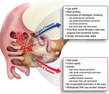
10
In addition to classifying ovarian tumors according to their FIGO stage, doctors have developed a classification system divding EOC into two types (Type I and Type II) based on the shared features of the different cancer types (55) (Figure 1.3). Type I carcinomas are low-grade cancers that are less aggressive, and are considered to be genetically stable, and surgically classified to arise form endometriosis or benign tumors. Type II cancers are high-grade EOCs classified to be biologically highly aggressive tumors, with a high tendency to metastasize from their primary lesions (56). Moreover, Type II carcinomas of the ovary have been genetically characterized to carry frequent mutations in TP53, BRCA1 and BRCA2 (56).
Figure 1.3. The two major histologic subtypes of EOC and their sub classifications
Type I tumors include low-grade, slow growing carcinomas known to develop from borderline tumors that develop from the ovarian surface epithelium, inclusion cysts, or endometriosis. Type II tumors include high-grade and rapidly growing carcinomas known to spread beyond the ovaries (57).
Additionally, pathologists classify EOC by grading the cancer, which allows examining the degree of similarity between EOC to healthy cells (58). When examined, the grading of the cancerous tissue helps pathologists make predictions on features of the cancer type such as its spreading potential, which in turn helps in improving prognosis (58). Healthy tissues consist of several cell types that are usually found clustered together, and when examining
11
tumor tissue, pathologists compare their cell characteristics to the normal healthy tissue and have developed a method to classify them accordingly: a) if the cancer cells display similar cellular architecture to normal cells, but contain a different type of cell grouping then it is referred to as a low-grade tumor or differentiated tumor (15); b) if the cancer cells look very different from healthy normal cells, they are then referred to as high-grade tumors or poorly differentiated tumors (15). There are not only two classifications for these cancer types, but some ovarian tumors are known as borderline cancers or borderline ovarian tumors (BOT) and are characterized by the absence of invasive stoma (50). BOTs have also been referred to as low-malignant potential tumors (LMP), they are also called borderline since these tumors present with cellular characteristics found in benign tissue or invasive cancers (50).
Importantly, epithelial EOC are mostly of the serous type and are graded as either:
1) LGSC -- low-grade serous carcinoma 2) HGSC -- high-grade serous carcinoma
While the other subtypes have been proposed to be graded, as follows:
Grade 1: The cancerous tissue is well differentiated, for which is appears to contain many normal-looking cells.
Grade 2: The tissue is moderately differentiated, and these cells appear to look more abnormal than normal.
Grade 3: The tissue is very poorly differentiated, and most of the cells in these tissues appear highly abnormal. (58)
1.1.5 Carcinogenesis and genetics of EOC
Like many other cancer types, EOC develops as a result of genetic mutations, and as acknowledged, these mutations play a major role in the acquisition of differentiated cellular functions that pertain and are not limited to enhanced growth and proliferation, increased cellular invasion and migration, angiogenesis, increased drug resistance and evasion of the immune system (59). There are two possible ways for the acquisition of genetic abnormalities, either through a) germline or inherited mutations, or b) acquired somatic mutations (60). These mutations are then known to result in either activating or inactivating tumor suppressors and oncogenes (60). One common example of inherited genetic
12
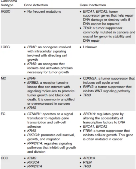
mutations in EOC is the inactivation of the tumor suppressor genes BRCA1 and BRCA2 (61). Population based studies on EOC patients indicate that between 10 and 15% of these patients have mutations in the BRCA1 or the BRCA2 genes (62, 63). Moreover, other genetic mutations have been described in EOC, as these gene mutations have been detected through the application of genome-wide association studies (64). These studies indicate inherited mutations of the genes MLH1, MSH2, MSH6, PMS2, EPCAM, and TP53 (64). Similarly, mutations acquired in EOC tumors are mostly found in HGSCs, with a common mutation in the TP53 gene (65). Furthermore, somatic mutations in the BRCA1 and BRCA2 genes also appear to occur in more than 30% of HGSCs (66). Likewise, rare somatic mutations have also been found in HGSCs, including activation and inactivation of the following genes: BRIP1, CHEK2, NF1, FAT3, CCNE1, PIK3CA, KRAS, MYC, PTEN, RB1, NF1, CDKN2A and RAD51C (56, 67). Table 1.2 summarizes the list of identified mutations in EOC and their suggested functions in the different EOC subtypes (50).
13
Table 1.2. Characteristic mutations found in EOCs
The table includes a list of the defined genetic alterations found in the different ovarian cancer subtypes. The gene list includes genes found either activated or inactivated in ovarian cancer (50).
In addition to genetic modifications found in EOC, epigenetic modifications have also showed to play additional roles in the EOC etiology (68). These types of modifications alter DNA accessibility to major transcription factors that in turn modify gene expression by either promoting or inhibiting their expression (68). The most common epigenetic modifications in EOC are DNA methylation, histone modifications, and more recently the suugested role of noncoding RNAs (ncRNAs) in the metastasis of EOC (68, 69). Multiple epigenomic studies have identified hyper and hypomethlyated genes and examined their role in the progression and carcinogenesis of EOC; most of them associated with poor
14
prognosis of the disease (70). One such example is the identified promoter methylation of the BRCA1 gene, found in more than 10% of HGSC patients (71). Studies from our lab also identified a role of aberrant DNA methylation in EOC, where data demonstrated that DNA hypermethylation occurs in low-malignant potential (borderline) tumors and these hypermethylated genes predominantly included key developmental/homeobox genes (72). Interestingly, contrary to DNA hypermethylation, significant DNA hypomethylation was observed almost exclusively in high-grade (grade 3) serous EOC tumors (72). These reports suggest that more focused research in the area of epigenetics may aid in the development of new EOC gene targets and more specific EOC therapies (68). Furthermore, studies in the field of proteomics and metabolomics have also helped in providing new understanding in EOC development, and specifying new detection and monitoring methods for this disease (73, 74). Proteomics studies have allowed for a close examination of important signaling pathways in EOC (74, 75). Thus, if research can have a better understanding of multiple signaling pathways suggested to play a role in EOC, then more sensitive and targeted therapies can be better explored and applied in the field.
1.1.6 EOC metastasis
EOC is one of the deadliest cancer types, especially since it presents at later or advanced stage of the disease (76). EOC patients show non-specific symptoms, making it harder to diagnose, in addition EOC is characterized with a highly aggressive metastatic profile, and is thus known to exhibit a high metastatic potential (76). Metastasis of EOC, like many other cancer types is known to progress in a manner reflected by documented changes in the cell’s biochemistry, morphology, migratory and invasive patterns (76). Nonetheless, the biological behavior of EOC is unique compared to other cancer types, since the process of EOC metastasis seems to occur more rapidly (77). It has been suggested that EOC from primary tumor sites can metastasize in a passive mechanism, which is made possible by the characterized physiological movement of peritoneal fluid to the peritoneum and omentum (77). The microenvironment where EOC cells to spread provides a unique placement for attachment and metastasis of these cells (76). For instance, the pelvic area provides EOC cells with high potential for metastasizing, since the organs in the pelvic region lack barriers that would usually hinder the spreading or metastasis of these tumor cells (76).
15
Additionally, the epithelial–mesenchymal transition (EMT) pathway is also another factor that promotes the invasive and migratory capacity of EOC cells (78). EMT contributes to the generation of circulating tumor cells from epithelial cancers such as EOCs, by promoting tumor cell intravasation to form mico-metastasis allowing for clonal outgrowth (79). Multiple studies have examined different gene pathways and protein types that regulate extracellular matrix degradation and coordinate cell junction assembly in EOC. Examinations of newly identified protein and gene receptors and their role in EMT induced EOC metastasis, include: the A-kinase anchor protein 4 (AKAP4) (80), Annexin A2 (81), Nidogen-1 (NID1) (82), the Golgi phosphoprotein 3 (GOLPH3) (83), the transcription factor E74-like factor 3 (ELF3) (84), in addition to two genes studied by our group: a) Hic-5 where we examine its role in EOC tumorigenesis through the regulation of the EMT pathway in a TGF-β-1 indpendent manner (TGF-β-1 is a transforming growth factor known to induce EMT, which is an essential process for cancer cell progression) (85, 86), b) the mannose receptor gene LY75, where our group examined its role in modulating EOC dissemination by regulating both the EMT and mesenchymal-epithelial transition (MET) pathways (87). Additionally, microRNAs are now more highly examined for their role in controlling signaling pathways that mediate EOC metastasis, studies have also examined several regulatory microRNAs that are shown to be involved in the formation of cancer stem cells, EMT, increasing migratory capacity of cancer cells, the enhanced formation of spheroids, apoptosis, autophagy, angiogenesis, formation and development of ascites. These EOC-related microRNAs include 145, mir-21, 31, 506, miR-101, miR-200, miR-214, and miR-25, each of them playing specific roles in the different EOC histotypes (76). Moreover, EOCs are also known for their peritoneal recurrence, and this occurs due to the availability of a protective niche for EOC cells, enhancing their ectopic survival by reprogramming genetic pathways that induces their capacity to become more aggressive when metastasized (88-91).
1.1.7 Risk factors of EOC
Research in EOC has led to the identification of several risk factors associated with EOC development, in addition to factors that have showed a protective role for the developing of the disease (92). Table 1.3 list the most described types of risks factors associated with