Université de Montréal
INTERLEUKIN-15 IN THE PATHOGENESIS OF
MULTIPLE SCLEROSIS AND ITS ANIMAL MODELS
par
Alma Nazlie Mohebiany
Département microbiologie et immunologie
Faculté de médecine
Mémoire présenté à la Faculté des études supérieures en vue de l’obtention
du grade de M.Sc. en microbiologie et immunologie
Août 2011
Université de Montréal
Faculté des études supérieurs
Ce mémoire intitulé :
INTERLEUKIN-15 IN THE PATHOGENESIS OF MULTIPLE
SCLEROSIS AND ITS ANIMAL MODELS
présenté par :
Alma Nazlie Mohebiany
a été évalué par un jury composé des personnes suivantes :
Dr. Nathalie Labrecque
(Président-rapporteur)Dr. Nathalie Arbour
(Directrice de recherché)Dr. Petronela Ancuta
(Membre de jury)Résumé
L'interleukine-15 (IL-15) contribue au développement et à l’activation des lymphocytes T CD8, des cellules immunes qui ont été impliquées dans plusieurs maladies auto-immunes telle la sclérose en plaques. Des niveaux élevés de l'IL-15 ont été trouvés chez les patients atteints de cette maladie comparativement aux témoins, mais aucune étude n'a examiné les effets de tels niveaux élevés sur les lymphocytes T CD8. Les objectifs de notre étude étaient 1- de caractériser l’expression de l'IL-15 par des lymphocytes B humains et de déterminer ses effets sur les fonctions des lymphocytes T CD8, et 2- d’évaluer l'expression in vivo de l'IL-15 dans des modèles murins de la sclérose en plaques.
Nous avons établi que les cellules B humaines augmentaient leur expression de l'IL-15 suite à une stimulation via le CD40. De plus, les fonctions effectrices des lymphocytes T CD8 ont été significativement augmentées lors des co-cultures avec des cellules B alloréactives exprimant l'IL-15. Dans les modèles murins de la sclérose en plaques, nous avons détecté au sein du système nerveux central des cellules immunes exprimant l’IL-15 ainsi que des cellules T CD8 exprimant le récepteur pour cette cytokine à différents stades de la maladie.
Nous avons démontré que les cellules B modulent des réponses des lymphocytes T CD8 via l’IL-15, ce qui suggère un rôle pour les cellules B dans la pathogenèse de la sclérose en plaques. Nous avons aussi mis en évidence la présence de cellules exprimant l’IL-15 dans le système nerveux central dans des modèles murins de cette maladie.
Mots clés
interleukine-15 cellules B cellules T CD8 sclérose en plaques
Summary
Interleukin-15 is a cytokine involved in the homeostatic proliferation and maintenance of CD8 T cells. Activated CD8 T cells are implicated in several autoimmune diseases, including Multiple Sclerosis (MS). Elevated levels of IL-15 have been reported in serum and on peripheral leukocytes of MS patients relative to controls, yet no study has addressed the effects of elevated IL-15 levels on CD8 T cells. To study the in vivo effects of any molecule, the animal model for MS, EAE, is used; the expression of IL-15 during the EAE disease course has not yet been elucidated. Thus the goals of our study were to characterize surface IL-15 expression on human B lymphocytes and determine the effects on human CD8 T cell functions; and to assess the in vivo expression of IL-15 in MS mouse models.
We found that B cells are capable of up-regulating the expression of surface IL-15 upon CD40 stimulation, and CD8 T cell effector functions were significantly enhanced upon co-culture with alloreactive IL-15-expressing B cells. In the MS mouse models we used, we found IL-15-expressing immune cells present within the central nervous system (CNS) at various points of disease, and that CNS-infiltrating CD8 T cells were potentially responsive to IL-15.
Here, we not only demonstrate the modulation of CD8 T cell responses by IL-15 presented by B cells, implying a role for B cells in MS pathogenesis, but also show the presence of IL-15-expressing cells within the inflamed CNS of EAE.
Key Words
Interleukin-15 B cells
CD8 T cells Multiple Sclerosis
TABLE OF CONTENTS
INTRODUCTION ...14
1.0.THEIMMUNESYSTEM...15
1.1 Adaptive immunity: Cell-mediated response...16
1.1.1 T cell development...16
1.1.2 The TCR complex and co-receptors ...17
1.1.3 T cell effectors ...18
1.1.3.1 CD4 T cells...18
1.1.3.2 CD8 T cells...19
1.2 Adaptive Immunity: Humoral response...21
1.2.1 B cell development ...21
1.2.2 B cell activation ...22
1.2.2.1 T-independent antigens...22
1.2.2.2 T-dependent antigens...22
1.2.2.3 Toll-like receptor activation ...23
1.2.3 B cell responses ...24
2.0INTERLEUKIN-15 ...26
2.1 Structure of IL-15 and IL-15Rα...27
2.2 Signal transduction ...28
2.2.1 Regulation of trans-presentation ...29
2.3 Expression of IL-15: regulation ...30
2.4 The role and effect of IL-15...31
2.5 IL-15 in disease ...33
2.5.1 Rheumatoid Arthritis ...33
2.5.2 Inflammatory Bowel Diseases ...34
2.5.3 Type 1/autoimmune diabetes ...34
2.5.4 CNS disorders ...34
3.0MULTIPLESCLEROSIS...35
3.1 Animal Models of MS ...36
3.1.1 Experimental Autoimmune Encephalomyelitis ...37
3.1.1.1 Characteristics of EAE ...37
3.1.1.2 Induction: Active vs. Passive EAE...38
3.1.1.3 Transgenic mouse models ...39
3.1.1.4 Limitations of EAE...39
3.2 Multiple Sclerosis and the Immune System ...40
3.2.1 The immune system and the CNS...40
3.2.2 Inflammation in MS...41
3.2.2.1 Innate immunity...41
3.2.2.2 MS and inflammation: the role of T cells...42
3.2.2.3 MS and inflammation: the role of B cells...46
3.3 Therapies...47
HYPOTHESIS AND OBJECTIVES...50
MATERIALS AND METHODS ...52
1.0ISOLATIONOFHUMANBLOODCELLS...53
1.1 Isolation of peripheral mononuclear cells (PBMC) from human blood ...53
1.2 Isolation of CD8 T cells and B cells...53
2.0IN-VITROASSAYS ...54
2.1 Upregulation of IL-15 on B cells...54
2.3 CD8 T cell and B cell co-cultures ...55
2.4 Flow Cytometry...55
2.5 Isolation of Human Brain Endothelial Cell (HBEC)* and Migration Assay...57
3.0IL-15ANDEAE ...57
3.1 Induction of Active Experimental Autoimmune Encephalomyelitis ...57
3.2 Scoring ...58
3.3 Perfusion ...58
3.4 Processing of organs: Lymphocyte isolation ...58
4.0IL-15EXPRESSION:QUANTITATIVEPCR ...59
4.1 RNA extraction: B cells ...59
4.2 RNA extraction: Mouse tissue ...60
4.3 Complementary DNA (cDNA)...60
4.4 Quantitative/Real-time Polymerase Chain Reaction ...61
5.0STATISTICALANALYSIS ...62
RESULTS ...63
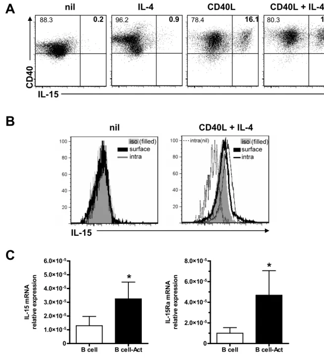
1.0B CELL ACTIVATION AND IL-15 PRODUCTION...64
1.1 Optimizing IL-15 production by B cells ...64
1.2 CD40L as a potent stimulus of B cells ...66
1.3 IL-15 on B cells enhances the cytotoxic profile of CD8 T cells ...70
1.4 IL-15 on B cells enhances the ability of CD8 T cells to cross an in vitro BBB...70
2.0EXPERIMENTALAUTOIMMUNEENCEPHALOMYELITISANDIL-15...73
2.1 Inducing Experimental Autoimmune Encephalomyelitis: the disease course ...73
2.2 IL-15 expression in EAE: mRNA...73
2.2.1 IL-15 in spleen and liver ...74
2.2.2 IL-15 in the CNS...75
2.3 IL-15 in EAE: infiltrating immune cells express IL-15 and IL-15Rα...78
2.4 CNS-infiltrating immune cells expressing IL-15 ...81
2.5 T cells found in the CNS of mice with active EAE are responsive to IL-15 ...84
DISCUSSION ...87
1.0EXPRESSIONOFIL-15BYHUMANBCELLS...88
1.1 Up-regulation of IL-15 expression...89
1.2 Effect of IL-15 on CD8 T cells ...91
2.0CHARACTERIZATIONOFIL-15INCLASSICALEAE ...93
2.1 Detection of IL-15 ...94
CONCLUSION ……...102
LIST OF FIGURES
FIG.1:DIAGRAM OF IL-15 GENE...28
FIG.2:TRANS-PRESENTATION OF IL-15...29
FIG.3:CD40L IS A POTENT STIMULUS FOR THE UPREGULATION OF IL-15 ON B CELLS...65
FIG.4:UPREGULATION OF IL-15 ON B CELLS VIA STIMULATION OF CD40 USING NIH-3T3 CD40L-EXPRESSING CELLS. ...67
FIG 5:SPECIFICITY OF THE UPREGULATION OF IL-15 ON B CELLS VIA STIMULATION OF CD40 USING NIH-3T3CD40L-EXPRESSING CELLS. ...69
FIG.6:IL-15 PRESENTED BY B CELLS IS FUNCTIONAL AND ENHANCES A CYTOTOXIC PROFILE IN
CD8T CELLS. ...71
FIG.7:IL-15 PRESENTED BY B CELLS ENHANCES THE ABILITY OF CD8T CELLS TO CROSS AN IN
VITRO BLOOD-BRAIN-BARRIER. ...72
FIG.8:TYPICAL CLINICAL SCORES AND DISEASE PROGRESSION OF ACTIVE EAE INDUCED IN
C57BL/6 AND SJL/J MICE. ...74
FIG.9:QUANTIFICATION OF IL-15 MRNA LEVELS IN SPLEEN AND LIVER OF B6 AND SJL MICE USING QPCR. ...76
FIG.10:EXPRESSION OF IL-15 IN VARIOUS PARTS OF THE CNS IN B6 AND SJL MICE. ...77
FIG. 11:IMMUNE CELLS FOUND IN THE CNS EXPRESS BOTH IL-15 AND ITS RECEPTOR ALPHA -CHAIN. ...79
FIG.12:IL-15 AND IL-15Rα-EXPRESSING CELLS ARE SPECIFICALLY FOUND IN THE CNS, AND ARE NOT PRESENT IN THE DLN OR SPLN...80
FIG.13:CHARACTERIZATION OF IL-15-EXPRESSING LYMPHOCYTES ISOLATED FROM CNS OF B6
MICE...82
FIG.14:CHARACTERIZATION OF IL-15-EXPRESSING LYMPHOCYTES ISOLATED FROM THE CNS OF
SJL MICE...83
FIG.15:T CELLS FOUND IN THE CNS ARE POTENTIALLY RESPONSIVE TO IL-15...85
LIST of TABLES
TABLE 1.ANTIBODIES USED FOR FLOW CYTOMETRY ANALYSIS...56
LIST OF ACRONYMS AND ABBREVIATIONS
A488 Alexa Fluor® 488 A700 Alexa Fluor® 700
AA amino acid
Ab antibody Ag antigen APC antigen presenting cells BCR B cell receptor
BAFF B lymphocyte activator of the TNF family
BBB blood-brain barrier
BFA Brefeldin A
B6 C57BL/6
cDNA Complementary DNA
CNS central nervous system
CSF cerebrospinal fluid
CFA complete Freund’s adjuvant
DC dendritic cells
D diversity DLN draining lymph nodes
DNA Deoxyribonucleic acid
EDTA ethylenediaminetetraacetic acid EAE experimental autoimmune encephalomyelitis G-CSF granulocyte colony-stimulating factor
GM-CSF granulocyte-macrophage colony-stimulating factor
GrB Granzyme B
HMGB1 High-mobility group protein B1 HBEC human brain endothelial cell
Hprt1 hypoxanthine phosphoribosyltransferase 1 Ig immunoglobulin
IBD inflammatory bowel disease IFN interferon
IL- interleukin-
ICAM intracellular adhesion molecule J joining
LSP long signal peptide
LPS lypopolysaccharide MHC major histocompatibility complex
MS Multiple Sclerosis MBP myelin basic protein
NCAM neural cell adhesion molecule
PB Pacific Blue
PBMC Peripheral blood mononuclear cells PFA paraformaldehyde
PAMP pathogen-associated molecular patterns PRR pattern recognition receptors PerCP-Cy5.5 Peridinin-cholorophyll proteins Cy5.5 PE Phycoerythrin
PECy7 Phycoerythrin-Cy7
polyI:C polyinosinic acid: polycytidylic acid PPMS primary progressive MS
PLP proteolipid protein
RAG recombinase-activating genes 1 and 2
RRMS relapsing-remitting MS
RPMI Roswell Park Memorial Institute
RT Room Temperature
RT-PCR Real-time polymerase chain reaction SPLN spleen
SPMS secondary progressive MS
SSP short signal peptide
STAT signal transducer and activator of transcription SJL SJL/J
TCR T cell receptor
TdT terminal deoxynucleotide transferase
TLR Toll-like receptors
TNF tumor necrosis factor
UTR untranslated region
V variable
DEDICATION
To my parents for seeing the end from the very beginning and encouraging me to reach it To E.T. and P.M. for the much needed push to start, helpful advice and support
To S.N.M. for believing in my capacity and keeping me company through many a sleepless night
To M.L. for your patience, flexibility and understanding when I needed to write
To B.B. for the never-ending support, patience, and steady encouragement; for helping me to see my progress and keep the end in sight
ACKNOWLEDGEMENTS
First and foremost, I would like to thank my supervisor, Nathalie Arbour, for giving me the opportunity to work with her and accompanying me in my learning, for being so understanding, and always being there to encourage and support.
I also thank my colleagues and lab-mates for their invaluable assistance and advice in times of need, for being there to discuss and troubleshoot experiments with and to make the time spent in the lab that much richer. Thank you to Camille Pittet and Raphael Schneider for first taking me under your wings and showing me the ropes, to Fatma Zaguia for your assistance with experiments and more, and to Emilie Viel for your words of encouragement. And many thanks to Diane Beauseigle, for your most invaluable help with experiments, for patiently answering my questions and the much appreciated support.
1.0. THE IMMUNE SYSTEM
The immune system is divided into two main branches: the innate immune system which serves as the first line of defense, and the adaptive immune system which tailors specific responses to and develops memory against pathogens. While there are two arms of the immune system defined, they usually act together: the activation of an antigen-specific response is initiated by components of the innate system, and the adaptive response will include innate effector responses to aid in promoting pathogen clearance. Thus, the cooperative effect of the two aspects of the immune system is necessary for a complete and effective immune response.
The innate immune system has various and diverse elements: there are physical barriers preventing the entry of pathogens into the body, from the epithelial cell layers with tight cell-cell contacts, to the mucus layers covering the respiratory and gastrointestinal tracts. It also includes various proteins and compounds or molecules produced by the innate system cells, including complement and chemokines and cytokines which attract and regulate the function of other immune cells. Lastly, the cells of the innate system express a range of pattern recognition receptors (PRRs), giving it the ability to respond to a number of pathogen-associated molecular patterns (PAMPs) on pathogens. Among the PRRs are Toll-like Receptors (TLRs) which recognize PAMPs such as lipopolysaccharide (LPS) from Gram negative bacteria, viral double-stranded RNA and unmethylated DNA with CpG motifs. Among the cells comprising the innate system, are natural killer cells, macrophages and dendritic cells (DC). The latter two cell types are phagocytic and, together with B cells, are classified as professional antigen presenting cells (APC). After processing of internalized proteins, these cells can present various antigens (Ag) to other immune cells on Major Histocompatibility Complex (MHC) Class II. DC, as well as activated B cells, have the ability to cross-present antigen on MHC Class I molecules as well. The efficacy of the first line of defense, namely the innate immune response, in addition to other factors, will contribute to whether or not the adaptive response is initiated.
The adaptive immune response is largely dependent on the receptors expressed on the T or B cell surface. The varied repertoire is due to the re-arrangement of the genes encoding the receptors; this resulting receptor diversity confers the ability of the adaptive
system to specifically respond to pathogenic insult, and to generate immune memory. The response of the adaptive system can be classified under two categories: the humoral response mediated by B cells, and the cell-mediated response mediated by T cells. In the former case, upon activation via the B cell receptor (BCR), these cells will secrete antibodies which promote opsonization and phagocytosis of foreign matter. Cellular immunity involves the activation of T cells via the T cell receptor (TCR) which occurs via recognition of antigen presented by MHC in addition to a co-stimulatory signal. Activation of the immune system will result in a strong and targeted response to allow pathogenic clearance including killing of pathogen-bearing cells; thus it is important that a balance is maintained with respect to the activation status of the immune cells, and that they do not become “mis”-activated.
1.1 Adaptive immunity: Cell-mediated response 1.1.1 T cell development
It is well-established that the thymus is crucial in providing the support T cell progenitors require for development, differentiation and selection to occur (Miller, 2002; Petrie, 2002). Progenitors, upon entering the thymus, are influenced by various factors; early expansion is influenced by IL-7, an essential factor in T cell development, (von Freeden-Jeffry et al., 1995), accompanied by the induction of various transcription factors, such as Notch (Zuniga-Pflucker, 2004) which serve to commit the precursor cells to the T cell lineage and induce expression of the genes required for TCR assembly.
The genes encoding TCRs are in a non-functional state; they exist as arrays of variable (V), diversity (D; present in only the β and δ chains of the TCR) and joining (J) segments; rearrangement is required for the creation of the genes encoding for the α and β or γ and δ chains of the TCR. Rearrangements are mediated by a protein complex encoded by recombinase-activating genes 1 and 2 (RAG). RAG binds and cleaves the DNA at specific sites flanking the V, D and J segments; the DNA ends are repaired and the resulting recombination, if functional, dictates the final structure and binding specificity of the TCR (Schatz and Ji, 2011). T cells expressing the αβ TCR will become double-positive, expressing both CD4 and CD8 (co-receptors of the TCR). The subsequent selection of T cells is crucial. Cells with TCRs that do not recognize or bind to self-MHC
molecules with low avidity are eliminated (positive selection), as are those with TCRs that recognize self-peptides presented by MHC molecules with too high avidity (negative selection) (Takahama, 2006), ensuring that self-reactive T cells do not mature. Self-reactive T cells escaping such selection can result in the development of autoimmune disease.
1.1.2 The TCR complex and co-receptors
The TCR is a heterodimer of di-sulfide linked α and β or γ and δ chains, and forms a complex together with CD3. CD3 is made up of the invariant proteins of the CD3 family (γ, δ and ε) and the ζ family (Clevers et al., 1988; Fernandez-Miguel et al., 1999). The CD3 chains possess sequence motifs for tyrosine phosphorylation, known as ITAMs (immunoreceptor tyrosine-based activation motif) (Davis, 2002); it is the CD3 component of the complex which is responsible for signaling upon stimulation of the TCR (Fernandez-Miguel et al., 1999). There are two pathways, it seems, which trigger T cell activation upon TCR stimulation: the first is tyrosine phosphorylation of the ζ chain ITAMs by src kinases (Salmond et al., 2009), and the second involves a conformational change in the CD3ε chain leading to Nck (an adaptor protein) recruitment (Davis, 2002). In either case, engagement of the TCR leads to the subsequent signaling via CD3, resulting in T cell activation; however, before signaling even occurs, the TCR needs to recognize and bind to a MHC-peptide complex. Aiding this process, the TCR/CD3 complex is further associated with other molecules, which further confer a degree of specificity to the T cell.
CD4 and CD8 are co-receptors to the TCR; they are trans-membrane glycoproteins, each with immunoglobulin-like (Ig-like) extracellular domains, and a short cytoplasmic tail (Leahy, 1995; Littman, 1987) which interacts with lck (Leahy, 1995), a src-family kinase, and thus they also play a role in signaling. There still are, however, differences in their structures. CD4 is a monomer with four Ig-like domains, and CD8 exists as a dimmer of either two α chains or an α and β chain (Leahy, 1995). These co-receptors play a role in MHC recognition by the TCR complex, increasing the affinity and stabilizing the interaction; CD4 and CD8 bind to non-polymorphic regions (von Freeden-Jeffry et al., 1995) of MHC class II or MHC class I respectively.
Stimulation of the TCR, while necessary, is not sufficient to fully induce a T cell response. Co-stimulation whether positive or negative, is also required. There are various co-stimulatory molecules, some of which are constitutively expressed and others which are induced. Among the co-stimulatory molecules expressed on T cells, are CD28, ICOS, CD30, 4-1BB, which result in positive signaling, and BTLA, CTLA-4 (CD152) and PD-1 regulators, which dampen activation of T cells (Beier et al., 2007). CD28 is constitutively expressed on T cells, and interacts with CD80/CD86 on APCs. In conjunction with TCR stimulation, CD28 ligation promotes IL-4 and IL-5 production, and induces increased levels of IL-2, which enhances proliferation in an autocrine manner and induces resistance to apoptosis (Beier et al., 2007).
1.1.3 T cell effectors
The effector functions of T cells are diverse and varied: they can function as helper cells by providing various signals to enhance responses mediated by several other immune cells; they may be directly involved in eliminating pathogens by killing infected cells, and T cells can also regulate and dampen immune responses, limiting tissue damage.
1.1.3.1 CD4 T cells
Naïve CD4 T cells (Th cells) differentiate into various helper T cell subsets upon activation. After stimulation by APCs, Th cells begin to produce IL-2; as they continue to respond to activating signals, they further differentiate (Chaplin, 2010). Th1 and Th2 are the classical helper cell subtypes; a specialized subset is the follicular helper cells. Regulatory T cells (Tregs) and recently, Th17 (Steinman, 2007) and Th9 (Veldhoen et al., 2008) cells have also been described.
Th1 cells drive cell-mediated responses, via activation of mononuclear phagocytes, natural killer cells and cytolytic T cells, licensing them to kill cells infected with intracellular pathogens. They produce IFNγ and IL-2, as well as lymphotoxin (Bonilla and Oettgen, 2010; Chaplin, 2010). Naïve helper CD4 T cells differentiate into Th1 cells under the influence of IL-12, IFNγ and the T-box expressed in T cell (T-bet) transcription factor (Bonilla and Oettgen, 2010).
Th2 cells differentiate under the influence of IL-4 and the transcription factor GATA-3 (Bonilla and Oettgen, 2010). They produce IL-4, 5, 10, and 13 and are responsible for enhancing antibody production by B cells, promoting clearance of parasites and extracellular pathogens.
Follicular helper cells are memory CD4 T cells which trigger the activation of B cells and reside in the lymph nodes and spleen; they express CXCR5, which mediates their recruitment to follicles (Bonilla and Oettgen, 2010). Th17 cells are induced in response to extracellular bacteria, and serve to help recruit neutrophils (Chaplin, 2010); this cell subset is also thought to play a role in autoimmunity (Bonilla and Oettgen, 2010). Th17 cell differentiation is induced by IL-1, IL-6 and TGF-β (Chung et al., 2009), in addition to autocrine IL-21 (Korn et al., 2009) under the direction of the transcription factors RORγt and RORα (Yang et al., 2008). IL-23 is also required for the expansion and maintenance of these cells (McGeachy et al., 2009), and differentiation is prevented by IL-12 and IFNγ (Harrington et al., 2005). Th17 cells produce IL-17 (A-F), a strong proinflammatory cytokine (Harrington et al., 2005; Korn et al., 2009; Steinman, 2007), as well as IL-6, GM-CSF and TNFα. Via their cytokine profile, Th17 cells are able to suppress regulatory T cells (Pasare and Medzhitov, 2003), stimulate macrophage differentiation (Hamilton and Anderson, 2004) and induce apoptosis (Hehlgans and Pfeffer, 2005).
1.1.3.2 CD8 T cells
CD8 T cells destroy infected cells or transformed cells; they are MHC class I restricted, and thus recognize antigens derived from polypeptides present in the cytosol. Upon activation, naïve CD8 T cells differentiate into cytotoxic T lymphocytes (CTLs) and are capable of killing target cells, in a contact-dependent manner. However it must be noted that CD8 T cells have functions other than killing; they produce cytokines such as IFNg and TNF as well as chemokines which recruit and or activate macrophages, neutrophils and microbicidal effector cells (Harty and Bevan, 1999; Harty et al., 2000) and which may also have the effect of interfering with pathogen attachment or gene expression (Harty et al., 2000).
The generation of effector CTL requires multiple signals; the signaling cascades of these events have been extensively studied in mouse models of viral infections. The first of
these is the interaction between the TCR and MHC class I molecules presenting Ag. A stimulatory signal is required for the full activation of a CD8 T cell. The “typical” co-stimulation of CD8 T cells is the interaction between CD80 or CD86 (members of the B7 family) and CD28, which results in IL-2 production (Lenschow et al., 1996; Sharpe and Freeman, 2002). Another co-stimulatory molecule is 4-1BB, or CD137 which is expressed by activated T cells and plays a role in augmenting CTL responses during suboptimal stimulation (Williams and Bevan, 2007). The ligation of 4-1BB by T cells results in the secretion of IL-2 and the induction of cell division (Bertram et al., 2002). While the signals mediated through TCR/MHC interaction and through co-stimulatory molecules are sufficient to activate (or suppress) CD8 T cells, the development of an optimal CTL response requires a cytokine signal (Williams and Bevan, 2007). IL-12 and type 1 interferons (IFNα/β) promote the expansion and differentiation of CTLs (Mescher et al., 2006; Thompson et al., 2006). IL-12 is important in the development of CTL cytotoxicity by inducing IFNγ production (Curtsinger et al., 2003) as well as the expression of granzyme B (Curtsinger et al., 2005). The expression of various transcription factors important in CTL differentiation is also induced by, or in part by, IL-12: T-bet (Joshi et al., 2007; Takemoto et al., 2006) is crucial for differentiation of CD8 T cells into effector cells (Sullivan et al., 2003); STAT4 activation is mediated by IL-12 and IFNα/β, leading to IFNγ production (Nguyen et al., 2002); and Type 1 interferons also promote the survival of activated CD8 T cells (Marrack et al., 1999). The balance between these cytokines, IL-12 and IFNα/β, varies depending on the infection (Cousens et al., 1999; Kolumam et al., 2005). Of course, this is a rather simplified perspective, and it is more than likely that other cytokines play a role and influence the CD8 T cell-mediated response. Furthermore, much of what is known about the CD8 T cell response is due to studies in various animal viral models, and how the immune response plays out in humans in the context of pathogen clearance and autoimmune disease may not be quite the same.
After a CD8 T cell-mediated immune response, the majority of effector cells die via apoptosis, mediated through Bim (Hildeman et al., 2002; Pellegrini et al., 2003) and potentially other mechanisms. The proportion of cells that survive become memory T cells, characterized by relatively enhanced proliferation, the ability to survive for long periods of time, and increased ability to re-express effector genes (Parish and Kaech, 2009). In
addition, memory CD8 T cells express receptors for IL-7, promoting survival, and IL-15, driving antigen-independent proliferation (Williams and Bevan, 2007).
1.2 Adaptive Immunity: Humoral response 1.2.1 B cell development
B cells are derived from hematopoietic stem cells in the bone marrow. During development, they acquire antigen specificity; pro-B cells express TdT (Terminal deoxynucleotide transferase), a specialized template-independent DNA polymerase that catalyzes polymerization of deoxynucleoside triphosphates in vitro (Benedict et al., 2000), and RAG1 and RAG2, which are necessary for gene recombination of the B cell receptor (BCR) (Mesquita Junior et al., 2010). The BCR is an immunoglobulin, consisting of two heavy and two light chains linked by disulfide bonds. There are 5 heavy chains: α, γ, δ, ε, and μ which define the classes of immunoglobulins: IgA, IgG, IgD, IgE and IgM respectively. The light chains are kappa (κ) and lambda (λ). The heavy chain is made up of various segments: VH, D, JH and CL (Bonilla and Oettgen, 2010); the segments VH, D, and JH are rearranged and assembled under the influence of RAG. The heavy chain complex is associated with an invariable chain and expressed on the cell surface as a pre-BCR with two peptide chains, Igα and Igβ. These peptides, which play a role in BCR trafficking and signal transduction, have an N-terminal extracellular Ig-like domain, a single trans-membrane domain and a cytoplasmic domain of 61 and 48 amino acids respectively (Defranco et al., 1995). Expression of the pre-BCR on the cell surface is followed by the rearrangement of the light chain κ; if a functional rearrangement fails, then the λ chain is rearranged. Antigen specificity is conferred by the variable regions of the heavy and light chains. The BCR of naïve B lymphocytes are IgM; co-expression of IgD marks a mature B cell (Zubler, 2001). Upon activation, the BCR undergoes class switching (Mesquita Junior et al., 2010).
Positive and negative selection follow the creation of a functional, intact BCR. Positive selection confers survival signals upon the B cells; negative selection takes place in the bone marrow. B cells that recognize self-antigen with high affinity will either undergo apoptosis or are given a second chance: RAG genes are re-activated and another light chain-VJ recombination is generated to substitute for an autoreactive combination
(Mesquita Junior et al., 2010). B cells recognizing self-antigens with weaker reactivity enter varying degrees of inactivation (i.e. anergy), but anergic cells still may be recruited in immune responses (Zubler, 2001). B cells migrating out of the bone marrow express various chemokine receptors and other molecules allowing them to migrate to specific areas within the body.
One subset of B cells, B1 in mice, migrate to the peritoneal tissues or cavities, the primary follicles of Peyer’s patches, and/or to the spleen and lymph nodes. Another B cell subset, B2, migrate to the splenic marginal zone, rich in macrophages and DCs, and favoring a T-independent response (Zubler, 2001).
1.2.2 B cell activation
The BCR complex consists of the membrane immunoglobulin and two peptide chains, Igα and Igβ, which are responsible for signaling via ITAM motifs. Cross-linking of the BCR leads to clustering of the cytoplasmic domains of Igα and Igβ. Subsequent phosphorylation of the ITAM motifs mediated by src-family tyrosine kinases further recruits other tyrosine kinases (Syk or Lyn) (Defranco et al., 1995), thereby activating factors that promote the transcription of genes involved in the proliferation and differentiation of the B cell (Mesquita Junior et al., 2010). The activation of B cells can take place in one of two ways, depending on the type of antigen it interacts with. One response requires T cell help, initiated by T-dependent antigens; the other, independent of T cell help, is initiated by T-independent antigens (Bonilla and Oettgen, 2010). More recently, the role of TLRs in the activation of B cells is of increasing interest.
1.2.2.1 T-independent antigens
Some macromolecules have repeating molecular patterns and can interact with and cross-link multiple immunoglobulin receptors on B cells. The (weak) activating signals transduced by cross-linking requires additional signals from cytokines or other cell contacts, provided by DCs (Bonilla and Oettgen, 2010).
1.2.2.2 T-dependent antigens
B cells, like T cells, require more than one signal for optimal activation. B cells are activated by activated T cells which recognize the peptide presented on B cells via MHC
class II. The BCR will have recognized and captured the antigen, resulting in cross-linking of the BCR and induction of intracellular signaling, which leads to the production of additional molecules to enhance the B-T cell interaction and allows B cells to receive additional signals needed for full B cell activation (Bonilla and Oettgen, 2010). The interaction between B and T cell ultimately leads to each activating the other through the expression of various molecules and cytokines. Once activated, T cells express co-stimulatory molecules such as ICOS, which interacts with the ligand B7RP-1 (ICOS-L) (Zubler, 2001). Interactions between various other molecules expressed on B and T cells lead to further stimulation and activation. Another important interaction is that between CD40L on activated T cells and CD40 on B cells, which also leads to induction of cytokine production (Mesquita Junior et al., 2010). Overall, the T-dependent activation of B cells is complex, and is not unidirectional, but rather each cell type induces the activation of the other.
1.2.2.3 Toll-like receptor activation
TLRs are a subset of pattern recognition receptors (PRRs), usually found expressed by innate cells, but are also found in other cell types including B cells. In general, PRRs recognize PAMPs found on microbial organisms, which are foreign and absent on host cells (Janeway, 2001). TLRs are type 1 trans-membrane glycoproteins with leucine-rich repeat motifs on their extracellular domain and a Toll/interleukin-1R-interacting domain which contributes to cytoplasmic signaling via the MyD88 pathway (Booth et al., 2011), with the exception of TLR3 which mediates signaling through TRIF (Brown et al., 2011). Human B cells express moderate to high levels of TLRs 1, 6, 7, 9 and 10, and low levels of TLR 2; TLRs 3 and 4 are not found on human B cells (Booth et al., 2011). The levels of TLR found on B cells vary according to the B cell subset, thus affecting the sensitivity to TLR-mediated signaling and activation. Memory B cells, in general, express higher levels of TLR, and activation via TLR interaction with their ligands induces differentiation into Ab-producing plasma cells. On the other hand, naïve B cells express lower levels of TLR and thus respond weakly to TLR agonists (Booth et al., 2011). It has been demonstrated that activation of naïve B cells may be dependent on stimulation via TLR, and thus it has been proposed that TLR stimulation may be a required 3rd signal following BCR cross-linking and T cell help (Ruprecht and Lanzavecchia, 2006).
Following efficient activation via the TLR, B cells are able to proliferate, secrete cytokines and express co-stimulatory molecules. TLR stimulation was also shown to inhibit apoptosis (Ruprecht and Lanzavecchia, 2006). In the case of CpG, acting on TLR9, stimulation promotes BCR class-switching to IgG2a, IgG2b and IgG3, and inhibits IgG1 and IgE switching (Lin et al., 2004; Liu et al., 2003). The secretion of immunoglobulins and cytokines requires additional signals concurrent to the CpG-induced signals. BCR cross-linking, CD40 ligation and factors such as BAFF and APRIL contribute to Ig secretion, whereas secretion of IL-6, IL-12 or IL-10 requires signals from CD40L or DCs (Booth et al., 2011).
Activation of B cells can occur in various ways, stimulation leading to different signaling pathways resulting in the expression of various cytokines and molecules important to mediating an immune response. Upon activation, B cells can either respond “directly” via antibody production, or also have a role in cytokine production which can modulate the immune response mediated by other immune cells.
1.2.3 B cell responses
The primary B cell response, relatively slow to develop, involves the activation of naïve B cells and their differentiation into either short-lived plasma blasts which produce antibodies, or into memory cells. Upon activation, B cells go down one of two pathways; they either further differentiation into short-lived antibody-producing plasma blasts, or enter follicles and establish a germinal center where they undergo class-switching of the BCR (Bonilla and Oettgen, 2010). The primary response is dominated by the production of IgM Ab with relatively low affinity for Ag; other Ab classes appear later and show higher affinity for the cognate antigen (Bonilla and Oettgen, 2010). In the germinal center, the BCR changes from IgM or IgD to IgG, IgA or IgE. The gene arrangement involved in class switching is analogous to TCR and BCR rearrangement and involves genomic DNA rearrangement and alternative RNA splicing (Bonilla and Oettgen, 2010; Mesquita Junior et al., 2010). The variable regions of the immunoglobulin remain the same, so that the specificity for antigen does not change. It is the constant portion of the heavy chain which is replaced by one of the other chains, thereby diversifying the immune response since the different Ig classes each have a different function (Mesquita Junior et al., 2010). Class
switching and the resulting heavy chain is under cytokine influence. Various cytokines induce a switch to IgG (Zubler, 2001), IL-4 and IL-13 induce a switch to IgE, which is inhibited by IFNγ, and IL-10 and TGFβ have been shown to promote IgA (Bonilla and Oettgen, 2010).
In addition to producing antibodies, B cells have a role in cytokine production. For instance, B cells produce IL-10, which is primarily anti-inflammatory, but can be pro-inflammatory under some conditions (Nikolajczyk, 2010). Activated B cells produce interleukins, TNF, macrophage inflammatory protein (MIP)1α and MIP1β. These factors modulate the migration of DC, activate macrophages, regulate T cell functions and also provide feedback signals further stimulating B cells (Dalakas, 2008).
Memory B cells can be of two sorts: the “conventional” memory B cells, or memory plasma cells (Yoshida et al., 2010). The former require restimulation with antigen, while plasma cells secrete specific antibodies for an extended time, long after the initial infection has been cleared, and persist independently of antigenic stimulation (Yoshida et al., 2010). The level of antigen required to elicit a response from memory B cells is lower than that for the response mediated by naïve B cells. In addition, the immune response mediated by memory B cells is faster, and antibody production reaches higher levels than the primary response (Mesquita Junior et al., 2010). While plasma cells depend on signals from BAFF and APRIL (see below), memory B cells require expression of a functional BCR complex, capable of mediating signaling (Yoshida et al., 2010). Interestingly, memory B cells can also be maintained via Epstein-Barr virus infection; the expression of viral receptors mimics regular activation signals, by-passing the direct need for BCR signaling (Yoshida et al., 2010).
B lymphocyte activator of the TNF family, or BAFF, is a potent stimulator of B cell maturation and survival. It is expressed by activated macrophages and DCs (Zubler, 2001) in membrane-bound or soluble form (Schneider et al., 1999; Thangarajh et al., 2004) and interacts with transmembrane activator and CamL interactor (TACI), expressed on activated B cells. TACI has been shown to have a role in B cell homeostasis in addition to being a key mediator of class switching (Castigli et al., 2005b; Seshasayee et al., 2003). TACI is expressed on mature B cells and plasma cells (Hsu et al., 2002), and mutations are associated with IgA deficiency (Castigli et al., 2005a). APRIL (a proliferation-inducing ligand) shares properties and structural homology with BAFF (Schneider et al., 1999;
Thangarajh et al., 2004), also interacts with TACI, and is expressed on a range of leukocytes (Bonilla and Oettgen, 2010; Zubler, 2001). BAFF and APRIL have been implicated in the pathogenesis of various autoimmune diseases including multiple sclerosis, systemic lupus erythematosus, and rheumatoid arthritis.
2.0 INTERLEUKIN-15
Interleukin-15 (IL-15) is a pleiotropic cytokine, expressed (at the mRNA level) in many tissues including kidney, lung, heart, skeletal muscle (Grabstein et al., 1994), and by various cell types under stimulatory conditions (Blauvelt et al., 1996; Jonuleit et al., 1997; Leclercq et al., 1996; Mohamadzadeh et al., 1995; Reinecker et al., 1996). However, in spite of this, the actual protein expression is not as widespread as the mRNA would indicate, and in fact translation is tightly regulated (Fehniger and Caligiuri, 2001). IL-15 is a member of the four α-helix family of cytokines which also include IL-2, IL-3, IL-6, IL-7, granulocyte stimulating factor (G-CSF) and granulocyte-macrophage colony-stimulating factor (GM-CSF) (Bazan, 1990a; Bazan, 1990b).
IL-15 was independently discovered by two groups based on its ability to mimic the effects of IL-2 in vitro (reviewed by (Waldmann, 2006)); however, in vivo, the effects of these two cytokines are very different (Fehniger and Caligiuri, 2001) demonstrated by using mice deficient in IL-15 (Kennedy et al., 2000) or IL-15Rα (Lodolce et al., 1998). The IL-15 and IL-2 receptors each have a unique alpha chain (Rα) which allows for the binding of the cytokine with high affinity. IL-2Rα expression is restricted to T cells for the most part, but is also expressed by DC (Wuest et al., 2011), while IL-15Rα overlaps with IL-15 in its expression at the mRNA level, and is found in almost every cell and tissue type (Anderson et al., 1995a; Giri et al., 1995). However, unlike the cytokine, IL-15Rα protein expression is not under similar regulation, and seems to parallel the transcript expression (Anderson et al., 1995b; Giri et al., 1995; Stonier et al., 2008). Interestingly, a pool of intracellular IL-15 is present in monocytes/macrophages, which is ready to be expressed on the surface as needed and independently of new protein synthesis (Neely et al., 2001). Signaling is mediated via a shared β chain (IL-2/15Rβ or CD122) (Bamford et al., 1994; Carson et al., 1994) and the common γ chain, also shared by 2, 4, 7, 9 and IL-21 (Stonier and Schluns, 2010).
2.1 Structure of IL-15 and IL-15Rα
The IL-15 gene (Fig.1) contains 9 exons, 7 of which are coding exons (Fehniger and Caligiuri, 2001). An alternative exon has also been described, encoding for an alternative leader peptide, between exons 4 and 5 (Nishimura et al., 1998; Onu et al., 1997; Tagaya et al., 1997). The original IL-15 encodes for an IL-15 precursor protein with a long (48-amino acid (AA)) leader peptide (LSP) and a 114-AA mature protein; the alternate sequence encodes for a short signal peptide (SSP) of 21-AA (Fehniger and Caligiuri, 2001). Both isoforms of IL-15 encode identical IL-15 protein, the only difference being in the signal sequence, and the localization of the final products: the LSP is secreted from the cell and/or placed into the cell membrane (Kurys et al., 2000; Onu et al., 1997), and the SSP remains localized to the cytoplasmic and nuclear compartments (Waldmann and Tagaya, 1999). The mature IL-15 protein is encoded by exons 5 to 8 of the IL-15 gene (Anderson et al., 1995b; Krause et al., 1996) and contains two disulfide bonds, one of which is homologous to that of IL-2 (Fehniger and Caligiuri, 2001). IL-15 has been predicted to have “helical moments” at four different sites in the protein (at AA 1-15, 18-57, 65-78 and 97-114), supporting the model of a four α-helix bundle structure (Fehniger and Caligiuri, 2001).
IL-15Rα is a type-1 trans-membrane protein with eight isoforms (Waldmann and Tagaya, 1999) and has been shown to be highly homologous to the IL-2Rα subunit (Anderson et al., 1995a; Giri et al., 1995). Both contain a short cytoplasmic tail, a transmembrane domain, a hinge region and a sushi domain (most isoforms), which is required for cytokine binding (Anderson et al., 1995a; Giri et al., 1995; Waldmann, 2006). However, in contrast to IL-2Rα which has low affinity for IL-2 in the absence of IL-2Rβγ, IL-15Rα binds its cytokine with high-affinity, without the need for the rest of the receptor complex (Fehniger and Caligiuri, 2001). Furthermore, IL-15Rα is capable of reverse signaling; it was demonstrated to mediate a non-proliferative signal in the absence of IL-2/15Rβ (Stevens et al., 1997) and hyper cross-linking of surface IL-15 resulted in activation of various signaling pathways including ERK 1/2 and MAPK (Neely et al., 2004). However, for signaling to occur, the Rα chain is not absolutely necessary: IL-15 may bind to IL-2/15Rβγ with intermediate affinity, in the absence of IL-15Rα (Giri et al., 1994).
Fig.1: Diagram of IL-15 gene. IL-15 has two isoforms due to an alternate exon (4A) resulting in a short (21-AA) signal peptide (IL-15SSP) rather than the long (48-AA) signal peptide (IL-15LSP). Adapted from (Bulfone-Paus et al., 2006).
2.2 Signal transduction
The IL-15R complex signals through Jak1 and 3 as well as signal transducer and activator of transcription (STAT) 3 and 5 (Giri et al., 1995; Waldmann et al., 1998). IL-15 was shown to activate the transcription factor NF-κB in neutrophils but not AP-1, while IL-15 stimulation of whole human peripheral blood lymphocytes activated both (Fehniger and Caligiuri, 2001). Additional signaling pathways may include src-related tyrosine kinases and through the Ras/Raf/MAPK pathway leading to fos/jun activation (McInnes and Gracie, 2004). However, it must be noted that signaling related to IL-15 does not occur in the conventional manner as with other cytokines.
A study in which T cell proliferation mediated by IL-15 did not require IL-15Rα expression on the responding T cells but rather required IL-15Rα expression by surrounding cells (Lodolce et al., 1998) was the first indication of a unique manner by
which IL-15 signaling is mediated. As the direct effect of IL-15 in inducing T cell proliferation had already been established, it was unlikely their observations were due to an indirect effect mediated by IL-15 (Stonier and Schluns, 2010). Another group soon proposed a mechanism of IL-15 trans-presentation (depicted in Fig. 2), demonstrating a prolonged effect on T cells by IL-15 relative to IL-2, despite IL-15 withdrawal from the media (Dubois et al., 2002). This was demonstrated to be mediated by the strong association of IL-15 with IL-15Rα, allowing the receptor alpha chain to maintain IL-15 on the cell surface and enhance the persistence of IL-15 signaling (Dubois et al., 2002). Furthermore, IL-15 and IL-15Rα were shown to associate intracellularly and were followed from the endoplasmic reticulum to the cell surface (Dubois et al., 2002).
Fig.2: Trans-presentation of IL-15. IL-15/IL-15Rα-expressing cell presents IL-15 in trans to effector cells expressing either IL-15Rβγ or IL-15Rαβγ. Adapted from (Bulfone-Paus et al., 2006)
2.2.1 Regulation of trans-presentation
At the most basic level, trans-presentation requires IL-15 and IL-15Rα to be transcribed in the same cell. However, it is not yet known if up-regulation of transcription will be sufficient to increase the IL-15 being trans-presented (Stonier and Schluns, 2010). It is known that there are several mechanisms in place at the post-transcriptional level
(discussed later) which would limit the availability of IL-15 for trans-presentation. Similar mechanisms regulating IL-15Rα have not been identified, which perhaps explains the higher prevalence of IL-15Rα compared to IL-15. Another level of regulation after limited protein expression is the transport of 15 to the cell surface. It has been reported that IL-15Rα is required for IL-15 to be transported from the endoplasmic reticulum to the cell surface (Duitman et al., 2008). Furthermore, IL-15Rα bound to IL-15 can be internalized and later return to the surface (Dubois et al., 2002) which may promote the need for regulating cytokine production, or may be an additional level of control, to limit the availability of IL-15 as needed.
Another factor to consider limiting the availability of IL-15 to cells could simply be cellular localization. For trans-presentation to occur, a responsive cell must be in close proximity to the cell presenting IL-15. It is thus highly probable that rather than randomly encountering IL-15 presenting cells, responsive cells would be directed to the source (Stonier and Schluns, 2010).
2.3 Expression of IL-15: regulation
IL-15 mRNA is found in many cell types, but protein expression is limited and regulated through multiple mechanisms. IL-15 production is regulated at both the transcriptional and posttranscriptional levels. Several binding sites for transcription factors were identified in the promoter regions of human and murine IL-15 (Fehniger and Caligiuri, 2001). There are three main “checkpoints” which regulate translation of IL-15 mRNA (Fehniger and Caligiuri, 2001): first, the existence of multiple start codons in the 5’ untranslated region (UTR); the LSP and SSP themselves; and negative regulation near the C terminus of the protein precursor.
The LSP IL-15 5’ UTR is relatively long and contains multiple AUGs upstream of the translation start site, which impedes translational efficiency (Bamford et al., 1996; Fehniger and Caligiuri, 2001). The removal of most of the upstream AUG sequences in the 5’ UTR resulted in a 5- to 10-fold increase in IL-15 protein expression (Bamford et al., 1996). It was also demonstrated that LSP IL-15 is translated with lower efficiency than SSP IL-15 (Tagaya et al., 1997); also considering the localization of each of the two isoforms, we see a tight regulation of protein expression. SSP IL-15 is more efficiently
translated, but is localized to the nucleus and cytoplasmic regions, and is not secreted. Not only is LSP IL-15 is less efficiently translated, but also is secreted from the cell and/or inserted into the cell membrane at low levels (Fehniger and Caligiuri, 2001; Kurys et al., 2000). Finally, it was suggested that a signal in the C terminus of the mature IL-15 protein is responsible for inefficient secretion (Fehniger and Caligiuri, 2001); since the activity of a LSP-IL-15-GFP construct was significantly higher than that of LSP-IL-15 (approximately 10 times more labeled IL-15 was secreted), the authors suggested the GFP tag masked a retention signal in the C terminus (Gaggero et al., 1999).
If all three of these “checkpoints” are removed – removing upstream AUG sequences, replacing IL-15 leader peptide with that of IL-2 and changing the C terminus end of the protein, synthesis of IL-15 increased by 250-fold (Bamford et al., 1998). The extent to which the expression of IL-15 is regulated indicates that overproduction would be dangerous. There is some evidence to support this: transgenic mice over-expressing IL-15 develop a fatal lymphocytic leukemia (Fehniger et al., 2001), and IL-15 is consistently upregulated in leukemic cells of childhood acute lymphoblastic leukemia patients with CNS involvement (Cario et al., 2007).
2.4 The role and effect of IL-15
IL-15 is important in the development, homeostasis and activity of NK cells and γδ T cells, and plays a role in the survival and expansion of naïve and memory CD8 T cells. It also has an effect on other immune cells, as well as on non-immune cells such as fibroblasts, epithelial and endothelial cells, and neuronal and glial cells.
IL-15 is important in the survival and expansion of naïve and memory CD8 T cells (Alves et al., 2003; Berard et al., 2003); homeostatic proliferation in response to IL-15 allows for the self-renewal and maintenance of memory CD8 T cells over long periods of time (Becker et al., 2002; Goldrath et al., 2002). Mice deficient in IL-15 or IL-15Rα have a marked decrease in the number of memory CD8 T cells and, to a lesser extent, in the number of naïve CD8 T cells, but have normal levels of CD4 and B cells (Kennedy et al., 2000; Lodolce et al., 1998); IL-15Rα deficient mice are, in addition, impaired in thymic CD8 T cell development (Lodolce et al., 1998).
Naïve T cells can proliferate in response to high concentrations of IL-2 and IL-15, as long as they also receive a signal via the TCR through MHC recognition (Cho et al., 2007; Ramsey et al., 2008). Also supporting a role for IL-15 in homeostatic proliferation, mice deficient in IL-15 have about half the number of naïve CD8 T cells relative to wild type mice (Berard et al., 2003; Ramanathan et al., 2006). Involved with the regulation of the naïve T cell response to IL-15 is SOCS-1, which is up-regulated in response to IL-15 (among other cytokines) (Yoshimura et al., 2007). Deficiency in SOCS1 increases the sensitivity of double positive thymocytes to both IL-7 and IL-15 (Chong et al., 2003; Ramanathan et al., 2006) and results in mature CD8 T cells that are hyper-sensitive to IL-15 (Davey et al., 2005; Ramanathan et al., 2006). Mice deficient in SOCS1 (restricted to T cells) have CD8 T cells of primarily a memory phenotype (Davey et al., 2005). Overall, as a regulator of the cellular response to IL-15, SOCS1 might be involved in controlling antigen-independent activation of naïve CD8 T cells under inflammatory conditions, to prevent expansion of potentially autoreactive CD8 T cells (Ramanathan et al., 2008).
Along with IL-7, IL-15 has a crucial role in the MHC-independent homeostasis of memory CD8 T cells (Surh and Sprent, 2008). While the expansion of memory CD8 T cells under the influence of IL-7 occurs only in lymphopenic hosts, IL-15 is able to support and favor the generation of memory CD8 T cells in mice with an intact T cell compartment (Surh and Sprent, 2008). One of the first indications that cytokines were involved in the maintenance of memory T cells, the injection of adjuvants resulted in an increase in the proliferation of memory CD8 T cells (Surh and Sprent, 2008). Further investigation showed that such adjuvants (e.g. LPS or PolyI:C) stimulated the production of type-1 interferons, which promoted the synthesis of IL-15 on APCs; this IL-15 was shown to act on memory CD8 T cells and enhance the rate of homeostatic proliferation (Zhang et al., 1998). Administering IL-15 to IL-15 deficient mice increased the proportion and number of CD8 memory T cells in the spleen and lymph nodes (Berard et al., 2003), and further outlining a crucial role for IL-15, memory CD8 T cells transferred into IL-15 deficient mice failed to proliferate and died rapidly (Judge et al., 2002).
2.5 IL-15 in disease
Activation of CD8 T cells, while beneficial in protective immune responses, can also contribute to chronic inflammatory disorders as well as to the progression of autoimmune diseases due to their ability to directly kill target cells. CD8 T cells have been implicated in various autoimmune diseases (reviewed by (Walter and Santamaria, 2005). IL-15, being one of the cytokines involved in the homeostatic maintenance of CD8 T cells, is an ideal candidate for potentially aberrant CD8 T cell activation. This cytokine facilitates the maintenance and triggering of autoreactive CD8 T cells (Itsumi et al., 2009; Peng et al., 2006; Ramanathan et al., 2011) and plays a crucial role in CD8 T cell migration and survival in organs (McGill et al., 2010; Verbist et al., 2011). Indeed, IL-15 has been implicated in various diseases, including those in which CD8 T cells have also been implicated: rheumatoid arthritis (RA), inflammatory bowel diseases (IBD), type 1 diabetes, psoriasis, as well as various CNS disorders (Davey et al., 2005; Liu et al., 2000; McInnes and Gracie, 2004; McInnes and Liew, 1998; Ramanathan et al., 2011; Rentzos and Rombos, 2011; Sakai et al., 1998). Furthermore, due to its ability to activate T cells, IL-15 is also under investigation regarding its potential in anti-tumor immunotherapy (Hanson et al., 2000; Jakobisiak et al., 2011; Klebanoff et al., 2011) and as a factor to improve vaccine efficacy (Hu et al., 2009; Huntington et al., 2011; Yu et al., 2011).
2.5.1 Rheumatoid Arthritis
IL-15 protein has been detected in synovium and in RA joint lesions (Thurkow et al., 1997). Synovial fluids were demonstrated to promote the migration and activation of T cells, which was partially inhibited by the addition of antibodies antagonistic to IL-15 (McInnes et al., 1996). Furthermore, IL-15 activated T cells from RA patients were able to stimulate macrophage cell lines as well as primary monocytes and macrophages to produce TNFα in vitro. Phase II clinical trials using an anti-IL-15 human monoclonal antibody (AMG 714) have been completed (NCT00433875, http://www.clinicaltrials.gov), and the same antibody is also being currently tested for psoriasis in a phase II trial (NCT00443326).
2.5.2 Inflammatory Bowel Diseases
IL-15 mRNA has been detected in macrophages and epithelial cells from patients with active IBD and has been detected at the protein level in the supernatants of rectal mucosal biopsy samples (Sakai et al., 1998). Mention et al. showed that IL-15 was not secreted, but rather was presented at the surface of enterocytes in celiac disease (Mention et al., 2003). In addition, T cells isolated from the lamina propria of IBD patients showed a greater responsiveness to IL-15 as compared to controls, not only producing IFNγ and TNF in response to IL-15 stimulation, but also inducing the production of pro-inflammatory cytokines in monocytes in vitro (Liu et al., 2000). In conjunction with retinoic acid, IL-15 induced the production of IL-12p70 and IL-23 by DC (DePaolo et al., 2011). Not only does the elevated levels of IL-15 seem to induce the production of various pro-inflammatory cytokines, but also is able to stimulate CD8 T cells to be cytotoxic. Studies have shown increased induction of NKG2D in response to elevated levels of IL-15 (Meresse et al., 2004; Roberts et al., 2001), and that NKG2D serves as a co-stimulator for CTL (Roberts et al., 2001), activating CTL functions independently of TCR specificity(Meresse et al., 2004).
2.5.3 Type 1/autoimmune diabetes
Serum IL-15 has been detected in patients with type 1 diabetes and levels were significantly higher as compared to controls (Kuczynski et al., 2005). In a mouse model of autoimmune diabetes, it was shown that stimulation with IL-15 and IL-21 enabled CD8 T cells to respond to weak antigens, inducing proliferation, cytokine secretion and cytolytic activity (Ramanathan et al., 2011). Furthermore, increased sensitivity to IL-15 via SOCS1 deficiency can result in the activation of autoreactive CD8 T cells in a model of type 1 diabetes (Davey et al., 2005).
2.5.4 CNS disorders
IL-15 has been demonstrated to be elevated in various CNS disorders: patients with Alzheimer’s disease or frontotemporal dementia had higher levels of IL-15 detected in their cerebrospinal fluid (CSF) as compared to patients with non-inflammatory neurological disease (Rentzos et al., 2006). An increase in circulating IL-15 was detected in levadopa-treated patients with advanced Parkinson’s disease and functional impairment
(Gangemi et al., 2003). In multiple sclerosis (MS), which not only involves the CNS, but is also an autoimmune disease, IL-15 and IL-15Rα have been detected at the mRNA level in the CNS (Hanisch et al., 1997; Kurowska et al., 2002; Lee et al., 1996), and at the protein level, expressed on the surface of monocytes from MS patients (Vaknin-Dembinsky et al., 2008).
A role for IL-15 in CNS disorders is further indicated upon examination of various animal models. The neuronal localization of IL-15 and IL-15Rα has been demonstrated in mice (Wu et al., 2010), and is expressed in the cerebellum and hippocampus(Gómez-Nicola et al., 2008a). IL-15 seems to be mainly expressed by astrocytes and microglia (Gómez-Nicola et al., 2008b), but has also been described to be expressed in some projection neurons (Gómez-Nicola et al., 2008a). In a mouse model of facial nerve axotomy, fewer T cells and activated microglia were found in IL-15 or IL-15Rα knockout mice than in wild type mice (Wu et al., 2010). This cytokine seems to play a role in the development of early inflammatory events. In a model of spinal cord and sciatic nerve constriction injury, IL-15 was expressed by astroglial and microglial cells in the spinal cord and acted as a cue for macrophage and T cell activation and infiltration (Gómez-Nicola et al., 2008b). Under inflammatory conditions, as demonstrated using a model of acute inflammatory injury using LPS injection, astrocytes and microglia expressed elevated levels of IL-15 (Gómez-Nicola et al., 2008a).
The role of IL-15 in MS pathogenesis is of specific interest, when the involvement of CD8 T cells is also considered, and warrants further investigation.
3.0 MULTIPLE SCLEROSIS
Multiple Sclerosis is considered to be a T cell-mediated autoimmune disease of the central nervous system, where myelin-specific T cells enter the CNS and cause damage in genetically susceptible individuals. The disease is characterized by the infiltration of various immune cells and their presence in and around lesions (Frohman et al., 2006), demyelination, and axonal injury (Lassmann et al., 2001; Trapp et al., 1998). While under “normal” conditions the CNS retains its immune privileged status, inflammatory processes breach the integrity of the blood-brain barrier thereby allowing entry of immune cells into the CNS.
The majority of cases (80-90%) of MS begin with a relapsing-remitting disease course (RRMS). As the disease progresses, the number of relapses decreases but patients enter a secondary progressive phase (SPMS) characterized by a decrease in inflammation but an increase in disability. A fraction of patients with MS do not have relapses, but rather have a primary progressive course of disease (PPMS) (Hemmer et al., 2002). In patients with RRMS, acute CNS lesions are commonly detected (Hemmer et al., 2002) characterized by perivascular infiltration by lymphocytes, demyelination, glial cell activation, axonal damage and the presence of myelin-laden macrophages (Frohman et al., 2006). In PPMS or SPMS, inflammatory activity is not as prominent as in RRMS, but rather brain atrophy is dominant and correlates with disability (Hemmer et al., 2002).
The inflammatory processes involved in multiple sclerosis cannot be described in a linear fashion, beginning with a triggering event followed by the response from the immune system subsequently leading to damage. The initial events or the factors involved leading to the development of multiple sclerosis still remains an enigma; there are various perspectives regarding this question: whether the causes are genetic (Hillert and Olerup, 1993; International Multiple Sclerosis Genetics Consortium, 2011), environmental (Gale and Martyn, 1995), viral (Kakalacheva et al., 2010; Santon et al., 2011; Sibley et al., 1985) or due to vitamin D levels (Albert et al., 2009; Smolders et al., 2009). However, while we do not know as yet the initiating events for MS, we can indirectly observe what is going on during disease progression through the examination of factors and cells present in the blood and CSF of patients, in post-mortem tissue, or by using animal models.
3.1 Animal Models of MS
Multiple sclerosis is a human disease, of unknown etiology. Furthermore, it is quite complex, involving contradictory processes: attacks and remission, demyelination and remyelination, inflammation and suppression of inflammation, all leading to progressive disability in patients. While no animal model can mimic all features of the disease, they are still useful in allowing us to learn about and understand various aspects of disease and the contributions from the immune system and CNS in neuroinflammation. Indeed, thus far they have contributed much to our understanding of some of the basic processes in MS. There are two main types of models (Pachner, 2011): autoimmune and virus-induced (although viral models also have autoimmune responses).
3.1.1 Experimental Autoimmune Encephalomyelitis
Experimental autoimmune encephalomyelitis (EAE), ironically, is an animal model developed based on observations in humans. In some instances of patients receiving Louis Pasteur’s rabies vaccine (Pachner, 2011), patients developed an ascending paralysis from the lower limbs to the upper limbs and neck, eventually affecting swallowing and breathing. In milder cases, paralysis was mild and recovery rapid (Baxter, 2007). Later investigation using brain extracts and rhesus monkeys presented the first instances of vaccine-induced encephalomyelitis (Baxter, 2007; Pachner, 2011). Following these first experiments, other groups boosted the immune response by injecting brain emulsions mixed with paraffin oil and Mycobacterium tuberculosis, an adjuvant described by Freund (Baxter, 2007). The resulting EAE could now be consistently induced in animal models. Depending on the animal used, the clinical signs and disease pathology will vary (Baxter, 2007).
Rats were the animal of choice in MS research (Croxford et al., 2010); the disease is consistent and rather straightforward to induce in Lewis rats. EAE is characterized by acute onset of disease followed by spontaneous recovery, resembling the relapses and remissions in MS. However, disease is localized to the spinal cord, and no demyelination is present (Croxford et al., 2010). Over the years, mice became a more common choice for induction of EAE and MS studies. It could simply have been a matter of what tools were available; more reagents were generated for use in mice than in rats, antibodies for use in flow cytometry were generated against mouse (and human) antigens, and, finally, the existence of various inbred genetic backgrounds and knockouts in mice made it possible to study the effects and involvement of specific factors in disease (Croxford et al., 2010), while minimizing variability in disease between groups (Pachner, 2011).
3.1.1.1 Characteristics of EAE
The disease course of EAE can be acute, chronic, or relapsing-remitting, depending on the genetic background, immunogen and the use of pertussis toxin (Stromnes and Goverman, 2006a). In general, myelin-specific CD4 T cells are activated in the periphery whereupon they cross the blood-brain barrier (BBB) into the CNS. APC present in the CNS re-activate infiltrating T cells via peptide presentation on MHC class II with co-stimulatory molecules thus initiating an immune response perpetuating inflammation and
damage (Stromnes and Goverman, 2006a). Events in EAE include the secretion of chemokines which recruit macrophages to sites of T cell activation, of pro-inflammatory molecules such as TNFα and IL-1, important in inflammation and CNS tissue damage (Stromnes and Goverman, 2006a). Some other features of EAE, also seen in MS, are destruction of the myelin sheaths, the presence of CNS lesions (more in the brain stem and spinal cord of EAE models), axonal injury, the infiltration of immune cells and the presence of immunoglobulin in the CNS and CSF (Baxter, 2007).
In rodent models of EAE, disease is typically characterized by an ascending paralysis, beginning with a limp tail and progressing to the hind and forelimbs. Disease course is typically scored on a scale from 0 to 5. There is no one universal scale for scoring EAE, but typically main “points” in disease are a limp tail, ataxia, hind limb weakness and/or paralysis in one or both limbs, forelimb weakness and/or paralysis and finally, a moribund state. Variations to the classical EAE disease course do occur (Muller et al., 2000), and is termed atypical EAE. Atypical disease usually involves active induction of EAE, but it has been observed in the adoptive transfer of wild-type or IFNγ deficient T cells (Stromnes and Goverman, 2006b). While in classical EAE, symptoms are indicative of inflammation and damage localized to the spinal cord, inflammation is localized more in the brain in atypical EAE (Muller et al., 2000; Stromnes and Goverman, 2006b).
3.1.1.2 Induction: Active vs. Passive EAE
Most protocols for the induction of EAE use purified encephalitogens, including myelin basic protein (MBP), proteolipid protein (PLP) and myelin oligodendrocyte glycoprotein (MOG); recombinant myelin proteins; or synthetic peptides derived from myelin proteins (Pachner, 2011; Stromnes and Goverman, 2006a).
Active EAE is induced via immunisation of susceptible rats or mice with CNS protein in complete Freund’s adjuvant, with or without simultaneous injection of pertussis toxin, depending on the strain, to promote opening of the BBB. It can take from 9-20 days, typically, for symptoms to begin to manifest (Stromnes and Goverman, 2006a).
Passive EAE involves the adoptive transfer of immune cells from immunized animals to naïve hosts. Transferred cells can be lymph node cells, T cells, or even specific subsets of cells, all of which are sufficient to induce disease (Stromnes and Goverman, 2006b). In passive EAE, cells can be labelled before transfer, so that localization and
activity can be monitored. Furthermore, the effector functions can be manipulated via pre-stimulation in vitro before transfer, so that the relevance of a particular cell subset may be examined in the disease pathogenesis (Stromnes and Goverman, 2006b). Most commonly, passive EAE models involve the transfer of CD4 T cells; fewer models involve transfer of CD8 T cells (Wekerle, 2008).
3.1.1.3 Transgenic mouse models
Transgenic mouse models are useful in that they can help expose and elucidate the potential role of components of the immune system in triggering disease induction. Often, these models involve constitutive expression of a molecule, or myelin-specific TCRs, resulting in spontaneous disease development.
One such model resulting in demyelination in mice, involves the constitutive expression of B7.2 (CD86), a co-stimulator of T cells, on microglia. This seems to induce the infiltration of the CNS by primarily CD8 memory-effector T cells, as well as CD4 T cells. Transfer of T cells into T cell-deficient B7.2 transgenic hosts induces disease, implying a role for the co-stimulatory signal on T cells in disease induction and development (Zehntner et al., 2003).
Another model expresses a myelin-specific T cell receptor towards MOG. While it would be expected that disease spontaneously develops in such mice, interestingly disease develops in deficient mice. Upon further examination, it was found that the MOG-specific TCR also recognizes neurofilament-M, a neuronal cytoskeletal protein (Krishnamoorthy et al., 2009). The authors suggest that cumulative responses – reactivity of specific T cells to more than one antigen – could result in spontaneous development of autoimmune disease in humans.
3.1.1.4 Limitations of EAE
Of course, being only a model for disease, there are some limitations for EAE as a model of MS. As the etiology of MS is currently unknown, EAE cannot be used to study potentially triggering events in the pathogenesis of MS especially as it is induced by immunization with myelin antigens or via adoptive transfer. Furthermore, there are no models which spontaneously develop disease except transgenic mice. Another factor to consider is that the myelin peptides used to initiate disease may be different from those actually inducing disease in multiple sclerosis and so the factors seemingly involved in