HAL Id: hal-03176302
https://hal.sorbonne-universite.fr/hal-03176302
Submitted on 22 Mar 2021
HAL is a multi-disciplinary open access
archive for the deposit and dissemination of sci-entific research documents, whether they are pub-lished or not. The documents may come from teaching and research institutions in France or abroad, or from public or private research centers.
L’archive ouverte pluridisciplinaire HAL, est destinée au dépôt et à la diffusion de documents scientifiques de niveau recherche, publiés ou non, émanant des établissements d’enseignement et de recherche français ou étrangers, des laboratoires publics ou privés.
Gradient of Neuroinflammation in Multiple Sclerosis
Emilie Poirion, Matteo Tonietto, François-Xavier Lejeune, Vito A.G.
Ricigliano, Marine Boudot de la Motte, Charline Benoit, Géraldine Bera,
Bertrand Kuhnast, Michel Bottlaender, Benedetta Bodini, et al.
To cite this version:
Emilie Poirion, Matteo Tonietto, François-Xavier Lejeune, Vito A.G. Ricigliano, Marine Boudot de la Motte, et al.. Structural and Clinical Correlates of a Periventricular Gradient of Neuroinflammation in Multiple Sclerosis. Neurology, American Academy of Neurology, 2021, pp.10.1212/WNL.0000000000011700. �10.1212/wnl.0000000000011700�. �hal-03176302�
Neurology Publish Ahead of Print DOI: 10.1212/WNL.0000000000011700
Structural and Clinical Correlates of a Periventricular Gradient of Neuroinflammation in Multiple Sclerosis
Emilie Poirion, PhD1,2; Matteo Tonietto, PhD1,3; François-Xavier Lejeune, PhD1; Vito A.G. Ricigliano, MD1; Marine Boudot de la Motte, MD1; Charline Benoit, MD1; Géraldine Bera, MD1; Bertrand Kuhnast, PhD3; Michel Bottlaender, MD, PhD3; Benedetta Bodini, MD, PhD1,4; Bruno Stankoff, MD, PhD1,4
The Article Processing Charge was funded by the Paris Brain Institute, ICM.
This is an open access article distributed under the terms of the Creative Commons Attribution-NonCommercial-NoDerivatives License 4.0 (CC BY-NC-ND), which permits downloading and sharing the work provided it is properly cited. The work cannot be changed in any way or used commercially without permission from the journal.
Neurology® Published Ahead of Print articles have been peer reviewed and accepted for publication. This manuscript will be published in its final form after copyediting, page composition, and review of proofs. Errors that could affect the content may be corrected during these processes.
1. Sorbonne University, Paris Brain Institute, Paris, France
2. Imaging department, Foundation A. de Rothschild Hospital, Paris, France 3. Paris-Saclay University, CEA, Orsay, France
4. Assistance Publique des Hôpitaux de Paris, France
Search Terms: (1) Multiple Sclerosis; (2) TSPO; (3) microglia; (4) disability worsening; (5) CSF
Title Character count: 81 characters Number of References: 50
Number of Tables: 2 Number of Figures: 5
Supplemental Data: https://doi.org/10.5061/dryad.z612jm69r Word count of Abstract: 250 words
Word count of Paper: 4525 words Corresponding author:
Bruno Stankoff
E-mail: bruno.stankoff@aphp.fr
Financial Disclosures
Dr Poirion reports no disclosures. Dr Tonietto reports no disclosures. Dr Lejeunereports no disclosures. Dr Riciglianoreports no disclosures.Dr Boudot de la Mottereports no disclosures. Dr Benoit reports no disclosures.
Dr Bera reports no disclosures. Dr Kuhnastreports no disclosures. Dr Bottlaenderreports no disclosures.
Dr Bodini reports fees for traveling and speaker’s honoraria from Novartis, Genzyme, Roche and Merck Serono, all outside the submitted work.
Dr Stankoff reports grants and personal fees for lectures from ROCHE, SANOFI-GENZYME, and MERCK-SERONO, personal fees for lectures from NOVARTIS, BIOGEN and TEVA, all outside the submitted work.
Statistical Analysis conducted by Emilie Poirion, reviewed by Matteo Tonietto and François-Xavier Lejeune, from the Brain Institute in Paris, France.
Study Funding by ANR (Agence Nationale de la Recherche), grant MNP2008-007125 to BS. It benefited additional funding from Foundation ARSEP, ECTRIMS, JNLF (Journées de Neurologie de Langue Française), FRM (Fondation pour la Recherche Médicale). APHP (Assistance Publique des Hôpitaux de Paris) sponsored the Study.
Abstract
Objectives: To explore in-vivo innate immune cell activation as a function of the distance from ventricular CSF in patients with Multiple Sclerosis (MS) using [18F]-DPA714 PET, and to investigate its relationship with periventricular microstructural damage, evaluated by magnetization transfer ratio (MTR), and with trajectories of disability worsening.
Methods: Thirty-seven MS patients and nineteen healthy controls underwent MRI and [18F]-DPA714 TSPO dynamic PET, from which individual maps of voxels characterized by innate immune cell activation (DPA+) were generated. White matter (WM) was divided in 3mm-thick concentric rings radiating from the ventricular surface toward the cortex, and the percentage of DPA+ voxels and mean MTR were extracted from each ring. Two-year trajectories of disability worsening were collected to identify patients with and without recent disability worsening.
Results: The percentage of DPA+ voxels was higher in patients compared to controls in the periventricular WM (p=6.10e-6), and declined with increasing distance from
ventricular surface, with a steeper gradient in patients compared to controls (p=0.001). This gradient was found both in periventricular lesions and normal-appearing WM. In the total WM, it correlated with a gradient of microstructural tissue damage measured by MTR (rs=-0.65, p=1.0e-3). When compared to clinically stable patients, patients with
disability worsening were characterized by a higher percentage of DPA+ voxels in the periventricular normal-appearing WM (p=0.025).
Conclusions: Our results demonstrate that in MS the innate immune cell activation predominates in periventricular regions and associates with microstructural damage and disability worsening. This could result from the diffusion of pro-inflammatory CSF-derived factors into surrounding tissues.
Introduction
Multiple sclerosis is a complex inflammatory disease responsible of an irreversible CNS injury resulting from a combination of focal lesions and more diffuse structural damage, both contributing to disability worsening 1.
Recent pathological investigations have revealed a possible relationship between cerebrospinal fluid (CSF) proximity and microstructural damage in the cortex 2,
potentially related to CSF derived pro-inflammatory cytokines 3. Advanced imaging tools have confirmed that regions facing the cerebrospinal fluid were particularly susceptible to tissue damage, by showing a correlation between periventricular lesion load and cortical thinning 4, as well as the presence of a periventricular gradient of microstructural damage
5–9
. Importantly, tissue damage in the periventricular white matter has been linked to that of the cortex, suggesting a shared mechanism of injury 4,5,8,10.
The biological mechanism underlying the relationship between cerebrospinal fluid proximity and tissue injury has not been elucidated in-vivo yet, but post-mortem evidence has suggested an involvement of activated microglial cells 2. Microglia activation can be visualized using positron emission tomography (PET) with radiotracers targeting the 18kDa translocator protein (TSPO), whose expression is upregulated in innate immune cells in inflammatory disorders like multiple sclerosis 11–17. In this study, we used a high-resolution research tomograph with a fluorinated second-generation TSPO tracer, the [18F]-DPA714 18,19, to explore whether periventricular white matter was characterized by a gradient of innate immune cell activation, and to investigate the relationship between this gradient and microstructural damage and disability.
Materials and methods
Standard protocol approvals, registrations, and patient consents
All subjects signed written informed consent to participate in a clinical and imaging protocol approved by the local ethics committee, according to the Declaration of Helsinki.
Subjects
We enrolled 41 patients with multiple sclerosis (13 relapsing-remitting multiple sclerosis - RRMS, 28 progressive multiple sclerosis - PMS) according to the revised McDonald criteria 20, and 20 age- and gender-matched healthy controls (HC).
Genomic DNA from blood samples was used to genotype the rs6971 polymorphism of the TSPO gene 21 revealing 31 high-affinity binders (22 patients, 9 HC), 25 mixed-affinity binders (15 patients, 10 HC), and 5 low-affinity binders (4 patients, 1 HC). This latter group was excluded from further analysis, leaving a total of 37 patients with multiple sclerosis and 19 healthy controls (Table 1).
Clinical assessment
At study entry, all patients underwent a neurological examination and were scored using the Expanded Disability Status Scale 22 (EDSS). For each patient, the EDSS score 2 years before study entry was retrospectively collected through the careful revision of primary medical files, and checked for consistency using our local database entered in the
European Database for Multiple Sclerosis records. Disability worsening over 2 years was evaluated as changes in EDSS between the inclusion visit and 2 years before study entry, and converted into EDSS step change 23. EDSS step change was used to classify patients as clinically worsening (EDSS step change ≥ 0.5) or clinically stable over the 2 years
preceding study entry. Patients were classified as untreated (off, if they had no disease modifying treatment during this period) or treated (on, if they received a disease modifying treatment) (Table 1 and SuppTable1;Data available from Dryad https://doi.org/10.5061/dryad.z612jm69r).
Images acquisition
Within a maximum of one month from study entry, all subjects underwent a MRI protocol on a Siemens 3T PRISMA scanner, equipped with a 32 channels head coil, and a 90-min dynamic [18F]-DPA714 PET exam on a high-resolution research tomograph (CPS Innovations, Knoxville, TN).
The MRI protocol included the following sequences: (i) 3D T1-weighted magnetization-prepared rapid gradient-echo (TR/TE/TI = 2300/2.98/900 ms, resolution: 1.0x1.0x1.1 mm3), (ii) T2-weighted imaging (TR/TE = 4500/14 ms, resolution: 0.9x0.9x3.0 mm3), (iii) fluid-attenuated inversion recovery (FLAIR, TR/TE/TI = 8880/129/2500 ms, resolution: 0.9x0.9x3.0 mm3), (iv) gradient-echo with (MTon) and without (MToff) magnetization transfer (TR/TE = 35/5 ms, resolution: 1.0x1.0x2.0 mm3).
The PET protocol consisted of an intravenous bolus injection of 198.4 ± 22.9 MBq of [18F]-DPA714 at the beginning of a 90-min dynamic acquisition, as previously detailed
24,25
. After reconstruction, the resulting dynamic PET images consisted of 27 time interval (time frames) images: six 1min frames for the initial 6 minutes (6x1), followed by 7x2-, and 14x5-minute frames, with a spatial resolution of ~2.5mm full width at half maximum.
Image analysis
In patients, T2 hyperintense lesions were manually contoured by an expert investigator (GB) on T2-weighted scans with reference to FLAIR sequences using a semi-automated edge-finding tool (JIM v6.0, Xinapse systems, Essex, UK). The corresponding lesion masks were generated and aligned to the individual T1-weighted scans using FLIRT (FMRIB’s [Oxford Centre for Functional MRI of the Brain’s] Linear Image Registration Tool, FMRIB Software Library, version 5.0.9) 26. After “lesion-filling” procedure in patients only 27, T1-weighted were segmented using a multi-atlas segmentation approach
28
, and analyzed with Freesurfer (version 6.0) to calculate the cortical thickness.
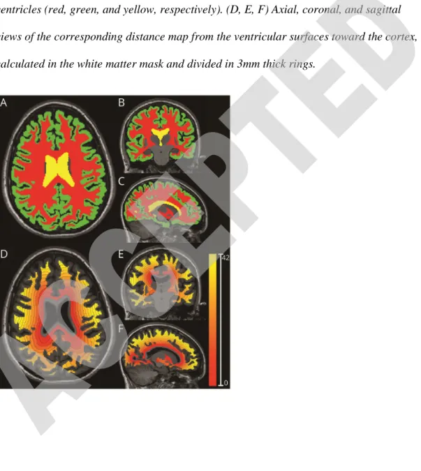
The following regions of interest were then defined on T1-weighted scans of all subjects: (i) white matter; (ii) cortex; and (iii) ventricular CSF, including lateral and third ventricles (Fig 1A-C). In patients with multiple sclerosis, the following regions of interest were also defined: T2 lesions and normal-appearing white matter, defined as the white matter outside visible lesions on T2-weighted. Manual corrections were performed when necessary to ensure anatomical accuracy. Distance maps from ventricular CSF towards the cortex in the white matter (either total white matter, or white matter divided in normal-appearing white matter and T2 lesions) were then calculated as the 3D Euclidean distance to the nearest nonzero voxel (Fig 1D-F).
T1-weighted scans were then normalized to the MNI152 standard space using a non-linear transformation from ANTs (Advanced Normalization Tools, v2.2) 29.
Subsequently, all regions of interest and distance maps were aligned to standard space. Based on the PET resolution, distance maps were then divided in 3mm thick concentric rings, radiating from the ventricular surface toward the cortex (Fig 1D-F). To minimize partial volume effect, the first 3mm close to ventricles and the last 3mm close to the white matter/cortex interface were removed from the white matter ring map.
PET image processing and calculation of innate immune cell activation in periventricular concentric rings
Voxel-wise [18F]-DPA714 distribution volume ratio (DVR) parametric maps (Fig 2A) were calculated using the Logan graphical method based on reference region, extracted using a supervised clustering algorithm 24,30. DVR maps were then aligned using FLIRT 26 to the corresponding T1-weighted images and normalized to the standard space using the previously calculated non-linear transformations 29 of the T1-weighted in native space onto the MNI152 standard space.
Voxels characterized by innate immune cell activation (hereinafter referred to as DPA+) were identified as those voxels whose DVR value exceeded of more than 20% the mean DVR value of the healthy controls in the same MNI position. The 20% relative threshold had been previously calculated by means of a voxel-wise non-parametric permutation-based t-test between the DVR maps of patients and healthy controls, as previously described 25. This step resulted in the generation of individual maps of innate immune cells activation, consisting of binary masks of DPA+ voxels (Fig 2B).
Given the impact of the TSPO affinity on the DVR estimates, voxel classification was separately conducted for high- and mixed-affinity binders.
For each subject, the percentage of voxels classified as DPA+ was extracted from each ring of the white matter, and of T2 lesions and normal-appearing white matter separately in patients only.
Magnetization transfer ratio in periventricular concentric rings
For each subject, both MToff and MTon sequences were rigidly aligned 26 to the corresponding T1-weighted scan in native space. Maps of magnetization transfer ratio (MTR) were calculated as MTR=(MToff-MTon)/MToff, measured in percentage units (pu), and normalized to the standard space using the previously calculated
transformations. In all subjects, mean MTR values within each ring of the white matter ring were extracted.
Due to the presence of artefacts, the MTR images of 9 subjects (4 patients, 5 HC) were excluded from further MTR-based analysis, leaving a total of 33 patients (women: 19, age: 48.1±11.6) and 14 healthy controls (women: 9, age: 41.0±12.4).
Statistical analysis
Statistical analyses were performed using R version 3.5.0. Demographic and clinical data are presented as mean ± standard deviation, while EDSS and EDSS step change are presented as median (range). For inferential statistics, results are reported as mean ± standard deviation, except otherwise specified. For all tests, the level of statistical significance was set at p < 0.05, and pairwise comparisons of estimated marginal means were performed with Bonferroni correction when necessary.
Wilcoxon-Mann-Whitney tests were used to assess differences in: (i) age between HC and patients with multiple sclerosis; and (ii) disease duration between RRMS and PMS. Fisher’s exact tests were used to determine whether there were significant differences in the proportion of: (i) gender and TSPO genotype between HC and patients with multiple sclerosis; and (ii) treatment and disability worsening category among RRMS and PMS. Ordered logistic regression models were used to test differences in EDSS and EDSS step-changes between RRMS and PMS.
Relationship between innate immune cell activation and distance from ventricular CSF
To evaluate the relationship between the percentage of DPA+ voxels, reflecting innate immune cell activation, and the distance from the ventricular CSF, we employed three separate linear mixed-effects models (package lme4), for the whole white matter,
normal-appearing white matter, and T2 lesions. Each model included the percentage of DPA+ voxels in each ring as the dependent variable, and the following independent variables: (i) ring distance from CSF; (ii) group (HC or patients with multiple sclerosis); and (iii) interaction between ring distance from CSF and group. To further investigate the contribution of T2 lesions to the spatial distribution of DPA+ voxels in the
normal-appearing white matter, we used an additional linear mixed-effects model in patients only, where the percentage of DPA+ voxels in the normal-appearing white matter was included as dependent variable, and the ring distance from CSF, as well as percentage of lesional voxels in each ring were included as independent variables. In all models, subjects and ring distance from CSF were considered as random effects to account for subject
variability. Since the percentages of DPA+ voxels were calculated in rings with different volumes, we assigned a weight to each data point corresponding to the number of voxels used to calculate each percentage, reflecting the degree of precision of each data point. As a result, percentages calculated on few voxels (i.e. rings near cortex due to presence of cortical gyri) had a low impact on the model fit.
In these models, the relationship between the percentage of DPA+ voxels and the ring distance from the ventricular CSF was described by the intercept and slope parameters of a linear model: (i) an intercept represents the estimated percentage of DPA+ voxels, reflecting the extent of activated innate immune cells, at the closest proximity to the CSF; (ii) a slope reflects the rate of change in the percentage of DPA+ voxels with distance from the ventricles. Both parameters were obtained as linear combinations of the model coefficients, which were simultaneously estimated at both the population level (e.g. in HC and patients with multiple sclerosis) and at the single subject level.
For each model, differences of intercept and slope between HC and patients with multiple sclerosis were tested with t-tests on the estimated population parameters.
To compare the relationship between the percentage of DPA+ voxels and the distance from the ventricular CSF between MS subgroups (RRMS and PMS) and healthy controls in white matter, normal-appearing white matter, and T2 lesions, linear regression models were employed including the intercept and slope parameters estimated at the single patient level as dependent variables, and group with the covariates age, gender, and presence of disease modifying treatment as independent variables.
Relationship between innate immune cell activation and microstructural damage
An identical statistical procedure to the one described above for the innate immune cell activation was then repeated to describe the relationship between MTR values, reflecting microstructural damage, and the distance from ventricular CSF in the white matter. In this case, each model returned the following parameters: (i) an intercept, reflecting the
estimated MTR value at the closest proximity to the CSF; and (ii) a slope, representing the rate of change in MTR values with distance from the ventricles. These parameters were simultaneously estimated at the population level and at the single subject level.
To investigate whether the innate immune cell activation was associated with microstructural damage at the closest proximity to the CSF in white matter, we used partial Spearman correlations between the intercepts of single patient percentage of DPA+ voxels and mean MTR, adjusted for age, gender and presence of disease modifying treatment.
To assess whether the rate of change in innate immune cell activation with distance from the ventricles was associated with the rate of change in microstructural damage in white matter, we used partial Spearman correlations between the slopes of single patient percentage of DPA+ voxels and mean MTR, adjusted for age, gender and presence of disease modifying treatment.
Spatial distribution of innate immune cell activation: relationship with cortical thickness and clinical scores
We investigated the relationship between DPA+ voxel intercept and slope in
periventricular areas (white matter, normal-appearing white matter, and T2 lesions) and cortical thickness using partial Spearman correlations, and their difference between clinically worsening and clinically stable patients using linear regression models. All analyses were adjusted for age, gender and presence of disease modifying treatment.
The difference between clinically worsening and clinically stable patients in the
percentage of DPA+ voxels intercepts and slopes in periventricular areas (white matter, normal-appearing white matter, and T2 lesions) were tested with linear regression models adjusted for age, gender and presence of disease modifying treatment.
Data Availability
The data that support the findings of this study are available from the corresponding author, upon reasonable request.
Results
Demographics
Demographic and clinical characteristics of the recruited subjects are reported in Table 1. No significant differences were found between healthy controls and patients with multiple sclerosis in age (p=0.96), gender (p=0.56), and TSPO genotype (p=0.41).
Patients with multiple sclerosis show a periventricular gradient of innate
immune cell activation
Patients with multiple sclerosis had a higher percentage of DPA+ voxels compared to healthy controls in the white matter at the closest proximity to the ventricles (Fig3A and table 2; intercept for patients: 39.0±17.2%, intercept for HC: 17.4±9.7%, t=5.01, p=6.10e-6). The percentage of DPA+ voxels decreased of -0.76±0.62% (t=-7.00, p=3.39e-8) for each mm of distance from the CSF in patients with multiple sclerosis while it remained stable in healthy controls (slope for HC: -0.22±0.22%.mm-1, t=-1.73, p=0.090), resulting in a significant difference between the gradient in the two groups of -0.54±0.16%.mm-1 (mean ± standard error, t=-3.46, p=0.001).
A similar regional distribution of innate immune cell activation was found when normal-appearing white matter and T2 lesions were analyzed separately (Fig 3B-3C,
respectively). Compared to the white matter of HC, patients with multiple sclerosis showed a higher percentage of DPA+ voxels at the closest proximity to the ventricles and a steeper slope in both normal-appearing white matter (intercept: 37.0±16.5%, t=4.70, p=1.83e-5; slope: -0.67±0.54%.mm-1, t=-3.19, p=2.37e-3) and T2 lesions (intercept: 53.3±21.9%, t=6.50, p=5.09e-8; slope: -0.78±0.72%.mm-1, t=-2.49, p=0.018) (Table2). Moreover, in the normal-appearing white matter of patients with multiple sclerosis, the percentage of DPA+ voxels was found to be associated not only with the distance from
ventricles, but also with the percentage of lesional voxels in each periventricular ring (t=-5.59, p=1.64e-6; t=6.63, p=1.00e-10; respectively).
When we performed the patient subgroup analysis, while the PMS patients showed a higher level of innate immune cell activation in the periventricular white matter, normal-appearing white matter and T2-lesions, and steeper slopes compared to RRMS, these differences were not statistically significant (Table 2, Supplemental Table2; Data available from Dryad https://doi.org/10.5061/dryad.z612jm69r).
Association between innate immune cell activation and microstructural
damage in periventricular white matter
In white matter, patients with multiple sclerosis had a lower mean MTR compared to healthy controls at the closest proximity to the ventricles (Fig4A, table2; intercept for patients: 44.6±2.2pu, intercept for HC: 47.1±1.1pu, t=-3.88, p=3.37e-4). The mean MTR increased of 0.044±0.068pu (t=3.12, p=3.89e-3) for each mm of distance from the CSF in patients with multiple sclerosis while it decreased of 0.049±0.017pu (t=-2.70, p=0.01) in HC, resulting in a significant difference between the two groups of 0.093±0.022pu.mm-1 (mean ± standard error, t=4.27, p=1.09e-04).
In patients with multiple sclerosis, a higher percentage of DPA+ voxels was significantly associated with a lower mean MTR at the closest proximity to ventricular CSF (rs=-0.53,
p=0.0024) (Fig 4B). Moreover, the decreasing rate of the percentage of DPA+ voxels with distance from the ventricles was associated with a corresponding increasing rate of mean MTR (rs=-0.65, p=1.0e-3) (Fig 4C).
The periventricular activation of innate immune cells is associated with
cortical thickness and is higher in clinically worsening patients
The association between a higher percentage of DPA+ voxels at the closest proximity to ventricular CSF and a reduced cortical thickness was not significant in the total white matter (rS=-0.33, p=0.058). However, this association was significant in the
normal-appearing white matter (rs=-0.34, p=0.0497) but not in T2 lesions (rs=-0.25, p=0.15). The
decreasing rate of the percentage of DPA+ voxels with distance from the ventricles was not associated with the cortical thickness (white matter: rs=0.20, p=0.27;
normal-appearing white matter: rs=0.23, p=0.19; T2 lesions: rs=0.17, p=0.33).
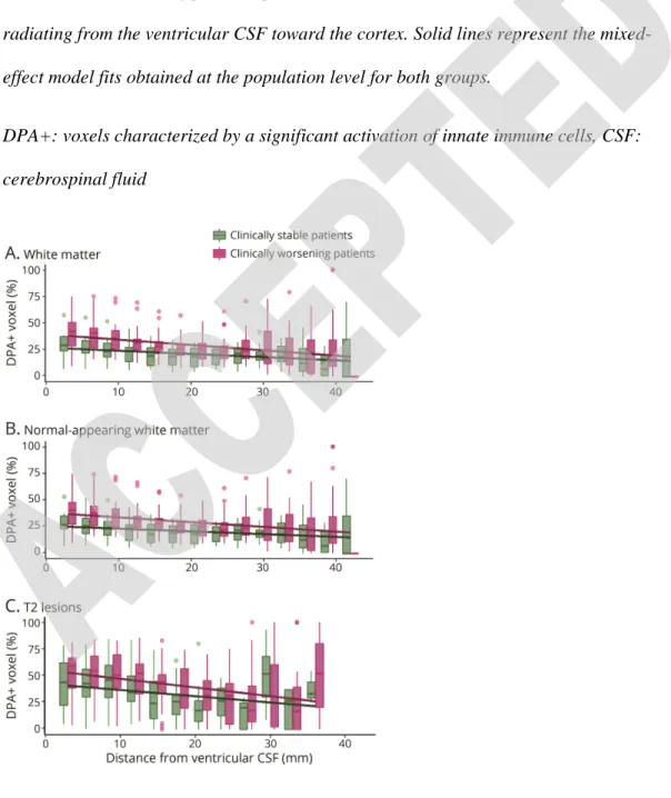
Compared to patients who remained stable over the two years preceding study entry, clinically worsening patients had a significantly higher percentage of DPA+ voxels at the closest proximity to ventricular CSF in the whole white matter (intercept for clinically worsening patients: 43.5±17.1%, intercept for clinically stable patients: 30.7±14.3%, t=2.20, p=0.035, Fig 5A). The difference in the slopes between clinically worsening and clinically stable patients was not significant (slope for clinically worsening patients: -0.85±0.65%.mm-1, slope for clinically stable patients: -0.61±0.54%.mm-1, t=-1.29, p=0.21).
When normal-appearing white matter and T2 lesions were analyzed separately, clinically worsening patients showed a higher percentage of DPA+ voxels at the closest proximity to the ventricles only in the normal-appearing white matter (intercept for clinically worsening patients: 41.6±16.5%, intercept for clinically stable patients: 28.6±13.3%, t=2.35, p=0.025, Fig 5B), but not in the T2 lesions (intercept for clinically worsening patients: 56.9±20.0%, intercept for clinically stable patients: 46.6±24.4%, t=1.21, p=0.24, Fig 5C). The difference in the rate of decrease in the percentage of DPA+ voxels with distance from the ventricular CSF was not significant between clinically worsening and clinically stable patients only in the normal-appearing white matter (slope for clinically
worsening patients: -0.75±0.58%.mm-1, slope for clinically stable patients: -0.51±0.46%.mm-1, t=-1.50, p=0.14), as well as in T2 lesions (slope for clinically worsening patients: -0.84±0.72%.mm-1, slope for clinically stable patients: -0.67±0.74%.mm-1, t=-0.60, p=0.55).
Discussion
In this study, we generated individual maps of TSPO binding based on [18F]-DPA714 PET to explore the regionalization of activated innate immune cells as a function of the distance from ventricular surface in a group of patients with multiple sclerosis compared with healthy controls. We demonstrated that in patients with multiple sclerosis the innate immune cell activation was higher at the closest proximity to the ventricular CSF, and declined with increasing distance from the ventricles, particularly in clinically worsening compared to clinically stable patients. Moreover, we found that the decreasing rate of active innate immune cells with distance from the ventricles was associated with a corresponding increase in the rate of microstructural damage, as measured by MTR.
Several biological mechanisms may underlie the particular brain regionalization of innate immune cell activation and corresponding microstructural damage that we found in our study in patients with multiple sclerosis. Periventricular tissues are known to be a preferential localization of multiple sclerosis plaques, that originate from an acute adaptive immunity response around post-capillary veins 31. A subsequent up- and down-regulation of lymphocytes T and B surface molecules was identified as a potential mechanism driving the persistence and compartmentalization of the inflammatory response in some established lesions 32,33. This compartmentalized inflammation may result in the secretion of pro-inflammatory mediators that could diffuse in surrounding periventricular areas and induce a persistent activation of innate immune cells. One interesting candidate molecular signal could be fibrine, which has been shown to be able
to invade the perivenular space, and to promote the activation of microglia and the
recruitment of peripheral inflammatory macrophages into the central nervous system 34,35.
In our study, when looking separately at normal-appearing white matter and T2 lesions in periventricular regions, we found a gradient of innate immune cell activation in both compartments. This higher activation of innate immune cells might therefore also be explained by the diffusion of soluble factors localized in the CSF, which could exert a deleterious role on tissues localized near the inner surface of the brain, both inside and outside demyelinating lesions. CSF-derived soluble factors from patients with multiple sclerosis have already been shown to induce neuronal damage in-vitro 36, with a few candidate molecules that have been identified, such as Ceramide 37, semaphorine 4A 38 or HERV-W Env proteins 39. Increased level of pro-inflammatory cytokines in the CSF of post-mortem cases of multiple sclerosis (IFNγ, TNF, IL2, IL22, CXCL13, CXCL10, LTα, IL6, IL10) were also shown to correlate with meningeal inflammation and extended grey matter tissue damage in subpial cortical regions 3. Interestingly, a regionalization of innate immune cell activation in cortical regions facing the inflamed CSF, which was spatially associated with enhanced demyelination and neuronal death in the outer cortical layers, was also described on post-mortem samples 2,3,40–42. As the CSF is a highly dynamic compartment with large exchanges between the sub-arachnoid space and the ventricles, our results could suggest that a similar mechanism to the one at play in cortical regions facing the CSF may also underlie the severe microstructural damage affecting
periventricular white matter. In addition, the infiltration of inflammatory cells through the more permeable blood to CSF barrier has been described as an early and sustained event in the experimental autoimmune encephalitis model of multiple sclerosis 43,44.Whereas their number is generally mild, the long-lasting presence of inflammatory cells in the CSF compartment of patients with multiple sclerosis could also contribute to the pathological process, enhancing the parenchymal infiltration of immune cells and/or inducing a diffuse
damage in tissues adjacent to the CSF, i.e. periventricular areas and subpial cortex. According to these hypotheses, inflammatory/cytotoxic mediators and/or inflammatory cells released in the CSF would induce a gradient of innate immune cells activation in surrounding tissues. It is possible however that in the normal-appearing white matter this gradient depends from the proximity to both CSF and white matter lesions, as we found an independent contribution of the distance from ventricles and of T2 lesions to the spatial distribution of DPA+ voxels.
Whatever the mechanism underlying the selective periventricular activation of innate immune cells may be, this gradient provides one explanation for the predominant tissue loss described in the same region by MRI 4–6,8,10, as a significant correlation between the periventricular gradient of innate immune cell activation and that of microstructural damage in the white matter was shown.
Although these hypotheses should be further explored by future experimental and pathological studies, a novel conception of the mechanisms underlying tissue damage in multiple sclerosis could be proposed, in which periventricular innate immune cell activation plays a key role in the cascade of events leading to neurodegeneration and, ultimately, to disease progression. Our results are in line with previously published data highlighting the clinical relevance of the periventricular gradient of microstructural damage specifically characterizing the normal-appearing white matter 6. Interestingly we found a significant association between periventricular neuroinflammation, disability trajectories and cortical damage in the normal-appearing white matter only, suggesting that the contribution of innate immune system to neurodegeneration may not be restricted to a more destructive fate or a lack of repair within multiple sclerosis lesions. How neuroinflammation in the periventricular regions could ultimately result in tissue damage and neurological disability remains unclear, but the cascade of events may potentially
involve oxidative and energetic dysregulation, followed by Wallerian or dying back axonal degeneration 45.
This study has potential limitations that should be taken into account in the interpretation of results. Firstly, TSPO PET tracers have a suboptimal specificity for innate immune cells, as they can also bind to reactive astrocytes and endothelial cells 46–48. Post-mortem evidence has indicated that TSPO+ vascular endothelium accounted for less than 5% of the TSPO+ cells, with no difference in vascular expression between patients with multiple sclerosis and healthy controls in the white matter 47. These findings, together with the methodology used to identify DPA+ voxels, that classifies each voxel according to the mean binding found in the same voxel of a group of control subjects, argue for a
negligible bias induced by the vascular binding in our study. By contrast, in active lesions and in the rim of chronic active lesions, astrocytes constitute up to 25% of TSPO
expressing cells 47, and our methodology do not allow to discriminate between the innate immune and the astrocytic contribution underlying the increased binding of [18F]-DPA714. Further studies are therefore needed to check whether TSPO expression in astrocytes is indeed linked to a proinflammatory state, contributing with microglial cells to the deleterious neuroinflammatory environment in MS, as already shown in the EAE model 49.
Another limitation is that TSPO tracers do not currently allow the differentiation between pro-inflammatory and regulatory innate immune cells 47. However, the correlation found between a higher activation of innate immune cells and a more severe microstructural damage in the periventricular region, together with the neuropathological evidence that in multiple sclerosis lesions homeostatic microglial cells are downregulated while pro-inflammatory microglial cells predominate 33, suggest that the TSPO binding detected in this study might mainly reflect the activation of “pro-inflammatory” innate immune cells.
Finally, the cross-sectional design of our imaging study does not allow to establish whether the periventricular gradient of innate immune activation chronologically follows or precedes (and is therefore potentially responsible for) a gradient of microstructural damage as demonstrated using MTR. Moreover we cannot fully exclude that part of the periventricular gradient of MTR change is directly linked to inflammation, as MTR was shown to be sensitive to neuroinflammation 50. Therefore, only longitudinal prospective studies combining imaging markers of innate immune cell activation and microstructural damage would have the potential to define the exact sequence of pathological events, and to identify the predictive value of the periventricular gradient of innate immune cell activation on the development of brain atrophy and disability progression.
Of note, the moderate quality of some MT acquisition has led to the exclusion of some subjects for the MTR analysis, limiting the investigation of the relationship between innate immune cell action and microstructural damage onto a subsample of our data, including 33 patients and 14 healthy controls. As the MT acquisition consists of 2 acquisitions, with and without the MT pulse, particular caution should be taken with the registration between them, as well as movement artefacts that might compromise data.
In conclusion, we were able for the first time to identify one pathological substrate for the periventricular gradient of microstructural damage that characterizes multiple sclerosis, consisting in a gradient of innate immune cell activation, reflected by [18F]-DPA714 binding. In combination with previous post-mortem investigations, these results suggest that tissue pathology in multiple sclerosis occurs preferentially at or near the inner surface of the brain, and is possibly linked to the proximity to the ventricular CSF and mediated by innate immune cell activation. Further work is needed to clarify the potential role of factors such as CSF mediators, and intrathecal inflammation, in the pathogenesis of lesional and non-lesional abnormalities in multiple sclerosis.
Acknowledgements
We thank the Centre d’Investigation Clinique team from ICM, Céline Louapre, Jean-Christophe Corvol for protocol organization, Caroline Papeix, Catherine Lubetzki, Elizabeth Maillart, Rana Assouad for helpful discussion and clinical help, C Baron, C Manciot, Vincent Lebon (SHFJ, CEA), Geraldine Gourbil (CIC) for their invaluable assistance. We address special thanks to Mattia Veronese, Federico Turkheimer for technical help and fruitful discussions. We also thank the staff of the CENIR (Research neuroimaging unit of the ICM), and the staff of the Unité de Recherche Clinique of Pitié Salpêtrière, in particular Anne Bissery and Laura Morizot.
Appendix 1: Authors
Name Location Contribution
Emilie Poirion, PhD Paris Brain Institute, Paris, France
Design and conceptualized study; Major role in the acquisition of data; Analyzed the data; Interpreted the data ; drafted the manuscript for intellectual content Matteo Tonietto, PhD Paris Brain Institute, Paris,
France
Analyzed the data; Interpreted the data ; drafted the manuscript for intellectual content
François-Xavier Lejeune, PhD
Paris Brain Institute, Paris, France
Analyzed the data
Vito A.G. Ricigliano, MD
Paris Brain Institute, Paris, France
Analysed the data
Marine Boudot de la Motte, MD
Paris Brain Institute, Paris, France
Analysed the data
Charline Benoit, MD Paris Brain Institute, Paris, France
Analysed the data
Géraldine Bera, MD Paris Brain Institute, Paris, France
Major role in the acquisition of data
Bertrand Kuhnast, PhD CEA, Orsay, France Major role in the acquisition of data Michel Bottlaender, MD,
PhD
CEA, Orsay, France Design and conceptualized study; Major role in the acquisition of data
Benedetta Bodini, MD, PhD
Paris Brain Institute, Paris, France
Design and conceptualized study; Interpreted the data ; drafted the manuscript for intellectual content
Bruno Stankoff, MD, PhD
Paris Brain Institute, Paris, France
Obtained funding for the study. Design and conceptualized study; Interpreted the data ; drafted the manuscript for intellectual content
References
1. Lassmann H. Pathogenic Mechanisms Associated With Different Clinical Courses of Multiple Sclerosis. Front Immunol. 2019;9:3116.
2. Magliozzi R, Howell OW, Reeves C, et al. A Gradient of neuronal loss and meningeal inflammation in multiple sclerosis. Ann Neurol. 2010;68:477–493. 3. Magliozzi R, Howell OW, Nicholas R, et al. Inflammatory intrathecal profiles and
cortical damage in multiple sclerosis. Ann Neurol. 2018;83:739–755.
4. Jehna M, Pirpamer L, Khalil M, et al. Periventricular lesions correlate with cortical thinning in multiple sclerosis: Cortical Thinning and Periventricular Lesions in MS. Ann Neurol. 2015;78:530–539.
5. Brown, Chowdhury A, Kanber B, et al. Magnetisation transfer ratio abnormalities in primary and secondary progressive multiple sclerosis. Mult Scler J.
2019;1352458519841810.
6. Brown JWL, Pardini M, Brownlee WJ, et al. An abnormal periventricular magnetization transfer ratio gradient occurs early in multiple sclerosis. Brain. 2017;140:387–398.
7. Liu Z, Pardini M, Yaldizli Ö, et al. Magnetization transfer ratio measures in normal-appearing white matter show periventricular gradient abnormalities in multiple sclerosis. Brain. 2015;138:1239–1246.
8. Pardini M, Sudre CH, Prados F, et al. Relationship of grey and white matter abnormalities with distance from the surface of the brain in multiple sclerosis. J Neurol Neurosurg Psychiatry. 2016;87:1212–1217.
9. Fadda G, Brown RA, Magliozzi R, et al. A surface-in gradient of thalamic damage evolves in pediatric multiple sclerosis. Ann Neurol. 2019;85:340–351.
10. Pardini M, Petracca M, Harel A, et al. The relationship between cortical lesions and periventricular NAWM abnormalities suggests a shared mechanism of injury in primary-progressive MS. NeuroImage Clin. 2017;16:111–115.
11. Airas L, Nylund M, Rissanen E. Evaluation of Microglial Activation in Multiple Sclerosis Patients Using Positron Emission Tomography. Front Neurol. 2018;9:181. 12. Banati R, Newcombe J, Gunn RN, et al. The peripheral benzodiazepine binding site
in the brain in multiple sclerosis. Quantitative in vivo imaging of microglia as a measure of disease activity. Brain. 2000;123:2321–2337.
13. Stankoff B, Poirion E, Tonietto M, Bodini B. Exploring the heterogeneity of MS lesions using positron emission tomography: a reappraisal of their contribution to disability. Brain Pathol Zurich Switz. 2018;28:723–734.
14. Datta, Colasanti A, Kalk N, et al. 11C-PBR28 and 18F-PBR111 Detect White Matter Inflammatory Heterogeneity in Multiple Sclerosis. J Nucl Med. 2017;58:1477–1482. 15. Politis M, Giannetti P, Su P, et al. Increased PK11195 PET binding in the cortex of
16. Rissanen E, Tuisku J, Rokka J, et al. In Vivo Detection of Diffuse Inflammation in Secondary Progressive Multiple Sclerosis Using PET Imaging and the Radioligand 11C-PK11195. J Nucl Med. 2014;55:939–944.
17. Herranz E, Giannì C, Louapre C, et al. The neuroinflammatory component of gray matter pathology in multiple sclerosis. Ann Neurol. 2016;80:776–790.
18. James ML, Fulton RR, Vercoullie J, et al. DPA-714, a new translocator protein-specific ligand: synthesis, radiofluorination, and pharmacologic characterization. J Nucl Med Off Publ Soc Nucl Med. 2008;49:814–822.
19. Rizzo G, Veronese M, Tonietto M, et al. Generalization of endothelial modelling of TSPO PET imaging: Considerations on tracer affinities. J Cereb Blood Flow Metab. Epub 2017 Nov 14.:0271678X17742004.
20. Polman CH, Reingold SC, Banwell B, et al. Diagnostic criteria for multiple sclerosis: 2010 Revisions to the McDonald criteria. Ann Neurol. 2011;69:292–302.
21. Owen, Gunn RN, Rabiner EA, et al. Mixed-Affinity Binding in Humans with 18-kDa Translocator Protein Ligands. J Nucl Med Off Publ Soc Nucl Med. 2011;52:24– 32.
22. Kurtzke JF. Rating neurologic impairment in multiple sclerosis: an expanded disability status scale (EDSS). Neurology. 1983;33:1444–1452.
23. Weinshenker BG, Issa M, Baskerville J. Meta-analysis of the placebo-treated groups in clinical trials of progressive MS. Neurology. 1996;46:1613–1619.
24. García-Lorenzo D, Lavisse S, Leroy C, et al. Validation of an automatic reference region extraction for the quantification of [18F] DPA-714 in dynamic brain PET studies. J Cereb Blood Flow Metab. 2017;38:333–346.
25. Bodini B, Poirion E, Tonietto M, et al. Individual mapping of innate immune cell activation is a candidate marker of patient-specific trajectories of disability worsening in Multiple Sclerosis. J Nucl Med. Epub 2020 Jan 31.
26. Jenkinson M, Smith S. A global optimisation method for robust affine registration of brain images. Med Image Anal. 2001;5:143–156.
27. Chard DT, Jackson JS, Miller DH, Wheeler‐Kingshott CAM. Reducing the impact of white matter lesions on automated measures of brain gray and white matter volumes. J Magn Reson Imaging. 2010;32:223–228.
28. Wang H, Suh JW, Das SR, Pluta JB, Craige C, Yushkevich PA. Multi-Atlas Segmentation with Joint Label Fusion. IEEE Trans Pattern Anal Mach Intell. 2013;35:611–623.
29. Avants BB, Epstein CL, Grossman M, Gee JC. Symmetric diffeomorphic image registration with cross-correlation: evaluating automated labeling of elderly and neurodegenerative brain. Med Image Anal. 2008;12:26–41.
30. Lavisse S, García-Lorenzo D, Peyronneau M-A, et al. Optimized Quantification of Translocator Protein Radioligand 18F-DPA-714 Uptake in the Brain of Genotyped Healthy Volunteers. J Nucl Med. 2015;56:1048–1054.
31. Adams CWM, Abdulla YH, Torres EM, Poston RN. Periventricular Lesions in Multiple Sclerosis: Their Perivenous Origin and Relationship to Granular Ependymitis. Neuropathol Appl Neurobiol. 1987;13:141–152.
32. Machado-Santos J, Saji E, Tröscher AR, et al. The compartmentalized inflammatory response in the multiple sclerosis brain is composed of tissue-resident CD8+ T lymphocytes and B cells. Brain J Neurol. 2018;141:2066–2082.
33. Zrzavy T, Hametner S, Wimmer I, Butovsky O, Weiner HL, Lassmann H. Loss of “homeostatic” microglia and patterns of their activation in active multiple sclerosis. Brain J Neurol. 2017;140:1900–1913.
34. Lee NJ, Ha S-K, Sati P, et al. Spatiotemporal distribution of fibrinogen in marmoset and human inflammatory demyelination. Brain. 2018;141:1637–1649.
35. Petersen MA, Ryu JK, Akassoglou K. Fibrinogen in neurological diseases: mechanisms, imaging and therapeutics. Nat Rev Neurosci. 2018;19:283–301. 36. Alcázar A, Regidor I, Masjuan J, Salinas M, Alvarez-Cermeño JC. Axonal damage
induced by cerebrospinal fluid from patients with relapsing-remitting multiple sclerosis. J Neuroimmunol. 2000;104:58–67.
37. Vidaurre OG, Haines JD, Katz Sand I, et al. Cerebrospinal fluid ceramides from patients with multiple sclerosis impair neuronal bioenergetics. Brain J Neurol. 2014;137:2271–2286.
38. Chiou B, Lucassen E, Sather M, Kallianpur A, Connor J. Semaphorin4A and H-ferritin utilize Tim-1 on human oligodendrocytes: A novel neuro-immune axis. Glia. 2018;66:1317–1330.
39. Küry P, Nath A, Créange A, et al. Human Endogenous Retroviruses in Neurological Diseases. Trends Mol Med. 2018;24:379–394.
40. Bevan RJ, Evans R, Griffiths L, et al. Meningeal inflammation and cortical demyelination in acute multiple sclerosis. Ann Neurol. 2018;84:829–842.
41. Howell OW, Reeves CA, Nicholas R, et al. Meningeal inflammation is widespread and linked to cortical pathology in multiple sclerosis. Brain. 2011;134:2755–2771. 42. Magliozzi R, Howell O, Vora A, et al. Meningeal B-cell follicles in secondary
progressive multiple sclerosis associate with early onset of disease and severe cortical pathology. Brain. 2007;130:1089–1104.
43. Engelhardt B, Carare RO, Bechmann I, Flügel A, Laman JD, Weller RO. Vascular, glial, and lymphatic immune gateways of the central nervous system. Acta
Neuropathol (Berl). 2016;132:317–338.
44. Kooij G, Kopplin K, Blasig R, et al. Disturbed function of the blood-cerebrospinal fluid barrier aggravates neuro-inflammation. Acta Neuropathol (Berl).
2014;128:267–277.
45. Lassmann H, van Horssen J, Mahad D. Progressive multiple sclerosis: pathology and pathogenesis. Nat Rev Neurol. Nature Publishing Group; 2012;8:647–656.
46. Lavisse S, Guillermier M, Hérard A-S, et al. Reactive Astrocytes Overexpress TSPO and Are Detected by TSPO Positron Emission Tomography Imaging. J Neurosci. 2012;32:10809–10818.
47. Nutma E, Stephenson JA, Gorter RP, et al. A quantitative neuropathological assessment of translocator protein expression in multiple sclerosis. Brain. 2019;142:3440–3455.
48. Wimberley C, Lavisse S, Brulon V, et al. Impact of Endothelial 18-kDa Translocator Protein on the Quantification of 18F-DPA-714. J Nucl Med Off Publ Soc Nucl Med. 2018;59:307–314.
49. Chechneva OV, Deng W. Mitochondrial translocator protein (TSPO), astrocytes and neuroinflammation. Neural Regen Res. 2016;11:1056–1057.
50. Moll NM, Rietsch AM, Thomas S, et al. Multiple sclerosis normal-appearing white matter: pathology-imaging correlations. Ann Neurol. 2011;70:764–773.
Figure Legends
Figure 1: Processing steps to generate 3mm-thick ring from cerebrospinal
fluid to adjacent white matter
(A, B, C) Axial, coronal, and sagittal views of a T1-weighted images segmented using a multi-atlas segmentation approach generating masks for the white matter, cortex, and ventricles (red, green, and yellow, respectively). (D, E, F) Axial, coronal, and sagittal views of the corresponding distance map from the ventricular surfaces toward the cortex, calculated in the white matter mask and divided in 3mm thick rings.
Figure 2: Illustrative example of [18F]-DPA714 DVR maps and the
corresponding individual map of DPA+ voxels.
(A) [18F]-DPA714 DVR map of a representative patient with multiple sclerosis (45 years old female patient with secondary progressive multiple sclerosis, disease duration: 22 years, EDSS at baseline: 6, EDSS step change in the two years preceding baseline: 1.5). The map was obtained using Logan graphical analysis with reference region extracted with a supervised clustering approach. (B) Corresponding individual map of DPA+ voxels (yellow) obtained by thresholding the DVR map of panel A (see text for details about the thresholding technique employed).
DVR: distribution volume ratio, EDSS=Expanded Disability Status Scale, DPA+: voxels characterized by a significant activation of innate immune cells
Figure 3: A periventricular gradient of innate immune cell activation in the
WM of patients with MS.
Boxplots represent the percentage of DPA+ voxels in the total white matter (A), the normal-appearing white matter (B), T2 lesions (C) of healthy controls (blue) and patients with multiple sclerosis (red) calculated in 3mm thick concentric rings radiating from the ventricular CSF toward the cortex. Solid lines represent the mixed-effect model fits obtained at the population level for both groups.
DPA+: voxels characterized by a significant activation of innate immune cells, CSF: cerebrospinal fluid.
Figure 4: Relationship between innate immune cell activation and
magnetization transfer ratio in the periventricular WM
(A) Boxplots represent the mean MTR in the white matter of healthy controls (blue) and patients with multiple sclerosis (red) calculated in 3mm thick concentric rings radiating from the ventricular CSF toward the cortex. Solid lines represent the mixed-effect model fits obtained at the population level for both healthy controls and patients with multiple sclerosis. In white matter, both the intercepts (B) and the slopes (C) of the percentage of DPA+ voxels and mean MTR values were inversely correlated with each other.
MTR: magnetization transfer ratio, CSF: cerebrospinal fluid, DPA+: voxels characterized by a significant activation of innate immune cells.
Figure 5: Periventricular innate immune cells activation associates with
clinical trajectories of disability worsening
Boxplots represent the percentage of DPA+ voxels in the total white matter (A), the normal-appearing white matter (B), T2 lesions (C) of clinically stable patients (green) and clinically worsening patients (pink) calculated in 3mm thick concentric rings radiating from the ventricular CSF toward the cortex. Solid lines represent the mixed-effect model fits obtained at the population level for both groups.
DPA+: voxels characterized by a significant activation of innate immune cells, CSF: cerebrospinal fluid
Tables
Table 1: Demographic and clinical characteristics of MS patients and healthy
controls
Demographic, clinical and radiological characteristics mean ± standard deviation or median [range]
Healthy controls Patients with multiple sclerosis (total) Relapsing-remitting multiple sclerosis Progressive multiple sclerosis Number 19 37 11 26 Age (years) 46.6±14.3 47.7±11.4 42.6±13.3 49.9±10.0 Gender (female/male) 13/6 21/16 8/3 13/13 Genotype (Mixed-/High-affinity binders) 10/9 15/22 4/7 11/15 Disease duration (years) - 9.3±5.4* 6.7±4.4 10.4±5.5 Baseline EDSS - 5 [2-7.5]# 3.5 [2-6] 6 [3-7.5]
EDSS step change - 1 [0-4.5] 1 [0-2.5] 1 [0-4.5] Individual trajectory (clinically stable/ clinically worsening) - 13/24 4/7 9/17 Disease Modifying Treatment (off/on) - 20/17$ 1/10 19/7
EDSS=Expanded Disability Status Scale
(*) p<0.05 by Wilcoxon-Mann-Whitney test, (#) p<0.001 by ordered logistic regression model, and ($) p<0.001 Fisher’s exact tests between MS subgroups.
Table 2: [18F]-DPA714 PET- and MTR-derived measurements at the population
level
Subjects group DPA+ voxel MTR white matter normal-appearing white matterT2 lesions white matter
intercept (%) slope (%.mm-1) intercept (%) slope (%.mm-1) intercept (%) slope (%.mm-1) intercept (pu) slope (pu.mm-1) HC 17.42±9.66 -0.22±0.22 47.05±1.05 -0.049±0.017 MS 39.01±17.15 -0.76±0.62 37.01±16.52 -0.67±0.54 53.28±21.88 -0.78±0.72 44.63±2.16 0.044±0.068 RRMS 33.32±16.12 -0.70±0.78 31.15±15.10 -0.60±0.63 50.51±22.38 -0.72±0.86 45.05±2.06 0.044±0.050 PMS 41.42±17.31 -0.79±0.58 39.48±16.74 -0.70±0.51 54.44±22.01 -0.81±0.67 44.45±2.22 0.044±0.075 Stable 30.68±14.33 -0.61±0.54 28.58±13.30 -0.51±0.46 46.56±24.42 -0.67±0.74 44.97±1.63 0.029±0.039 Worsening 43.52±17.12 -0.85±0.65 41.57±16.52 -0.75±0.58 56.91±19.97 -0.84±0.72 44.44±2.45 0.053±0.080
DPA+: voxels characterized by a significant activation of innate immune cells, MTR: Magnetization Transfer Ratio; HC: Healthy Controls; MS: patients with MS; RRMS: Relapsing-Remitting MS; PMS: Progressive MS
DOI 10.1212/WNL.0000000000011700
published online March 18, 2021
Neurology
Emilie Poirion, Matteo Tonietto, François-Xavier Lejeune, et al.
Multiple Sclerosis
This information is current as of March 18, 2021
Services
Updated Information &
ull
http://n.neurology.org/content/early/2021/03/18/WNL.0000000000011700.f
including high resolution figures, can be found at:
Subspecialty Collections http://n.neurology.org/cgi/collection/pet PET http://n.neurology.org/cgi/collection/multiple_sclerosis Multiple sclerosis http://n.neurology.org/cgi/collection/mti_ MTI http://n.neurology.org/cgi/collection/mri MRI http://n.neurology.org/cgi/collection/all_immunology All Immunology collection(s):
This article, along with others on similar topics, appears in the following
Permissions & Licensing
http://www.neurology.org/about/about_the_journal#permissions
entirety can be found online at:
Information about reproducing this article in parts (figures,tables) or in its
Reprints
http://n.neurology.org/subscribers/advertise
Information about ordering reprints can be found online:
0028-3878. Online ISSN: 1526-632X.
Kluwer Health, Inc. on behalf of the American Academy of Neurology.. All rights reserved. Print ISSN: is now a weekly with 48 issues per year. Copyright Copyright © 2021 The Author(s). Published by Wolters
® is the official journal of the American Academy of Neurology. Published continuously since 1951, it
![Figure 2: Illustrative example of [18F]-DPA714 DVR maps and the corresponding individual map of DPA+ voxels](https://thumb-eu.123doks.com/thumbv2/123doknet/14617987.546689/32.918.138.482.612.910/figure-illustrative-example-dpa-dvr-corresponding-individual-voxels.webp)

![Table 2: [18F]-DPA714 PET- and MTR-derived measurements at the population level Subjects group DPA+ voxel MTR white matter normal-appearing white matter](https://thumb-eu.123doks.com/thumbv2/123doknet/14617987.546689/39.918.143.806.172.541/table-derived-measurements-population-subjects-matter-normal-appearing.webp)
