Mechanisms of active laryngeal closure during non-invasive
intermittent positive pressure ventilation in non-sedated lambs
Roy Bianca 1, Samson Nathalie 1, Moreau-Bussière François 2, Ouimet Alain 3, Dorion Dominique 3, Mayer Sandeep 3, Praud Jean-Paul 1,3
1: Neonatal Respiratory Research Unit, Departments of Pediatrics and Physiology; 2: Department of Anesthesiology; 3: Department of Surgery
Université de Sherbrooke.
Short title: Mechanisms of glottal closure during nasal ventilation
Address for correspondence Jean-Paul Praud MD PhD
Departments of Pediatrics and Physiology Université de Sherbrooke
J1H 5N4 – Quebec, Canada
Email: [email protected]
Phone: 1 819 346 1110, ext 14851 Fax: 1 819 564 5215
ABSTRACT
The present study stems from our recent demonstration that a progressive increase in nasal intermittent positive-pressure ventilation (nIPPV) leads to active glottal closure in non-sedated, newborn lambs. The aim of the study was to determine whether the mechanisms involved in this glottal narrowing during nIPPV originate from upper airway receptors and/or from bronchopulmonary receptors. Two groups of newborn lambs were chronically instrumented for polysomnographic recording: the first group of 5 lambs underwent a two-step bilateral thoracic vagotomy using video-assisted thoracoscopic surgery (bilateral vagotomy group) while the second group comprised of 6 lambs underwent chronic laryngo-tracheal separation (isolated upper airway group). A few days later, polysomnographic recordings were performed to assess glottal muscle EMG during step-increases in nIPPV (volume control mode). Results show that active glottal narrowing does not develop when nIPPV is applied on the upper airways only, and that this narrowing is prevented by bilateral vagotomy when nIPPV is applied on intact airways. In conclusion, active glottal narrowing in response to increasing nIPPV originates from bronchopulmonary receptors.
Keywords: bronchopulmonary receptors, volume control ventilation, video-assisted thoracic surgery, upper airways, vagotomy.
INTRODUCTION
Nasal intermittent positive pressure ventilation (nIPPV) is increasingly used to treat acute and chronic respiratory insufficiency, including in the neonatal period, in an effort to decrease the complications related to endotracheal tube ventilation (8). However, a major difference in the application of IPPV via the nasal vs. endotracheal route is generally overlooked, namely the presence of the larynx, a closing valve, which can prevent nIPPV from reaching the lungs. A few studies have shown that increasing nIPPV in either the volume control or pressure support mode induces active glottal closure in both adult humans and newborn lambs (4, 9). Our studies also showed that this active glottal narrowing in lambs was related to both a decrease in glottal dilator EMG activity (cricothyroid, CT) and an increase in glottal constrictor EMG activity (thyroarytenoid muscle, TA) (9). However, the mechanisms by which this active glottal narrowing is induced during nIPPV are totally unknown. Results from our previous study strongly suggest that afferent messages from either central or peripheral chemoreceptors are not involved. Theoretically, the reflex mechanism responsible for glottal narrowing could originate from upper airways receptors, which include pressure, temperature (flow) and drive receptors (12) or from bronchopulmonary receptors, which include the slow adapting (stretch) receptors, the rapidly adapting pulmonary (or irritant) receptors and the bronchopulmonary C-fiber endings (17). Finally, the parietal rib cage mechanoreceptors, including the neuromuscular spindles, Golgi tendon organs and articulation receptors may also bear some responsibility. The aim of the present study conducted in lambs was thus to determine if the mechanisms involved in active glottal narrowing during increasing nIPPV originate from upper airway receptors and/or bronchopulmonary receptors.
MATERIALS AND METHODS
Experiments were conducted in 14 mixed-bred lambs aged from 2 to 6 days and weighing 4.1 kg (SD 0.8; range 2.9 from 5.4) on the day of the experiment. All lambs were born at term by spontaneous vaginal delivery at the sheepfold of our usual provider. The protocol of the study was approved by the ethics committee for animal care and experimentation of the Université de Sherbrooke.
Surgical preparation
Lambs were separated in 2 groups prior to surgery, including 8 lambs instrumented for bilateral vagotomy and 6 lambs with isolated upper airways. Surgical implantation of electrodes for subsequent polysomnographic recording was identical in all lambs (see Common instrumentation below). However, the remaining surgery differed, depending on the experimental group.
Common instrumentation. Aseptic surgery was performed at 1-2 days of life under general anesthesia (2 % isoflurane + 30 % N20 + 68 % O2). Endotracheal intubation was preceded by an intramuscular injection of atropine sulphate (0.1 mg/kg), ketamine (10 mg/kg) and antibiotics (5 mg/kg gentamicin and 7,500 IU/kg Duplocillin). One dose of ketoprofen (3 mg/kg IM) was given immediately before surgery for analgesia and repeated if needed on the next day. An intravenous injection of Ringers lactate (10 mL/Kg) was administered prior to surgery and a mixture of 5% dextrose in NaCl 0.9% was systematically infused perioperatively (80 mL/Kg/ day). Chronic instrumentation was performed as previously described (9,16). Briefly, bipolar gold-plated stainless steel electrodes were inserted into the two thyroarytenoid muscles (TA, a glottal adductor) and bipolar enameled chrome wires were sewn into the two cricothyroid muscles (CT, a glottal dilator) for recording
electromyographic activity (EMG). Two right-angled, platinum needle-electrodes were inserted into the parietal cortex through the skull, for electrocorticogram (EEG) recording. One platinum needle-electrode was also inserted under the scalp as a ground. Leads from all electrodes were subcutaneously tunneled to exit on the back of the lamb and housed in a pocket on a vest. Finally, a catheter was placed into the brachial artery for measuring pH, PO2, PCO2, HCO3- and hemoglobin saturation throughout surgery and on ensuing days thereafter. Heart rate, rectal temperature, pulse oximetry, end tidal CO2 and blood pH were continuously monitored throughout surgery. Post-operative care included daily intramuscular injection of 5 mg/kg gentamicin and 0.05 ml/kg duplocilline until the end of the experimentation; in addition, the arterial catheter was flushed twice a day with a heparinized solution. Surgical instrumentation for bilateral vagotomy. All 8 lambs underwent a 2 step, intrathoracic bilateral vagotomy, which first involved Video-Assisted Thoracoscopic Surgery (VATS) (KARL STORZ GmbH & Co. KG, Tuttlingen, Germany). After generating a right pneumothorax by CO2 insufflation, the endoscope and surgical instruments were introduced into the pleural space through 2 small (5-10 mm) parietal incisions. The bare portion of an enameled chrome wire (0.12 mm diameter, Leico Industries Inc., New York, NY, USA) was positioned around the vagus nerve, just caudally to the origin of the recurrent laryngeal nerve. The remaining enameled wire was glued in polyethylene tubing, with the two ends exiting through the skin. Once completed on the right side, the same procedure was repeated on the left. Finally, 2 to 5 days after surgery, a bilateral vagotomy was performed as previously described for sectioning the superior laryngeal nerve (3). The two bare ends of chrome wire protruding from the right thorax with the polyethylene tubing were attached to an electrocauter (Electrosectilis, Model 770, Britcher Corp., CA, USA).
Traction was then applied to the wire during electrocautery, thus sectioning the vagus nerve. The procedure was completed in less than 5 sec and resulted in minimal discomfort for the lamb (startle at most). The procedure was then repeated on the left side. Bilateral vagotomy was confirmed by multiple means, including i) by pulling off the unbroken wires from the thorax just after electrocautery, ii) by immediately observing a decrease in respiratory rate, and iii) by systematic verification at necropsy. This unique model allowed studying the same lambs before and after bilateral vagotomy, each lamb acting as its own control.
Surgery for the isolated upper airway group. All lambs in this group underwent chronic separation of their upper airways from lower airways. The separation was performed directly under the larynx, just above the first tracheal ring. The caudal end of the larynx was attached to a 2 cm-long Dacron aortic prosthesis, whose caudal end was attached to a neck stoma. In addition, a tracheostomy was performed between the fifth and sixth rings of the trachea to which a 2 cm-long endotracheal tube was sutured and glued externally around the tracheostomy, in such a way that there was no permanent endotracheal tube. Finally, the rostral end of the trachea was sutured. This unique model allowed to perform nasal mask ventilation to the isolated upper airways while the lamb was spontaneously breathing through the tracheostomy, or to perform mechanical ventilation directly through the tracheostomy, without any upper airway ventilation. To our knowledge, this chronic, non-sedated animal model with isolated upper airways, which has taken several months to design with the help of an experienced ENT surgeon (DD), is unique.
Ventilatory equipment. Intermittent positive pressure ventilation was performed using a Siemens Servo 300 ventilator and Servo Screen (Siemens Corporation, New York, NY) with heated (32ºC) and humidified air. Nasal ventilation was performed through a custom-designed nasal mask, as previously described (9). Briefly, the mask was built from a plaster shell, which had a double nasal canula, a naso-gastric tube and a plastic catheter for mask pressure recording and was filled with dental paste to best fit the muzzle of each lamb. A small, non-diffusing gas bag (200 ml) was attached to the external end of the Dacron tube, as a surrogate for the lamb’s lungs.
Recording equipment. Just prior to polysomnographic recordings, 2 needle electrodes were inserted subcutaneously on each side of one eye for electrooculogram (EOG) recording and a pulse oximeter (SpO2) probe was attached at the base of the tail. In addition, elastic bands for respiratory inductance plethysmography (Respitrace, NIMS, Miami Beach, FL) were installed on the thorax and the abdomen to monitor respiratory movements and assess lung volume variations qualitatively. Mask pressure was monitored by using calibrated pressure transducers (MP 45-30-871, Validyne, Northridge, CA). All recordings were carried out in non-sedated lambs, using our custom-designed radiotelemetry system. The transmitter used for this study was composed of differential channels (EEG, EOG, ECG and 4 EMGs) as previously described (6, 7). All transmitted signals were fed from the receiver to the acquisition system. The raw EMG signals were sampled at 500 Hz, rectified, integrated and averaged (moving time average = 100 ms). The telemetry transmitter was connected to the electrode leads and housed in the pocket of a vest worn by the lamb. Polysomnographic signals were recorded on a PC, using
Acknowledge software (version 3.7.3, BioPac Systems, Inc., Santa Barbara, CA, USA).
Design of the study
On arrival in our in-house animal quarters, only lambs of the bilateral vagotomy group were housed with their mother. Lambs from the isolated upper airway group were housed in a Plexiglas chamber (1.2 m3, in agreement with recommendations by the Canadian Council for Animal Care for sheep housing) through which water-saturated air was continuously flowed (10L/min) using an Allegiance AirlifeTM Nebulizer (no. 5207) and a home humidifier. Tracheal secretions were systematically suctioned at least three times a day, according to American Thoracic Society recommendations (15). Lambs from this group were also fed ad libitum three times a day with ewe’s milk at 8:00 am, 12:00 pm and 4:00 pm. The study was performed without sedation at least 48 h after surgery. The study was designed to allow simultaneous recording of EEG, EOG, and EMG activity, variations of mask pressure, respiratory movements and SpO2 while using incremental levels of ventilation during wakefulness and quiet sleep. The lambs were comfortably positioned in a sling with loose restraints. Two experimenters were present throughout all of the recordings to note lamb behavior, set ventilator parameters and prevent disconnection from the ventilator.
Bilateral vagotomy group. A first polysomnographic recording was performed during nasal ventilation with the vagi intact (= instrumented but not cut). On the following day, the same experiment was repeated during polysomnographic recording after performing the bilateral vagotomy (see above). The protocol design for nasal ventilation has been previously described (9). Following an initial recording with no CPAP (continuous positive airway pressure), i.e., with the nasal mask on but
without any mechanical ventilatory support, “baseline” tidal volume (Vt) and respiratory rate (RR) were obtained with a CPAP of 4 cmH2O applied via the nasal mask. Three levels of ventilation were tested in the volume control mode (VC), while maintaining a positive end-expiratory pressure (PEEP) at 4 cmH2O. For the first level of volume control ventilation (VC#1), tidal volume and respiratory rate were set at the same values as when the lamb was breathing spontaneously with CPAP 4 cmH2O. Thereafter, tidal volume was increased in a stepwise manner to 10 mL/ Kg (VC#2), 15 mL/ Kg (VC#3) and 20 mL/Kg (VC#4). Every effort was made to record approximately 100 respiratory cycles during both wakefulness and quiet sleep, at each level of ventilation. At any given time during the experiment, ventilation was halted if the lamb displayed discomfort or agitation and/or there was an obvious abdominal distension or presence of liquid reflux via the nasogastric tube. While both the VC mode and pressure support IPPV were tested in our previous study (9), only the VC mode was tested in the present study, since pressure support was not feasible on isolated upper airways (no inspiratory trigger from the lamb to the ventilator).
Isolated upper airways group. Two polysomnographic recordings were performed on the same day in random order, during nasal mask and tracheostomy ventilation. Each level of IPPV (VC mode) was sequentially tested as described above for the bivagotomy group. After completion of the first round of ventilation via the nasal or tracheostomy route, the lambs were allowed to rest for 30 min before the same protocol was repeated on the other portion of the airways.
States of alertness. Standard electrophysiological and behavioral criteria were used to define W, QS and AS from EEG, EOG and continuous observation (13). Arousal from QS was characterized by sudden disappearance of high-amplitude, low-frequency waves on the EEG trace whereas arousal from AS was recognized by direct observation of the lamb and disappearance of intense EOG activity.
Respiratory parameters. Twenty consecutive breaths, which had to be preceded and followed by 20 seconds of stable respiratory pattern, were selected for analysis in each lamb at every ventilatory level in W and QS. Inspiratory duration was defined for analysis of glottal muscle EMG as the insufflation time by the mechanical ventilator, except when IPPV was applied on the isolated upper airways (see below). Amplitude of the inspiratory TA and CT EMGs were analyzed and averaged, together with the inspiratory mask pressure to recognize mechanical insufflation, using Acknowledge (version 3.7.0 Biopac Systems) and Microsoft Excel software. The maximal amplitude of the phasic inspiratory CT EMG measured in W and in the no CPAP condition was averaged and used as a reference value (100%) for subsequent measurements of CT EMG in the various ventilatory modes and states of alertness in each lamb. Since, typically, no phasic TA EMG was recorded during inspiration, the maximal amplitude of the phasic TA EMG was averaged from 4 swallowing activities in the no CPAP condition and used as the reference value (100%) for subsequent measurements of TA EMG. Of note, for lambs in which IPPV was applied on the isolated upper airways, regular phasic inspiratory CT EMG and expiratory TA EMG were still present with spontaneous breathing via the open tracheostomy, which occurred irrespective of the timing of mechanical insufflations. Hence, a different analysis was necessary for the 20 breaths selected as above during stable respiration via the tracheostomy. Firstly, the number of mechanical insufflations with
inspiratory CT or TA EMG was counted. Thereafter, the cycles with phasic CT or TA EMG obviously occurring with spontaneous inspiration or expiration respectively were then discarded. When in doubt, the cycles were not discarded. Finally, the number of mechanical insufflations with phasic inspiratory CT or TA EMG was expressed as a percentage of the total number of mechanical insufflations in each condition.
Statistical analysis. Amplitude of TA and CT EMG were first averaged in each lamb for each ventilation step, each experimental condition and W or QS, then in all lambs as a whole. Results were finally expressed as a mean with standard deviation (SD). Statistical analyses were conducted using generalized estimating equations (GENMOD procedure of SAS software, version 8) for repeated measures and Poisson distribution. The working correlation matrix was of the exchangeable type. A difference was deemed statistically significant if p value was lower than 0.05.
RESULTS
Since results for inspiratory TA and CT EMGs were not significantly different between W and QS in both the current study (W vs. QS: inspiratory TA EMG, p = 0.83; inspiratory CT EMG, p = 0.51) and our previous study (9), results obtained in both states of alertness are reported together.
Lambs with bilateral vagotomy
From a total of 8 lambs, which initially underwent surgery, the study was completed in 5 lambs, due to technical problems with chronic electrodes or the vagotomy, which was not complete on one side. Total duration of polysomnographic recordings was 392 min.
With intact vagi, and while breathing with the nasal mask on but without CPAP, regular phasic inspiratory CT EMG was consistently observed in all 5 lambs. By contrast, phasic TA EMG was observed during expiration only for most respirations in 2 lambs, but more irregularly in the remaining 3 lambs. Moreover, no phasic inspiratory TA EMG was observed in any of the lambs (Figure 1). Overall, CT and TA EMG were not modified after bilateral vagotomy, while on CPAP 0 (Table 1). While inspiratory TA EMG was still absent when changing from no CPAP to CPAP 4 breathing before bilateral vagotomy, a significant decrease in inspiratory CT EMG followed the application of nasal CPAP 4 (p = 0.003). However, inspiratory CT EMG was still consistently present with CPAP 4. Moreover, expiratory TA EMG was only present in one lamb. Similar changes were observed when switching from nasal CPAP 0 to CPAP 4 after bilateral vagotomy, i.e. no inspiratory TA EMG, and a significant decrease in inspiratory CT EMG (p = 0.009) (Table 1).
The progressive increase in nasal ventilation before bilateral vagotomy was paralleled by an increase in inspiratory TA EMG, in phase with mechanical insufflations (p = 0.05, VC#2 vs. CPAP 4) (Figure 2). Conversely, inspiratory CT EMG progressively decreased with increasing nasal ventilation (p < 0.0001, VC#2 vs. CPAP 4) (Figure 2).
Following bilateral vagotomy, the increase in inspiratory TA EMG previously observed with increasing nasal ventilation was inhibited (Figure 3A). However, the decrease in inspiratory CT EMG was still present (p = 0.003, VC#2 vs. CPAP 4) (Figure 4A). Figure 1 illustrates the effects of bilateral vagotomy on TA and CT EMG in one lamb.
Lambs with isolated upper airways
Total duration of polysomnographic recordings was 586 min in 6 lambs. Baseline recording was performed with lambs breathing through their tracheostomy, with a nasal mask in place but no CPAP. As expected, regular phasic inspiratory CT EMG as well as regular phasic expiratory TA EMG were observed in 5 lambs. However, no inspiratory TA EMG or expiratory CT EMG was observed (Figure 5).
Mechanical ventilation applied on the lower airways (via tracheostomy). While the addition of CPAP 4 via the tracheostomy induced no changes in inspiratory TA (p = 0.96), a statistically significant decrease in inspiratory CT EMG was observed (p = 0.0001). Both the expiratory TA EMG and the inspiratory CT EMG were still present in the same 5 lambs.
The step-increase in IPPV via the tracheostomy induced a significant increase in inspiratory TA EMG (VC#2 vs. CPAP 4, p = 0.002). Simultaneously, inspiratory CT EMG significantly decreased (VC#2 vs. no CPAP, p = 0.005).
Mechanical ventilation applied on the isolated upper airways (via the nasal mask). Application of nIPPV on the isolated upper airways did not induce any increase in inspiratory TA EMG activity, as compared to no CPAP (1.2 % vs. 0.4% of breathing cycles, p = 0.5) (Figure 3B). In addition, inspiratory TA EMG was significantly lower when IPPV was applied onto the isolated upper airways comparatively to that applied on the lower airways via the tracheostomy (1.2 % vs. 55% of breathing cycles, p < 0.0001) (Figure 3B). In addition, no decrease in inspiratory CT EMG activity was noted when IPPV was applied on the isolated upper airways, as compared to no CPAP (100 % vs. 91% of breathing cycles, p = 0.3) (Figure 4B). Finally, the percentage of breathing cycles with inspiratory CT EMG was significantly higher when IPPV was applied onto the isolated upper airways as opposed to application on the lower airways via the tracheostomy (100 % vs. 18%, p = 0.01) (Figure 4B). Figure 5 illustrates the differences in TA and CT EMG in one lamb when IPPV is applied on the lower airways vs. on the upper airways.
DISCUSSION
The present study provides new insight on the mechanisms involved in active laryngeal closure during non-invasive intermittent positive pressure ventilation. Indeed, the results herein strongly suggest that the increase in glottal constrictor muscle EMG which, in lambs, develops during wakefulness and quiet sleep when increasing nIPPV in volume control mode, originates mainly from bronchopulmonary receptors, with no role for upper airway receptors. In addition, results show that the simultaneous decrease in glottal dilator muscle EMG does not originate from upper airway or bonchopulmonary receptors. Such unique results obtained in newly developed ovine models further illustrate the influence of lower airway receptors on upper airway function.
Increase in thyroarytenoid muscle inspiratory EMG activity
The involvement of bronchopulmonary receptors in the increase in inspiratory TA EMG during nIPPV is shown by 1) the absence of any increase in inspiratory TA EMG when nIPPV is applied on isolated upper airways, in contrast with 2) the increase in inspiratory TA EMG when IPPV is applied via a tracheostomy and 3) the prevention of this increase by bilateral vagotomy, which prevents vagal afferent messages from bronchopulmonary origin from reaching the brainstem respiratory centers. In addition, the absence of any increase in inspiratory TA EMG activity when nIPPV is applied on the isolated upper airways strongly argues against the involvement of any type of upper airway receptor.
Our study was not aimed however at determining which type of bronchopulmonary receptor(s) is involved in the increase in inspiratory TA EMG with nIPPV. Both the slowly and rapidly adapting receptors are stimulated by an increase in tidal volume
(14). Stimulation of the rapidly adapting receptors is further suggested by the observation of frequent swallows at the highest volumes (VC #3) tested in the present study (14). Further partitioning the responsibility of slowly vs. rapidly adapting bronchopulmonary receptors may prove to be a difficult task, although attempts could be made with SO2 inhalation (via a tracheostomy), after verification that SO2 is also capable of inhibiting slowly adapting receptors in lambs, as reported in rabbits (10). Finally, we propose the likely non-involvement of C fiber endings since we previously showed that stimulation of pulmonary C fiber endings in lambs rather leads to an increase in expiratory TA EMG (2). This hypothesis could be easily tested using our neonatal ovine model with blocked C fibers (2).
Decrease in cricothyroid muscle inspiratory EMG activity
Similarly to TA EMG, no significant variation in inspiratory CT EMG was observed when nIPPV was administered directly on the upper airways, suggesting that the decrease in CT inspiratory EMG observed when increasing nIPPV onto intact airways does not originate from upper airway receptors. In contrast to the observed increase in inspiratory TA EMG however, bilateral vagotomy did not prevent the decrease in inspiratory EMG, suggesting that bronchopulmonary receptors are not involved in the inhibition of CT EMG with nIPPV. Results from our previous study showed that, overall, mean values of arterial blood gases were not modified during the observed decrease in inspiratory CT EMG with increasing nIPPV, suggesting that peripheral and central chemoreceptors are not the major actors responsible for this decrease in inspiratory CT EMG (9). Finally, while receptors of the chest wall, such as the neuromuscular spindles or Golgi tendon organs of the intercostal muscles, could be involved in the decrease in CT EMG, this remains purely speculative.
Validation of two new neonatal ovine models
A recent review on bronchopulmonary receptors/reflexes has highlighted the importance of gaining further knowledge on the modifications of upper airway function induced by mechanisms originating from the intrathoracic airways and the lungs (1). This is likely to be especially relevant in the neonatal period, where vagal afferent messages originating from bronchopulmonary receptors seem preponderant, as compared to later in life (5). Two unique animal models were specifically)developed for tackling these issues and validated in the present study. A 2-step bilateral vagotomy, enabling to use each lamb as its own control, was developed with a pediatric surgeon (AO) with extensive expertise in video-assisted thoracic surgery in children. Among the several advantages offered by this model over previous bivagotomized lamb models (11,18), the use of video-assisted surgery is especially attractive, for it is far less invasive and painful than a standard thoracotomy. Hence, the overall experiment is performed under both more physiological and ethical conditions. In the second model, a chronically isolated upper airway lamb preparation was developed with the help of an ENT surgeon (DD) with extensive experience in upper airway reconstruction. With careful postoperative care, lambs in this group appear to display normal activity along with the absence of any breathing problems.
Overall, the development of these two unique animal models not only represents an important aspect of the present study, but also paves the way for further studies on the interrelationships between upper and lower airways receptors.
In conclusion, using two unique and specifically designed lamb models of bilateral vagotomy or chronically isolated upper airways, we have shown that active glottal closure, occurring when nasal IPPV is increased, originates from bronchopulmonary receptors. Beyond the overall clinical relevance of this knowledge in the care of newborn infants treated with nIPPV, the demonstration of further interrelationships between the lower and upper airways is of significant physiological importance.
ACKNOWLEDGMENTS
The authors would like to express their profound gratitude to Jean–Philippe Gagné for expert assistance and technical help, to Marie-Pierre Garant for statistical analyses, and to the Karl-Storz company for the loan of the equipment used for Video-Assisted Thoracoscopic Surgery.
GRANTS
The present research is supported by grants from the Canadian Institutes of Health Research (MOP 15558) and the Foundation of Stars. Jean-Paul Praud is a member of the FRSQ-funded Centre de recherche clinique Étienne-Le Bel and a national scholar of the Fonds de recherche en santé du Québec. Nathalie Samson is a recipient of a Canada Graduate Scholarships Doctoral Award from the CIHR.
REFERENCES
1. Bailey EF and Fregosi RF. Modulation of upper airway muscle activities by bronchopulmonary afferents. J.Appl.Physiol. 101: 2: 609-617, 2006.
2. Diaz V, Arsenault J and Praud J-P. Consequences of capsaicin treatment on pulmonary vagal reflexes and chemoreceptor activity in lambs. J.Appl.Physiol. 89: 5: 1709-1718, 2000.
3. Fortier PH, Reix P, Arsenault J, Dorion D and Praud J-P. Active upper airway closure during induced central apneas in lambs is complete at the laryngeal level only. J.Appl.Physiol. 95: 1: 97-103, 2003.
4. Kuna ST, McCarthy MP and Smickley JS. Laryngeal response to passively induced hypocapnia during NREM sleep in normal adult humans. J.Appl.Physiol. 75: 3: 1088-1096, 1993.
5. Lalani S, Remmers JE, Green FH, Bukhari A, Ford GT and Hasan SU. Effects of vagal denervation on cardiorespiratory and behavioral responses in the newborn lamb. J.Appl.Physiol. 91: 5: 2301-2313, 2001.
6. Letourneau P, Dumont S, Kianicka I, Diaz V, Dorion D, Drolet R and Praud J-P. Radiotelemetry system for apnea study in lambs. Respir.Physiol. 116: 1: 85-93, 1999.
7. Letourneau P and Praud J-P. A radiotelemetry system for polysomnographic recordings in lambs. Methods 30: 2: 115-121, 2003.
8. Manzar S, Nair AK, Pai MG, Paul J, Manikoth P, Georage M and Al-Khusaiby SM. Use of nasal intermittent positive pressure ventilation to avoid intubation in neonates. Saudi Med.J. 25: 10: 1464-1467, 2004.
9. Moreau-Bussiere F, Samson N, St-Hilaire M, Reix P, Lafond JR, Nsegbe E and Praud J-P. Laryngeal response to nasal ventilation in nonsedated newborn lambs. J.Appl.Physiol. 102: 6: 2149-2157, 2007.
10. Mortola JP, Fisher JT and Sant'Ambrogio G. Vagal control of the breathing pattern and respiratory mechanics in the adult and newborn rabbit. Pflugers Arch. 401: 3: 281-286, 1984.
11. Praud J-P, Canet E and Bureau MA. Chemoreceptor and vagal influences on thyroarytenoid muscle activity in awake lambs during hypoxia. J.Appl.Physiol. 72: 3: 962-969, 1992.
12. Reix P, St-Hilaire M and Praud J-P. Laryngeal sensitivity in the neonatal period: from bench to bedside. Pediatr.Pulmonol. 42: 8: 674-682, 2007.
13. Renolleau S, Letourneau P, Niyonsenga T, Praud J-P and Gagne B. Thyroarytenoid muscle electrical activity during spontaneous apneas in preterm lambs. Am.J.Respir.Crit.Care Med. 159: 5 Pt 1: 1396-1404, 1999.
14. Sant'Ambrogio G and Widdicombe J. Reflexes from airway rapidly adapting receptors. Respir.Physiol. 125: 1-2: 33-45, 2001.
15. Sherman JM, Davis S, Albamonte-Petrick S, Chatburn RL, Fitton C, Green C, Johnston J, Lyrene RK, Myer C,3rd, Othersen HB, Wood R, Zach M, Zander J
and Zinman R. Care of the child with a chronic tracheostomy. Am.J.Respir.Crit.Care
Med. 161: 1: 297-308, 2000.
16. St-Hilaire M, Samson N, Nsegbe E, Duvareille C, Moreau-Bussière F, Micheau P, Lebon J and Praud J-P. Postnatal maturation of laryngeal chemoreflexes in the preterm lamb. J. Appl. Physiol. 102: 4: 1429-1438, 2007.
17. Widdicombe J. Airway receptors. Respir.Physiol. 125: 1-2: 3-15, 2001.
18. Wong KA, Bano A, Rigaux A, Wang B, Bharadwaj B, Schurch S, Green F, Remmers JE and Hasan SU. Pulmonary vagal innervation is required to establish adequate alveolar ventilation in the newborn lamb. J. Appl. Physiol. 85: 3: 849-859, 1998.
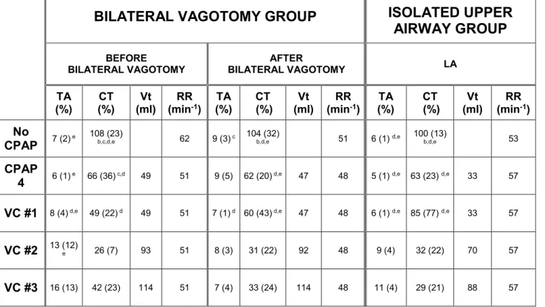
Table 1: Mean values of TA (laryngeal constrictor) and CT (laryngeal dilator) EMG activity and respiratory parameters during no CPAP, CPAP 4 cmH2O, nasal volume-controlled ventilation in the bilateral vagotomy group and lower airway ventilation in the isolated upper airway group.
BILATERAL VAGOTOMY GROUP
ISOLATED UPPER
AIRWAY GROUP
BEFORE
BILATERAL VAGOTOMY BILATERAL VAGOTOMYAFTER LA
TA
(%) (%) CT (ml) Vt (minRR -1) (%) TA (%) CT (ml) Vt (minRR -1) (%) TA (%) CT (ml) Vt (minRR -1)
No
CPAP 7 (2) e 108 (23)b,c,d,e 62 9 (3) c 104 (32)b,d,e 51 6 (1) d,e 100 (13) b,d,e 53
CPAP
4 6 (1) e 66 (36) c,d 49 51 9 (5) 62 (20) d,e 47 48 5 (1) d,e 63 (23) d,e 33 57 VC #1 8 (4) d,e 49 (22) d 49 51 7 (1) d 60 (43) d,e 47 48 6 (1) d,e 85 (77) d,e 33 57
VC #2 13 (12)e 26 (7) 93 51 8 (3) 31 (22) 92 48 9 (4) 32 (22) 70 57
VC #3 16 (13) 42 (23) 114 51 7 (4) 33 (24) 114 48 11 (4) 29 (21) 88 57
TA, CT EMG: thyroarytenoid, cricothyroid inspiratory electrical activity, expressed as a percentage of baseline EMG; Vt: tidal volume; RR: respiratory rate; CPAP: continuous positive airway pressure; VC: volume control, intermittent positive pressure ventilation. LA : lower airways.
FIGURE LEGENDS
Figure 1: Electrical activity (EMG) of thyroarytenoid (a laryngeal constrictor) and cricothyroid (a laryngeal dilator) muscles in one lamb during baseline breathing (no CPAP, left) and nasal intermittent positive pressure ventilation (nIPPV), before (middle) and after (right) bilateral vagotomy.
Recordings were obtained during quiet sleep. Note: 1) the increase in thyroarytenoid muscle (TA) EMG during inspiration (I) from no CPAP to nIPPV before bilateral vagotomy; 2) the absence of an increase in TA EMG after bilateral vagotomy; 3) the disappearance of inspiratory cricothyroid muscle EMG in nIPPV, which is not affected by bilateral vagotomy; 4) the decrease in respiratory rate after bilateral vagotomy on the SUM signal. Abbreviations: TA: thyroarytenoid muscle EMG; ∫TA: moving time averaged TA; CT: cricothyroid muscle EMG; ∫CT: moving time averaged CT; SUM: sum signal of the respiratory inductance plethysmograph, illustrating the variations of lung volumes with respiration (inspiration upwards); EEG: electroencephalogram; EOG: electrooculogram.
Figure 2: Variations in inspiratory TA and CT EMG with increasing intermittent positive pressure ventilation applied through a nasal mask and before vagotomy (n = 5 lambs). No CPAP: baseline breathing; CPAP 4: continuous positive airway pressure, 4 cmH2O; VC #1, 2 and 3: progressively increasing intermittent positive pressure ventilation in the volume control mode. Voltage amplitude of inspiratory TA EMG is expressed as a
percentage of the mean amplitude observed with swallows during baseline recording. Voltage amplitude of inspiratory CT EMG is expressed as a percentage of the mean amplitude observed in inspiration during baseline spontaneous breathing. * : p < 0.05.
Figure 3: The increase in inspiratory thyroarytenoid muscle (TA) EMG observed with intermittent positive pressure ventilation (IPPV) originates from bronchopulmonary receptors, and not from the upper airways.
A: effects of vagotomy: the significant increase in mean inspiratory TA EMG observed when ventilating intact lambs via a nasal mask (left) (p = 0.0008 vs. baseline breathing) is inhibited by bilateral vagotomy (right) (p = 0.4). Voltage amplitude of inspiratory TA EMG is expressed as a percentage of the mean amplitude observed with swallows during baseline recording. B: lambs with isolated upper airways: the significant increase in the number of breathing cycles with inspiratory TA EMG observed from no CPAP condition when IPPV is applied solely on the lower airways via a tracheostomy (p < 0.0001 vs. baseline breathing) (middle box, LA-VC) is absent when ventilating the isolated upper airways (right, UA-VC) (p = 0.5). * : p < 0.05. Abbreviations: no CPAP: baseline breathing; LA-VC: ventilation in volume control mode on the lower airways; UA-VC: ventilation in volume control mode on the upper airways.
Figure 4: Decrease in inspiratory cricothyroid muscle (CT) EMG from baseline breathing to intermittent positive pressure ventilation (IPPV).
A, upper graphs: the observation that the inhibition of inspiratory CT EMG in IPPV is not prevented by vagotomy suggests that this inhibition does not originate from the bronchopulmonary receptors. B, lower graphs: similarly, the observation that the
number of breathing cycles with inspiratory CT EMG is still significantly decreased when IPPV is applied on the isolated upper airways only suggest that it does not originate from the upper airways. * p < 0.05. Abbreviations: no CPAP: baseline breathing; LA-VC: IPPV, volume control mode, via a tracheostomy on the lower airways; UA-LA-VC: ventilation, volume control mode, via a nasal mask on the upper airways.
Figure 5: Recordings obtained in one lamb with chronic laryngo-tracheal separation. In control conditions (left panel, no CPAP), both expiratory thyroarytenoid muscle (TA) EMG and inspiratory cricothyroid muscle (CT) EMG are present. During intermittent positive pressure ventilation (IPPV) applied on the lower airways via a tracheostomy (middle panel, LA), inspiratory TA EMG is observed simultaneously to mechanical insufflations, while CT EMG has disappeared. Oppositely, during IPPV applied on the isolated upper airways via a nasal mask (right panel, UA), while the lamb is spontaneously breathing through a tracheostomy, no changes are observed for TA and CT EMG from baseline. See figure 1 for abbreviations. Airway pressure is measured at the nasal mask for both no CPAP and UA conditions and at the tracheostomy site for LA condition.
Figure 1:
TA
∫TA
∫CT
CT
SUM
EEG
EOG
No CPAP Before bilateral vagotomy After bilateral vagotomy
3 s 3 s 3 s I I I I I I I I I I
Figure 2:
Inspiratory TA EMG
Inspiratory CT EMG
no CPAP CPAP 4 VC#1 VC#2 VC#3 0 20 40 60 80 100 120 140 160
*
*
*
*
*
*
*
% no CPAP CPAP 4 VC#1 VC#2 VC#3 0 5 10 15 20 25 30 35*
*
*
*
%*
Figure 3:
B
A
VC
Before bilateral vagotomy
0 2 4 6 8 10 12 14 16
*
%
After bilateral vagotomy
no CPAP VC 0 2 4 6 8 10 12 14 16 % % LA-VC UA-VC no CPAP 0 20 40 60 80
Inspiratory TA EMG in the isolated upper
airway group
.*
*
Figure 4:
A
After bilateral vagotomy
no CPAP VC 0 20 40 60 80 100 120
Before bilateral vagotomy
0
20
120