Délivré par :
Institut National Polytechnique de Toulouse (INP Toulouse)
Discipline ou spécialité :
Pathologie, Toxicologie, Génétique et Nutrition
Présentée et soutenue par :
M. IMOURANA ALASSANE-KPEMBIle lundi 25 novembre 2013
Titre :
Unité de recherche : Ecole doctorale :
TOXICITE INTESTINALE DES MYCOTOXINES: ANALYSE DES
INTERACTIONS ENTRE TRICHOTHECENES B.
Sciences Ecologiques, Vétérinaires, Agronomiques et Bioingénieries (SEVAB) INRA (INRA)
Directeur(s) de Thèse :
MME ISABELLE OSWALD M. OLIVIER PUEL
Rapporteurs :
M. FRANCOIS SICHEL, UNIVERSITE DE CAEN
Mme MARIE-CHRISTINE CHAGNON, UNIVERSITE DE BOURGOGNE
Membre(s) du jury :
2 M. ALAIN PERIQUET, UNIVERSITE TOULOUSE 3, Membre
2 M. FRANÇOIS ABIOLA, MINISTERE DE L'ENS. SUP. ET RECH. BENIN, Membre
2 Mme ISABELLE OSWALD, INRA TOULOUSE, Membre
1
de ce jury,
Le Pr François Adébayo ABIOLA qui a accepté de présider le jury, malgré un "emploi du temps de ministre". Je voudrais à travers sa personne exprimer toute ma gratitude à mes "Vénérables maîtres" de l’Ecole vétérinaire de Dakar. Le Pr Marie-Christine CHAGNON et le Pr François SICHEL qui me font l’honneur d’évaluer ce travail en qualité de rapporteurs ;
Le Pr Alain PERIQUET qui a bien voulu être examinateur de ce travail. Ses enseignements de Master Qualité et sécurité des aliments constituent les tous premiers éléments qui ont nourri cette réflexion sur l’évaluation de la toxicité combinée des contaminants alimentaires.
Le Dr Isabelle OSWALD, Directeur de cette thèse, pour tant de choses : son accueil au sein d’une équipe "qui bouge", l’équipe d’Immuno-mycotoxicologie TOXALIM-INRA, la pertinence de ses avis, toutes ces rencontres très
enrichissantes que j’ai eu l’occasion de faire, et pour avoir encadré mes premiers pas de chercheur.
Le Dr Olivier PUEL, co-directeur de cette thèse pour toute sa disponibilité, ses conseils et ses qualités humaines.
Je tiens à dire toute ma sympathie à tous les membres de l’équipe IMT pour la bonne camaraderie et l’esprit d’entraide qui y sont entretenus. J’ai une pensée particulière pour Soraya la "maman de substitution", Joëlle pour le cœur qu’elle met à faire "le job", Philippe "la tour de contrôle" et Anne-Marie dont le professionnalisme inspire beaucoup d’humilité. Je remercie "les filles" Selma, Patricia, Julie, Sabria, Alix, Joanna, Delphine, Rhoda et également (les quelques garçons égarés comme moi au sein de cette équipe) Pascal et Sophal, pour leur bonne humeur communicative et
2
enrichissantes et sa grande gentillesse.
Ce travail doit beaucoup à une rencontre aussi fortuite qu’exceptionnelle, celle du Pr David Ting-Chao CHOU.
Once upon a time, there was a Master who held two bottles of antitumor ingredients. The red bottle contained drug A, and the blue bottle contained drug B. He then gave the two bottles to his disciples, John and Paul. The Master asked them to conduct drug combination studies in different proportions and to determine whether they were synergistic or antagonistic by using any of their best choice of assay method and by using the best choice of theory for their dose-effect data analysis…
Chou TC Pharmacol Rev. 2006; 58(3):621-81
The Master waited several years until I met him somewhere between York Avenue and 1st Avenue, while looking for my way in that very misleading field of drug
combination studies, then he retired. Thanks so much !
Yac, Roger, Gorgui, J’ai eu à maintes reprises l’occasion au cours de ces années d’éprouver la solidité des liens d’amitié et de fraternité qui nous lient, et jamais je ne les ai pris en défaut.
Je remercie mes parents et mes frères qui m’ont accompagné au quotidien de leurs mots d’encouragement et de leur amour.
Ce travail n’aurait tout simplement pas été possible sans toi, Sokh. Tu as assuré ! Chapeau bas!
Quant à vous deux, moussaillons "Sambito" et "Moyenne sœur", prenez-en de la graine !
3
L’intestin est la première barrière de l’organisme contre les contaminants alimentaires, dont les mycotoxines. Le déoxynivalenol (DON) est un contaminant majeur des céréales, souvent retrouvé en association avec d’autres trichothécènes B (TCTs B), le 3- et 15-acétyldéoxynivalénol (3-ADON et 15-ADON), le nivalénol (NIV) et la fusarénone X (FX). Au niveau cellulaire, le DON interagit avec l’ARN ribosomique, bloquant ainsi la synthèse protéique et activant la cascade de la voie de signalisation de MAPKinases impliquée dans des mécanismes de la réponse inflammatoire. Au niveau intestinal, cette mycotoxine pourrait donc perturber le renouvellement continu de l’épithélium, et l’homéostasie de la réponse inflammatoire. On suggère ainsi qu’elle pourrait jouer un rôle dans la pathogénie des maladies inflammatoires chroniques de l’intestin. Si les effets du DON sont relativement connus, ceux du NIV et de leurs dérivés acétylés sont moins bien documentés. De même, peu de données existent quant à la toxicité combinée de ces mycotoxines dont la co-occurrence est avérée.
Sur des modèles in vitro de cellules épithéliales intestinales humaines et porcines et sur un modèle ex vivo d’explants de jéjunum de porc, nous avons comparé les toxicités individuelles de cinq TCTs B (DON, 3- et 15-ADON, NIV et FX) et analysé leur toxicité combinée en termes de synergie, additivité ou antagonisme vis-à-vis de l’intestin.
Les résultats montrent qu’à des concentrations de l’ordre du micromolaire, les TCTs B inhibent la croissance des cellules épithéliales intestinales par ordre croissant de toxicité 3-ADON, DON, 15-ADON, NIV et FX. Aux faibles doses correspondant à des niveaux d’exposition rencontrés chez le consommateur français ou européen, des synergies d’un facteur 3 à 10 ont été observées. Ces travaux ont également permis de caractériser l’activité pro-inflammatoire au niveau intestinal des TCTs B, et l’analyse benchmark de données de transcriptomique a montré que l’exposition de l’intestin à des doses aussi faibles que 0.04µM de FX, 0.1µM de DON ou 0.1µM de NIV s’accompagne d’une activation significative des mécanismes de l’inflammation. Ces
4
renouvellement de l’épithélium intestinal et l’activité pro-inflammatoire au niveau intestinal pourraient être des marqueurs très sensibles dans le cadre de l’évaluation de la toxicité individuelle et des interactions entre TCTs B.
5
As for other food-born contaminants, the gastro-intestinal tract represents the first barrier against deoxynivalenol (DON). This mycotoxin frequently co-occurs with other type B trichothecenes (TCTs B) namely 3-acetyldeoxynivalenol (3-ADON), 15-acetyldeoxynivalenol (15-ADON), nivalenol (NIV) and fusarenon-X (FX). At the cellular level, DON binding to ribosomal RNA results in the inhibition of protein synthesis and triggers the mitogen-activated protein kinases (MAPKs) pathway that have been linked to immune response mechanisms. Thus, intestinal epithelial cell renewing is considered a putative target in DON toxicity. Moreover, based on the ability of DON to disturb the state of homeostasis of the inflammatory response in the intestine mimicking what is found in inflammatory bowel diseases (IBD), it is proposed that this mycotoxin may play a role in such diseases. However, very few is known about the intestinal toxicity of the other co-occuring TCT B, and their combined effects eventually.
By means of in vitro human and porcine intestinal epithelial cells models and an ex vivo porcine jejunal explants model, we assessed the individual toxicity of five TCT B (DON, 3- and 15-ADON, NIV and FX) toward the intestine and we analyzed their combined toxicity in terms of additivity, synergy or antagonism.
The tested TCT B significantly impaired the intestinal epithelial cell growth in the micromolar range, in increasing order of potency 3-ADON, DON, 15-ADON, NIV and FX. The toxicity of low doses of TCT B was synergistic. For mycotoxin concentrations corresponding to exposure levels reported for French and European consumers, the amplitude of this synergy ranged between 3 and 10. Benchmark dose analyses of the transcriptional data also showed that the exposure of the intestine to mycotoxin concentrations as low as 0.04µM for FX, 0.1µM for DON and 0.1µM for NIV could be associated to a significant activation of the inflammatory response mechanisms. Taken together, these results suggest that epithelial cell renewing and pro-inflammatory effects at the intestinal level may be consider very sensitive
7
Articles
Alassane-Kpembi, I., Kolf-Clauw, M., Gauthier, T., Abrami, R., Abiola, F.A., Oswald, I.P., Puel, O., 2013. New insights into mycotoxin mixtures: The toxicity of low doses of Type B trichothecenes on intestinal epithelial cells is synergistic. Toxicol Appl Pharmacol 272, 191-198.
Kolf-Clauw, M., Sassahara, M., Lucioli, J., Rubira-Gerez, J., Alassane-Kpembi, I., Lyazhri, F., Borin, C., Oswald, I.P., 2013. The emerging mycotoxin, enniatin B1, down-modulates the gastrointestinal toxicity of T-2 toxin in vitro on intestinal epithelial cells and ex vivo on intestinal explants. Arch Toxicol. Sous presse.
Alassane-Kpembi I., Puel O., and Oswald I.P. 2013. Toxicological interactions between deoxynivalenol, nivalenol and their acetyl derivatives in intestinal epithelial cells. Soumis pour publication à Toxicology.
Alassane-Kpembi I., Puel O., and Oswald I.P. 2013. Transcriptional Regulation of the Inflammatory Response in the Intestine upon Exposure to Trichothecene Mycotoxins Deoxynivalenol, Nivalenol and Fusarenon-X: a Benchmark Dose Analysis. Manuscrit en préparation.
Revues et chapitres de livre
Alassane-Kpembi, I. and Oswald, I. P. 2013. Effects of feed contaminants on the intestinal health of monogastric farm animals. In: Niewold, T. (ed.) Intestinal health, key to optimise production. Wageningen Academic Publishers, The Netherlands. Sous presse.
Alassane-Kpembi I., Abiola F.A., Streit E., Schatzmayr G., Tassis P., Tzika E., Marin D., Taranu I., Tabuc C., Nicolau A., Aprodu I., Puel O., and Oswald I.P. 2013.
8
Communications orales (Congrès nationaux ou internationaux)
Alassane-Kpembi I., Gauthier T., Lyazrhi F., Puel O., Abiola F. A., Kolf-Clauw M. et Oswald I.P. Analyse des interactions entre trichothécènes B sur une lignée de cellules intestinales. Journées mycotoxines, Bordeaux (France), 19 Janvier 2012.
Alassane-Kpembi I., Abiola F.A., Puel O., and Oswald I.P. 2013. The type of interaction between type B trichothecenes on the intestine varies with the dose. 12th European Fusarium Seminar, Bordeaux, 12-16 May 2013.
Alassane-Kpembi I., Abiola F.A., Puel O., and Oswald I.P. 2013. New insights into mycotoxin mixtures: the toxicity of low doses of Type B trichothecenes against intestinal epithelial cells is synergistic. 35th Mycotoxin Workshop, Ghent (Belgium), 22-24 May 2013.
Alassane-Kpembi I., Abiola F.A., Puel O., and Oswald I.P. 2013. The issue of toxicological interactions in in vitro assessment of the combined toxicity of mycotoxins. 2nd FOODSEG Symposium Safe Food for Europe: Recent scientific and regulatory developments, Bucharest (Romania), 14-15 June 2013.
Posters
Alassane-Kpembi I., Gauthier T., Lyazrhi F., Puel O., Abiola F. A., Kolf-Clauw M. et Oswald I.P. Cytotoxicité des mycotoxines de la famille des trichotécènes : analyse des interactions. Journées de l’école Doctorale SEVAB 2011, Toulouse (France), 27 Octobre 2011.
Alassane-Kpembi I., Gauthier T., Lyazrhi F., Puel O., Abiola F. A., Kolf-Clauw M. et Oswald I.P. Cytotoxicité combinée du DON et de ses dérivés acétylés sur des
9
Alassane-Kpembi I., Gauthier T., Lyazrhi F., Puel O., Abiola F. A., Kolf-Clauw M. et Oswald I.P. Interactions des trichothécènes B : Evaluation de la cytotoxicité combinée du DON et de ses dérivés acétylés. Journées d’animation scientifique du département de Santé Animale INRA, Fréjus (France), 22 – 25 Mai 2011.
10
µM : Micromolaire IL : Interleukine
15-ADON : 15-acetyldéoxynivalénol IPEC-1 : Intestinal Porcine Epithelial Cells-1 3-ADON : 3-acetyldéoxynivalénol JNK : Jun N-terminal kinase
AFB1 : Aflatoxine B1 kDa : Kilo Dalton
BMD : Benchmark dose MAPK : Mitogen-activated protein kinase BMDL : Benchmark dose Lower bound
confidence limit MRP-2 : Multridrug resistance-associated protein 2
CI : Combination index MTT : 3,(4,5-dimethylthiazol-2-yl) 2,5-diphenyltetrazolium bromide
COX : Cyclo-oxygénase NF-κB : Nuclear factor kappa-B CPA : Cyclopiazonic acid NIV : Nivalenol
DJT : Dose journalière tolérable NOAEL : No-observed adverse effect level
Dm: Median-effect dose NOD : Nucleotide-binding oligomerization domain
DON : Déoxynivalenol NR : Neutral red
DRI : Dose reduction index OMS : Organisation mondiale de la santé ERK : Extarcellular signal-regulated
protein kinase OTA : Ochratoxine A
fa : fraction affected PODs : Points of Departure
FAO : Food and agriculture organization RT-PCR : Reverse transcriptase polymerase chain reaction
FB1 : Fumonisine B1 SCF : Scientific committee on food
FX : Fusarénone-X SOCS : Suppressor of cytokines signaling IBD : Inflammatory bowel disease TCT : Trichothécene
IC50 : Inhibitory concentration 50% Th1 et Th2 : Lymphocytes T-auxilliaire (helper) 1 et 2
IFN : Interféron TLR : Toll-like receptor
Ig : Immunoglobuline TNF-α : Tumor necrosis factor α ZEA : Zéaralénone
11
Remerciements 1
Résumé 3
Abstract 5
Liste des publications et communications 7
I. Introduction 12
1. Contexte de l'étude 13
2. Etude bibliographique 16
2.1 Effets des contaminants alimentaires sur la santé intestinale
des animaux monogastriques 17
2.2 Approches méthodologiques et pertinence biologique des études d’évaluation in vitro des interactions toxicodynamiques
des mycotoxines 50
II. Travail expérimental 100
Objectifs de la thèse 101
Chapitre I: Toxicité comparée et combinée des TCT B pour le
renouvellement de l'épithélium intestinal 103
Résumé de l'étude 104
Article N° 1 105
Article N° 2 113
Chapitre II: Caractérisation de l'activité intestinale pro-inflammatoire
des TCT B 144
Résumé de l'étude 145
Article N° 3 146
III. Discussion générale 185
IV. Conclusions-perspectives 208
Bibliographie 212
12
13
1. CONTEXTE DE L’ETUDE
Les moisissures, en particulier celles du genre Aspergillus, Fusarium, Penicillium, Claviceps et Alternaria, qui colonisent les denrées végétales (céréales, fruits, noix, amandes, grains, fourrages) au champ ou en cours de stockage produisent une multitude de métabolites secondaires. La sécrétion de ces composés, de structures chimiques très variées, vise entre autres à faciliter aux moisissures la colonisation du substrat, en leur procurant par exemple un avantage par rapport autres micro-organismes avec lesquels ils seraient en compétition. Au nombre de ces métabolites secondaires, certains qui présentent une toxicité plus ou moins marquée pour les humains et les animaux sont appelés mycotoxines. On estime que 25% des récoltes mondiales véhiculent ces contaminants naturels dont la plupart ne sont pas dégradés par la chaleur et restent présents même après la disparition des champignons (CAST, 2003). Les principales classes de mycotoxines aujourd’hui recensées sont les aflatoxines, les trichothécènes (TCTs), les fumonisines, les ochratoxines, les alcaloïdes de l’ergot de seigle et la zéaralénone.
En France, comme dans les grands bassins de production de céréales dans le monde, les conditions climatiques sont propices au développement des champignons du genre Fusarium, et en particulier F. graminearum et F. culmorum. Ces céréales sont ainsi susceptibles d'être contaminées par du déoxynivalenol (DON), une mycotoxine du groupe des TCT. D’un enjeu de santé publique au départ restreint à la niche écologique des espèces de champignon productrices, l’accroissement du volume des échanges commerciaux internationaux et la place des céréales dans ces échanges, ont fait de la contamination des aliments par le DON une préoccupation mondiale. Depuis 2010, le DON se classe ainsi en tête de peloton des mycotoxines les plus fréquemment rencontrées dans le monde, avec plus de 60% des échantillons d’aliment testés positifs (Schatzmayr et Streit, 2013).
14
Outre le DON, F. graminearum et F. culmorum produisent d’autres mycotoxines telles que la zéaralenone, la fusarine C, la chrysogine ainsi que de nombreux autres TCT B (15-acetyldeoxynivalenol (15-ADON), 3-acetyldeoxynivalenol (3-ADON), nivalenol (NIV) et fusarénone X (FX), mais aussi des toxines dont la structure reste à élucider. On estime que seulement 20% des métabolites de Fusarium sont identifiés structurellement. En conséquence, l'homme et l'animal ne sont généralement pas exposés à une mycotoxine, mais à un cocktail de toxines en même temps. De nombreuses enquêtes menées à l’échelle mondiale montrent ainsi que la co-contamination par plusieurs mycotoxines est la règle plutôt que l’exception (Rodrigues et Naehrer, 2012; Streit et al., 2012; Streit et al., 2013). Il est légitime de s’interroger sur le risque cumulatif associé à cette exposition simultanée à plusieurs toxiques, et ses implications sur l’innocuité d’aliments contaminés à des niveaux jugés acceptables pour chaque toxique pris individuellement. La plupart des études de toxicité ayant analysé les effets des mycotoxines individuellement, peu d’éléments existent à ce jour pour une évaluation du risque qui prenne en compte de possibles effets toxiques additifs, synergiques, voire antagonistes. Il est donc important d’étudier les effets des co-contaminations mycotoxiques pour leur prise en compte par la réglementation.
Le porc est particulièrement exposé aux mycotoxines, du fait de son alimentation qui est essentiellement composée de céréales. Contrairement aux polygastriques doués de bonnes capacités de détoxification d’un certain nombre de mycotoxines, le porc est une espèce particulièrement sensible. Il est également très similaire à l’humain du point de vue transcriptomique, protéomique et immunitaire. A ce titre, il représente un bon modèle d’étude de l’impact des mycotoxines contenues dans les aliments sur la santé (Nejdfors et al., 2000).
Dans le tractus gastro-intestinal la biodisponibilité des mycotoxines, comme celles des autres contaminants alimentaires, est maximale, et l’intestin représente la première cible, mais également la première ligne de défense de l’organisme contre ces toxiques. Chez les mammifères adultes l’intestin est l’organe qui présente le pouvoir de
15
régénération le plus élevé, et on estime que l’épithélium intestinal est entièrement renouvelé entre 72h et 96h (Heath, 1996). Il est, de fait, très sensible à la toxicité du DON et des autres mycotoxines du groupes des TCT qui sont des inhibiteurs connus de la synthèse protéique (Ueno, 1985).
Notre projet de recherche ayant abouti à la rédaction de cette thèse dans le cadre de la préparation du Doctorat de l’Université de Toulouse, avait pour objectif de caractériser la toxicité individuelle et combinée de cinq TCT B, le DON, le NIV et leurs dérivés acétylés (3-ADON, 15-ADON et FX) vis à vis de l’intestin, en utilisant deux modèles de complexité croissante:
(i) Le renouvellement de l’épithélium intestinal
16
2. ETUDE BIBLIOGRAPHIQUE
La synthèse bibliographique se présente sous la forme d’un chapitre de livre consacré à l’effet des contaminants alimentaires notamment les mycotoxines sur la santé intestinale, et d’un article de revue qui traite des différentes approches méthodologiques et des conclusions des études in vitro des interactions toxicodynamiques entre mycotoxines.
L’intestin est particulièrement exposé aux contaminants aussi bien naturels que d’origine anthropogénique qui peuvent être véhiculés par les aliments. Chez les animaux à cycle court et plus particulièrement les monogastriques, la santé intestinale est un déterminant majeur de l’efficacité alimentaire et donc des performances zootechniques enregistrés dans les élevages. De même, la physiologie de ces animaux est à bien des égards comparable à celle de l’homme. Partant des mycotoxines comme exemple de contaminant naturel et des dioxines comme exemple de contaminant d’origine anthropogénique, nous nous sommes intéressés dans la première partie de cette synthèse bibliographique aux effets des contaminants alimentaires sur les principales fonctions physiologiques de l’intestin rapportés dans des études de toxicologie ayant des monogastriques comme modèle d’étude.
L’intestin est très vraisemblablement soumis aux effets simultanés de plusieurs contaminants. La deuxième partie de synthèse aborde donc la complexité des approches méthodologiques dans les études d’interaction de type toxicodynamique des xénobiotiques en général, et de façon plus spécifique, les conclusions biologiques d’études consacrées à l’évaluation in vitro de la toxicité combinée des mycotoxines.
17
2.1. Effets des contaminants alimentaires sur la santé intestinale des animaux monogastriques
De par l’importance de sa surface d’échange avec le milieu extérieur, la muqueuse intestinale est particulièrement exposée à diverses agressions de nature chimique ou microbiologique. Les trois principales fonctions de cette muqueuse peuvent être affectées par les contaminants alimentaires. Elle sert d’abord de barrière physique entre le l’organisme et le contenu de la lumière intestinale. Elle est responsable de la digestion puis de l’absorption des nutriments. Elle sert enfin de métronome de la réponse immunitaire locale en participant à la mise en place de l’homéostasie nécessaire à l’orientation vers la tolérance aux antigènes associés à la flore commensale et aux aliments, et aux mécanismes actifs de défense mis en jeu en présence d’antigènes associés à la flore pathogène. Nous présentons dans cette revue les effets décrits pour deux types de contaminants alimentaires (les mycotoxines et les dioxines) sur ces fonctions. Cette étude fait la synthèse des modes d’action de ces deux classes de contaminants au niveau de l’intestin, et montre clairement que des niveaux d’exposition usuellement rencontrés sont susceptibles d’impacter négativement l’intégrité et la fonctionnalité de cet organe.
Cette revue rédigée sous la direction du Dr Isabelle OSWALD, a été acceptée comme chapitre de l’ouvrage intitulé "Intestinal health, key to optimise production" (Ed. Theo Niewold) Wageningen Academic Publishers à paraître en Janvier 2014.
18
Effect of feed contaminants on intestinal health of monogastric farm animals
Imourana ALASSANE-KPEMBIa,b,c and Isabelle P. OSWALDa,b
aINRA, UMR 1331 ToxAlim, Research centre in Food Toxicology, F-31027, Toulouse
France
bUniversité de Toulouse, INP, UMR 1331, Toxalim, F-31027, Toulouse, France c
19
Abstract
As the most extensively exposed surface in the body, the intestinal mucosa has to face important chemical and biological challenges. The intestinal mucosa has three main physiological functions. It establishes a physical barrier between the internal milieu and the luminal content. The intestinal mucosa is also responsible for luminal nutrients digestion and their subsequent absorption. The mucosal epithelium is at the interface of immune system and luminal contents, including dietary antigens and microbial products. This implies a local defence mechanisms regulation that involves integrating all the signals that come from the external and internal world to preserve immune homeostasis steady-state conditions. Either of these intestinal physiological functions may be targeted by feed contaminants. These contaminants may be naturally occurring compounds or substances from anthropogenic sources. In the present chapter, we present mycotoxins and dioxins, which are representative examples of both classes of contaminants. Data gathered show clearly that dietary exposure to realistic doses of these contaminants impairs intestine functionality and its integrity as well. The mechanisms of action of mycotoxins and dioxins targeting the gastrointestinal tract are clarified and evidences for their deleterious effects for monogastrics intestinal health is provided.
20
I. Introduction
The gastrointestinal tract has the most extensively exposed surface in the body and is constantly exposed to potentially harmful food-born substances from diverse sources (Yegani and Korver, 2008). Gut damages caused by these contaminants may lead to poor intestinal health. Three main functions that may be targeted by feed contaminants are devolved to the intestinal mucosa (Rescigno, 2011; Turner, 2009). First, it establishes a physical barrier between the internal milieu and the sometime hostile luminal content. The intestinal mucosa is also responsible for nutrients digestion and their subsequent absorption, which require a selectively permeable barrier. For these two functions, the mucosal epithelium is de facto at the interface of immune system and luminal contents, including dietary antigens and microbial products. That raises the third function for intestinal mucosa, which is in charge of integrating all the signals that come from the external and internal world to preserve intestinal immune homeostasis steady-state conditions.
Naturally occurring contaminants and pollutants from anthropogenic sources are the two classes of contaminants that may be present in feed. Natural contaminants from both plants and fungal specific primary and secondary metabolisms may be found in feed. These substances may have anti-nutritional or specific toxic effects for farm animals. Toxins from fungal and plant origin include and are not limited to mycotoxins, lectins, cyanogens, gossypol, glucosinolates and phyto-oestrogens (for a review see D'Mello 2004). Environmental naturally occurring contaminants like heavy metals and metalloids are widespread and can be found from traces to macro levels. Their significant contamination through soil of seeds and plants fed to animals may also result in chronic toxicity. Contaminants from anthropogenic source are in general substances manufactured for industrial use that are not naturally occurring, but may enter accidentally or deliberately the environment and result in environmental, agricultural, industrial or other contaminations. In other cases industrial activity may increase the mobility of naturally-occurring chemicals, or increase their amount
21
available to circulate in the environment, allowing them to contaminate feed at higher levels than would otherwise occur. Examples of these anthropogenic contaminants include persistent organic pollutants (especially dioxins), pesticides and radionuclides.
In this chapter, we present mycotoxins and dioxins which are illustrative examples of both feed contaminants classes, and their reported effects as well as mechanisms of action targeting the gastrointestinal tract and potentially deleterious for the intestinal health of monogastric farm animals.
Mycotoxins in Feed
Mycotoxins are secondary fungal metabolites produced under specific environmental conditions by a variety of mould spoiling agricultural commodities. As secondary metabolites, they are not essential for life, but may provide the fungus with an ecological advantage in certain environments. Factors contributing to the presence of mycotoxins include ecological and storage conditions that are most times beyond human control. Toxigenic molds are known to produce one or more of these mycotoxins and a substrate can be spoiled by more than one mold. Some 300 compounds have been recognized as mycotoxins of which a dozen is considered as threat to human and animal health. It has been estimated that at least 25% of the yearly worldwide grain production is contaminated (CAST, 2003). The mycotoxins of biological and economical significance in animal agriculture, i.e., aflatoxins, fumonisins, trichothecenes, zearalenone, and ochratoxins have been extensively reviewed and will be only shortly described in this chapter (Bennett and Klich, 2003; Bryden, 2012).
Aflatoxins
Aflatoxins were isolated and identified in England in the earlier 1960’s as the cause of a mysterious outbreak of hepatic necrosis affecting thousands of poultry. Similar incidents were then reported in pigs. Investigations revealed that toxicity was
22
associated to the presence of Aspergillus flavus in the feed and further that extracts of the culture of the fungus were capable of inducing the syndrome. Later many other species in the sections Flavi, Nidulantes and Ochraceorosei were also identified as aflatoxin-producers (for detailed review see Varga et al., 2011). Structurally aflatoxins are difurocoumarin derivatives that fluoresce under ultraviolet light. The most important aflatoxin in terms of toxic potency and occurrence is aflatoxin B1which has been classified as a group 1 carcinogen by the International Agency for Research on Cancer (IARC). AFB1 is hepatotoxic and hepatocarcinogenic, but many other effects can be associated to its toxicity: immunosuppression, reduced growth rate and reproductivity, lowered milk and egg production (Rawal et al., 2010).
Fumonisins
Fumonisins are products of polyketide and amino acid metabolism and have a linear structure with amine and tricarballylic ester functions. They are produced by Fusarium verticillioides and many other Fusarium species. Some twelve fumonisins have been isolated, but fumonisin B1 (FB1) is the most abundantly produced and the most toxic. The toxicity of FB1, including its effect on the intestine, is mainly exerted through the ability of this toxin to disrupt sphingolipid metabolism (Bouhet and Oswald, 2007). The IARC has classified FB1 as possibly carcinogenic to humans (group 2B). This toxin may also be implicated in the aetiology of human oesophagal cancer and neural tube defects. FB1 also causes leukoencephalomalacia in equines, pulmonary oedema and hydrothorax in swine, nephrotoxicity and hepatotoxicity in many species.
Trichothecenes
Trichothecenes are closely related sesquiterpenoid mycotoxins with a 12,13-epoxy ring and a variable number of hydroxyl, acetoxy or other substituents. They are classified as macrocyclic or non-macrocyclic depending on the presence of a macrocyclic ester or an ester-ether bridge between C-4 and C-15. Food and feed-born
23
trichothecenes are mainly non-macrocyclic compounds produced by fungi of the Fusarium genus. They can be subclassified in type A which have a hydrogen or ester type side chain at the C-8 position and type B which have a ketone group instead. Trichothecenes are well-characterized inhibitors of the protein and nucleic acids synthesis. In acute toxicity tests, type A members such as T-2 toxin have been found to be more toxic than type B components such as deoxynivalenol (DON) and nivalenol (NIV). However in practical situations of chronic intake, the effects and syndromes arising from DON are likely to be more important. In animal studies, chronic exposure to trichothecenes caused impaired weight gain, anorexia, hematotoxicity and immune dysregulation.
Zearalenone
Zearalenone (ZEA) frequently co-occurs with certain type B trichothecenes as they are simultaneously produced by the same fungi. ZEA is a non-steroidal oestrogen and its alcohol metabolites (α-zearalenol and β-zearalenol) have increased oestrogenic activity corresponding to their binding affinities for hepatic, uterine, mammary and hypothalamic oestrogen receptors. Dietary exposure to low doses of ZEA leads to hyperoestrogenic syndroms and subsequent reproductive dysfunctions in pigs.
Ochratoxins
Ochratoxins are isolated from fungi belonging to Aspergillus and Penicillium geni. They are chemically described as 3,4-dihydromethylisocoumarin derivatives linked with an amide bond to the amino group of L-β-phenylalanine. The most commonly occurring and most toxic member is ochratoxin A (OTA) which toxicological profile includes nephrotoxicity, hepatotoxicity, teratogenicity and immunotoxicity. In addition, OTA has been demonstrated to be carcinogenic among laboratory animals, which justifies that IARC has rated OTA as a possible human carcinogen (group 2B). Among farm animals, pigs are particularly sensitive to the toxin for its tissue
24
accumulation, due to a rather long serum half-life and the entero-hepatic recirculation. The feed occurrence of ochratoxins is not only important from the animal health and performance perspective, but also from the potential human indirect exposure through the animal derived foods consumed.
Dioxins in feed
Dioxins and dioxin-like chemicals are structurally related and environmentally persistent compounds that share a common mechanism of action, and as a consequence a common spectrum of biological responses. The term “dioxin” commonly refers to polychlorinated dibenzo-p-dioxins (PCDDs) and dibenzofurans (PCDFs) that are trace level unintentional by-products of most forms of combustion and several industrial chemical processes including chlorine bleaching of wood pulp and the manufacture of organochlorine pesticides, phenoxyacid herbicides and fungicides. PCDDs and PCDFs may also be present as contaminants in polychlorinated biphenyls (PCBs), which are considered dioxin-like compounds. While PCDDs and PCDFs are produced unintentionally as unwanted by-products, PCBs were manufactured for use in transformers, insulators, and many others technological applications. PCB production was banned by the Stockholm Convention on Persistent Organic Pollutants in 2001. Exposure levels are expressed in toxic equivalents (TEQ) of the most toxic congener, 2,3,7,8-tetrachloro-dibenzo-p-dioxin (2,3,7,8-TCDD). The main health issues concerning the dioxins have been reviewed elsewhere (Schecter et al. 2006). These compounds have a high-affinity binding to the aryl hydrocarbon receptor (AhR) which is an intracellular ligand–activated transcription factor involved in regulation of the expression of a large number of genes and which also interacts with regulatory proteins such as specific cellular kinases, and some of cell cycle control as well as apoptosis proteins. Even tiny doses of dioxins can cause death in certain laboratory animals and wildlife species, and 2,3,7,8-TCDD is considered the most toxic man-made chemical. Its LD50 for guinea pigs is evaluated to 1µg/kg body weight. The observed effects in humans and experimental animals
25
include toxicity to the immune system, reproductive and developmental abnormalities, and endocrine disruption with diabetes and thyroid disorders (Bursian et al., 2013a; b; Pavuk et al., 2003; Weisglas-Kuperus et al., 2000). The IARC has classified 2,3,7,8-TCDD as Group 1, carcinogenic to humans. Polygastrics grazing pastures that are close to industrial area are typically considered farm animals exposed to dioxin risk (Kamphues and Schulz, 2006), but the Belgian PCB/dioxin incident revealed that poultry and pigs may also be at risk (Bernard et al., 2002; Covaci et al., 2008). For example, an incident occurred at the end of January 1999 when a mixture of PCBs contaminated with dioxins was accidentally added to a stock of recycled fat used in the production of animal feeds. Signs of poultry poisoning were noticed by February, 1999, and it appeared that more than 2500 Belgian, French and Netherlands farms could have been supplied with contaminated feeds. Important features of the crisis were the creation of the Federal Agency for Food Safety in Belgium and the introduction in 1999 of norms for PCBs in feedstuffs and food in Belgium followed by the introduction in 2002 of European harmonized norms for PCDD/Fs in animal feed and food of animal origin. The Belgian crisis was followed few years later by an Irish pork PCB/dioxin contamination which source was traced to an animal feed production facility using the hot gases from the combustion of contaminated fuel oil to dry animal feed (Marnane, 2012).
Effects of feed contaminants on intestinal epithelium renewing and intestinal barrier function
Intestinal cell proliferation
The intestinal epithelium is the most vigorously self-renewing tissue of adult mammals (Heath, 1996). The mammalian intestinal epithelium is renewed every 4–5 days. This high cell turnover allows rapid resealing of the epithelial barrier following injury or damage in order to maintain an effective barrier function. The normal homeostasis may be impaired by feed contaminants affecting self-renewing capacities of the
26
intestinal epithelium. The non-transformed porcine intestinal epithelial cell lines IPEC -1 and IPEC-J2 (see chapter Techniques for investigating gut function in vivo, ex vivo and in vitro in monogastric farm animals) have been used to assess mycotoxins toxicity on pig intestine (Bouhet et al., 2004; Diesing et al., 2011; Vandenbroucke et al., 2011). Proliferating intestinal epithelial cells appears more sensitive to trichothecenes than differentiated cells, suggesting that exposure to these feed contaminants could dramatically impair the physiologically constant state of regeneration of the intestinal epithelium. Moreover, co-exposure to low concentrations of different trichothecenes leads to synergistic cytotoxicity in intestinal epithelial cells (Alassane-Kpembi et al., 2013).
Intestinal Barrier function
Testing high concentrations of DON (2000ng/mL) on both intestinal cell lines resulted in disintegration of tight junction protein zonula occludens-1 (ZO-1), increase of cell cycle phase G2/M and early activation of caspase-3, while low concentration (200ng/mL) showed no effect on these parameters (Diesing et al., 2011). Reduced expression of other tight junction proteins, claudin 3 and claudin 4, was also reported in IPEC-1 monolayer epithelium exposed to DON (Pinton et al., 2010; 2009). Other mycotoxins T2-toxin, FB1 and ZEA, also impair the integrity of IPEC-J2 monolayer via altered viability and reduced trans-epithelial electrical resistance (TEER), and promote the trans-epithelial passage of the antibiotics (Goossens et al., 2012). It has also been observed that a prolonged exposure to FB1 prevents the establishment of the TEER and alters the resistance of an already established monolayer IPEC-1 epithelium (Bouhet et al., 2004).
The mechanism by which mycotoxins could alter pig intestinal barrier function has also been investigated using intestinal cells and intestinal explants culture. The activation of the p44/42 ERK signaling pathway by DON and its acetyl derivatives
27
intestinal barrier function (Pinton et al., 2012). Selective removal of claudin isoforms was also demonstrated with OTA which decreased intestinal barrier properties by repressing claudin 3 and 4, but not claudin 1 (McLaughlin et al., 2004). Limited data related with chicken intestinal barrier function suggest that acute exposure to AFB1 moderately affect TEER (Yunus et al., 2011b).
Beside the mycotoxins, PCBs constitute the other group of frequently occurring feed contaminants exerting an ascertained disruptive effect on intestinal epithelial integrity, even though no study to our knowledge assessed the specific sensitivity of farm animal models to this class of compounds. The disruptive effect of highly chlorinated PCBs on gut integrity has been demonstrated in human colon adenocarcinoma cells (Caco-2) and in C57BL/6 mice (Choi et al., 2010). The authors showed that exposure to each of PCB congeners PCB153, PCB118, PCB104, and PCB126 increased the permeability of the intestinal barrier to fluorescein isothiocyanate (FITC)-labelled dextran (4 kDa) and disrupted expression of tight junction proteins ZO-1 and occludin.
Histo-morphological alterations of intestine induced by feed contaminants
A broad spectrum of histo-morphological alteration patterns is related to the presence of contaminants in feedstuffs. DON and its acetyl derivatives, FB1 and ZEA are associated with mucosal morphological abnormalities in poultry and pigs. Villi height decrease or atrophy as well as villi fusion and other regressive lesions in the gastro-intestinal tract are frequently reported (Awad et al., 2006; Kolf-Clauw et al., 2009; Sklan et al., 2003; Yunus et al., 2012; Zielonka et al., 2009). However hyperplasia of goblet cells was observed in 1-day-old broilers fed ad libitum for two weeks a 300 mg/Kg FB1-contaminated diet (Brown et al., 1992). A chronic exposure of Ross 308 male chicks to increasing levels of AFB1, was associated with a dose dependent reduction of duodenum and jejunum weight (Yunus et al., 2011a). Interestingly, a compensatory increase in the length of the duodenum and jejunum was noted after 4 weeks of exposure to high concentration of toxin. Contrary to the observations in
28
broilers, a linear increase in the crypts depth in distal jejunum was noted with increasing levels of AFB1 in the diet of layers (Applegate et al., 2009).
Polychlorinated dibenzo-p-dioxins (PCDDs) are also associated with regressive intestinal lesions. A survey of environmental contamination with 2,3,7,8-TCDD and other PCDDs indicated that the intestine weight in two great blue heron (Ardea herodias) colonies regressed negatively on TCDD level (Sanderson et al., 1994).
Modulation of digestive functionality of the intestine by feed contaminants Absorptive functionality
The frequent regressive intestinal lesions may explain at least in part the reduced absorption of nutrients observed with exposure to several feed contaminants. Smith et al. (2012) reviewed evidence from human and animal studies that mycotoxins may share a downstream pathway for children stunting by targeting the intestinal tract and inducing Environmental Enteropathy. Aflatoxin reduced iron absorption in chicken, regardless of the presence of anemia (Lanza et al., 1981). Surprisingly broiler chicks fed a 10 mg AFB1/Kg diet for one week showed an increase of the both mediated and passive components of glucose and methionine in vitro absorption, while lower exposure (1.25 to 5 mg) for longer period (3 week) showed no effect compared with control birds receiving no aflatoxin (Ruff and Wyatt, 1976).
Glucose uptake was assessed into laying hens jejuna epithelia in presence of DON (Awad et al., 2007). DON decreased glucose uptake almost as efficiently as phlorizin. In the presence of phlorizin, pharmacological inhibitor of sodium glucose-linked transporter 1 (SGLT-1), DON had no additional effect on the glucose uptake. SGLT-1 is the main apical transporter for active glucose uptake in the small intestine. It works like a symporter that uses the electrochemical gradient of Na+ to drive the glucose absorption. This similarity between the effects of phlorizin and DON on glucose uptake evidences their common ability to inhibit Na+-D-glucose co-transport.
29
These authors also evaluated mRNA expression of SGLT-1 in broiler chicken fed a diet naturally contaminated with DON (1 and 5 mg/kg), (Awad et al., 2011). After 5 weeks, the mRNA of SGLT-1 was down-regulated in duodenal and jejunal tissues of DON-supplemented groups. Ussing chamber experiments conducted at the same time confirmed the glucose-induced inhibition of currents in intestinal tissues. Taken together, these results suggest that gene expression mediate the inhibitory effects of DON on intestinal glucose uptake. Based on this transcriptomic approach, Dietrich et al (2012) showed that not only DON impairs sugars (glucose and fructose) intestinal absorption in broilers, but that mycotoxin might also alter the uptake of palmitate and monocarboxilates in the jejunum at realistic doses of DON (2.5 to 5 mg/kg).
Conversely, sodium-dependent glucose absorption might be up-regulated in pig after acute or long term exposure to the mycotoxin FB1 (Lalles et al., 2009; Lessard et al., 2009). Studies in mice found that exposure to fumonisins in utero increases the frequency of developmental defects, and administration of folate or a complex sphingolipid is preventive (Marasas et al., 2004). The folate uptake is mediated by the folate receptor, which like many glycosylphosphatidylinositol-anchored proteins, is enriched in cholesterol and sphingolipids. In the human intestinal epithelial Caco-2 model, the fumonisin induced-depletion in sphingolipids was shown to impair the folate receptor function (Stevens and Tang, 1997). The authors suggested that dietary exposure to FB1 could adversely affect folate uptake and potentially compromise cellular processes dependent on this vitamin. In pigs, sphingolipids depletion was observed in intestinal epithelium after a seven-days-dietary exposure to 1.5mg/kg b.w. FB1 (Loiseau et al., 2007).
Active intestinal absorption of glucose and leucine may also be impaired after oral treatment with 2,3,7,8-TCDD or PCBs (Ball and Chhabra, 1981; Madge, 1976a; Madge, 1976b).
30
Activity of digestive enzymes
Modulation of digestive enzyme production and/or activity is one of the biological effects of food contaminants targeting the gastrointestinal tract. Several studies concluded that enzyme activity is modulated following mycotoxin consumption, even though contradictory effects were reported. In pigs the activity of aminopeptidase N, but not of sucrase, was markedly lowered by consumption of corn culture extracts containing fumonisins in a nine-day feeding trial (Lessard et al., 2009). A 2-week feeding study with laying hens showed that the specific activity of the intestinal maltase increased quadratically by feeding up to 1.2 mg/kg of aflatoxin and declined at 2.5 mg/kg of aflatoxin (Applegate et al., 2009). Activities of digestive enzymes from duodenum contents including protease, chymotrypsin, trypsin and amylase were increased in AFB1-treated group for ducks in a six week- feeding trial (Han et al., 2008).On the contrary α-amylase and lipase activities in duodenum were lower in hens fed the aflatoxin-contaminated diet while pancreas amylase, trypsin and chymotrypsin activity were increased (Matur et al., 2010). In line with lipase activity decrease, there was a highly significant increase of lipid excretion in feces of young broiler chickens at 1.25 µg/g and above dietary aflatoxin exposure (including a three-fold increase at 10 µg/g) (Osborne and Hamilton, 1981). The paradoxical upward trend of pancreatic enzyme secretion might be ascribed to pancreas cells damage as acute and chronic pancreatitis are associated with great proenzyme release (Han et al., 2008; Matur et al., 2010). It’s noteworthy that Osborne and Hamilton (1981) reported pancreatomegaly in chicken for 2.5 µg/g dietary aflatoxin exposure and above.
The intestinal mucosa can also be regarded as the largest endocrine organ, which utilizes some 100 identified messengers (Ahlman and Nilsson, 2001; Furness et al., 1999). The enterochromaffin cells constitute the largest endocrine cell population in the gastrointestinal tract. On their apical side, gut endocrine cells have microvilli, which may serve to taste the intraluminal milieu (Newson et al., 1982). As an example cholecystokinin (CCK), also known as cholecystokinin-pancreozymin, is released
31
from duodenal endocrine cells after a meal. This hormone is produced by enteroendocrine I cells, which are distributed throughout the proximal intestine epithelium. It mediates digestive enzymes production from the pancreas and activates neurons of the gallbladder wall with subsequent emptying of bile for breakdown of fats and proteins in the meal. CCK also inhibits gastric emptying and initiates a satiety response via vagal afferents.
Chronic exposure to different types of commonly encountered environmental polychlorinated aromatic hydrocarbons (i.e. PCBs, dioxins, and 2,3,4,7,8-pentachlorodibenzofuran PeCDF) may exert an endocrine disruptive effect on intestinal CCK (Lee et al., 2000). These authors demonstrated that chronic ingestion of PCB-126,2,3,4,7,8-pentachlorodibenzofuran (PeCDF), or TCDD alone; a mixture of these three chemicals; or a mixture of PCB-126 and PCB-153 decreases intestinal stores of CCK peptide in a specific manner in rats. In addition, TCDD treatment of intestinal CCK cells lowered levels of prohormone convertase-1 and -2, which are involved in processing pro-CCK to mature, biologically active CCK.
Modification of the intestinal microflora by feed contaminant
As other fungi secondary metabolites especially antibiotics, several mycotoxins have shown their antimicrobial potential (Ali-Vehmas et al., 1998; Burmeister and Hesseltine, 1966). As a consequence, mycotoxins may modify the intestinal microflora. In pigs trichothecenes DON and T-2 toxin modified the dynamics of the intestinal bacteria communities, causing an increase of the number of aerobic bacteria (Tenk et al., 1982; Waché et al., 2009). A significant decrease of the amount of Salmonella Typhimurium bacteria present in the cecum content, and a tendency to a reduced colonization of the jejunum, ileum, cecum, and colon contents was showed in presence of 15 and 83µg T-2 toxin per kg feed in pigs (Verbrugghe et al., 2012). Microarray analysis revealed that T-2 toxin causes an intoxication of Salmonella Typhimurium, represented by a reduced motility and a down-regulation of metabolic
32
and Salmonella Pathogenicity Island 1 genes. However, in vitro T-2 toxin promoted the susceptibility of intestinal epithelial cells to Salmonella Typhimurium invasion and its translocation over an intact intestinal porcine epithelial cell monolayer. The enhanced Salmonella invasion in and translocation over the intestinal epithelial IPEC-1 and IPEC-J2 cells was also observed with non-cytotoxic doses of DON (Pinton et al., 2009; Vandenbroucke et al., 2011). Besides trichothecenes, the ability of OTA and patulin to increase the translocation of commensal bacteria across intact epithelium was also reported (Maresca et al., 2008).
Effect of feed contaminant on secretion of some intestinal defence components Mucus
In order to protect the mucosa, the host produces a complex layer of mucus that covers the gastrointestinal tract (Johansson et al., 2011; McGuckin et al., 2011). The mucus is organized in two layers that are organized around the highly glycosylated MUC2 mucin, forming a large, net-like polymer. The mucus gel provides a matrix for the retention of antimicrobial molecules in the mucosal environment. In addition, mucin glycoproteins that form the major macromolecular constituents of mucus can themselves have direct antimicrobial properties that limit the growth of microorganisms in the mucus. The mucin glycoproteins are produced by goblet cells. Feed contaminants may modulate mucus secretion.
ZEA and FB1 were found to exert a proliferative effect or an increasing activity on the goblets cells and their content of mucinogen vesicles in pigs and chickens (Brown et al., 1992; Obremski et al., 2005). On the contrary, low doses of mixtures of T-2 toxin, DON and ZEA reduced the number of goblet cells and the tightness of the intestinal glycocalyx in pigs (Obremski et al., 2008).
33
Anti-microbial peptides
The other major secretory cells within the gastrointestinal tract are the Paneth cells, which are identified by their characteristic intracellular granules containing a range of antimicrobial molecules that are secreted into the mucus to ensure sterility of the stem cell niche. Among them are defensins, a family of cationic antimicrobial peptides containing a specific six-cysteine motif also produced by epithelial cells (Yang et al., 2007). On porcine IPEC-J2 cells, an up-regulation of porcine beta-defensins 1 and 2 mRNA expression following exposure to DON, NIV, ZEA, individually and in mixtures has been observed (Wan et al., 2013). Supernatants from IPEC-J2 cells exposed to toxins, singly or in combination, however, possessed significantly less antimicrobial activity against Escherichia coli than untreated supernatants. The results
suggested interactive effects when cells were exposed to mycotoxin combinations.
Secretory IgA
Secretory antibodies, immunoglobulin A (IgA) and IgG that are very important components of the mucosal barrier, are secreted into the mucus by the epithelial cells (Strugnell and Wijburg, 2010). These antibodies influence the commensal microbiota and contribute substantially to the capacity of the mucus to retain and clear potential pathogens. Specific immunoglobulin (Ig) receptors are responsible for the immune response by regulating the Ig transport and cellular concentrations in various tissues. These receptors transport Igs across epithelial tissues to their sites of action. The Fc receptor (FcRn) is specific for IgG, whereas the polymeric immunoglobulin receptor (pIgR) recognizes dimeric IgA and pentameric IgM. Expression of the pIgR gene in epithelial cells of mucosal and glandular tissues is an unconditional pre-requisite for acquiring mucosal immunity, while FcRn could be important in immune activation and tolerance (Dickinson et al., 1999; Verbeet et al., 1995). In sheep, the silage contaminating mycotoxin mycophenolic acid (MPA) lowered FcRn expression in the liver, which may result in a lower IgG serum-to-bile transport, while pIgR expression
34
in ileum was stimulated (Dzidic et al., 2004). For viruses that invade via the mucosal route, both IgA and IgG can provide protection and mediate viral clearance. In a mouse model, T-2 toxin impaired the gastrointestinal tract clearance of the enteric reovirus serotype 1 and increased its fecal shedding (Li et al., 2006). These effects corresponded to a transient suppression of the induction of the specific IgA in feces and decreased secretion of reovirus-specific IgA and IgG2a in Peyer's patch and lamina
propria fragment cultures.
Also in mouse model, a single oral administration of low dose 2,3,7,8-TCDD resulted in a marked decrease in IgA secretion in the gut, showing that relatively low dose of dioxins may impair intestine mucosal immunity (Ishikawa, 2009; Kinoshita et al., 2006). Altogether, these results bring strong evidence that feed contaminants may modulate the secretory antibody-dependant intestinal immune response.
Modulation of intestinal immune response by feed contaminants
When keeping the boundaries with the external world, the intestinal mucosa has to integrate internal and external signals for coordinating an innate and/or adaptative immune response (Maldonado-Contreras and McCormick, 2011). The deregulation of this intestinal mucosa immunomodulatory function may lead to intestinal inflammation or failure to face the continuous challenge of the transient and/or resident intestinal microflora. A finely tuned cross-talk between several cell lineages of the intestinal mucosa determines the homeostatic balance. This cross-talk is mediated by cytokines and chemokines which are small peptide molecules. Feed contaminants are able to modulate the production of these molecules. A heat map for the frequently regulated intestinal cytokines and chemokines in farm or experimental animals dietary exposed to DON, FB1, AF, T-2 toxin, OTA, alone or in combination has been proposed (Grenier and Applegate, 2013). A marked intestinal pro-inflammatory cytokines up-regulation, especially IL-6 and IL-8, is associated to mycotoxin exposure. The double-mechanism by which DON up-regulates the
35
chemokine IL-8 production is thought to involve the activation via MAPKinases and Protein Kinase R pathways of NFκB-dependent transcription of the IL-8 gene, and the HuR/ELAVL1 RNA binding protein-dependent stabilization of IL-8 mRNA (Pestka, 2010). Conversely, FB1 is known to decrease the basal expression and synthesis of IL-8 chemokine in pig intestine (Bouhet et al., 2006). It makes sense that the subsequent reduction in inflammatory cell recruitment during intestinal infections explains at least in part the higher susceptibility of FB1 dietary exposed pigs (Oswald et al., 2003). The same picture of down-regulation of the pro-inflammatory response can be observed in animal models exposed to TCDD or its congeners (Monteleone et al., 2012; Monteleone et al., 2011).
Impairment of the intestinal adaptative immunity is also attributed to exposure to feed contaminants. A decrease in intestinal IFN-γ production, the Th1 signature cytokine, is observed when trichothecene-exposed animals are orally challenged with enteric reovirus (Li et al., 2005; Li et al., 2006). As a consequence, those animals fail to clear the virus from their intestine.
Conclusion
Intestinal health is of great interest for all animals, especially monogastrics. Lengthy literature has been dedicated to the impact of chemicals on intestinal health. Data gathered show clearly that realistic doses of naturally occurring as well as anthropogenic feed contaminants impair intestinal functionality via different pathways. Physical barrier functionality of the gut, its digestive/absorptive role as well as its local defence mechanisms regulation assignment are sometimes simultaneously affected in numerous situations of dietary exposure to contaminants.
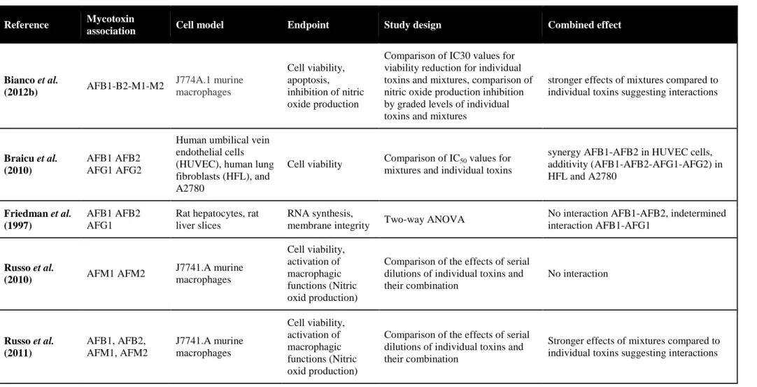
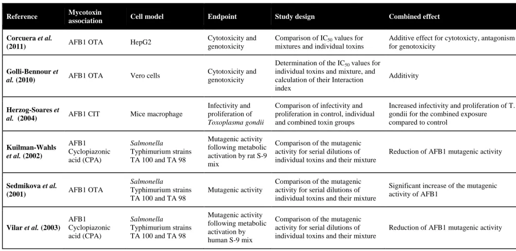
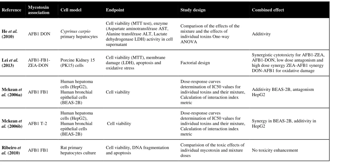
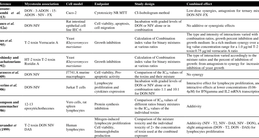
Moreover several contaminants may be present at the same time in the feed and interact together. More attention needs to be paid to the toxicological effect of contaminant mixture to determine whether they act additively, in synergy or
36
antagonism. There is a growing body of evidence pointing that mycotoxins may impair in a synergistic manner intestinal integrity (Grenier and Oswald 2012; Wan et al., 2013; Alassane-Kpembi et al., 2013). The characterization of these toxicological interactions deserves to be extended to other contaminant mixtures to improve our understanding of intestinal health risk associated with the presence of feed contaminants.
37
References
Ahlman, H. and Nilsson, O., 2001. The gut as the largest endocrine organ in the body. Annals of Oncology 12 Suppl 2: S63-68.
Alassane-Kpembi I., Kolf-Clauw M., Gauthier T., Abrami R., Abiola F.A., Oswald I.P. and Puel O., 2013. New insight into mycotoxin mixtures: the toxicity of low doses of Type B trichothecenes against intestinal epithelial cells is synergistic. Toxicology and Applied Pharmacology (in press).
Ali-Vehmas, T., Rizzo, A., Westermarck, T. and Atroshi, F., 1998. Measurement of antibacterial activities of T-2 toxin, deoxynivalenol, ochratoxin A, aflatoxin B1 and fumonisin B1 using microtitration tray-based turbidimetric techniques. Zentralblatt für Veterinärmedizin A 45: 453-458.
Applegate, T.J., Schatzmayr, G., Prickel, K., Troche, C. and Jiang, Z., 2009. Effect of aflatoxin culture on intestinal function and nutrient loss in laying hens. Poultry Science 88: 1235-1241.
Awad, W.A., Bohm, J., Razzazi-Fazeli, E. and Zentek, J., 2006. Effects of feeding deoxynivalenol contaminated wheat on growth performance, organ weights and histological parameters of the intestine of broiler chickens. Journal of Animal Physiology and Animal Nutrition 90: 32-37.
Awad, W.A., Aschenbach, J.R., Setyabudi, F.M., Razzazi-Fazeli, E., Bohm, J. and Zentek, J., 2007. In vitro effects of deoxynivalenol on small intestinal D-glucose uptake and absorption of deoxynivalenol across the isolated jejunal epithelium of laying hens. Poultry Science 86: 15-20.
Awad, W.A., Vahjen, W., Aschenbach, J.R. and Zentek, J., 2011. A diet naturally contaminated with the Fusarium mycotoxin deoxynivalenol (DON)
38
downregulates gene expression of glucose transporters in the intestine of broiler chickens. Livestock Science 140: 72-79.
Ball, L.M. and Chhabra, R.S., 1981. Intestinal absorption of nutrients in rats treated with 2,3,7,8-tetrachlorodibenzo-p-dioxin (TCDD). Journal of Toxicologyand Environmental Health 8: 629-638.
Bennett, J. and Klich, M., 2003. Mycotoxins. Clinical Microbiology Reviews 16: 497-516.
Bernard, A., Broeckaert, F., De Poorter, G., De Cock, A., Hermans, C., Saegerman, C. and Houins, G., 2002. The Belgian PCB/dioxin incident: analysis of the food chain contamination and health risk evaluation. Environmental Research 88: 1-18.
Bouhet, S., Hourcade, E., Loiseau, N., Fikry, A., Martinez, S., Roselli, M., Galtier, P., Mengheri, E. and Oswald, I.P., 2004. The mycotoxin fumonisin B1 alters the proliferation and the barrier function of porcine intestinal epithelial cells. Toxicological Sciences77: 165-171.
Bouhet, S., Le Dorze, E., Peres, S., Fairbrother, J.M. and Oswald, I.P., 2006. Mycotoxin fumonisin B1 selectively down-regulates the basal IL-8 expression in pig intestine: in vivo and in vitro studies. Food and Chemical Toxicology 44: 1768-1773.
Bouhet, S. and Oswald, I.P., 2007. The intestine as a possible target for fumonisin toxicity. Molecular Nutritionand Food Research 51: 925-931.
Brown, T.P., Rottinghaus, G.E. and Williams, M.E., 1992. Fumonisin mycotoxicosis in broilers: performance and pathology. Avian Diseases 36: 450-454.
39
Bryden, W., 2012. Mycotoxin contamination of the feed supply chain: Implications for animal productivity and feed security. Animal Feed Science and Technology 173: 134-158.
Burmeister, H.R. and Hesseltine, C.W., 1966. Survey of the sensitivity of microorganisms to aflatoxin. Applied Microbiology 14: 403-404.
Bursian, S.J., Kern, J., Remington, R.E., Link, J.E. and Fitzgerald, S.D., 2013a. Dietary exposure of mink (Mustela vison) to fish from the upper Hudson River, New York, USA: Effects on organ mass and pathology. Environmental Toxicologyand Chemistry 32: 794-801.
Bursian, S.J., Kern, J., Remington, R.E., Link, J.E. and Fitzgerald, S.D., 2013b. Dietary exposure of mink (Mustela vison) to fish from the upper Hudson River, New York, USA: Effects on reproduction and offspring growth and mortality. Environmental Toxicologyand Chemistry 32: 780-793.
CAST, 2003. Mycotoxins: risks in plant, animal, and human systems. Council for Agricultural Science and Technology, Ames, Iowa, USA, 199 pp.
Choi, Y.J., Seelbach, M.J., Pu, H., Eum, S.Y., Chen, L., Zhang, B., Hennig, B. and Toborek, M., 2010. Polychlorinated biphenyls disrupt intestinal integrity via NADPH oxidase-induced alterations of tight junction protein expression. Environmental Health Perspectives 118: 976-981.
Covaci, A., Voorspoels, S., Schepens, P., Jorens, P., Blust, R. and Neels, H., 2008. The Belgian PCB/dioxin crisis-8 years later: An overview. Environmental Toxicologyand Pharmacology 25: 164-170.
D'Mello, J.P.F., 2004. Contaminants and toxins in animal feeds. In: Food and Agriculture Organization of the United Nations (Ed.), Assessing quality and
40
safety of animal feeds. Publishing Management Service, Information Division, FAO, Viale delle Terme di Caracalla, 00100 Rome, Italy, Rome (Italy), pp. 107-128.
Dickinson, B.L., Badizadegan, K., Wu, Z., Ahouse, J.C., Zhu, X., Simister, N.E., Blumberg, R.S. and Lencer, W.I., 1999. Bidirectional FcRn-dependent IgG transport in a polarized human intestinal epithelial cell line. The Journalof Clinical Investigation 104: 903-911.
Diesing, A.K., Nossol, C., Panther, P., Walk, N., Post, A., Kluess, J., Kreutzmann, P., Danicke, S., Rothkotter, H.J. and Kahlert, S., 2011. Mycotoxin deoxynivalenol (DON) mediates biphasic cellular response in intestinal porcine epithelial cell lines IPEC-1 and IPEC-J2. Toxicology Letters 200: 8-18.
Dietrich, B., Neuenschwander, S., Bucher, B. and Wenk, C., 2012. Fusarium mycotoxin-contaminated wheat containing deoxynivalenol alters the gene expression in the liver and the jejunum of broilers. Animal 6: 278-291.
Dzidic, A., Mohr, A., Meyer, K., Bauer, J., Meyer, H.H. and Pfaffl, M.W., 2004. Effects of mycophenolic acid (MPA) treatment on expression of Fc receptor (FcRn) and polymeric immunoglobulin receptor (pIgR) mRNA in adult sheep tissues. Croatian Medical Journal 45: 130-135.
Furness, J.B., Kunze, W.A. and Clerc, N., 1999. Nutrient tasting and signaling mechanisms in the gut. II. The intestine as a sensory organ: neural, endocrine, and immune responses. The American Journalof Physiology 277: G922-928.
Goossens, J., Pasmans, F., Verbrugghe, E., Vandenbroucke, V., De Baere, S., Meyer, E., Haesebrouck, F., De Backer, P. and Croubels, S., 2012. Porcine intestinal epithelial barrier disruption by the Fusarium mycotoxins deoxynivalenol and
T-41
2 toxin promotes transepithelial passage of doxycycline and paromomycin. BMC Veterinary Research 8: 245.
Grenier, B. and Oswald, I.P., 2011. Mycotoxin co-contamination of foods and feeds: meta-analysis of publications describing toxicological interactions. World Mycotoxin Journal 4:285-313.
Grenier, B. and Applegate, T.J., 2013. Modulation of intestinal functions following mycotoxin ingestion: meta-analysis of published experiments in animals. Toxins 5: 396-430.
Han, X.-Y., Huang, Q.-C., Li, W.-F., Jiang, J.-F. and Xu, Z.-R., 2008. Changes in growth performance, digestive enzyme activities and nutrient digestibility of cherry valley ducks in response to aflatoxin B1 levels. Livestock Science 119: 216-220.
Heath, J.P., 1996. Epithelial cell migration in the intestine. Cell Biology International 20: 139-146.
Ishikawa, S., 2009. Children's immunology, what can we learn from animal studies (3): Impaired mucosal immunity in the gut by 2,3,7,8-tetraclorodibenzo-p-dioxin (TCDD): a possible role for allergic sensitization. The Journal of Toxicological Sciences 34 Suppl 2: SP349-361.
Johansson, M.E., Larsson, J.M. and Hansson, G.C., 2011. The two mucus layers of colon are organized by the MUC2 mucin, whereas the outer layer is a legislator of host-microbial interactions. Proceedings of the National Academyof Sciences ofthe United Statesof America 108 Suppl 1: 4659-4665.