Science Arts & Métiers (SAM)
is an open access repository that collects the work of Arts et Métiers Institute of
Technology researchers and makes it freely available over the web where possible.
This is an author-deposited version published in: https://sam.ensam.eu
Handle ID: .http://hdl.handle.net/10985/10054
To cite this version :
Jean-Yves LE POMMELLEC, Vianney PIRON, Mohamed Lamine ASKOURA, Jean-Pierre L'HUILLIER - Assessment of the effective attenuation coefficient of scattering media illuminated by a LED array : Effect of the beam size - In: European Conferences on Biomedical Optics 2015, Allemagne, 2015-06 - Proc. SPIE 9542, Medical Laser Applications and Laser-Tissue Interactions - 2015
Any correspondence concerning this service should be sent to the repository Administrator : archiveouverte@ensam.eu
media illuminated by a LED array : Effect of the beam size
Jean-Yves Le Pommellec
a*, Vianney Piron
a, Mohamed-Lamine Askoura
b, Jean-Pierre L’Huillier
aaL’UNAM Ecole Nationale Supérieure des Arts et Métiers (ENSAM), Laboratoire Arts et Métiers ParisTech Angers
(LAMPA), 2 Boulevard du Ronceray, BP 93525, 49035 Angers cedex 01, France.
bL’UNAM Université, SFR 4207 QUASAV, Groupe ESA (Ecole Supérieure d’Agriculture), UPSP GRAPPE
(Groupe de Recherche Agro-Alimentaire sur le Produit et Procédés), 55 Rue Rabelais-BP 30748, 49007 ANGERS Cedex 01, France.
ABSTRACT
The knowledge of the light fluence rate distribution inside a biological tissue irradiated by a Laser (or LED) is fundamental to achieve medical treatments. In this paper, a semi-infinite tissue model was considered, and the steady-state photon diffusion equation was solved by means of the 2-D Fourier Transform. This method can be applied to any irradiation source (radial symmetry or not) at the surface of the tissue. Two particular beam shapes are studied: planar irradiation and flat beam with finite radius. The total fluence rate along the depth in tissues was computed by adding the collimated and the diffuse components. The analytical solution was also used to study the effect of the beam radius on the light attenuation. Measurements were performed using a tank filled with a liquid-simulating medium (Milk), illuminated with a LED array (660 nm , 100mm×100mm). Several circular diaphragms were used to obtain uniform beams with well defined radii. An optical fibre (with an isotropic tip) was used to measure the fluence rate inside the medium. Preliminary experimental results are in agreement with theoretical predictions.
Key Words : Tissue-optics, Diffusion approximation, 2-D Fourier transform modelling, Effective attenuation coefficient, Beam size
1. INTRODUCTION
Irradiation of biological tissue by laser light has become an increasingly important tool used for both medical investigations and treatments The therapeutic and diagnostic applications based upon light interaction in these fields depend mainly on how the energy fluence rate is distributed spatially within the tissue target. Clinical examples where the distribution of light inside various tissues plays a key role are typically Photodynamic tumor therapy1 , the photothermal treatments2,3, and also fluorescence diagnostics with transillumination imaging.4,5
Photon migration through highly scattering media such as biological tissues is well described by the radiative transfer equation (RTE) 6. However the RTE is an integro-differential equation and it is often difficult to derive analytical solutions for solving the main bio-optical problems.
The Monte Carlo method7 provides accurate predictions of light transport, but suffers from large computation times and complex coding8 in case of heterogeneous tissue structure. For this reason, this stochastic method is predominantly useful for validation studies. Interestingly, under few assumptions (diffusion dominant over absorption, source-detector distances > 10 mm) the RTE can be simplified to a more tractable equation named diffusion equation 9,10. A large class of biological tissues, which are illuminated with light sources whose wavelength ranges from ~ 600 nm to ~ 900 nm11 , may avantageously be modeled using the diffusion equation. Finite Element Method (FEM) may be used to solve light propagation problem through turbid media12 from the consideration of the diffusion approximation. However, semi-analytical expressions have been also proposed for cases of cylindrical beam sources.
*
2
The Green’s function method13 provides a solution for a tissue slab irradiated with a beam of finite radius. The determination of the eigen values of the problem involves finding the zeros of the transcendal functions, and then requires numerical implementation. Furthermore, the Hankel transform method14,15 gives another solution for a semi-infinite medium illuminated with a Gaussian beam.
The primary objective of this work was to present a general analytical model based on the 2-D Fourier Transform applied to the steady diffusion equation. This model can be used to determine the fluence rate at each point of a semi-infinite tissue model irradiated with any source beam source. Two particular cases are studied: planar irradiation and flat beam with finite radius. The effect of the source radius on the in-depth light attenuation was evaluated. Measurements were performed in a liquid-simulating phantom (Milk).
2. MATHEMATICAL STATEMENT
2-1. Photon diffusion equation
For the purpose of this study , the tissue is considered as a semi-infinite homogeneous medium. The incident radiation is assumed to be collimated and normal to the interface and the positive
z
direction corresponds to the tissue depth. The total fluence rate
(
x
,
y
,
z
)
(
W
.
mm
2)
at position(
x
,
y
,
z
)
of the tissue is divided into collimated
c(
x
,
y
,
z
)
and diffuse components
d(
x
,
y
,
z
)
11,14,15
(
x
,
y
,
z
)
c(
x
,
y
,
z
)
d(
x
,
y
,
z
)
(1) The collimated component satisfies Beer’s law :
c(
x
,
y
,
z
)
c(
x
,
y
,
0
)
exp
tz
(2) Where
c(
x
,
y
,
0
)
is the collimated photon flux at the point(
x
,
y
,
0
)
of the surface of the medium,s a
t
is the total attenuation coefficient,
a(
mm
1)
is the absorption coefficient and(
)
1 mm
s
is the scattering coefficient.The diffuse component
d(
x
,
y
,
z
)
is the solution of the diffusion equation :
2
d(
x
,
y
,
z
)
eff2
d(
x
,
y
,
z
)
3
s
s
g
a
c(
x
,
y
,
0
)
exp(
tz
)
(3) whereg
is anisotropy factor and
eff
3
a
a
(
1
g
)
s
is the effective attenuation coefficient. 2-2. Boundary conditionsFor a semi-infinite medium under collimated irradiation, the indices of refraction are mismatched and no diffuse intensity enters the medium from outside. The boundary condition at the surface is 11,15
(
,
,
0
)
)
1
(
2
)
,
,
(
2
)
0
,
,
(
0y
x
g
g
A
z
z
y
x
AD
y
x
c s a s z d d
(4) whereD
1
3
a
(
1
g
)
s
is the diffusion coefficient.The value of
A
is linked to the Fresnel coefficientR
F(
)
and consequently is a function of the refractive index of the scattering medium :
/2 0 2 / 0 2)
(
cos
sin
2
1
cos
sin
3
1
d
R
d
R
A
F FFor the computations , the value of the refractive index was set at 1.4 thus yielding A = 2.94912.
2-3. Fourier transform formulation
The diffuse fluence rate due to a flat beam irradiation (radius
r
0) is determined by solving Eq. 3 subject to the boundary condition (4), using the two dimensionnal Fourier transform defined as:
F
d
k
x,
k
y,
z
d
x
,
y
,
z
exp
2
i
k
xx
k
yy
dx
dy
(5) The diffuse fluence rate can be easily computed if we work in cylindrical rather than Cartesian coordinates using the relations :r
x
2
y
2 and
k
x2
k
2y .r
z
z
z
J
r
d
eff t d
0 0 2 2 2)
(
2
)
4
exp(
)
(
)
exp(
)
(
2
)
,
(
(6) where
)
4
(
)
2
(
3
)
(
2 2 20 1 0 t eff a t sg
r
J
r
(7)
)
8
(
4
2
1
)
2
(
6
)
(
)
2
1
(
)
(
2 2 0 1 0
eff s tD
A
r
J
r
g
D
A
AD
In Eq. (6) ,
J
0 is the Bessel function of first kind and zero - order, whereas in Eqs (7-8)J
1 is the Bessel function of first kind and one – order.The Fourier-transform diffusion model was developed on the base of the software " Mathcad", and implemented on a personal computer.
In the case of
r
0
, the total fluence rate (
c
d)
is identical to that provided by Deulin and L’Huillier16 for the 1-D configuration (Planar irradiation). For a large penetration depth
(
z
)
~
exp(
effz
)
. Consequently,eff
can be accurately assessed from measurement of the fluence rate inside tissues irradiated with a wide-plane collimated beam as reported by Choukeife and L’Huillier162-4. Fluence rate distribution measurement
The fluence rate distribution inside the tissue can be determined by displacing (step by step), along the beam axis, an isotropic-end optical fibre. For this probe, it has been shown (Star et al19 ) that the detected signal is proportional to the fluence rate in the medium.
4
Figure 1: Measurement with an isotropic-end optical fibre
3 TYPICAL RESULTS
3-1. Theoretical results
Theoretical results are given for a tissue illuminated by a flat beam irradiation. The flux density collected by an isotropic optical fibre was calculated from equations (1), (2) and (6). In Figure 2, the logarithm of the normalized fluence rate :
ln
(
r
0
,
z
)
/
(
r
0
,
z
0
)
is plotted against depth for a semi-infinite tissue sample, irradiated with a collimated laser beam, with
a
0
.
01
mm
1,1 '
1
mm
s
and g = 0.8. The in-depth variations of the fluence rate is shown for a flat beam having three different radii : (5 mm, 10 mm and 20 mm). Close to the surface, the flux density increases by a factor depending on the field size. For large depths, the flux decreases as)
(
exp
z
where
is the attenuation coefficient along the beam axis.0 5 10 15 20 4 2 0 Depth (mm) N o rm a li ze d f lu e n c e ra te
20 mm
5 mm
Figure 2 : Plots of the normalized fluence rate as a function of the depth inside the tissue LASER BEAM
g
s a,
,
OPTICAL FIBRE PHOTODETECTOR Z10 20 30 40 50 0.16 0.18 0.2 0.22 Beam radius (mm) A tt e n u a ti o n c o ef fi ci e n t
0.176
0.174
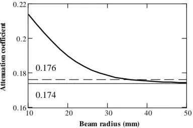
Figure 3 : Plots of the attenuation coefficient as a function of the beam radius
The effect of the incident beam size on the light attenuation along the beam axis is depicted Figure 3. As shown
decreases as the beam radius increases. Once the beam size reaches a large value (45 mm), this coefficient reaches the minimum value
eff
0.174 mm-1 corresponding to the 1-D solution.16. For a beam radiusr
0
35 mm the attenuation is
0.176 mm-1 .The relative difference (%) betweeen this value and the attenuation coefficient (
eff
0.174 mm-1) is equal to 1,2%. Consequently the error on the
eff value is equal to 1.2% if the beam radius isr
0
35 mm. Clearly, as this limit radius (called critical radius) is reached, the penetration of the beam is maximum.3-2. Experimental results
Figure 4 Experimental set up
Experiments were conducted to check the validity of the theoretical predictions.The measurements of the attenuation of the fluence rate in a scattering and absorbing medium as a function of depth are performed with the set up shown in Figure 4. A LED array (660 nm, 100mm×100 mm, 1.5 mW/mm2) is used to illuminate the transparent base of a tank filled with a tissue simulating "phantom" (Milk – UHT). Several circular diaphragms were used to obtain uniform circular beams with radii varying from 10 mm to 50 mm. The fluence rate was measured with an
Isotropic fiber optic
probe
Diaphragm
LED Array
Tank
Milk
Transparent base
Photodetector
6
isotropic light dosimetry probe ( Optical fiber with an isotropic tip) connected to a photodiode and displaced by steps along the axis from the irradiation surface downward into the medium.
0 20 40 60 80 100 0.5 1 1.5 Radial distance (mm) N o rm a li ze d d e te ct e d p o w er
ideal distributi on
measured li ght
intensity
Fig 5 Light intensity distribution measured along the radius of a circular diaphragm
Figure 5 shows the light intensity distribution measured along the radius of a typical circular diaphragm (r0 = 40 mm). As illustrated in this figure, the intensity generated appears as uniformly distributed over a large area
except for radial distances close to the beam edge.
0 5 10 15 20 3 2 1 0 Depth z (mm) N o rm a li ze d f lu e n c e ra te
30 ,40, 50 mm
20 mm
10 mm
Figure 6 Experimental depth behaviour of the normalized fluence rate
The fluence rate was measured with an isotropic-end optical fibre displaced along the axis of the system. The logarithm of the normalized fluence rate is plotted (Figure 6) as a function of the distance between the fiber tip and the transparent base of the tank, for five circular diaphragms (
r
0
10, 20 , 30 , 40, 50 mm). According to theoretical predictions, it appears that the logarithm of the fluence rate evolves linearly with respect to the depth z. Otherwise, it is still shown that the attenuation coefficient (defined as the slope of the curve) decreases as the radius increases and then tends to a minimum value for large beam size. For a broad incident beam (40 mm) the attenuation coefficient 0.098 mm-1 is close to the true effective attenuation coefficient 0.096 mm-1.4 CONCLUSION
In this paper, a semi-analytical model based on the two-dimensionnal Fourier Transform of the steady-state diffusion equation has been described. This model was used to compute the total Fluence rate in case of a semi-infinite tissue model illuminated with a uniform laser of finite radius or with a planar source. This approach is as versatile as the other techniques (Green’s Function method or Hankel transform method), but has the advantage of being much faster. The simulations show that the effective attenuation coefficient of the studied tissue can be well assessed when a sufficient source radius is reached. Preliminary experiments allow us to calibrate theoretical specifications. Further investigations are now required to optimize such a procedure in cases of varied tissue-optical parameter sets.
REFERENCES
[1] Wilson BC, Patterson MS. The physics, biophysics and technology of photodynamic therapy. Phys Med Biol 2008 ; 53: R61-R10
[2]. Welch AJ, van Gemert MJC. The Optical and Thermal Responses of Laser-Irradiated Tissue. Plenum Press New York 1995
[3]. L’Huillier JP. Theoretical analysis of the role played by tissue-optical parameters in the laser ablation process. Proc SPIE 1997; 3195: 151-165
[4]. Leblond F., Davis SC. , Valdès PA., Pogue BW. Pre-clinical whole-body Fluorescence imaging : Review of instruments, methods and applications. J.Photochem. Photobiol.B : Biology 2010, 98, 77-94.
[5]. L’Huillier JP., Vaudelle F. Improved localization of hidden fluorescent in highly scattering slab media based on a two-way transmittance determination. Opt.Express. 2006, 14(26), 12915-12929.
[6]. Chandraesekhar S. Radiative transfer. Oxford UP New York 1960
[7]. Wang L , Jacques S.L, Zheng L. Monte Carlo modelling of light transport in multi-layered tissues. Computer methods and programs in Biomedecine 1997; 47 : 131-146
[8]. Mansouri C, L’Huillier JP, Kashou NH, Humeau A. Depth sensitivity analysis of functional near-infrared spectroscopy measurement using three-dimensional Monte Carlo modelling based magnetic resonance imaging. Lasers in Med Sci 2010 25:431-438.
[9]. A Ishimaru. Wave Propagation and scattering in Random Media. Academic New York, 1978 [10]. Prahl S.A. Light transport in tissue, Phd Thesis, University of Texas at Austin, 1988.
[11]. Boulnois JL. Photophysical processes in recent medical laser developments : a review. Lasers in Med Sci 1986; 1 : 47-66.
[12]. Deulin X, L’Huillier JP. Finite element approach to photon propagation modelling in semi-infinite homogeneous and multilayered tissue structures. EPJ Appl Phys 2006; 33 :133-146
[13]. Reynolds L, Johnson C. Diffuse reflectance from a finite blood medium : applications to the modelling of the fiber optics catheters. Applied Optics 1976; 15 :2059-2067
[14]. Grossweiner L.I., Karagiannes J.L, Johnson P.W, Zhang Z.Y, Gaussian-beam spread in biological tissues. Applied Optics 1990; 29 : 379-383
[15]. Carp SA, Prahl SA, Venugopalan V. Radiative transport in the delta P1 approximation : accuracy of fluence rate and optical penetration depth predictions in turbid semi-infinite media. J Biomed Opt 2004; 9: 632-647
8
[16]. Star W.M, Marijnissen PA. Calculating the response of isotropic light dosimetry probes as a function of the tissue refractive index. Applied Optics 1989; 28 : 2288-2291.
[17]. Choukeife. JE., L’Huillier JP. Measurements of scattering effects within tissue-like media at two wavelengths of 632.8 nm and 680 nm. Lasers Med Sci 1999, 14:286-296.