On the application of surface enhanced Raman scattering to study the interaction of DsRed fluorescent proteins with silver nanoparticles embedded in thin silica layers
Texte intégral
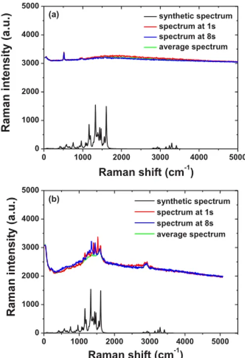
Figure
Documents relatifs
Dissolved organic matter controls of arsenic bioavailability to bacteria.. Martin Pothier, Véronique Lenoble, Cédric Garnier, Benjamin Misson, Charlotte Rentmeister,
We aimed to: (a) identify the more important topics for biodiversity conservation in this biome, (b) evaluate the relative importance of stakeholder type and region in topic
(2016) Multilocus Variable Number of Tandem Repeat Analysis Reveals Multiple Introductions in Spain of Xanthomonas arboricola pv. pruni, the Causal Agent of Bacterial Spot Disease
A diferencia del espacio, que es un campo de disputa a la vez material e imaterial (político, cultural, etc.), como lo acabamos de precisar, el territorio es una porción delimitada
(2008) Distinct Genetic Loci Control Plasma HIV-RNA and Cellular HIV-DNA Levels in HIV-1 Infection: The ANRS Genome Wide Association 01 Study.. This is an open-access
This paper investigates the density of hypersurfaces in a projective normal simplicial toric variety over a finite field having a quasismooth intersection with a given quasismooth
Our results show spatial home gardening patterns differentiation at three intertwined levels: At the micro-level of domestic space (according to the size and share of vegetable
However, known factorizations of matrix polynomi- als, such as the Smith form [10], involve unimod- ular matrices but usual factorizations such as QR, eigenvalue or singular
