Université de Montréal
Increasing Axonal Arborization Size of Dopamine Neurons
to Produce a Better Mouse Model of Parkinson’s Disease
by Pamela Cassidy
Département de Pharmacologie Faculté de Médicine
Mémoire présenté à la Faculté des études supérieures en vue de l’obtention du grade de Maitre en Sciences (M.Sc.)
en Pharmacologie, option neuropharmacologie
April, 2018 © Pamela Cassidy, 2018
ii
RÉSUMÉ
Dans la maladie de Parkinson, les neurones dopaminergiques (DA) de la substance noire compacte (SNc) sont particulièrement vulnérables dû a leurs très grande taille de l'arborisation axonale, des besoins énergétiques très élevés et du stress oxidatif chroniquement élevé associés à ce phénotype. Étrangement, les modèles génétiques murins de la maladie de Parkinson ne montrent pas de dégénérescence spontanée des neurones DA. Notre hypothèse principale est que suite à une lésion partielle des neurones DA de la SNc, les neurones survivants montreront un bourgeonnement axonal compensatoire, ce qui résultera, chez la souris adulte, en une population de neurones DA dotée d'une arborisation axonale beaucoup plus grande et d'une vulnérabilité basale accrue. Une lésion unilatérale d'approximativement 50% des neurones DA était induite dans la SNc par une injection unilatérale de 6-hydroxydopamine (6-OHDA) chez des souris de 5 jours des deux sexes. Les souris ont alors ensuite évaluées à l'âge de 3 mois. Dans une première étape, nous avons quantifié la taille de l'arborisation axonale des neurones DA en infectant une sous-population de ces neurones avec un virus AAV-EYFP. Dans une deuxième étape, la vulnérabilité des neurones DA était évaluée à l’age de P135, en injectant les souris avec une virus d’alpha-synucléin à P90. Nos résultats montrent qu'à la suite d'une lésion partielle des neurones DA de la SNC, les neurones survivants se compensent en augmentant leur taille d’arborization axonale par environ 2 fois. Cette compensation se traduit par environ 3-fois l'augmentation de la vulnérabilité de ces neurones lorsqu'ils sont exposés à un stress secondaire. .
Mots-clés : Maladie de Parkinson, dopamine, modèle animal, néonatal, 6-hydroxydopamine,
arborisation axonale, croissance compensatoire, vulnérabilité, substance noire, aire tegmentaire ventrale
iii
ABSTRACT
In Parkinson’s disease (PD), dopaminergic neurons of the substantia nigra pars compacta (SNC) are one of the key subsets of neurons particularly vulnerable to degeneration. Research has only started to elucidate some of the potential causes of this selective vulnerability, but the exceptionally large and complex axonal arborization of these neurons appears to play an important role due to its impact of cellular bioenergetics and oxidative stress. Unfortunately, genetic mouse models of PD have until now not been able to replicate the spontaneous loss over time of SNC dopaminergic (DA) neurons. This could be due in part to the fact that DA neurons in mice do not have an axonal arborization as developed as that of DA neurons in the human brain. We hypothesized that manipulations which increase axonal arborization size in SNC dopamine neurons in mice will increase their vulnerability to cellular stress, resulting in a greater risk of cell death. Our objective was to force SNC neurons to develop a larger than normal axonal arborization, thereby increasing their energy expenditures and vulnerability. An approximate 60% lesion of dopaminergic neurons was induced in the SNC through unilateral injection of 6-hydroxydopamine (6-OHDA) in male and female neonatal mice. Axonal arborization size was quantified at P90 using a conditional AAV-EYFP virus and confocal microscopy. In the second phase of the project, we examined the vulnerability of SNC DA at P135 neurons to subsequent viral overexpression of alpha-synuclein. Our results show that a partial lesion of SNc dopamine neurons in neonatal mice induced an approximate 2-fold increase in compensatory sprouting of surviving neurons and, by consequence, greatly increased the density of their axonal projections to the striatum. This compensatory process increased the vulnerability of the surviving SNC DA neurons to alpha-synuclein toxicity by nearly 3-fold. These findings are compatible with our initial hypothesis and suggest that producing mice that have DA neurons with larger axonal arborizations may facilitate the development of better mouse models of PD.
Keywords: Parkinson’s disease, dopamine, animal model, neonatal, 6-hydroxydopamine,
axonal arborization, compensatory sprouting, vulnerability, Substantia Nigra, Ventral Tegmental Area
iv
TABLE OF CONTENTS
RÉSUMÉ ... ii ABSTRACT ... iii TABLE OF CONTENTS ... iv LIST OF FIGURES ... xLIST OF TABLES ... xii
LIST OF ACRONYMS ... xiii
ACKNOWLEDGEMENTS ... xvii
INTRODUCTION... 1
THE DOPAMINERGIC SYSTEM ... 3
NEUROMODULATORY MECHANISMS ... 3
DOPAMINE ... 3
SEROTONIN... 5
NOREPINEPHRINE ... 6
THE NEUROMOLECULAR MECHANISMS OF NEUROTRANSMITTERS ... 6
GLUTAMATE ... 6
GAMMA-AMINOBUTYRIC ACID (GABA) ... 7
ACETYLCHOLINE ... 8
DAT CHARACTERIZATION AND FUNCTION ... 8
CHARACTERISTICS OF DOPAMINE NEURONS ... 9
THE MESODIENCEPHALIC DOPAMINERGIC SYSTEM ... 11
VENTRAL MIDBRAIN DOPAMINERGIC NEURONS ... 15
THE BASAL GANGLIA CIRCUIT ... 16
v
HETEROGENEITY OF MIDBRAIN DOPAMINE NEURONS ... 19
PARKINSON’S DISEASE ... 23
TYPES OF PD ... 23
SYMPTOMS OF PD ... 23
PREMOTOR PHASE OF PD... 23
CARDINAL MOTOR SYMPTOMS OF PD... 25
BRADYKINESIA ... 26
TREMOR... 26
RIGIDITY ... 27
CURRENT TREATMENT OPTIONS ... 27
PHARMACOLOGICAL THERAPIES ... 27
LEVODOPA ... 28
DOPAMINE RECEPTOR AGONISTS ... 28
COMT & MAO-B INHIBITORS... 29
ANTICHOLINERGICS ... 29
SURGICAL THERAPIES ... 29
CELL REPLACEMENT THERAPY ... 30
THE PATHOPHYSIOLOGY OF PD ... 33
LEWY BODY PATHOLOGY ... 33
ALPHA-SYNUCLEIN FUNCTION/PHYSIOLOGY ... 35
ALPHA-SYNUCLEIN PATHOLOGY ... 36
ALPHA-SYNUCLEIN AND MITOCHONDRIAL DYSFUNCTION ... 37
ALPHA-SYNUCLEIN AND DOPAMINE ... 38
RISK FACTORS OF PARKINSON’S DISEASE ... 39
vi
ENNIRONMENTAL RISK FACTORS ... 40
GENETIC RISK FACTORS ... 40
TRANSGENIC MODELS OF PD ... 41
PARKIN GENE MODEL ... 42
PINK1 GENE MODEL ... 42
DJ-1 GENE MODEL... 43
NEUROTOXIC ANIMAL MODELS OF PD ... 43
THE 6-HYDROXYDOPAMINE (6-OHDA) MODEL OF PD ... 44
6-OHDA MECHANISM OF ACTION ... 45
MPTP, RESERPINE, ROTENONE & PARAQUAT ... 46
PARKINSONIAN BEHAVIOR IN UNILATERAL MOUSE MODELS OF PD ... 47
REDUNDANCY OF AXONAL CONNECTIONS IN THE DOPAMINE SYSTEM ... 49
COMPENSATORY MECHANISMS IN THE DOPAMINE SYSTEM IN RESPONSE TO PARTIAL LESIONS ... 52
COMPENASTORY SPROUTING OF DOPAMINERGIC NEURONS ... 52
INCREASED DOPAMINE RECEPTOR SENSITIVITY ... 55
NON-DOPAMINERGIC COMPENSATORY MECHANISMS ... 57
COMPENSATORY MECHANISMS OF THE SEROTONERGIC SYSTEM ARE CLOSELY TIED TO DOPAMINERGIC DENERVATION ... 57
GABA & GLUTAMATERGIC COMPENSATORY MECHANISMS ... 58
COMPENSATORY VARIATIONS BETWEEN NEONATAL AND ADULT LESIONS OF THE NIGROSTRIATAL PATHWAY ... 59
ADAPTATIONS OF THE DOPAMINERGIC SYSTEM IN LESIONED NEONATAL MICE ... 59
ADAPTATIONS OF THE SEROTONERGIC SYSTEM IN LESIONED NEONATAL MICE ... 61
vii
VULNERABILITY OF SNC DOPAMINE NEURONS ... 62
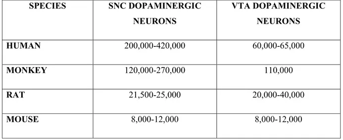
NUMBER, SIZE AND COMPLEXITY OF DA NEURONS ACROSS SPECIES ... 62
MORPHOLOGICAL CHARACTERISTICS OF SNC DA NEURONS CONTRIBUTE TO VULNERABILITY ... 64
ENERGETIC METABOLISM AND OXIDATIVE STRESS ... 66
CALCIUM-MEDIATED CELLULAR STRESS ... 67
LIMITATIONS OF CURRENT ANIMAL MODELS OF PD ... 69
OBJECTIVES AND HYPOTHESIS... 70
METHODOLOGY ... 72 SUBJECTS ... 72 NEONATAL SURGERIES ... 73 INK INJECTIONS ... 74 P60 STEREOTAXIC SURGERIES ... 75 DRUGS ... 78 6-HYDROXYDOPAMINE (6-OHDA) ... 78 DESIPRAMINE ... 78 AAV-EYFP VIRUS ... 78 ALPHA-SYNUCLEIN VIRUS ... 78 PERFUSION ... 78 CRYOSTAT ... 79
IMMUNOHISTOCHEMISTRY FOR IMMUNOFLUORESCENCE ... 79
IMMUNOSTAINING FOR STEREOLOGY ... 80
QUANTIFYING AXONAL ARBORIZATION SIZE OF DOPAMINE NEURONS ... 80
CONFOCAL MICROSCOPY ... 80
viii
BEHAVIOURAL TESTING ... 83
ROTAROD ... 83
GRIP STRENGTH TEST ... 84
CYLINDER TEST ... 84
LOCOMOTION ... 84
STATISTICAL ANALYSIS ... 85
RESULTS ... 86
STEREOLOGICAL COUNTS OF TH+ NEURONS IN THE SNC AND VTA ... 87
INCREASE IN AXONAL ARBORIZATION SIZE FOLLOWING PARTIAL LESION .. 89
ANALYSIS OF MOTOR FUNCTION IN PARTIALLY LESIONED AND UNLESIONED MICE ... 104
ROTAROD PERFORMANCE ... 104
PREFERENTIAL FOREPAW USE ... 106
ASYMMETRICAL ROTATION BEHAVIOUR ... 107
GRIP STRENGTH TEST ... 110
CELL DEATH AND STRIATAL DENERVATION IN RELATION TO PERFORMANCE ON BEHAVIOUR TASKS ... 113
ACTIMETRY MEASURES OF BEHAVIOR ... 118
INCREASED VULNERABILITY OF DA NEURONS ... 125
DISCUSSION ... 129
VTA COMPENSATION AND VULNERABILITY OF DOPAMINERGIC NEURONS ... 130
COMPENSATION OF SNC DA NEURONS AND VULNERABILITY ... 132
A MODEL MORE REPRESENTATIVE OF THE HUMAN PATHOLOGY OF PD .. 133
ix
BEHAVIOURAL EFFECTS OF NEONATAL PARTIAL LESIONS AS COMPARED
TO ADULT LESIONS ... 135
OVERCOMPENSATION EFFECTS AND NEURAL REWIRING OF THE MOTOR CORTEX ... 137
COMPENSATION FOLLOWING SEVERE DA DEPLETION ... 139
CELL LOSS, STRIATAL DENERVATION AND BEHAVIOUR ... 141
NEUROINFLAMMATORY RESPONSE AND DOPAMINERGIC CELL LOSS IN THE SUBSTANTIA NIGRA ... 141
LIMITATIONS OF THE PRESENT EXPERIMENT ... 143
VARIABILITY OF 6-OHDA INJECTIONS ... 143
INDIVIDUAL DIFFERENCES IN BEHAVIOURAL PERFORMANCE ... 144
COMPENSATORY MECHANISMS FOLLOWING PARTIAL LESION ... 145
FUTURE DIRECTIONS ... 146
CONCLUSION ... 151
x
LIST OF FIGURES
Figure 1: Selective neuronal vulnerability in Parkinson’s disease ... 10
Figure 2: Dopaminergic pathways in the human brain ... 12
Figure 3: Axonal arborization of single nigrostriatal neurons in rats ... 14
Figure 4: TH-positive neurons in the murine ventral mesencephalic dopaminergic complex . 16 Figure 5: Schematic Representation of the Basal Ganglia Nuclei ... 17
Figure 6: Classic Pathology of PD ... 33
Figure 7: Braak Staging System of Parkinson’s Disease ... 34
Figure 8: Mechanism of action of neurotoxins used in PD models ... 44
Figure 9: Comparison of the chemical structures of 6-OHDA and dopamine ... 46
Figure 10: Reconstruction of SNC-d DA Neuron... 65
Figure 11: Project Timeline ... 73
Figure 12: Representation of sites sampled for confocal microscopy ... 81
Figure 13: TH-immunofluorescent staining of mesencephalon and striatum following 6-OHDA injection... 87
Figure 14: Stereological counts of SNC & VTA DA neurons ... 89
Figure 15: Immunofluorescent images of the TH/GFP signal in striatal and mesencephalic slices of lesioned mice ... 90
Figure 16: 20X confocal images of the EYFP-infected dSTR ... 92
Figure 17: 20X Confocal images of the EYFP-infected vSTR... 93
Figure 18: Axonal arborization size of SNC-targeted DA neurons projecting to the dSTR and vSTR ... 95
Figure 19: Axonal arborization size of VTA-targeted DA neurons projecting to the dSTR and vSTR ... 96
Figure 20: Linear regression analysis of SNC cell death and dSTR TH-innervation in relation to change in axonal arborization size of SNC DA neurons projecting to the dSTR ... 97
Figure 21: Linear regression analysis of VTA cell death and vSTR TH-innervation vs change in arborization size of VTA-targeted DA neurons projecting to the vSTR ... 99
Figure 22: Linear regression analysis of lesion size and TH-innervation of dSTR in relation to axonal arborization size of VTA DA neurons projecting to the dSTR ... 101
xi
Figure 23: Linear regression analysis of cell death in the SNC and VTA in relation to
TH-innervation in the dSTR and vSTR respectively ... 102
Figure 24: Rotarod performance in unlesioned and partially lesioned, female and male mice at P30 & P60... 105
Figure 25: Preferential forepaw use in unlesioned and partially lesioned female and male mice at P30 & P60 ... 106
Figure 26: Spontaneous contralateral rotations in unlesioned and lesioned, female and male mice at P30 & P60 – No apomorphine ... 108
Figure 27: Apomorphine-induced ipsilateral rotations in unlesioned and lesioned, female and male mice at P30 & P60 ... 109
Figure 28: Grip strength test in unlesioned and lesioned mice, female and male, P30 & P60 ... …111
Figure 29: Grip strength test between females and males, P30 & P60, in unlesioned mice... 112
Figure 30: Linear regression analysis of measured parkinsonian behaviors in relation to dopaminergic cell death and dSTR innervation ... 116
Figure 31: Ambulatory time in P30 & P60 mice ... 121
Figure 32: Ambulatory activity in P30 & P60 mice ... 122
Figure 33: Total distance covered in P30 & P60 mice ... 123
Figure 34: Non-ambulatory behaviour time and non-ambulatory activity in P30 & P60 mice ... 124
Figure 35: Vulnerability of compensating SNC and VTA DA neurons following exposure to a secondary stressor ... 127
xii
LIST OF TABLES
Table 1: Number of SNC and VTA DA Neurons in Humans, Monkeys, Rats and Mice ... 64 Table 2: Stereotaxic Coordinates of Unilateral 6-OHDA SNC Injection ... 74 Table 3: Stereotaxic Coordinates of SNC and VTA AAV-EYFP Viral Injections ... 76 Table 4: Stereotaxic Coordinates of Bilateral SNC & VTA Injections of the Alpha-Synuclein
Overexpressing Virus ... 77 Table 5: Stereological Counts of TH+ Cells in Unlesioned and Lesioned hemispheres of SNC
xiii
LIST OF ACRONYMS
5-HT: Serotonin
6-OHDA: 6-hydroxydopamine AA: Ascorbic Acid
AADC: aromatic-L-amino-acid decarboxylase
AAV EYFP: Adeno-associated Virus Enhanced Yellow Fluorescent Protein Acb: Nucleus Accumbens
ACh: Acetylcholine
AMPA: alpha-amino-3-hydroxy-5-methyl-4-isoxazolepropionic acid AS: Alpha-synuclein
ATP: Adenosine triphosphate CB: calbindin
Cl-: Chloride
CN: Caudate nucleus
CNS: Central nervous system
COMT: Catechol-O-methyl transferase CPu: Caudate putamen
CSF: Cerebrospinal fluid DA: Dopamine
DAB: 3’3-diaminobenzadine DAT: dopamine transporter DBS: Deep brain stimulation DIC: Dat-Ires-Cre
DJ-1: Protein deglycase DJ-1 DNA: Deoxyribonucleic acid dSTR: dorsal striatum
ECF: Extracellular fluid ER: Endoplasmic reticulum
xiv GAD: Glutamate decarboxylase
GDNF: Glial cell-derived neurotrophic factor GFP: Green fluorescent protein
GPe: external segment of the Globus pallidus GPi: internal segment of the Globus Pallidus hNSCs: Human neural stem cells
iGluR: ionotropic glutamate receptor KO: Knockout
L: Lesioned LB: Lewy body LC: Locus coeruleus L-DOPA: Levodopa
LRRK2: Leucine-rich repeat kinase 2 LTD: Long-term depression
LTP: Long-term potentiation
mAChR: muscarinic acetylcholine receptor MAO-B: Monoamine oxidase B
MFB: Medial forebrain bundle
mGluR: metabotropic glutamate receptor MPTP: 1-methyl-1,2,3,4-tetrahydropyridine mRNA: messenger RNA
MSN: Medium spiny neurons mtDNA: Mitochondrial DNA NA: Noradrenaline
Na+: Sodium
nAChR: nicotinic acetylcholine receptor NMDA: N-methyl-D-aspartate
NSCs: Neural stem cells
xv NT: Neurotransmitter
OXPHOS: oxidative phosphorylation PBS: Phosphate buffered saline PD: Parkinson’s Disease PFA: Paraformaldehyde
PINK-1: PTEN-induced putative kinase 1 Put: Putamen
RBD: REM sleep behaviour disorder ROS: Reactive oxygen species RRF : retrorubral field
SN: Substantia nigra SN: Substantia nigra
SNARE: Soluble NSF attachment protein receptor SNC: Substantia nigra pars compacta
SNCA: alpha-synuclein gene SNC-d: dorsal tier of the SNC SNC-v: ventral tier of the SNC SNr: Substantia nigra pars reticulata SPNs: Spiny projection neurons STh: subthalamus
STN: Subthalamic nucleus TH: Tyrosine hydroxylase
UCHL1: Ubiquitin carboxy-terminal hydrolase L1 UL: Unlesioned
VGAT: Vesicular glutamate transporter VGLUT2: Vesicular glutamate transporter-2 VMAT2: Vesicular monoamine transporter 2 vSTR: ventral striatum
VT: Volume transmission VTA: Ventral tegmental area
xvi WT: Wild-type
xvii
ACKNOWLEDGEMENTS
I would like to take this opportunity to express my sincerest appreciation for the opportunity that Dr. Trudeau and the Université de Montréal have provided for me to complete this master’s degree. The guidance and support I received throughout this project from Dr. Trudeau and his research team were invaluable. I would like to give a special thanks to our lab technician Marie-Josée Bourque, who not only provided assistance when it was needed, but a support system for overcoming difficulties and challenges as well. This experience would not have been the same without her, as she is the heart of our team. I would also like to acknowledge Nicolas Giguère, another member of the Trudeau Laboratory. Although working on different projects, he always went out of his way to lend a helping hand and mentor the newcomers. This truly is a special team and I am so glad to have been a part of it.
INTRODUCTION
Parkinson’s disease (PD) is the second most common neurodegenerative disease after Alzheimer’s disease, holding a lifetime risk of 4-5% (Mullin & Schapira, 2013). The number of cases of PD is expected to double by 2050, and current therapies are only symptom-treating and do not stop the progression of the disease. Characterized by a progressive dopaminergic denervation of the striatum, PD is caused by a gradual loss of dopamine (DA) neurons in the substantia nigra (SN). The dopaminergic deficit resulting from the progressive cell loss in PD results in significant motor impairments, including: resting tremor, rigidity, bradykinesia and postural instability (Mullin & Schapira, 2013; Iancu et al., 2005). Often, these motor disturbances are preceded by other, subtler non-motor symptoms such as anosmia, sleep disturbances, pain and other sensory abnormalities and impaired cognition (Le, Sayana & Jankovic, 2014; Mullin & Schapira, 2013).
The causes of PD are varied and complex and have been a primary focus of the scientific community in recent time. Whereas genetic and environmental factors have been shown to contribute to the development of PD, the specific mechanisms resulting in the selective degeneration of SN DA neurons continue to be elucidated. To date however, there is evidence to support several factors contributing to this degeneration, including: protein mishandling, oxidative stress, increased energetic demands and mitochondrial dysfunction (Surmeier et al., 2011; Berman & Hastings, 1999; Van Laar et al., 2008; Bolam & Pissadaki, 2012).
Contrary to prior beliefs, PD is now understood to be a dynamic and widespread disease. Whereas dopaminergic cell death in the SN is responsible for most of the motor symptoms observed in PD, cell death is not confined to this region. Lewy body (LB) pathology and cell death have been suggested to begin in the brain stem and undergo a slow progression toward the forebrain. The cardinal motor symptoms of PD are expressed when this pathology reaches the midbrain (Braak, 2003; Bolam & Pissadaki, 2012). It has been estimated that the primary symptoms of PD only become observable when the density of dopaminergic terminals in the striatum decreases by approximately 70% below normal levels, associated with the loss of 80-90% of DA cell bodies (Hefti, Melamed & Wurtman, 1980; Tadaiesky et al., 2008).
2
Recent evidence has started to accumulate regarding potential compensatory mechanisms which help explain this delay of symptom manifestation. To date, many various short-term and long-term mechanisms have been discovered, one of which involves compensatory sprouting. It is currently believed that the gradual cell death of SNC DA neurons triggers a compensatory axonal sprouting response (Finkelstein et al., 2000; Bolam & Pissadaki, 2012). In the context of such compensatory sprouting, the axonal arborization size of the surviving DA neurons is likely to be increased, leading to reinnervation of the partially denervated striatum.
PD does not spontaneously develop in rodents as it does in humans. Whereas their shorter lifespan is considered a contributing factor, a more recent theory is that dopaminergic SN neurons in humans are so much larger and more complex than those of rodents, and the bioenergetic needs are so much greater, that human SNC DA neurons are much more vulnerable to environmental and genetic stressors than those of rodents (Oorschot, 1996; Yin et al., 2009; Bolam & Pissadaki, 2012). Based on work carried out in culture, it has been hypothesized that this increase in axonal arborization size contributes to the increased vulnerability of SNC DA neurons through elevated bioenergetic demands, increased oxidative stress, and mitochondrial dysfunction (Pacelli et al., 2015).
Current neurotoxic and genetic models of PD fail to take this increased size and vulnerability into consideration. Therefore, the hypotheses for the current thesis project were two-fold: first, that a partial unilateral lesion of the SNC of neonatal mice will induce compensatory sprouting, resulting in adult mice with fewer DA neurons and much larger axonal arborizations, more representative of what is occurring in the human pathology. Second, that this larger arborization size would increase the vulnerability of these neurons upon exposure to secondary stressors. It is expected that this new model of PD would be more representative of the human pathology and would give us the opportunity to further our understanding of the mechanisms involved in the pathological progression of this disease, as well as the ability to identify more effective treatment strategies than are currently available.
3
THE DOPAMINERGIC SYSTEM
The dopaminergic system, discovered roughly 50 years ago, has become one of the most extensively studied neurotransmitter systems in the brain (Greer & Williams, 1963; Barbeau et al., 1963; O’Reilly, Loncin & Cooksey, 1965; Björklund & Dunnett, 2007). Whereas impressive strides have been made in this field since that time, the dynamic and adaptive properties of DA neurotransmission mean there is much yet to be discovered and explained. The separation of the midbrain DA projections into functionally and anatomically distinct components proposed decades ago remain valid today, however, we have come to understand that these systems are a lot more complex and intertwined than previously believed. A solid understanding of the dopaminergic system, its organization, and its role in essential functions such as motor control, and emotional and cognitive processes, is fundamental to our understanding of neurodegenerative diseases such as PD. Amongst all DA terminal regions of the brain, the striatum, divided into the caudate putamen (dorsal striatum) and nucleus accumbens (ventral striatum), is of important focus in PD research. This is due to its involvement in the aforementioned essential functions, the disruption of which results in some of the cardinal motor symptoms of this disease.
NEUROMODULATORY MECHANISMS
DOPAMINE
Dopamine is produced when L-tyrosine is converted to L-DOPA (3,4-dihydroxyphenylalanine) by the enzyme tyrosine hydroxylase (TH), which is subsequently converted into DA by the enzyme aromatic-L-amino-acid decarboxylase (AADC). Once DA has either been synthesized in the cytoplasm of DA neurons or undergone cellular reuptake by the dopamine transporter (DAT), it is then packaged into vesicles by vesicular monoamine transporter 2 (VMAT2) and stored until its release (Morales & & Margolis, 2017). Dopamine neither excites nor inhibits its target cells and is therefore often considered more of a neuromodulator than a neurotransmitter (NT). Excitability of GABAergic and cholinergic
4
interneurons and striatal MSNs is modulated by a dense innervation of mesencephalic dopaminergic axons. Each cell type has a differential class and combination of DA receptor expression, which affect the characteristics of dopaminergic modulation (Do et al., 2013). Synapsing primarily with GABA and glutamate neurons, DA also modulates the efficacy of signal transmission mediated by other neurotransmitters. Its effect is exerted through two distinct mechanisms: the phasic synaptic mode of dopaminergic signal transmission, and the tonic-nonsynaptic mode of dopaminergic transmission.
In the phasic synaptic mode of transmission, the sensitivity of the response of DA-receptive neurons to NT stimulation is altered by DA. The neuronal function modulated by DA is dependent on the DA-receptor subtype that is activated on the postsynaptic cell. Consequently, excitatory neurotransmission can be either facilitated or inhibited depending on the type of DA receptor subtype activated. For example, D1-type receptor activation enhances glutamatergic excitatory effects upon its binding to NMDA receptors, whereas activation of the D2 receptor results in an inhibition of glutamatergic effects upon its binding to the AMPA receptor (Cepeda, Buchwald & Levine, 1993; Di Chiara, 1997).
In the tonic-nonsynaptic mode of dopaminergic transmission, DA can modulate the neurotransmitter release that is induced by cellular excitation. Both dopaminergic and nondopaminergic cells possess extrasynaptic DA receptors which are activated upon DA release into the synaptic cleft. Activation of D1 and D2 extrasynaptic receptors can modulate either the release of DA itself, or the release of other neurotransmitters (i.e. acetylcholine, glutamate and GABA) by nondopaminergic neurons. Release of these neurotransmitters can be either enhanced or inhibited by activation of D1 and D2 extrasynaptic receptors, respectively (Starke, 1981; Chesselet, 1984; Di Chiara, 1997).
DA receptors are classified into two families: the D1-like family, which include receptor subtypes D1 and D5; and the D2-like family, which include subtypes D2, D3 and D4. These five G-protein coupled receptor subtypes mediate the diverse physiological actions of DA. D1-like receptors are coupled to the Gs (stimulatory) protein that activates adenalyl cyclase, whereas the other receptors are coupled to Gi (inhibitory) proteins that inhibit adenalyl cyclase and
5
activate potassium (K+) channels (Missale et al., 1998; Carrion et al., 2010). DA receptors have a rather heterogenous pattern of distribution: D1-like receptor concentration is relatively higher than D2 in the prefrontal cortex, whereas D2-like receptor concentration is higher in the caudate nucleus, putamen and nucleus accumbens. Important to note is that whereas D1 and D2-like receptors exert opposing effects, they often exhibit a synergistic relationship when more complex outputs are considered (Sesack et al., 2003; Carrion et al., 2010).
DA receptors in the striatum are not distributed homogenously among dopaminergic cells, therefore, the flow of information can be modulated by DA across the striatum in two ways: by stimulating either D1-family receptors or D2-family receptors via the tonic-nonsynaptic mechanism of action. Stimulation of the D1 family of receptors results in enhanced NT release and could allow DA to facilitate the transmission of information from the striatum to other brain areas. Stimulation of the D2-family receptors however, reduces NT release, thereby allowing DA to reduce unnecessary surrounding signals and enhancing the transmission of information across the striatum. (Cepeda, Buchwald & Levine, 1993; Martin & Waszczak, 1994; Di Chiara, 1997)
SEROTONIN
Whereas loss of nigrostriatal DA neurons is the defining feature of PD, increasing evidence indicates that degeneration of serotonin (5-HT) and norepinephrine neurons contribute to some clinically significant non-motor symptoms of PD. Serotonin-related alterations have also been shown to play a significant role in emotional, cognitive and motor-related PD symptoms (Carter & Pycock, 1979; Meyer et al., 2004; Brichta, Greengard & Flajolet, 2013). These neurons have been shown to have dynamic compensatory properties in response to DA depletion (Berger & Glowinski, 1978; Arai et al., 1994; Maeda et al., 2003).
HT neurons are located primarily in the raphe nuclei of the brainstem and provide 5-HT innervation to the entire brain, but particularly to structures of the basal ganglia. PD patients can present with serotonergic depletion as high as 85% in regions such as the SN, striatum, hypothalamus and thalamus (Kish et al., 2008; Huot, Fox & Brotchie, 2011). To date, 14 5-HT receptor subtypes have been identified, 13 of which are metabotropic and one ionotropic
6
(Nichols & Nichols, 2008; Huot, Fox & Brotchie, 2011). These receptor subtypes are implicated in various functions and are heterogeneously distributed throughout the brain. They also have various roles in modulating glutamatergic, serotonergic and dopaminergic neurotransmission in the basal ganglia.
NOREPINEPHRINE
PD is characterized not only by disrupted function of the SNC and basal ganglia networks, but also of cortical networks, particularly the primary motor cortex (Guo et al., 2015). Postmortem studies have revealed degeneration in the locus coeruleus (LC), which is responsible for supplying the cortex with noradrenaline (NA). The cerebellar cortex, thalamus and motor cortex have all been reported to have reduced levels of NA (Scatton et al., 1983; Sommerauer et al., 2018; Pifl, Kish & Hornykiewicz, 2012). Braak staging of PD reports the LC as being affected by alpha-synuclein aggregates in stage 2, which precedes and could surpass SNC involvement at a later timepoint in the disease progression. It is believed that this NA pathology is, in part, responsible for the mood, postural instability and gait abnormalities observed in PD (Pifl, Kish & Hornykiewicz, 2012; Brichta, Greengard & Flajolet, 2013). Some studies have shown that deficiencies in NA innervation can exacerbate dopaminergic cell loss in some neurotoxic models of PD (Mavridis et al., 1991; Fornai et al., 1996).
THE NEUROMOLECULAR MECHANISMS OF NEUROTRANSMITTERS
GLUTAMATE
Glutamate is an excitatory neurotransmitter that functions to activate the postsynaptic cell. Glutamate is important in regulating DAs modulatory functions. Glutaminase is the enzyme that produces glutamate from glutamine. Vesicular glutamate transporter-2 (VGLUT2) is the primary transporter expressed in glutamatergic neurons of the A10 area. The effect of the NT is transmitted through two types of receptors: ionotropic glutamate receptors (iGluRs) and metabotropic glutamate receptors (mGluRs). Both receptors are widely expressed in various brain regions, including the basal ganglia. The way they mediate synaptic transmission depends
7
on the type of receptor activated: fast synaptic transmission and slow synaptic transmission is the result of iGluRs and mGluRs respectively (Brichta, Greengard & Flajolet, 2013). mGluRs can be located either presynaptically, postsynaptically, or both. They also have the ability to mediate both excitatory and inhibitor effects. Hyperactivation of glutamatergic projections in certain areas (STN and PPN) have implicated their involvement in the control of posture and gait in PD.
GAMMA-AMINOBUTYRIC ACID (GABA)
Gamma-aminobutyric acid (GABA) is an inhibitory neurotransmitter which depresses the activity of the postsynaptic cell. It is synthesized from glutamate by the enzyme glutamate decarboxylase (GAD1 or GAD2) and accumulated into vesicles by the vesicular GABA transporter (VGAT). Ionotropic GABAA and metabotropic GABAB receptors are the primary
inhibitory receptors in the human basal ganglia (Waldvogel et al., 1999, 2004). GABAA
receptors are the most widespread inhibitory receptors in the CNS and have a variety of classes, depending on subunit formation. These receptors are localized on postsynaptic membranes of inhibitory synapses and facilitate fast-response, inhibitory neurotransmission.
GABAergic spiny projection neurons (SPNs) are one of the principal neurons in the striatum and can constitute up to 90% of the striatal neuron population. They are also one of the major targets of DA innervation. GABAergic SPNs can be divided into two populations: the direct pathway SPNs and the indirect pathway SPNs (Gerfen & Surmeier, 2011; Brichta Greengard & Flajolet, 2013). Each of these pathways exhibit a differential expression of DA receptors. D1 receptors and D2 receptors are selectively expressed by direct and indirect pathway SPNs respectively. Signaling alterations in these pathways change the output of the basal ganglia, which results in some of the motor symptoms of PD (Gerfen & Surmeier, 2011; Brichta Greengard & Flajolet, 2013). Research studying advanced PD has shown that GABAergic SPNs in this phase of the disease are characterized by truncated dendrites and a reduced number of spines.
8 ACETYLCHOLINE
Acetylcholine (ACh) is another neuromodulatory system affected in PD. Cholinergic degeneration has been shown to contribute to cognitive impairments, psychosis, gait impairments and REM-sleep disturbances. These ACh interneurons are large, aspiny and possess a dense widespread innervation of dendritic arbors in the striatum where they are tonically active (Bolam, 1984). There are two types of ACh receptors: nicotinic and muscarinic. Nicotinic ACh receptors (nAChR) are able to locally regulate DA release and enhance DA release (Zhou et al., 2001; Do et al., 2013). There are four types of muscarinic ACh receptors (mAChR) which are present in striatopallidal and striatonigral MSNs as well as glutamatergic terminals of cortical projections (Ding et al., 2010; Do et al., 2013).
ACh neurons play a significant role in motor control by modulating striatal output (Perez-Lloret, Peralta & Barrantes, 2016). Some studies have shown that DA release can be triggered by Ach release in nigrostriatal varicosities acting on nAChRs. The opposite effect can be triggered by ACh release activating mAChRs. DA has also been shown inhibit cholinergic interneurons, which become hyperactivated in PD. On the other hand, this cholinergic hyperactivity further potentiates the reduction in dopaminergic activity, making it a vicious cycle (Brichta, Greengard & Flajolet, 2013; Perez-Lloret, Peralta & Barrantes, 2016). The resulting increase in striatal ACh levels is believed to contribute to the development of motor signs seen in PD.
DAT CHARACTERIZATION AND FUNCTION
The dopamine transporter (DAT) is part of a large family of transporters (Na+ and Cl- dependent), including other monoamine transporters. The transport process of DAT involves translocating the DA substrate as well as 2Na+ and 1Cl- ions across the DA cell membrane (Storch, Ludolph & Schwarz, 2004). It’s major physiological role is rapid reuptake of DA from the synaptic cleft, thereby terminating DA neurotransmission. It controls both the intensity and duration of neurotransmission by modulating the dopaminergic concentration in the extracellular space (Storch, Ludolph & Schwarz, 2004). DAT is a bi-directional transporter and is influenced by several factors, including the presynaptic protein alpha-synuclein.
9
DAT mRNA in the mammalian brain is localized in the cell bodies of DA neurons. In the midbrain, the highest DAT mRNA expression levels are in the SNC and VTA. DAT labelling analyzed in post-mortem human brains indicated that the labelling was most evident in the striatum and moderate in the cell bodies of the SNC, VTA and retrorubral field (RRF). Consistent with this, antibodies highly specific to DAT revealed that the DAT protein was most concentrated in the striatum and nucleus accumbens (Storch, Ludolph & Schwarz, 2004).
CHARACTERISTICS OF DOPAMINE NEURONS
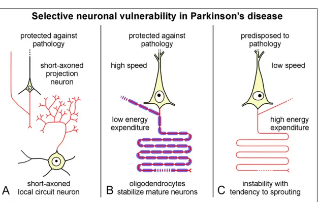
Cell death in this neurodegenerative disease is associated with neurons that possess long, unmyelinated axons in their region of arborization (Braak et al., 2003a, 2003b; Kapfhammer & Schwab, 1994; Bolam & Pissadaki, 2012), which are primary characteristics of DA neurons. Nerve cells possessing long robust axons that are insulated by thick myelin sheaths however, have been shown to exhibit protection against the formation of Lewy neurites (LN) and Lewy bodies (LB) during the course of PD (Braak et al., 2003a, 2003b; Kapfhammer & Schwab, 1994). Several factors have been suspected to contribute to the potential neuroprotective effects of a thick myelin sheath: the speed of conduction, energy expenditure required for the transmission of impulses, and the interaction of the axon with oligodendroglial cells (Figure 1).
A thicker myelin sheath not only increases conduction speed, but also requires less energy for impulse transmission (Braak et al., 2003; Braak et al., 2004). Also, as demonstrated by Kapfhammer & Schwab (1994), the degree of myelination reflects and determines the potential of a brain region for modification of its neural connections. Upon analysis of neurite growth inhibitors (present in myelin) and GAP-43 (marker protein for fiber growth and synaptic plasticity), they demonstrated that myelinated neuronal connections are rather stable, with synaptic plasticity and sprouting actively inhibited by myelination. Poorly myelinated areas however, appeared to be more conducive to sprouting and synaptic plasticity due to the absence of neurite growth inhibitors and increased expression of GAP-43. Their studies also
10
demonstrated that both the SN and striatal tissue are regions of low myelination (Kapfhammer & Schwab, 1994).
Figure 1: Selective neuronal vulnerability in Parkinson’s disease
(Figure 1) Selective neuronal vulnerability in PD. Short-axon projections and local circuit neurons demonstrate increased protection against pathology (A). Projection neurons with long axons and sturdy myelination are also resistant to PD. This is due to the high conductivity, low energy output and superior stability of the parent neuron against axonal sprouting that results from heavy axonal myelination (B). Projection cells possessing long, thin, poorly myelinated axons are among the most vulnerable to PD pathology, due to low conductivity, high energy, and a tendency for sprouting (C). Figure reproduced with permission of Braak et al., 2004.
Another reason projection neurons with either no or poor axonal myelination are more prone to pathological sprouting (Khafhammer & Schab, 1994), is that they have an exceptionally high energy turnover. This could result in their being subjected to continuous oxidative stress, which plays a prominent role in the pathogenesis of idiopathic PD (Beal, 1995; Giasson et al., 2000; Scudamore & Ciossek, 2018).
11
THE MESODIENCEPHALIC DOPAMINERGIC SYSTEM
Dopaminergic neurons, in the adult brain, are localized in the mesencephalon, diencephalon, and olfactory bulb (Arias-Carrion et al., 2010). The majority of these cells however, reside in the ventral part of the mesencephalon. Traditionally, midbrain DA neurons projecting from the SN and VTA were subdivided into four groups: mesocortical, mesolimbic, nigrostriatal, and tuberoinfundibular pathways (Figure 2).
The mesocortical pathway consists of dopaminergic neurons from the VTA projecting to the prefrontal, cingulate and perirhinal cortex. The mesolimbic pathway, also referred to as the reward pathway, connects the VTA to the nucleus accumbens and olfactory tubercle of the ventral striatum. This mesocorticolimbic system has been shown to be involved in the modulation of emotion-related behaviour, including motivation, cognitive control and emotional response, and addiction (Smith & Villalba, 2008; Arias-Carrion et al., 2010). The nigrostriatal pathway connects the SNC, via dopaminergic projections, to the caudate nucleus and putamen of the dorsal striatum. This pathway is essential for the initiation and control of movement and is a part of the basal ganglia motor loop. Finally, dopaminergic neurons projecting from the arcuate nucleus in the tuberal region of the hypothalamus to the median eminence make up the tuberoinfundibular pathway. At this site, dopamine release regulates prolactin secretion from the anterior pituitary gland (Smith & Villalba, 2008; Arias-Carrion et al., 2010). From their various nuclei, dopaminergic axons progress medially where they join together and project through the medial forebrain bundle (MFB) to the internal capsule where they then branch off to form synapses in their target regions.
12
Figure 2: Dopaminergic pathways in the human brain
(Figure 2) Demonstrates the mesocortical (blue), mesolimbic (green) and nigrostriatal (red)
pathways. The mesolimbic and mesocortical pathways originate from the VTA and are important for modulating emotion-related behaviors. The mesocortical pathway sends dopaminergic projections to the prefrontal, cingulate and perirhinal cortices. The mesolimbic pathway projects from the VTA primarily to the nucleus accumbens, however it also innervates the amygdala and hippocampus. The nigrostriatal system, originating in the SNC, projects to the caudate putamen and plays an essential role in voluntary movement. Figure reproduced with
permission from Arias-Carrion, et al., (2010).
These dopaminergic pathways are both anatomically and functionally distinct, though recent evidence is suggesting that this separation is not as exclusive as once thought, and that some of these projections contain cells of origin that are intermixed in the SN-VTA complex (Björklund & Dunnett, 2007). It has been suggested that the caudate putamen may not be the only structure innervated by midbrain DA neurons, and that subsections of other basal ganglia
13
structures such as the globus pallidus, the ventral pallidum and the subthalamic nucleus also receive innervation from these neurons (Gauthier et al., 1999; Lanciego, Luquin & Obeso; 2012). This gives midbrain DA neurons the ability to also directly modulate the activity of basal
ganglia output neurons.
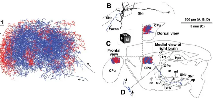
A tracer study of single anterogradely-labeled axons conducted by Gauthier et al., (1999) reconstructed 10 axons whose parent cell body resided in the SNC. They found that these axons appeared to be composed of at least two distinct projection subsystems: those with a profuse arborization in the striatum but poor arborization in the extrastriatal components of the basal ganglia, and those with limited branching in the striatum but a highly patterned set of collateral systems influencing multiple components of the basal ganglia.
Upon characterization of these two axonal subgroups, Gauthier et al. (1999) found that the first type was characterized by only one short thin collateral in the globus pallidus before entering the striatum. It then divided into 4-5 major branches and broke up into many thin and highly varicose collaterals. In contrast, the second subgroup was characterized by axons branching to various extrastriatal structures prior to entering the striatum, where it demonstrated poor arborization. Fibers of this type possessed 2-3 thin and varicose collaterals to either the entopenduncular nucleus, globus pallidus, or both. These collaterals were also shown to arborize in the STN as well. Contrary to the first type, these collaterals displayed infrequent branching and lacked a dense terminal field (Figure 3).
This study demonstrated that the nigrostriatal dopamine system is not a monolithic entity as was once thought, but that SNC neurons possess the ability to influence major components of the basal ganglia in a highly specific manner (Gauthier et al., 1999). Other studies have concurred that SNC neurons projecting to the pallidum are largely distinct from those that terminate in the striatum (Smith et al., 1989; Schneider & Dacko, 1991). Not only does extrastriatal innervation appear to play an important role in the functional organization of the basal ganglia, but, when combined with other findings suggesting that fibers terminating in the pallidum display a larger resistance to the MPTP neurotoxin (Smith et al., 1989; Schneider & Dacko, 1991), important functional implications for parkinsonism need to be considered.
14
Current PD models tend to target the SNC in its entirety, whereas targeting specific subpopulations could be more representative of the disease.
Figure 3: Axonal arborization of single nigrostriatal neurons in rats
(Figure 3) Schematic representation of the axonal arbor observed in a single rat nigrostriatal DA neuron. The first axonal subgroup type (A) is characterized by a short thin collateral projecting
15
from the SNC to the GP, then entering the striatum. Once in the striatum, it divided into 4-5 major branches before branching off into many thin, highly varicose collaterals. The second axonal subtype (B) sent axonal branches into various extrastriatal regions before entering the striatum where it showed little arborization. This subtype of fibers had 2-3 thin and varicose collaterals that were sent into either the entopenduncular nucleus, globus pallidus and/or the STN. Contrary to the dense arborization observed in the first subtype (A), this type had infrequent branching and lacked a dense terminal field. Figure reproduced with permission from
Gauthier et al., 1999.
VENTRAL MIDBRAIN DOPAMINERGIC NEURONS
Dopaminergic neurons of the ventral midbrain can be subdivided into three categories: A8 (retrorubral field; RRF), A9 (SNC) & A10 (VTA). The primary composition of these regions is dopamine neurons accompanied by small groups of GABAergic interneurons and glutamatergic neurons (Olson & Nestler, 2007; Smith & Villalba, 2008; Li et al., 2013; Morales & Margolis, 2017). A variety of neuropeptides have been identified in neuronal subsets of the SNC and VTA, however, the main chemical phenotype contributing to the segregation of ventral midbrain regions is the differential expression of calbindin, a calcium binding protein. Exhibiting a strong expression in VTA, RRF and dorsal tier of the SNC (SNC-d), it appears to lack expression in ventral tier SNC (SNC-v) neurons (Figure 4). Increasing evidence indicates that VTA and SNC-d DA neurons are much less vulnerable to neurodegeneration than SNC-v neurons, so given the differential CB expression, it has been proposed that its presence could play a neuroprotective role in PD whereas its absence could partially account for the increased vulnerability in SNC-v DA neurons (Smith & Villalba, 2008; Vogt Weisenhorn, Geisert & Wurst, 2016). The varying characteristics of midbrain DA neurons result in differential neurodegeneration of the areas, resulting in complex topographical and regional patterns of cell death. Patterns of nigral and striatal degeneration have recently been closely linked with the levels of expression of CB.
16
Figure 4: TH-positive neurons in the murine ventral mesencephalic dopaminergic complex
(Figure 4) TH positive neurons are shown in the murine ventral mesencephalic dopaminergic complex, from rostral to intermediate to caudal (A-C). Histological sections are mirror images of the colored sketches the bilateral organization of the complex. Figure reproduced with
permission from Vogt Weisenhorn, Geisert & Wurst, 2016.
THE BASAL GANGLIA CIRCUIT
The basal ganglia refer to a group of subcortical nuclei responsible primarily for motor control, as well as other roles such as motor learning, executive functions and behaviors, and
17
emotions. Disruption of this network form the basis for several movement disorders. The basal ganaglia-thalamocortical motor circuit, consisting of the cortex, the basal ganglia and thalamus, is particularly pertinent in PD, as it is clearly perturbed and plays a key role in regulating motor behavior (Lanciego, Luquin & Obeso, 2012).
Figure 5: Schematic Representation of the Basal Ganglia Nuclei
(Figure 5) Schematic representation of the basal ganglia nuclei (left) with regions clearly defined
(Right). Caudate nucleus (CN), Putamen (Put), Accumbens (Acb), external segment of the
globus pallidus (GPe), internal segment of the globus pallidus (GPi), Subthalamic nucleus (STN), Substantia nigra pars compacta (SNC), Substantia nigra pars reticulata (SNr). Figure
reproduced with permission from Lanciego, Luquin & Obeso, 2012.
The basal ganglia includes many regions: the caudate nucleus (CN), Putamen (Put), Accumbens (Acb), external segment of the globus pallidus (GPe), internal segment of the globus pallidus (GPi), STN, SNC and SNr (Lanciego, Luquin & Obeso (2012). The striatum, serving as the primary input of the basal ganglia, plays a significant role in decision-making, particularly in action selection and initiation, which requires the convergence of sensorimotor, cognitive and motivational information (DeLong et al., 1990; Smith et al., 1998). Glutamatergic inputs from the thalamus and cortex are received by the striatum, which in turn, sends GABAergic projection outputs to the GP and SNr. Excitatory synaptic connections are made on MSNs by both cortical
18
and thalamic inputs. The cortical afferents of these MSNs are from the sensory, motor and associational cortex, whereas the thalamic afferents are from the intralaminar thalamic nuclei (Doig et al., 2010; Do et al., 2013). Various types of neurons form numerous connections in the dSTR, including with cholinergic, dopaminergic and serotonergic axons that strongly innervate the dSTR. Given the complexity of the neuronal circuits in this region, disruption of the signaling in the striatum can cause the movement impairments observed in PD (Lovinger et al., 2010; Do et al., 2013).
THE CORTICOSTRIATAL CIRCUIT: DIRECT/INDIRECT PATHWAY
The basal ganglia have two major pathways regulating motor function: the direct (striatonigral) and indirect (striatopallidal) pathways, both modulated by dopamine. MSNs of the direct pathway exhibit high levels of both D1 and M4 receptor expression, and project to the GPi/SNr. MSNs of the indirect pathway however, exhibit strong D2 and adenosine receptor expression and project to the GPe (Starr, 1998; Do et al., 2013). A balance between these two pathways is necessary to maintain proper functioning and modulation of cortical regions concerned with motor control.
Abnormalities in this circuit are largely responsible for the hypokinetic and hyperkinetic states observed in PD. Basal ganglia-thalamocortical motor circuit alterations can result from the loss of dopaminergic neurons of the SNC. As discussed, the neurodegeneration of the SNC results in depleted striatal dopamine, and this in turn causes a shift in the balance of basal ganglia activity toward the indirect circuit (Starr, 1998; Hill, Wyse & Anderson; 2009; Lanciego, 2013). This shift leads to an increase in activity of the STN, which overstimulates the GPi/SNr. Increased output from these regions results in excessive inhibition of the thalamocortical pathway, which reduces cortical neuronal activation associated with movement initiation (Starr, 1998; Hill, Wyse & Anderson, 2009; Lanciego, 2013). In other words, inhibition of this pathway results in the hypokinetic motor symptoms of PD.
19
HETEROGENEITY OF MIDBRAIN DOPAMINE NEURONS
Whereas neurons of the SNC and VTA are primarily dopaminergic, increasing evidence indicates that neurons of these regions are more heterogenous than once thought. Dopaminergic neurons from the VTA have long been theorized to play distinct roles in positive and negative reinforcement, decision making, working memory, incentive and stimulus salience, and aversion (Morales & Margolis, 2017). Recent optogenetic experiments have revealed that different functions of the VTA are actually associated with distinct neuronal networks and mediated by a diverse population of VTA DA neurons (Stamatakis et al., 2013; Wang et al., 2015; Yoo et al., 2016). Phenotypic characterization of VTA neurons that subpopulations of TH neurons can vary with regards to biochemical composition, morphological features, functional properties, axonal projections and synaptic connectivity (Li et al., 2013; Wang et al., 2015; Yoo et al., 2016).
Other research has indicated that certain motivated behaviors can be produced by subpopulations of GABA and glutamate neurons in the VTA, independently from dopamine, or even more interestingly, by VTA neurons containing multiple NTs. To date, various subpopulations of VTA neurons have been shown to release dopamine and glutamate from distinct compartments within a single axon (Zhang et al., 2015; Morales & Margolis, 2017). Other subpopulations of these neurons can release glutamate and GABA from a single axon terminal (Root et al., 2014), or co-release DA and GABA from the same vesicle (Berrios et al., 2016). Based on shared characteristics, these various subsets of DA neurons tend to be concentrated in particular subregions of the VTA (Morales & Margolis, 2017).
Typically, dopaminergic neurons can be identified by labeling TH expression, VMAT2 or DAT. Upon closer examination of these heterogenous VTA neurons however, subsets of TH expressing neurons have been discovered that express neither VMAT2 or DAT, which are essential for vesicular repackaging and DA reuptake (Lammel et al., 2008; Stamatakis et al., 2013). In the mouse, subpopulations in the VTA midline nuclei have even been found to express TH mRNA with no detectable levels of TH protein (Yamaguchi et al., 2015; Morales & Margolis, 2017). The purpose of these unique TH neurons in the VTA is currently unknown.
20
VTA GABA neurons, as identified by GAD or VGAT expression, are distributed throughout the rat VTA and have also been shown to be heterogenous in their composition as well. Various experiments have indicated that only some of these neurons contain corticotropin-releasing factor-binding protein (CRF-BP) or cholecystokinin (CCK) (Wang & Morales, 2008; Morales & Margolis, 2017). Electrophysiological and behavioral studies have indicated that CRF-BP, through its interactions with CRF and CRF peptides, can affect neurotransmission of dopaminergic and GABAergic neurons in the VTA (Wang & Morales, 2008). CCK is a neuropeptide that commonly colocalizes with TH neurons in the VTA but has recently been shown to colocalize with GABA neurons in the VTA lacking TH expression (Olson & Nestler, 2007). Other studies have shown that some subsets of VTA GABA neurons respond specifically to DRD2 or mu-opioid receptor activation, whereas in the past, activation of these two receptors were used to distinguish between dopaminergic and GABAergic neurons, and were assumed mutually exclusive (Margolis et al., 2012).
VTA glutamate neurons have been identified as VTA neurons expressing mRNA encoding VGLUT2. These neurons have been implicated in mediating rapid excitatory signalling and are particularly prevalent within the midline nuclei. This subpopulation of neurons actually outnumbers TH-expressing neurons in some portions of the VTA and have also been found in the hypothalamus (Kawano et al., 2006; Yamaguchi et al., 2011; Morales & Margolis2017).
Although the majority of VTA neurons signal by releasing DA, GABA or glutamate, recent studies have identified subpopulations of VTA neurons that exhibit combinatorial NT characteristics (Yamaguchi et al., 2011; Li et al., 2013; Zhang et al., 2015; Morales & Margolis, 2017). Whereas much about the mechanisms and function of these combinatorial neurons remain to be elucidated, these neurons have been shown to co-release DA and glutamate, DA and GABA, or both glutamate and GABA.
Preferentially concentrated in the midline nuclei of the VTA, combinatorial TH and VGLUT2 all express the enzyme AADC and are therefore capable of synthesizing DA (Yamaguchi et al., 2011). Electrophysiological and voltammetry studies have confirmed that these neurons do in fact release both glutamate and DA (Li et al., 2013). Some of these neurons
21
however, have been shown to lack expression of DAT or VMAT2. Upon examination of ultrastructural, biochemical and electrophysiological properties of VGLUT2 inputs from VTA neurons in the nucleus accumbens (nAcc). Zhang et al., (2015) demonstrated a segregation in VGLUT2-TH neurons of dopaminergic and glutamatergic vesicles into different axonal microdomains within a given axon. This study ruled out the theory that nAcc vesicles co-express glutamate and DA and demonstrated that these combinatorial neurons possess two distinct contiguous domains specialized for DA or glutamate release in both mice and rats (Zhang et al., 2015; Morales & Margolis, 2017). Residing in axon terminals, these glutamate vesicles establish asymmetric synapses, commonly associated with excitatory transmission, which lie adjacent to DA axonal segments. This gives individual VGLUT2-TH axons within the nAcc the ability to participate in synaptic or non-synaptic (volume) transmission, meaning. It can therefore provide both excitatory and inhibitory signalling via glutamate and dopamine respectively.
As stated, another form of combinatorial neurons in the VTA have been found to express both TH and GAD. These TH-GAD neurons appear to have a number of subpopulations possessing varied combinations of phenotypes. Some subpopulations have been shown to rarely express VMAT2 and do not appear to exhibit axonal release of DA (Root et al., 2014), others appear to be able to release GABA from the axons of TH-expressing neurons that contain VMAT2 but lack GAD1, GAD2 and VGAT. In cases where VGAT is lacking, it has been suggested that GABA transporters, proposed to be present in DA axon terminal membranes, transport cytoplasmic GABA from the extracellular environment into the cytoplasm of the axon (Tritsch et al., 2014; Morales & Margolis, 2017). In this theory, VMAT2 would then pack cytoplasmic GABA into synaptic vesicles. Unlike the VGLUT2-TH neurons, this does result in the vesicular coexistence of GABA and DA. Also unique to these TH-GAD neurons is the ability of these DA neurons to synthesize GABA directly (Kim et al., 2015; Morales & Margolis, 2017). These neurons therefore, exhibit the ability to release GABA through both synthesis and transporter uptake mechanisms. It is currently not known whether these axons also have the ability to release glutamate.
A subpopulation of non-TH-expressing neurons in the VTA have been identified which co-express VGLUT2, GAD1, GAD2 and VGAT. Some of the axon terminals of these neurons
22
have been shown to contain both VGLUT2 and VGAT and have the ability to establish both asymmetric (excitatory) and symmetric synapses (inhibitory) (Root et al., 2014). These axons work with postsynaptic neurons possessing both GABAA receptors and GluR1-containing
AMPA receptors to evoke either fast inhibition (followed by excitation) or fast excitation (followed by inhibition), the combination of which could allow pronounced temporal specificity. Finally, a small subpopulation of TH-expressing neurons was identified in the mouse and rat VTA which express VGUT2, GAD1 and GAD2, however it remains unknown to date whether these neurons have the ability to release all three transmitters (Root et al., 2014; Stamatakis et al., 2013; Yamaguchi et al., 2011; Morales & Margolis, 2017).
Interestingly, whereas GABA and glutamate neurons are present in the SNC, there is no evidence to date indicating the presence of combinatorial neurons in this region. Other phenotypic heterogeneity has been identified in SNC neurons however. DAT has been found to be much more highly expressed in SNC neurons as compared to VTA (Blanchard et al., 1994; Storch, Ludolph & Schwarz, 2004), and it is believed that this is one contributing factor to their increased vulnerability. GABAA receptor subunits have also been found to be heterogeneously
distributed at the regional, cellular, and subcellular levels in the human SNC and SNr. The predominant GABA receptor subunits in the SNC are not the same as in the SNr (Waldvogel et al., 2008).
As demonstrated, the characteristics and organization of midbrain DA neurons is much more complex than previously thought. These details however, can allow for much more targeted approaches to pre-clinical PD research and can help elucidate some of the remaining unknowns in the field. Given how little is known about the function or purpose of these heterogenous VTA neurons, one has to wonder if these unique properties contribute at all to the resistance VTA neurons have in PD neurodegeneration.
23
PARKINSON’S DISEASE
TYPES OF PD
PD is classified into two categories: familial and sporadic/idiopathic. Familial PD occurs in approximately 10% of cases, whereby the onset of disease is due to rare familial genetic mutations (Gillies et al., 2014). On the other hand, sporadic PD is due to exogenous causes influenced by many factors: cellular and molecular processes underlying the degeneration of the nigrostriatal dopamine system, mitochondrial dysfunction, excessive production of reactive oxygen species (ROS), and the formation of pathological protein aggregates (Klein & Schlossmacher, 2007; Gillies et al., 2014). The emerging consensus supported by many researchers in the field today is that PD is a complex, multisystem disease, resulting from the interaction of numerous genetic and environmental factors.
SYMPTOMS OF PD
PREMOTOR PHASE OF PD
The premotor phase of PD is considered as the first stage of symptomology in PD, and occurs when psychopathological processes begin to appear, which can occur many years before the onset of motor manifestations (Cummings & Masterman, 1999; Tolosa, Compta & Gaig, 2007; Zeimssen & Reichmann, 2007; Tadaiesky et al., 2008). Premotor complications include depression, anxiety and cognitive decline can be detrimental to a patient’s quality of life. Mood disorders such as depression and anxiety can occur in up to 40% of patients with early stage PD (Tolosa, Compta & Gaig, 2007; Tadaiesky et al., 2008). Whereas these particular mood disorders are relatively well understood on their own, recent studies have started to suggest that the pathophysiology underlying mood disorders in PD may not use the same mechanisms as these disorders in the general population (Lieberman, 2006; Tadaiesky et al., 2008). Although their pathophysiology in the context of PD is not completely understood, dysfunction in striatal, frontal and limbic dopaminergic, cholinergic, serotonergic, noradrenergic and GABAergic pathways are thought to be involved in their genesis (Wolters, 1999; Schrag, 2004).
24
Dementia and less severe cognitive impairments are also common in PD and can be an important predictor for quality of life. Impaired procedural memory has been associated with striatal alterations, whereas executive dysfunction and working memory impairments have implicated the involvement of the prefrontal cortex-basal ganglia loop (Jackson et al., 1995; Packard & Knowlton, 2002; Lewis et al., 2003; Tadaiesky et al., 2008).
The development and testing of neuroprotective drugs could be greatly facilitated by the ability to diagnose PD prior to motor symptom onset. Ideally, early diagnosis could allow for pre-treatment of the disease to either halt or delay progression. Whereas degeneration of SNC DA neurons is the primary characteristic of PD, neuronal dysfunction or degeneration in other regions within the nervous system likely account for premotor symptoms of the disease (Slow, Postuma & Lang, 2014). One such example is olfactory dysfunction, a key element of the preclinical stage of PD and one of the earliest nonmotor features of the disease. Olfactory dysfunction has been found to occur in an estimated 90% of sporadic PD cases (Doty, Deems & Stellar, 1998; Gagnon et al., 2009; Postuma et al., 2009). To a certain extent, olfactory dysfunction can be predictive of the future development of PD, although given that it is a symptom affected in multiple neurodegenerative diseases there is a lack of specificity. The impairment is rarely complete, is not affected by DA therapies, and has been correlated with abnormal sympathetic cardiac function. Evidence suggests that deficits in cholinergic, noradrenergic and serotonergic function may contribute this olfactory dysfunction (Wolters, 1999; Postuma & Gagnon, 2010).
The specific mechanisms responsible for olfactory dysfunction however, are currently unknown. What is known is that there is significant degeneration and cell loss that occurs in the LC, an area that is responsible for sending noradrenergic projections to the olfactory bulb several olfactory-related structures (Zarow et al., 2003; Doty, 2012). A close relationship has also been established between olfactory function and central cholinergic processes in PD as well as an intimate relationship between serotonin and the olfactory bulb. Marked reductions in both types of these processes have been observed in PD (Scatton et al., 1983; Bohnen et al., 2003; Doty, 2012). Alpha-synuclein pathology is also prevalent throughout the olfactory system in the early
25
stages of the disease, which is in accordance with the Braak staging system of PD (Braak et al., 2001; Sylveira-Moriyama, et al., 2009).
Another premotor symptom of PD that holds potential for early identification is sleep disorders. In PD, some of these disorders include REM sleep behavior disorder (RBD), insomnia and restless leg syndrome. Manifesting years prior to the appearance of PD motor symptoms, studies have shown that RBD in particular could be used as an important preclinical marker for the disease (Slow, Postuma & Lang, 2014). This is a parasomnia characterized by reduced REM sleep muscle atonia and dream-enacting behaviours. RBD prevalence in PD patients has been estimated to range between 30-50% during the course of the illness. Longitudinal studies have found that RBD patients who develop a neurodegenerative condition will manifest overwhelming symptoms of synucleopathy (Postuma et al., 2009; Slow et al., 2014).
Synucleopathy is found throughout the brainstem and can help explain the increased incidence of olfactory and autonomic dysfunction in RBD patients, as these structures are affected in the early stages of the disease. As such, these two dysfunctions have recently become considered early pre-motor symptoms of PD (Braak et al., 2001; Slow, Postuma & Lang, 2014). Cognition, psychosis and depression also all had increased prevalence in PD patients with RBD. Studies have suggested that the presence of RBD in patients can be used to predict future development of PD, as well as help to identify motor and non-motor manifestations associated with the disease (Postuma et al., 2008, 2012; Slow, Postuma & Lang, 2014).
CARDINAL MOTOR SYMPTOMS OF PD
The four cardinal features of PD include tremor at rest, rigidity, akinesia (or bradykinesia) and postural instability. Freezing and flexed posture are also considered as features of parkinsonism (Jankovic, 2008). Because there is no definitive test for the diagnosis of PD, current diagnostic criteria for PD should include a combination of motor and nonmotor impairments. Studies have estimated that by the time motor impairments become evident in the
26
progression of this disease, dopaminergic cell death of approximately 80-90% of SNC neurons has already occurred, accompanied by a 50-80% reduction in striatal DA levels (Fearnley & Jees, 1991; Tagliaferro & Burke, 2016).
BRADYKINESIA
Bradykinesia, or slowness of movement, is one of the most common and easily recognizable symptoms of PD. Although its pathophysiology is not completely understood, studies have shown that this is the symptom which best correlates with degree of dopamine deficiency (Lozano et al., 1995; Cutsuridis V & Perantonis S, 2006; Jankovic, 2008). Bradykinesia is a hallmark symptom of basal ganglia disorders and leads to difficulties in the planning, initiation and execution of movement. Recent evidence suggests that the neuropathology responsible for bradykinesia results from a disruption in normal motor cortex activity mediated by reduced dopaminergic function (Lozano et al., 1995; Cutsuridis V & Perantonis S, 2006; Jankovic, 2008). Electromyographic studies of PD patients has demonstrated that the deficit observed in initiation and maintenance of movement has to do with a reduction in muscle force, thought to be caused by deficits in the putamen and globus pallidus (Cutsuridis V & Perantonis S, 2006; Jankovic, 2008).
TREMOR
Another common and easily recognized symptom of PD is resting tremor. Tremors are typically unilateral and are most prominent at the distal part of an extremity. Resting tremors in PD can involve the hands, lips, chin, jaw and legs and tends to disappear during sleep. Studies have indicated that 100% of PD patients had a tremor at some point during the disease; however, the occurrence of resting tremor is highly variable among patients and disease progression (Martin et al., 1973; Rajput, Rozdilsky & Rajput, 1991; Jankovic, 2008). Hughes et al., (1993) reported that resting tremor appeared at onset of disease in 69% of patients, and that tremor was lost in 9% of patients late in the disease.
27 RIGIDITY
Rigidity is characterized by increased muscle tone which produces increased resistance to movement. This stiffness, caused by an excessive and continuous flexing of the muscles, can cause joint pain and is a frequent clinical manifestation of PD. Rigidity in PD is often accompanied by the “cogwheel” phenomenon, whereby reinforcing manoeuvres increase rigidity during the passive movement of a limb (either proximally or distally), particularly when associated with an underlying tremor (Jankovic, 2008).
CURRENT TREATMENT OPTIONS
Despite tremendous research efforts, the two primary objectives of therapeutic PD research have yet to be achieved: prevention of its onward progression and reparation of systems already damaged (Tagliaferro & Burke, 2016). There is currently no cure for PD and current treatment options only offer symptom management. These treatments include pharmacological therapies, surgical interventions and cell replacement therapies.
PHARMACOLOGICAL THERAPIES
Given that DA cannot cross the blood brain barrier, drug treatment options are aimed at either indirectly replenishing DA stores or mimicking the action of DA. These dopaminergic medications can be effective, for a time, at reducing the cardinal symptoms of PD. Whereas there are certain benefits to all the current PD drug treatments, their limited effectiveness and negative side-effects leave a lot to be desired. One of the most serious downsides to current drug treatments are psychiatric manifestations. Patients with PD are more likely to develop depression than the general population, which can be treated with anti-depressants (Guttman, Kish & Furukawa, 2003). Drug-induced psychosis can be a major management problem however, with patients developing visual hallucinations, paranoia and other psychotic symptoms. These issues can be resolved with the reduction or elimination of certain parkinsonian therapies, which often results unfortunately, in a worsening of the original parkinsonian symptoms. Therefore, given that the only current treatment options available focus