IDENTIFICATION OF A NEW CELL LINE PERMISSIVE TO
PORCINE REPRODUCTIVE AND RESPIRATORY SYNDROME
VIRUS REPLICATION
par
Jian-Jun Jia, DVM
Département de pathologie et microbiologie Faculté de médecine vétérinaire
Mémoire présenté à la Faculté de médecine vétérinaire en vue de l’obtention du grade de
maître ès sciences (M.Sc) en sciences vétérinaires
option microbiologie
Août 2009
Université de Montréal
Faculté de médecine vétérinaire
Ce mémoire intitulé
IDENTIFICATION OF A NEW CELL LINE PERMISSIVE TO
PORCINE REPRODUCTIVE AND RESPIRATORY SYNDROME
VIRUS REPLICATION
présenté par
Jian-Jun Jia, DVM
a été évalué par un jury composé des personnes suivantes
Josée Harel, présidente-rapporteuse
Carl A Gagnon, directeur de recherche
Le syndrome reproducteur et respiratoire porcin (SRRP) est une des maladies les plus dévastatrices économiquement pour l'industrie mondiale du porc. L'agent étiologique du SRRP est le virus du SRRP (VSRRP) lequel est connu pour avoir une spécificité d'hôte très restreinte et pour sa transmission par voie aerosol. Les antigènes et les ARN du VSRRP ont été trouvés dans des cellules épithéliales du tractus respiratoire de porcs infectés par le virus. L’interaction entre les macrophages alvéolaires porcins (PAMs) et le VSRRP a été démontrée comme jouant un rôle important dans l’infection causée par le virus. Malgré cela, l’interaction prenant place entre les cellules épithéliales du tractus respiratoire porcin et le virus ne devrait pas être négligée. Jusqu’à présent, la réplication du VSRRP in vitro dans des cellules épithéliales du tractus respiratoire porcin n’a pas été conduite avec succès et les tentatives pour le faire ont échoué. Une nouvelle lignée de cellules épithéliales de poumon de porc (SJPL) est maintenant disponible et sera utilisée dans cette étude afin de déterminer si elle est permissive à la réplication du VSRRP et si elle peut être un modèle approprié pour l’étude de la pathogénèse virale du VSRRP. L’expérimentation a démontré que cette nouvelle lignée cellulaire était permissive à l’infection et à la réplication du VSRRP. Afin de corroborer ces résultats, la cinétique de réplication du virus à été effectuée avec les cellules MARC-145 et SJPL. Aucune différence significative dans la production virale totale n’a été trouvée entre les deux lignées cellulaires. Les cellules SJPL ont permis la réplication de plusieurs souches Nord-Américaines du VSRRP, quoiqu’elles sont légèrement moins efficaces que les cellules MARC-145 pour l’isolement du virus. De plus, les cellules SJPL sont phénotypiquement différentes des cellules MARC-145. Plus précisément, les cellules SJPL sont plus sensibles à l’activation par le VSRRP des pro-caspases 3/7 et plusieurs inducteurs apoptotiques. Elles ont également montré de 8 à 16 fois plus de sensibilité à l’effet antiviral causé par l’IFN-α sur la réplication du virus contrairement aux cellules MARC-145. Ces résultats démontrent que les cellules SJPL pourraient représenter un substitut intéressant aux cellules MARC-145 pour la production d’antigènes pour un vaccin anti-VSRRP. Également, dû à leurs origines (poumon de l’hôte naturel), elles pourraient s’avérer être un modèle in vitro plus approprié pour l’étude de la pathogénèse du VSRRP.
Mots clés : Virus du syndrome reproducteur et respiratoire porcin; VSRRP; cellule
Porcine reproductive and respiratory syndrome (PRRS) is one of the most economically devastating diseases for the pig industry worldwide. The etiological agent of PRRS is the PRRS virus (PRRSV), which is known to have a very restricted host specifity and to be airborne transmitted. PRRSV RNAs and antigens were found in epithelial cells of the respiratory tract of swine in PRRSV-infected pigs. Even if the interaction between porcine alveolar macrophages (PAMs) and PRRSV plays an important role in the PRRSV infection, the role of the interaction between epithelial cells of the swine respiratory tract and PRRSV should not been neglected. However, no epithelial cells of the swine respiratory tract have been demonstrated to allow PRRSV replication in vitro and attempts to generate such a cell line have failed. The goal of this study is to determine whether epithelial cells of the swine respiratory tract are permissive to PRRSV replication and are a suitable model for studying the viral pathogenesis of PRRSV. We have discovered that the SJPL cell line, an epithelial cell line of the respiratory tract of swine, is permissive to PRRSV infection and replication. To corroborate these results, PRRSV replication kinetics were evaluated in a subclone of the African green monkey kidney MA104 cells (MARC-145), which has been known to be fully permissive to PRRSV infection and replication, and in SJPL cells. No significant difference was found between the two cell lines for overall viral production. Moreover, the SJPL cells were able to permit the replication of several PRRSV North-American strains but they were slightly less efficient for virus isolation than MARC-145 cells. In addition, SJPL is phenotypically different from MARC-145. Specifically, the SJPL cells were more sensitive to procaspases 3/7 activation by PRRSV and several apoptotic inducers compared to MARC-145 cells. In addition, the SJPL cells showed 8 to 16 times more sensitivity to the antiviral effect of IFN-α against PRRSV replication than MARC-145 cells. Altogether, the SJPL cells could be an interesting substitute to MARC-145 cells for PRRSV vaccine antigen production, and could be a more relevant in vitro model, because of their origin (lung of the natural host), to study the pathogenesis of PRRSV.
Key words: Porcine reproductive and respiratory syndrome virus; PRRSV; porcine
TABLE OF CONTENTS
RÉSUMÉ ... i ABSTRACT ... iv TABLE OF CONTENTS ... vi LIST OF TABLES ... ix LIST OF FIGURES ... x LIST OF ABBREVIATIONS ... xi DEDICATION ... xiii ACKNOWLEDGMENTS ... xiv Ι. INTRODUCTION ... 1II. LITERATURE REVIEW ... 4
1. Disease history and terminology ... 5
2. Clinical manifestation ... 6
3. Etiology ... 7
3.1. Taxonomy ... 7
3.2. Viral genomic organization ... 8
3.3. Virus biological and physical properties ... 12
3.4. Virus genetic variation ... 12
4. Pathogenesis ... 14
4.1. Virus entry into susceptible cells ... 14
4.2. Virus transmission ... 16
4.3. PRRSV cell and tissue tropism ... 18
4.4. PRRSV-induced apoptosis ... 19
6. Disease control and eradication ... 26
6.1. Control and eradication strategies ... 26
6.2. Treatments and prevention ... 28
6.2.1. PRRS Vaccines ... 29
7. Diagnosis of PRRSV infection ... 30
III. MATERIALS, METHODS & RESULTS ... 32
Identification of a new porcine lung epithelial cell line permissive to porcine reproductive and respiratory syndrome virus infection and replication ... 33
1. Introduction ... 35
2. Materials and methods ... 38
2.1. Cells and viruses ... 38
2.2. Immunofluorescence assay (IFA) ... 39
2.3. Virus production during multiple cell passages ... 40
2.4. Virus replication kinetics assay ... 40
2.5. Virus isolation ... 41 2.6. Apoptosis ... 42 2.7. Inhibition of PRRSV replication ... 42 2.8. Statistical analysis ... 43 3. Results ... 45 3.1. SJPL cells susceptibility to PRRSV ... 45
3.2. Infectious PRRSV particle production in infected SJPL cells ... 45
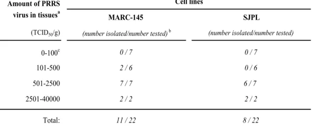
3.3. Virus isolation efficiency ... 46
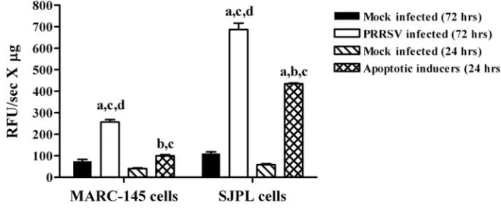
3.4. SJPL cells susceptibility to PRRSV apoptosis... 47
3.5. Inhibition of PRRSV replication ... 48
5. Acknowledgments ... 52
6. References ... 53
IV. Discussion ... 66
V. Conclusion ... 73
VI. References ... 75
LIST OF TABLES
Literature review
Table I. Comparison of ORFs2 to 7 encoded protein between NA and EU strains of
PRRSV ... 10
Submitted manuscript
Identification of a new porcine lung epithelial cell line permissive to porcine reproductive and respiratory syndrome virus infection and replication
Table I. PRRSV virus isolation efficiency from swine samples using SJPL cells
compared to MARC-145 cells ... 63
Table II. Inhibition of PRRSV infection in PCV-2 co-infected cells... 64
LIST OF FIGURES
Literature review
Figure 1. Nidovirales order classification ... 8
Figure 2. Schematic representation of the virion of PRRSV ... 11
Figure 3. PRRSV genomic organizations . ... 11
Figure 4. Pathogenesis of PRRSV infection ... 18
Figure 5. Temporal sequence of events after infection of a pig with PRRSV . ... 26
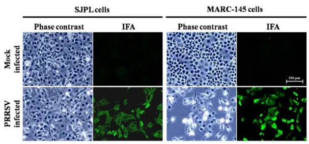
Submitted manuscript Identification of a new porcine lung epithelial cell line permissive to porcine reproductive and respiratory syndrome virus infection and replication Figure 1. Detection of the N viral protein in PRRSV-infected SJPL cells by an immunofluorescence assay ... 59
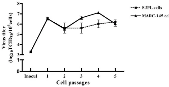
Figure 2. PRRSV infectious particles production in infected SJPL cells following five consecutive passages. ... 60
Figure 3. PRRSV replication kinetics in infected SJPL cells. MARC-145 and SJPL cells were infected at 1 MOI with PRRSV IAF-Klop strain. ... 61
LIST OF ABBREVIATIONS
aa Amino acids
ASFV African swine fever virus,
Cap Capsid protein
CMI response Cell mediated immune response
CPE Cytopathic effect
DNA Deoxyribonucleic acid
EAV Equine arteritis virus
ELISA Enzyme-linked immunosorbent assay
FBS Fetal bovine serum
IFA Immunofluorescence assay
IPMA Immunoperoxidase monolayer assay
IFN-γ/α Interferon-gamma/alpha
IHC Immunohistochemistry
ISH In situ hybridization
LDV Lactate dehydrogenase-elevating virus
LV Lelystad virus
MAb Monoclonal antibody
MARC-145 African green monkey kidney- derived MA-104
cell
MLV Modified live Virus
MOI Multiplicity of infection
MSD Mystery swine disease
NAb Neutralizing antibody
nm Nano meter
ORF Open reading frame
PAM Porcine alveolar macrophage
PBMC Peripheral blood mononuclear cells
PBS Phosphate buffered saline
PCV-2 Porcine circovirus type 2
PEARS Porcine epidemic abortion and respiratory
syndrome
PFA Paraformaldehyde
PHFS Porcine high fever syndrome
pi Post-infection/inoculation
PRDC Porcine respiratory disease complex
PRRSV Porcine reproductive and respiratory syndrome
virus
RNA Ribonucleic acid
SJPL Saint-Jude porcine lung cells
SHFV Simian hemorrhagic fever virus
SIRS Swine infertility and respiratory syndrome
SIV Swine influenza virus
SPF Specific pathogen free
TBS Tris buffered saline
TCID50 Tissue culture infectious dose with a 50% end
point
TNF-α Tumor necrosis factor-alpha
TUNEL Terminal deoxynucleotidyl transferase dUTP
nick end labeling
UTR Untranslated region
To my parents, my brothers and sisters
For believing in me and For supporting me
ACKNOWLEDGMENTS
These last two years will definitely stay engraved in my memory for the rest of my life, and that is due to all the great people that surrounded me and supported me in this journey. First and foremost, I would like to express my sincere gratitude to Dr Carl A Gagnon, professor of University of Montreal, who has been my supervisor since the beginning of my study for giving me the opportunity to live this experience in his laboratory amongst the great people I am now proud to call my friends. He provided me with a great deal of helpful suggestions, important advice and constant encouragement during the course of this work. He also provided me with great insight and knowledge of the research world. He inspired me with his great kindness and endless patience.
Special thanks go to all the members of the laboratory from whom the valuable suggestions and selfless help that I received in the past years. I would be grateful to all members of the research group of infectious disease of the swine for their support of all nature, especially to Dr David W. Silversides and Dr Josée Harel for participating on my advisory committee as well as on the jury of this document.
Finally I would give great thanks to my parents for giving me love, courage and power to face all the difficulties and challenges; my sisters, brothers and my friends for giving me support in pursuing my masters’ studies.
Porcine reproductive and respiratory syndrome (PRRS) is one of the most economically devastating diseases for the pig industry worldwide (Garner et al., 2001; Neumann et al., 2005; Pejsak et al., 1997). The disease was first reported in the United States in 1987 (Keffaber, 1989; Loula, 1991) and in Europe in the early 1990s (OIE, 1992). Since then, it has spread throughout the world and has caused huge economic losses in swine industry. The etiologic agent, PRRS virus (PRRSV) was identified by investigators in the Netherlands and USA in 1991 (Benfield et al., 1992; Wensvoort et al., 1991b). PRRS has become a well-recognized global swine disease (Albina, 1997; Botner et al., 1994; Hopper et al., 1992; Kuwahara et al., 1994; Tian et al., 2007). In the recent years, new PRRSV variants emerged in Vietnam and China causing unprecedented large-scale outbreaks and catastrophic clinical syndromes (Feng et al., 2008; Tian et al., 2007).
PRRSV is believed to replicate in specific cells both in vivo and in vitro. The presence of PRRSV antigens and RNAs has been shown in different cells types in vivo by immunohistochemistry (IHC) or in situ hybridization (ISH) (Halbur et al., 1995a; Magar et al., 1993; Pol et al., 1991; Rossow et al., 1996; Sur et al., 1997). In vitro, PRRSV replicates in primary cultures of PAMs as well as freshly isolated blood monocytes or monocytic derived dendritic cells (Voicu et al., 1994; Wang et al., 2007; Wensvoort et al., 1991b). Only two other non-porcine permissive cell lines permit the replication of PRRSV, the MARC-145 and CL2621 cells (subclones of MA104 monkey kidney cell line) (Bautista et al., 1993; Benfield et al., 1992; Kim et al., 1993) which are routinely used for in vitro propagation of PRRSV and for large scale production of PRRSV vaccine. It is well known that the respiratory tract is the primary route of PRRSV infection and transmission, and intranasal inoculation was used for experimental infections to support this idea (Brockmeier et al., 2000; Magar et al., 1995; Meredith, 1993; Wensvoort et al., 1992). Since PRRSV antigens could be found in the epithelial cells of the respiratory tract of infected swine, it can be speculated that these cells may favor the propagation of PRRSV in vitro. However, to our best knowledge, until now, no epithelial cell of the respiratory tract of swine has been reported to be permissive to PRRSV replication in vitro.
The goals of the present study were: (1) to determine if the PRRSV natural cell host of the respiratory tract of swine, the epithelial cells, could support PRRSV replication in vitro; (2) to establish a new in vitro PRRSV permissive cell model for studying the viral pathogenesis of PRRSV.
1. Disease history and terminology
In the late 1980's, catastrophic outbreaks of a previously unrecognized disease in pigs were reported in the United States (Keffaber, 1989; Loula, 1991) where it became widespread, with subsequent extension into Canada (Bilodeau et al., 1991). First described in herds in North Carolina, the syndrome included severe reproductive losses, extensive postweaning pneumonia, reduction of growth performance, and increased mortality (Hill, 1990). In the absence of a recognized cause, the name "Mystery Swine Disease" (MSD) came into common usage (Hill, 1990). In Europe, clinical outbreaks notably similar to MSD were reported in November 1990 near Munster, Germany (OIE, 1992), in the Netherlands in January 1991 and in Belgium in March 1991 (OIE, 1992), but no link was found between outbreaks in Germany and MSD in the U.S. (Anon, 1991). Subsequently, disease was found in Spain (Plana et al., 1992), Great Britain (Edwards et al., 1992), France (Baron et al., 1992), Denmark (Botner et al., 1994), Poland (Pejsak and Markowska-Daniel, 1996) and Czech Republic (Valicek et al., 1997). In Asia, outbreaks occurred in Japan in 1988 (Hirose et al., 1995), in Taiwan in 1991 (Chang et al., 1993) and in China in 1995 (Tong and Qiu, 2003). Thus, the pandemic had spread to most of the major swine producing countries of the world during a short period of time. Initially, a variety of etiologies for MSD were proposed (Bane and Hall, 1990; Daniels, 1990; Hoeffling, 1990; Joo, 1988; Joo, 1990; Quaife, 1989; Reotutar, 1989). In Canada, a new subtype of Influenza A virus was isolated from piglets suffering from severe respiratory disease and added to the list as a possible agent of MSD (Dea et al., 1992; Elazhary et al., 1991). Identifying the etiology was complicated by the fact that one or more of the suspected pathogens, as well as other infectious agents, were commonly isolated from cases of MSD. The lack of a specific etiologic agent combined with various clinical signs led to the use of several disease names, such as blue ear disease (Paton et al., 1991; Wensvoort et al., 1991a), mystery swine disease (MSD) (Hill, 1990; Reotutar, 1989), porcine epidemic abortion and respiratory syndrome (PEARS) (Pol et al., 1991; Terpstra et al., 1991), swine infertility and respiratory syndrome (SIRS) (Benfield et al., 1992; Christianson et al., 1992; Collins et al., 1992), pig plaque (Keffaber, 1989) and new pig disease (Meredith, 1992). One virus first isolated in the Netherlands (Wensvoort et al., 1991b)
was designated Lelystad (LV) and later another virus isolated from sick swine by a team of researchers from South Dakota State University, the University of Minnesota, and Boehringer Ingelheim Animal Health was named Swine Infertility and Respiratory Syndrome (SIRS) virus. Both virus isolates were shown to induce reproductive failure and respiratory signs under experimental conditions (Collins et al., 1992; Terpstra et al., 1991), but in May of 1992, participants at the International Symposium on SIRS in Minneapolis, Minnesota, chose to name the disease the porcine reproductive and respiratory syndrome (PRRS), and since then the agent has been referred to as the PRRS virus (PRRSV).
Today, PRRS is endemic in the global swine producing countries and has become one of the most important pathogens causing economic losses in the swine industries (Albina, 1997; Blaha, 2000; Neumann et al., 2005). PRRSV was diagnosed in Africa for the first time in June 2004 following outbreaks in Western Cape Province, South Africa (OIE, 2005b). Serologic tests did not identify additional infected sites at that time but new outbreaks were identified in October 2005 (OIE, 2005a) and again in August 2007 (Beltran-Alcrudo et al., 2007). Chile is on the verge of becoming the first country to eradicate PRRSV. Chilean producers are currently in the process of culling all sows that were present at the time of infection (Anon, 2007). Sweden claimed to be free of PRRS until 2007 when the disease was recognized as an emerging disease (Carlsson et al., 2009). Most recently, new PRRSV variants emerged and circulated in Vietnam and China causing unprecedented large-scale outbreak and catastrophic clinical syndromes (Feng et al., 2008; Tian et al., 2007; Zhou et al., 2008). Some countries, including Switzerland, New Zealand, and Australia, claim to be free of the disease (Cannon et al., 1998; Elvander et al., 1997; Garner et al., 1996; Motha et al., 1997).
2. Clinical manifestation
PRRS is characterized by anorexia, fever and abortion late in gestation, premature births, stillbirths, and mummified fetuses. However, the two most prevalent clinical signs are severe reproductive failure in sows and gilts (characterized by late-term abortions, an increased number of stillborns, mummified and weak-born pigs) (Bilodeau et al., 1991; Christianson et al., 1992; Keffaber, 1989; Pol et al., 1991; Terpstra et al., 1991) and respiratory problems in pigs of all ages associated with a
non-specific lymphomononuclear interstitial pneumonitis (Bilodeau et al., 1991; Collins et al., 1992; Halbur et al., 1995b; Halbur et al., 1996b; Rossow et al., 1994). Furthermore, the intensity of the disease appears to vary among isolates and variation in PRRSV virulence has been observed in experimentally infected animals (Halbur et al., 1995b; Mengeling et al., 1996). Studies showed that pigs experimentally infected with different isolates developed major differences in clinical disease, rectal temperatures, and gross and histological lung lesions; mildly virulent isolate infections induced transient pyrexia, dyspnea and tachypnea, whereas highly virulent isolate infections exhibited labored breathing, pyrexia, lethargy, and anorexia (Halbur et al., 1995a; Halbur et al., 1995b; Halbur et al., 1996b). Moreover, highly virulent isolates of PRRSV infection resulted in longer periods of viremia, increased severity of clinical signs and mortality, and significantly higher viral loads in blood and tissues (Johnson et al., 2004). Several other factors such as animal age and bacterial co-infection can influence virus replication and clinical signs. Infection of younger animals showed a longer viremia, as well as higher excretion rates and replication rates in macrophages compared to the older pigs (Thanawongnuwech et al., 1998; van der Linden et al., 2003). Additionally, certain bacteria appeared to enhance the duration and severity of PRRSV induced clinical signs (Brockmeier et al., 2000; Thacker et al., 1999). Host immune status may also affect the severity of the clinical signs. Previous exposure to PRRSV can prevent the development of PRRS clinical signs by subsequent infection with the homologous PRRSV (Shibata et al., 2000).
3. Etiology 3.1. Taxonomy
The first PRRSV isolates obtained in Europe and North America were designated Lelystad and ATCC VR-2332 respectively. Now PRRSV is divided into two distinct genotypes, the European (EU) type (or type I) and North American (NA) type (or type II). The EU and NA genotypes of PRRSV share only 63% nucleotide (nt) homology (Allende et al., 1999; Collins et al., 1992; Meulenberg et al., 1993; Nelsen et al., 1999). Although distinct genetically and antigenically, both types exhibit the same genome organization and nearly the same pathogenesis. PRRSV is an enveloped, single-stranded, positive-sense RNA virus classifying in the Arteriviridae family
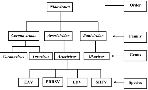
within the genus Arterivirus, along with equine arteritis virus (EAV), lactate dehydrogenase-elevating virus (LDV) of mice, and simian hemorrhagic fever virus (SHFV), because of their similar morphology, genome organization, transcription strategy, macrophage tropism, the ability to induce prolonged viremia and persistent infections (Benfield et al., 1992; Cavanagh, 1997; Plagemann and Moennig, 1992). The family Arteriviridae, Toroviridae, and Coronaviridae are the members of a single established order, Nidovirales (Cavanagh, 1997) (Figure 1).
Figure 1. Nidovirales order classification (Cavanagh, 1997)
3.2. Viral genomic organization
As described in the earlier studies, mature PRRSV virions contain a spherical icosahedral capsid core of 20-30 nm in diameter, which is surrounded by a lipid envelope containing the viral membrane proteins, yielding a relatively smooth spherical virion of about 60 nm in diameter (Benfield et al., 1992; Dea et al., 2000; Doan and Dokland, 2003a; Doan and Dokland, 2003b). Recently, Spilman et al. (Spilman et al., 2009) described the structure of PRRSV virions based on cryo-electron microscopy (EM) analysis and tomographic reconstruction of virions grown in MARC-145 cells. They reported that the virus has a pleomorphic morphology, a
Species Family Order Genus PRRSV LDV SHFV
EAV
Nidovirales Coronaviridae Coronavirus Arteriviridae Roniviridae
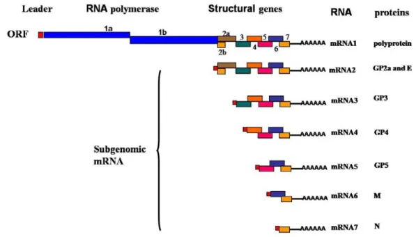
spherical to oval shape with a size ranging from about 50 to 65 nm, a hollow, layered core of around 40 nm diameter and a smooth outer surface studded with a few envelope protein complexes. The structural analysis indicated that the PRRSV core consists of an helical nucleocapsid wrapped up into a hollow ball (Spilman et al., 2009), contrary to previous studies (Benfield et al., 1992; Dea et al., 2000; Doan and Dokland, 2003a; Doan and Dokland, 2003b). These results were not surprising since other members of the Nidovirales, such as Coronavirus, are known to possess a helicoidal capsid (Figure 2). The 5’-capped and 3’-polyadenylated viral genome is approximately 15 kb in length (Meulenberg et al., 1993; Snijder and Meulenberg, 1998; Wootton et al., 2000). The viral genome contains nine known overlapping open reading frames (ORFs), designated ORF1a, ORF1b, ORF2a, ORF2b, and ORFs3 through 7(from the 5’ to 3’end of the genome), which are transcribed into a nested set of subgenomic mRNAs (sg mRNAs) as shown in Figure 3 (Dea et al., 2000; Meulenberg et al., 1993; Wootton et al., 2000; Wu et al., 2001). The replicase-associated genes which occupy approximately 75% of the viral genome, ORF1a and ORF1b, code for polyproteins pp1a and pp1ab by ribosomal frame shifting, and these proteins are directly translated from the incoming genomic viral RNAs (Snijder and Meulenberg, 1998). The pp1a is predicted to be cleaved at eight sites to form nine nonstructural proteins (nsp): nsp1α, nsp1β, and nsp2 to nsp8 (den Boon et al., 1995; Snijder and Meulenberg, 1998). Proteolytic cleavage of the ORF1b portion of the pp1ab generates products of nsp9 through nsp12 (van Dinten et al., 1996). The 13 nonstructural proteins (nsp) are believed to be involved in genome replication and transcription (Bautista et al., 2002; van Dinten et al., 1999). The C-terminus of ORF1a overlaps the N-terminus of ORF1b by 16 nucleotides. A heptanucleotide slippery sequence, UUUAAAC, located just upstream of the UAG stop codon of ORF1a, and a pseudo-knot structure downstream of the slippery sequence is believed to be essential for the expression of ORF1b of PRRSV via a mechanism of ribosomal frame-shifting (Allende et al., 1999; Meulenberg et al., 1993; Nelsen et al., 1999). The 3’ end of the genome (ORFs2 through 7) encodes four glycosylated membrane associated proteins GP2a, GP3, GP4, GP5 (encoded by sg mRNAs 2a, 3-5), two unglycosylated membrane proteins E and M (encoded by sg mRNAs 2b and 6), and a nucleocapsid protein (N) (encoded by sg mRNA 7) (Table 1) (Bautista et al., 1996; Mardassi et al., 1996; Meng et al., 1995a; Meulenberg and Petersen-den Besten, 1996; Meulenberg et
al., 1995; Mounir et al., 1995; Snijder and Meulenberg, 1998; Wu et al., 2001; Wu et al., 2005). Three N-glycosylated minor envelope proteins (GP2a, GP3, and GP4) form heterotrimers by disulfide linkage (Wissink et al., 2005). The nature of GP3 is still controversial, as there are conflicting data regarding its presence as a constituent of the envelope of virus particles. It has been convincingly demonstrated that GP3 is a 45- to 50-kDa structural protein of the PRRSV LV (type I or European) strain (van Nieuwstadt et al., 1996). However, the GP3 has been reported as being a non-structural protein of the PRRSV type II IAF-Klop strain, with a subset of viral GP3 being released into the cell culture medium as a non-virion associated and membrane-free form (Gonin et al., 1998; Mardassi et al., 1998). In the recent years, accumulated data have suggested that GP3 is a structural protein of the PRRSV NA type (Cancel-Tirado et al., 2004; Jiang et al., 2008). Most recently, it was reported that GP3 is a minor structural component of the PRRSV type II (FL12 strain) virion, similar to what has been previously described for PRRSV type I (de Lima et al., 2009). The N protein is not N-glycosylated, although it contains 1 or 2 potential N-glycosylation sites (Meulenberg et al., 1995). All structural proteins are translated from a nested set of 3’-coterminal subgenomic mRNAs, which contain a common leader sequence (Meulenberg et al., 1995; Snijder and Meulenberg, 1998; Wu et al., 2001).
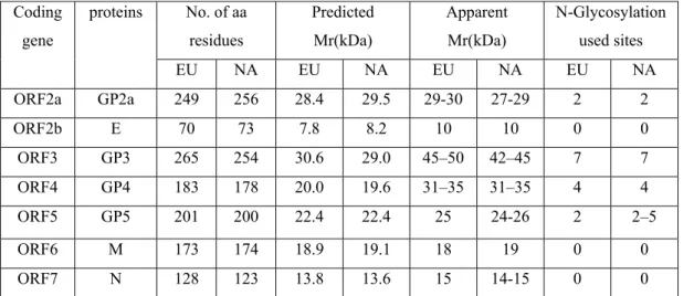
Table I. Comparison of ORFs2 to 7 encoded protein between NA and EU strains of
PRRSV Coding gene proteins No. of aa residues Predicted Mr(kDa) Apparent Mr(kDa) N-Glycosylation used sites EU NA EU NA EU NA EU NA ORF2a GP2a 249 256 28.4 29.5 29-30 27-29 2 2 ORF2b E 70 73 7.8 8.2 10 10 0 0 ORF3 GP3 265 254 30.6 29.0 45–50 42–45 7 7 ORF4 GP4 183 178 20.0 19.6 31–35 31–35 4 4 ORF5 GP5 201 200 22.4 22.4 25 24-26 2 2–5 ORF6 M 173 174 18.9 19.1 18 19 0 0 ORF7 N 128 123 13.8 13.6 15 14-15 0 0
Adapted from Dea S., et al., 2000 with some modifications (Wu et al., 2001). EU: European strains, NA: North American strains, E protein was previously called GP2b
GP5 + M Heterodimer
(Dea et al., 2000; Delpute et al., 2002; Snijder et al., 2003) GP3 : virion structural natur is controversal:
a) Incorporated into virions (Meulenberg et al., 1995), membrane-associated as heterotrimers with GP2a and GP4 (Wissink et al., 2005); a minor structural component of the PRRSV type II (FL12 strain) virion (de Lima, et al 2009) b) nonstructural and secreted from PRRSV infected cells (IAF-Klop strain)
(Mardassi et al., 1998)
E + GP2a-GP3-GP4
Heterotrimer and/or heteromultimeric complex (Wieringa et al., 2004; Wissink et al., 2005)
GP3 GP5 M GP4 GP2a N E ssRNA GP5 + M Heterodimer
(Dea et al., 2000; Delpute et al., 2002; Snijder et al., 2003) GP5 + M
Heterodimer
(Dea et al., 2000; Delpute et al., 2002; Snijder et al., 2003) GP3 : virion structural natur is controversal:
a) Incorporated into virions (Meulenberg et al., 1995), membrane-associated as heterotrimers with GP2a and GP4 (Wissink et al., 2005); a minor structural component of the PRRSV type II (FL12 strain) virion (de Lima, et al 2009) b) nonstructural and secreted from PRRSV infected cells (IAF-Klop strain)
(Mardassi et al., 1998)
GP3 : virion structural natur is controversal:
a) Incorporated into virions (Meulenberg et al., 1995), membrane-associated as heterotrimers with GP2a and GP4 (Wissink et al., 2005); a minor structural component of the PRRSV type II (FL12 strain) virion (de Lima, et al 2009) b) nonstructural and secreted from PRRSV infected cells (IAF-Klop strain)
(Mardassi et al., 1998)
E + GP2a-GP3-GP4
Heterotrimer and/or heteromultimeric complex (Wieringa et al., 2004; Wissink et al., 2005)
E + GP2a-GP3-GP4
Heterotrimer and/or heteromultimeric complex (Wieringa et al., 2004; Wissink et al., 2005)
GP3 GP5 M GP4 GP2a N E GP3 GP5 M GP4 GP2a N E ssRNA
Figure 2. Schematic representation of the virion of PRRSV. The virion is spherical to
oval in shape, enveloped, and possesses a non-segmented single-strand RNA genome that is encapsidated by the nucleocapsid protein (N), yielding a helicoidal capsid structure. (Kindly provided by Nedzad Music with modifications).
Figure 3. PRRSV genomic organizations (Dea et al., 2000; Meulenberg et al., 1993;
3.3. Virus biological and physical properties
Being an enveloped virus, infectivity of PRRSV outside of the host is affected by temperature, pH and exposure to detergents. It has been reported that infectivity of PRRSV was unchanged after 1 month incubation at 4oC or 4 months at -70oC (Benfield et al., 1992). However, the infectivity decreases with increasing temperature. Specifically, infectivity was reduced 50% after incubation for 12 hours at 37oC and was completely inactivated after 48 hours of incubation at 37oC and 45 minutes incubation at 56oC (Benfield et al., 1992). The PRRSV remains stable at pHs ranging from 6.5 to 7.5 (Bloemraad et al., 1994). Detergents are effective at reducing infectivity of the virus and lipid solvents such as chloroform and ether are particularly efficient at disrupting the viral envelope and inactivating the virion (Benfield et al., 1992). The virus survives in water for up to 11 days, but drying quickly inactivates it (Benfield et al., 1999). Buoyant densities of the infectious viral particles are 1.13–1.15 g/ml in sucrose and 1.18–1.19 g/ml in CsCl (Benfield et al., 1992; Mardassi et al., 1994a; Wensvoort, 1993).
3.4. Virus genetic variation
As for other envelope RNA viruses, a high degree of genomic variability has been reported for the Arterivirus (Snijder and Meulenberg, 1998), including PRRSV (Mardassi et al., 1994b; Meng et al., 1995a; Meng et al., 1995b; Murtaugh et al., 1995; Nelsen et al., 1999). Sequence comparisons have shown that there are significant genetic differences between the prototype strains from North America (ATCC VR-2332) and Europe (Lelystad virus - LV) (Meulenberg et al., 1993; Murtaugh et al., 1995; Nelsen et al., 1999), which share only about 63% nucleotide identity (Allende et al., 1999; Nelsen et al., 1999). At the beginning of the global PRRSV epidemic, EU types were detected only in Europe, while US types were restricted to North and Central America (Andreyev et al., 1997) and Asia (Shibata et al., 1996). Now, EU type PRRSV has been found in the US (Fang et al., 2004; Ropp et al., 2004), while US type has been introduced to Europe through the use of a live vaccine (Botner et al., 1999; Botner et al., 1997; Nielsen et al., 2001; Nielsen et al., 2002; Storgaard et al., 1999). Until now, the EU type strain has never been reported in the field in Canada.
phylogenetic analysis places the most recent common ancestor (MRCA) for the EU and US genotypes at at least 100 years back in time (Forsberg, 2005; Hanada et al., 2005; Stadejek et al., 2002), providing strong support for the hypothesis that EU and US viruses evolved in parallel in North America and Europe prior to their cotemporal species jump into pigs and emergence as clinical entities in the later 1980s. Originally, EU genotype viruses were thought to form a very homogeneous, ‘Lelystad-like’ group (Drew et al., 1997; Le Gall et al., 1998; Suarez et al., 1996b; Wensvoort et al., 1991b). Recently, the view that EU genotype viruses are genetically homogeneous was challenged by the studies of unusually diverse EU genotype PRRSV strains, first in Denmark (Oleksiewicz et al., 2000) and later in Italy (Forsberg et al., 2002), the Czech Republic (Indik et al., 2000), Poland (Stadejek et al., 2002), Spain (Mateu et al., 2003), Germany and the Netherlands (Pesch et al., 2005) and even Thailand (Thanawongnuwech et al., 2004a).
Genetic analyses have shown the existence of two major virus genotypes, the EU and the NA, with extensive genetic variability both within and between these genotypes. The leader sequence of PRRSV strains varies significantly. The 190 bp leader sequence of ATCC VR-2332 strain is 31 bp shorter than that of LV, and possesses a sequence identity of 61% with LV (Nelsen et al., 1999). The leader sequence of another NA strain, the 16244B strain, is 189 bp in length and also differs considerably in nucleotide sequence compared to LV (Allende et al., 1999). Like the leader sequence, the ORF1 gene sequence also differs extensively between the U.S. and the European strains (Allende et al., 1999; Nelsen et al., 1999). The ORF1a of ATCC VR-2332 strain shares only about 55% nucleotide sequence identity when compared to LV. ORF1b is more conserved than ORF1a and shares about 63% nucleotide sequence identity compared to LV.
Marked differences were also found between EU and NA isolates in some structural genes (Kapur et al., 1996; Murtaugh et al., 1995). GP5 is the most variable structural protein (Mardassi et al., 1995; Meng et al., 1995b) with the highest degree of diversity within one genotype. Among NA isolates, nucleotide homology of the GP5 coding region was found to be 90% or even less (Andreyev et al., 1997; Dee et al., 2001; Meng, 2000) and from 51 to 59% when NA viruses are compared to LV virus (Andreyev et al., 1997; Kapur et al., 1996; Meng et al., 1994; Murtaugh et al., 1995). N protein encoding region (ORF7) is highly conserved among NA isolates, with 95 to 100% amino acid homology, but a comparison of NA viruses and LV revealed only 57
to 59% amino acid homology (Meng et al., 1995a; Murtaugh et al., 1995). M (Matrix) protein encoding region (ORF6) is the most conserved gene among NA isolates, with 96% to 100% amino acid (aa) identity, and is the most conserved gene between NA and EU isolates, with 70 to 81% identity (Kapur et al., 1996; Meng et al., 1995b; Murtaugh et al., 1995).
Based on sequence analysis, the degree of aa identity amongst the NA PRRSV isolates varied from 91 to 99% for GP2 (GP2a and E), 86 to 98% for GP3, and 92 to 99% for GP4 (Kapur et al., 1996; Mardassi et al., 1995; Meng et al., 1995b; Morozov et al., 1995). A comparison of LV with isolate ATCC VR-2332 revealed aa identities of 63, 76, 58, and 68% for GP2a, E, 3, and 4, (Murtaugh et al., 1995; Ropp et al., 2004). With approximately 54 to 60% aa identity between the NA and EU isolates of PRRSV, GP3 is regarded as the second most variable protein amongst PRRSV strains (Mardassi et al., 1995; Murtaugh et al., 1995), with most of the variations located at the N-terminus. In fact, only 29% aa identity is found within the 35 most N-terminal residues between strains from the two continents. Interestingly, despite these extensive aa changes, the potential N-linked glycosylation sites, as well as the general hydropathy profiles of the ORF3 product, are highly conserved. In addition, the GP3 of the NA strains have a C-terminal deletion of 12 aa compared to LV (Mardassi et al., 1995; Meng et al., 1995b; Morozov et al., 1995). According to a study using UK PRRSV isolates, the ORF3 product has a hydrophilic hypervariable region proximate to the C-terminal region that overlaps with ORF4, resulting in a hypervariable region located at the N-terminal extremity of GP4 (Drew et al., 1997; Katz et al., 1995).
4. Pathogenesis
4.1. Virus entry into susceptible cells
PRRSV cell interactions and how the virus enters the cells were first reported in 1996 (Kreutz and Ackermann, 1996). It was speculated that since the direct fusion of the PRRSV envelope with the cellular membrane was not observed at any time, PRRSV entry most probably occurs by receptor-mediated endocytosis. In 1998, this hypothesis was confirmed (Duan et al., 1998) and a PRRSV receptor was identified on PAM by generation of PAM-specific monoclonal antibodies. Now it is generally
believed that despite the very restricted cell tropism of PRRSV, the virus is able to replicate in several non-permissive cell lines upon transfection of its viral RNA. Cell tropism is determined by the presence or absence of specific receptors on the cell surface or other proteins involved in virus entry (Kreutz, 1998; Meulenberg et al., 1998).
So far, several viral receptor candidates or viral binding proteins for PRRSV have been described, including heparan sulphate for binding and sialoadhesin (CD169) for internalization on macrophages, binding protein vimentin on MARC-145, CD163 on MARC-145 and PAMs and CD151 on MARC-145 (Calvert et al., 2007; Delputte et al., 2002; Kim et al., 2006; Kristiansen et al., 2001; Shanmukhappa et al., 2007; Vanderheijden et al., 2001; Vanderheijden et al., 2003). In addition, a yet unidentified 150 kDa polypeptide doublet and a 210 or 220 kDa glycoprotein, which can be speculated to be sialoadhesin, were found to be involved in PRRSV infection of macrophages (Duan et al., 1998; Wissink et al., 2003). In the current model for PRRSV infection of macrophages, heparan sulfate serves as an attachment factor that binds to viral structural M protein or the M-GP5 complex but is not required for internalization (Delputte et al., 2005; Delputte et al., 2002). Subsequently, PRRSV will engage sialoadhesin in a more stable interaction involving sialic acids present on the virion and the N-terminal sialic acid-binging domain of sialoadhesin, followed by internalization (Delputte et al., 2005; Delputte et al., 2004; Delputte and Nauwynck, 2004; Delputte et al., 2007b; Gorp et al., 2008). Upon internalization, the virus is transported towards an endosomal compartment where a drop in pH is required for proper virus replication (Kreutz and Ackermann, 1996; Nauwynck et al., 1999).
The CD163, a cellular protein in the scavenger receptor cystein rich (SRCR) super family and a type I membrane glycoprotein, has been described functioning as the macrophage receptor for hemoglobin-haptoglobin complex binding and as a cellular receptor for PRRSV infection (Calvert et al., 2007; Kristiansen et al., 2001) . CD163 was also essential in PRRSV infection of MARC-145 cells and rendered many non-permissive cells susceptible to PRRSV infection upon expression (Calvert et al., 2007). Moreover, a more recent research demonstrated that co-expression of recombinant CD163 and sialoadhesin in non-permissive cells increased virus production 10-100 times compared with cells expressing only CD163, sustaining the requirement of both molecules for efficient PRRSV infection (Gorp et al., 2008).
The CD151 molecule was identified by RNA-ligand screening of a MARC-145 cell expression library to be a PRRSV 3' UTR RNA-binding protein (Shanmukhappa et al., 2007). In several RNA viruses, the interaction between 5’ and/or 3’ UTR RNA and host cell proteins was already reported to play an important role in virus replication mechanisms, such as the transcription, translation, orientation and transport of viral RNA (Loffler et al., 1997; Yu and Leibowitz, 1995). The CD151 is a member of the tetraspanin superfamily, which has several cellular functions that include cell signaling, cell activation and platelet aggregation (Fitter et al., 1999; Hasegawa et al., 1998; Sincock et al., 1999). Transfection of CD151 rendered BHK-21, a non-susceptible cell line, non-susceptible to PRRSV infection. The transfection of siRNA against CD151 inhibited PRRSV infection into MARC-145 cells. Additionally, polyclonal anti-CD151 antibody (Ab) completely blocked PRRSV infection into MARC-145 cells (Shanmukhappa et al., 2007). These results suggest that CD151 plays a critical role in PRRSV infection in vitro. Since CD151 is a transmembrane protein, it is reasoned that this molecule serves as the entry molecule (Shanmukhappa et al., 2007).
4.2. Virus transmission
It is known that one of the main characteristics of PRRSV is its high transmissibility, which almost certainly contributed markedly to its quick spread around the world. Pigs are susceptible to infection by a number of routes, including oral, intranasal, intramuscular, intraperitoneal, and vaginal routes (Rossow et al., 1994) (Figure 4).
It is well documented that PRRSV can be transmitted by direct contact, through close ‘snout to snout’ contact between pigs or by contact with organic secretions excreted by infected pigs (Christopher-Hennings et al., 1995; Rossow et al., 1994; Wills et al., 1997a; Wills et al., 1997b; Yoon et al., 1993). In addition, there seems to exist various other indirect ways by which PRRSV can disseminate to a susceptible population, including contaminated fomites (Otake et al., 2002b; Otake et al., 2002c), arthropods (Otake et al., 2002d; Otake et al., 2003) and aerosols (Otake et al., 2002a).
A. Direct transmissions
The PRRSV has been recovered from a variety of porcine secretions and excretions including semen, saliva, feces, and milk and colostrum (Rossow et al.,
1994; Swenson et al., 1994; Wagstrom et al., 2001; Wills et al., 1997b; Yoon et al., 1993). Specifically, infectious PRRSV and PRRSV RNA have been detected in the semen of experimentally infected boars up to 43 and 92 days post-infection, respectively (Christopher-Hennings et al., 1995; Swenson et al., 1994). Fecal shedding remains a highly debated issue; several studies report the presence of PRRSV in feces from 28 to 35 days following experimental infection whereas others report no detection of virus in fecal samples (Wills et al., 1997b; Yoon et al., 1993).
B. Indirect transmissions
Several indirect transmissions by fomites have been identified. Specifically, boots and coveralls have been identified as potential sources of PRRSV transmission to naïve pigs (Otake et al., 2002c). Due to the propensity for PRRSV replication and circulation in the bloodstream, needles have also been recognized as an indirect means of PRRSV transmission between pigs, demonstrating the need for proper needle management (Otake et al., 2003). Mechanical transmission of PRRSV was demonstrated throughout a coordinated sequence of events involving fomites (boots, coolers and containers, shipping parcels, vehicles) (Dee et al., 2003; Dee et al., 2002). However, studies have demonstrated that certain intervention strategies, such as the use of disposable footwear, boot baths, the wearing of gloves and double-bagging products designated for entry into farms significantly reduced the level of PRRSV contamination on the surface of objects and mechanical spread of the virus (Dee et al., 2004).
Even if PRRSV is relatively fragile in the environment, appropriate weather (wind, temperature, humidity etc) may favour the transmission of the virus trough aerosols up to 4.7 km distance (Dee et al., 2009) .
Insects (mosquitoes (Aedes vexans) and houseflies (Musca domestica)) are commonly observed in swine facilities during the summer months and have been shown to mechanically transmit PRRSV from infected to naïve pigs under experimental conditions (Otake et al., 2002d; Otake et al., 2003). Transport of PRRSV by insects throughout an agricultural area has been reported for up to 2.4 km following contact with an infected pig population (Schurrer et al., 2004). However, the significance of these vectors under field conditions, i.e. in commercial pig farms, still needs to be determined.
Previous studies investigated the role of various mammals (rodents, raccoons, dogs, cats, opossums, skunks) and birds (house sparrows and starlings) in the
transmission of PRRSV(Wills et al., 2000b); none were capable of serving as mechanical or biological vectors (Wills et al., 2000b).
Figure 4. Pathogenesis of PRRSV infection (adapted from www.porcilis-prrs.com)
4.3. PRRSV cell and tissue tropism
PRRSV is generally believed to have a very restricted cell tropism both in vivo and in vitro. In vivo, the virus mainly infects well-differentiated cells of the monocyte-macrophage lineage, in particular porcine alveolar monocyte-macrophages (PAM), the primary target cells of virus and interstitial macrophages in other tissues such as heart, thymus, spleen and Peyer's patches, hepatic sinusoids, renal medullary interstitium, and adrenal gland (Beyer et al., 2000; Duan et al., 1997; Halbur et al., 1996a; Halbur et al., 1995b). In addition to macrophages, PRRSV RNA and nucleocapsid protein were found by in situ hybridization (ISH) in testicular germ cells, endothelial cells in the heart, interdigitating cells in the thymus, dendritic cells in the spleen and Peyer's patches (Halbur et al., 1996a; Sur et al., 1997). In experimentally infected gnotobiotic pigs, PRRSV antigen were found in bronchiolar epithelial cells, arteriolar endothelial cells, monocytes as well as interstitial, alveolar, and intravascular macrophages using an
immnogold-silver immunohistochemical staining (Rossow et al., 1996). The antigens and RNA of European, North American and Korean strains of PRRSV were found in bronchiolar epithelial cells (Pol et al., 1991), epithelium-like cells of alveolar ducts (Magar et al., 1993), and pneumocytes (Cheon et al., 1997; Pol et al., 1991) in the naturally infected pigs, whereas they were not found in these types of cells in the experimentally infected pigs (Teifke et al., 2001). Tissues such as lung, lymphoid tissues, Peyer’s patches, and kidney were also the preferred organ targets of PRRSV infection (Haynes et al., 1997; Sur et al., 1996). Studies showed that PRRSV distribution was also isolate or strain-dependent, for instance, more virulent strains had more positive PRRSV cell distribution in more tissues and organs (Haynes et al., 1997). It has been reported that PRRSV can be isolated from the ovary and may be responsible for episodes of female reproductive failure, and PRRSV antigens & RNAs were detected in ovarian follicles in gilts as well (Collins et al., 1992; Kranker et al., 1998; Prieto et al., 1996; Prieto et al., 1997a; Prieto et al., 1997b; Prieto, 1996; Sur et al., 2001).
In vitro, PRRSV can grow in a few cell lines. So far, primary cultures of PAMs as well as freshly isolated blood monocytes or monocytic derived dendritic cells (Voicu et al., 1994; Wang et al., 2007; Wensvoort et al., 1991b) are known to be the only porcine cells that can effectively be used for viral growth. Other non-porcine permissive immortalized cell lines that permit the complete replication cycle of PRRSV are African green monkey kidney cells or derivatives thereof such as MARC-145 or CL2621 (Bautista et al., 1993; Benfield et al., 1992; Kim et al., 1993).
4.4. PRRSV-induced apoptosis
Apoptosis, necrosis/oncosis, autophagy and pyroptosis are now generally recognized distinct processes leading to eukaryotic cell death, with clearly distinguishable morphological and biochemical features (Bergsbaken et al., 2009; Fink and Cookson, 2007; Labbe and Saleh, 2008; Wyllie et al., 1980). However, apoptosis and necrosis/oncosis are better-recognized molecular mechanisms of eukaryotic cell death and can simultaneously occur in tissue or cells exposed to the same stimuli (Shimizu et al., 1996). Whether a cell undergoes apoptosis or not depends on a delicate balance of anti- and pro-apoptotic stimuli (Costers et al., 2008). Mechanistically, apoptosis results from the activity of a distinct subset of caspases (cysteine-dependent
aspartate-specific proteases). Initiator caspases are activated primarily by two mechanisms (Green, 2003): 1) ligation of cell surface death receptors, including the tumor necrosis factor-alpha (TNF-α) receptor and Fas, leads to caspase-8 activation via the extrinsic pathway, and 2) mitochondrial release of cytochrome c activates caspase-9 via the intrinsic pathway. The pathways cross-talk, as caspase-8 can promote cytochrome C release and caspase-9 activation. Both caspase-8 and caspase- 9 activate executioner caspases, including caspase-3, which cleave cellular substrates to produce the features associated with apoptosis. These characteristics include cytoplasmic and nuclear condensation, oligonucleosomal DNA cleavage and maintenance of an intact plasma membrane. Apoptotic cells package their contents into membrane-bound apoptotic bodies and expose surface molecules like phosphatidylserine to target phagocytic uptake and removal such that apoptosis is generally non-inflammatory in vivo (Elmore, 2007; Fink and Cookson, 2007). Apoptosis is considered to be an important host defense mechanism that interrupts viral replication and eliminates virus-infected cells (Thomson, 2001). Apoptosis can be induced in virus-infected cells by host immune cells, such as cytotoxic T-lymphocytes and natural killer cells through soluble factors or through direct cell-to-cell contact. In addition, apoptosis can be induced in virus-infected cells as a response to viral replication (Matsumoto et al., 2005; Tanaka et al., 1998). Induction of apoptosis before completion of viral replication would severely limit progeny virus production and virus spread in the host. Consequently, viruses have evolved strategies that inhibit apoptosis during replication, thereby ensuring cell survival until sufficient virus progeny is produced (Teodoro and Branton, 1997; Thomson, 2001). Many viruses have adapted by encoding anti-apoptotic gene products that permit their seemingly undetected replication. Some viruses (e.g., African Swine Fever Virus, ASFV) encode proteins that prevent apoptosis through inactivation of p53 or binding of Bax (Afonso et al., 1996; Brun et al., 1996; Neilan et al., 1993; Revilla et al., 1997; Young et al., 1997). Yet other viruses (such as Baculovirus) possess mechanisms to inhibit apoptosis by expressing caspase inhibitors that interfere with caspase function (Manji and Friesen, 2001). Thus, viruses have evolved numerous mechanisms acting at many different targets to interfere and block apoptosis.
Several studies demonstrated that PRRSV infection induced apoptosis both in vitro and in vivo (Choi and Chae, 2002; Kim et al., 2002; Labarque et al., 2003; Miller
and Fox, 2004; Sirinarumitr et al., 1998; Suarez et al., 1996a; Sur et al., 1997; Sur et al., 1998). PRRSV infection of MARC-145 and porcine alveolar macrophages (PAMs) resulted in apoptosis characterized by morphological changes, DNA fragmentation and specific caspase activation. PRRSV induces apoptosis both directly (in infected cells) and indirectly (in bystander cells) and within both infected tissue cultured cells and animals, apoptotic cells are observed and contribute to the pathology observed in the animal (Sirinarumitr et al., 1998).
The evidence of direct induction of apoptosis coming from in vitro studies demonstrated that artificial expression of GP5 using viral vectors can induce apoptosis within cell monolayers, but the mechanism was not well elucidated (Gagnon et al., 2003; Suarez et al., 1996a). Expression of PRRSV GP5 (ORF5) gene in cell monolayers using a vaccinia virus expression vector induced apoptosis, while the vaccinia vector alone did not (Suarez et al., 1996a). Gagnon et al. (2003) expressed the GP5 protein using an adenovirus expression system and detected an increase in caspase 3 activity in cell monolayers transfected with recombinant vector (Gagnon et al., 2003). The apoptosis inducing region of GP5 has been mapped to the N-terminal 119 amino acids by Fernandez et al. (Fernandez et al., 2002). Recently, the question of if and when PRRSV modulates apoptosis in PRRSV-infected macrophages was investigated (Costers et al., 2008). This study showed that during a PRRSV infection two oppositely directed sets of reactions are switched on in PRRSV-infected macrophages in vitro: at first, reaction favor anti-apoptosis, but finally, PRRSV-infected macrophages die by apoptosis (Costers et al., 2008). Both anti- and pro-apoptotic effects were not only observed in PRRSV-infected macrophages, but also in PRRSV-infected MARC-145 cells (Costers et al., 2008). In conclusion, this study showed that PRRSV replication results in activation of anti- and pro-apoptotic pathways. Early in infection, the balance tends towards anti-apoptosis, whereas late in infection, the balance is driven towards pro-apoptosis. In addition, this study indicates that the ability of PRRSV to modulate apoptosis in the infected cell is intrinsic to the virus, and not dependent on the cell type (Costers et al., 2008). Lee et al. (2007) provided further evidence of apoptosis induced by PRRSV directly in PRRSV-infected MARC-145 cells and ultimately, the authors elucidated that PRRSV induced apoptosis is through a mitochondria-mediated pathway. In summary, these authors demonstrated that (i) PRRSV infection causes characteristic morphological and biochemical changes of apoptosis such as chromatin condensation, DNA fragmentation, externalization of
phosphatidylserine (PS), caspase activation, and poly (ADP-ribose) polymerase (PARP) cleavage; (ii) PRRSV induces caspase-dependent apoptosis and activates both caspase-8 and caspase-9; (iii) a crosstalk between extrinsic and intrinsic pathways existed since caspase-8 activated through ligation of the death ligand with the death receptor, possibly TNFR-α/TNFR1 and Fas/FasL, and mediates caspase-9 activation via Bid cleavage; (iv) PRRSV caused an increased ratio of Bax/Bcl-2 which is followed by the disruption of the mitochondrial transmembrane potential and cytochrome c release; (v) oxidative stress induced by PRRSV is involved in apoptosis; and (vi) PRRSV infection causes secondary necrosis (Lee and Kleiboeker, 2007).
Other studies have shown that PRRSV induces apoptosis mostly in uninfected bystander cells both in vitro and in vivo. In previous studies, the majority of apoptotic cells were detected in lung lavages, lungs, and lymphoid tissues by using IHC, TUNEL, DNA electrophoresis, and electron microscopy, but upon performing dual-labeling experiments it was concluded that the majority of apoptotic cells were not infected with PRRSV (Choi and Chae, 2002; Kim et al., 2002; Labarque et al., 2003; Miller and Fox, 2004; Sirinarumitr et al., 1998; Suarez et al., 1996a; Sur et al., 1997; Sur et al., 1998). Chang and collaborators have reported that alveolar macrophages from PRRSV-infected pigs showed a significantly increased apoptotic rate (22-34%) compared to porcine circovirus 2 infected alveolar macrophages (3%) (Chang et al., 2005). Given the fact that only 5-10% of alveolar macrophages were PRRSV-infected, theses authors suggested that TNF-α or GP5 released from PRRSV-infected cells caused apoptosis in bystander cells. Another study from the same group demonstrated that increased FasL expression in PRRSV-infected macrophages caused apoptosis in co-cultured swine splenic lymphocytes (Chang et al., 2007).
5. Host immunology
A complex immunological interaction exists between PRRSV and pigs that involves both induction and subversion of host defenses (Murtaugh et al., 2002). Immunization to PRRSV infection is a double-edged sword. On the one hand, PRRSV has a predilection for immune cells and the disease manifestations can be linked directly to changes in the immune system. PRRSV appears to replicate exclusively in cells of the immune lineage, notably macrophages; the direct replication of which may
lead to immunosuppression, precipitate secondary infection and/or mediate disease. On the other hand, the virus stimulates immunity post-infection that protects an animal from re-infection. Thus, the immune system appears to be intimately involved in both the disease process and protection from disease (Molitor et al., 1997). PRRS is one of the most challenging subjects of research in veterinary viral immunology and our current knowledge on the basic mechanisms for PRRSV protective immunity is still fragmentary.
Because porcine alveolar macrophages (PAMs) have a pivotal role for an effective innate and adaptive immune response (Janeway et al., 2005a; Janeway et al., 2005b), any suppression of macrophage function plus a fall in macrophage numbers due to viral infection could increase the host’s susceptibility to secondary infections. For the innate immune response, the main role of PAMs is to ingest and subsequently kill pathogens, and release cytokines such as TNTα and IL-1, which can activate pathways of both the innate and adaptive immune response (Fearon and Locksley, 1996; Janeway et al., 2005a; Janeway et al., 2005b). By acting as antigen-presenting cells, macrophages, along with dendritic cells (DCs), also trigger the adaptive immune response (Janeway et al., 2005a). PRRSV can also multiply inside DCs, which can affect the activation of these cells and prevent triggering of the adaptive immune response (Loving et al., 2007; Wang et al., 2007). Several studies have also shown weak and atypical innate immune responses, such as weak IFN-α responses (van Reeth, 1999) and high induction of interleukin(IL)-10 in PRRSV-infected porcine monocytes, macrophages and dendritic cell (Flores-Mendoza et al., 2008; Suradhat et al., 2003) and in vivo in PRRSV-infected pigs (Suradhat, 2003; Sutherland et al., 2007; Thanawongnuwech et al., 2004a; Thanawongnuwech et al., 2004b)
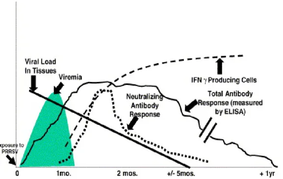
Pigs mount a rapid antibody response to infection by PRRSV, which is detectable from day 5 post-infection, but these early antibodies are mainly directed to the N- and M-proteins and are non-neutralizing. Neutralizing antibodies (NAbs) begin to appear between 7-28 days post-infection, but their titers remain low, and substantial variation in the neutralizing antibody response has been reported (Loemba et al., 1996; Plagemann, 2006). Typical titers of serum NAbs, which are considered unusually low in comparison with those induced by other viruses, are between 2 and 12 (Labarque et al., 2000; Loemba et al., 1996). Thus the humoral immune response to PRRSV in pigs is characterized by early production of strong, non-NAbs, which are detected from 5-6
days post-infection (pi), followed by the delayed appearance of neutralizing antibodies between 3 and 4 weeks post-infection, which then persist at low levels (Figure 5) (Lopez and Osorio, 2004).
The protective capacity of NAbs is debated. The early development of non-NAbs and later development of non-NAbs may have a significant effect on the development of PRRSV persistent infections. It has been shown that non-NAbs enhance viral replication in alveolar macrophages, a phenomenon known as antibody-dependent enhancement (ADE) (Yoon et al., 1996; Yoon et al., 1997). The non-neutralizing humoral response may act as a Trojan horse for PRRSV by coating the virus and enhancing the internalization of viral particles into macrophages (Mateu and Diaz, 2008). In contrast, development of NAbs is not sufficient to completely eliminate the virus (Mateu and Diaz, 2008). Likewise, viremia may be resolved in the absence of detectable levels of neutralizing antibodies (Diaz et al., 2006). Nonetheless, NAbs may play a central role in protecting swine against reinfection with PRRSV since passive transfer of antibodies fully protected pregnant sows against a challenge of virulent PRRSV and blocked transplacental infection (Osorio et al., 2002). Similarly, pigs receiving an amount of NAbs sufficient to reach a serum titer of 8 consistently did not develop viremia, whereas serum titers of 32 produced sterilizing immunity (Lopez and Osorio, 2004; Osorio et al., 2002). However, other authors do not report such a strong correlation between NAbs and the absence of viremia (Jiang et al., 2007a; Jiang et al., 2007b; Jiang et al., 2007c; Plagemann, 2006; Zuckermann et al., 2007). Delay in the neutralizing antibody response to PRRSV has been postulated (in addition to other hypotheses) to be due to the presence of a nearby immunodominant “decoy” epitope (aa 27–30 of GP5), which may evoke a robust, early, and non-protective immune response that masks and/or impairs the response to the major neutralizing epitope (aa 37-45 of GP5) (Ostrowski et al., 2002). An alternative explanation of the peculiar nature of the PRRSV-neutralizing response could be a so-called glycan-shielding phenomenon as proposed for the human and simian immunodeficiency viruses. “Glycan shielding” may be a primary mechanism to explain evasion from NAbs, ensuring in vivo persistence of these viruses (Wei et al., 2003). Also, observations using field strains of PRRSV appear to support the role of N-linked glycosylation sites in interfering with the neutralizing antibody response. Spanish PRRSV strains have evolved from 1991 to 2005 and there has been a trend to gain two glycosylation sites in N37 (Asp) and N53 (Asp) flanking the major
neutralizing epitope of GP5 compared to LV strain, consistent with selection of strains inducing weaker NAb responses (Mateu et al., 2006). Together, these findings support the suggestion that natural infection with PRRSV may involve an immune evasion strategy in which few NAbs are produced, and/or large amounts of PRRSV NAbs in sera of PRRSV-infected animals may be unable to react with virions due to blocking or shielding of the neutralizing epitope by the glycan moieties on GP5 (Ansari et al., 2006) .
Cell-mediated immune (CMI) response to PRRSV determined by lymphocyte blastogenesis and adaptive cytokine production is delayed, primarily detectable in the in vitro recall response of PBMC around 1-2 weeks after infection (Bassaganya-Riera et al., 2004; Bautista and Molitor, 1997; Charerntantanakul et al., 2006; Lopez Fuertes et al., 1999; Meier et al., 2003; Royaee et al., 2004). Infection with PRRSV has been shown to increase the numbers of various peripheral blood mononuclear leukocyte subsets (Albina et al., 1998b; Diaz et al., 2005), beginning with an increase in CD8α+ cells, 1week after infection, followed by an increase in the numbers of CD4+ and γδ T cells, 7 weeks post-infection. The latter was shown to coincide with an increase in the number of interferon-γ (INF-γ) producing cells in the peripheral blood (Batista et al., 2004), an indicator for proliferation of cytotoxic cells. Over the same period, the number of effector cells specific for PRRSV was shown to increase, reaching maximal levels at 7 weeks post-infection (Bautista and Molitor, 1997). Protective immunity against PRRSV is not clearly understood. Various studies suggest that the cell-mediated immune response is not sufficient to completely eliminate the virus and to prevent persistent infection (Batista et al., 2004; Lowe et al., 2005; Murtaugh et al., 2002). A delay in the appearance of the cellular immune response suggests that PRRSV infection involves a mechanism of immunosuppression or immunomodulation (Done, 1995; Murtaugh et al., 2002).
Figure 5. Temporal sequence of events after infection of a pig with PRRSV (Adapted
from Lopez and Osorio, 2004).
6. Disease control and eradication 6.1. Control and eradication strategies
The rapid spread and economic impact of PRRS have made it a frequent topic of research, especially regarding its control (Neumann et al., 2005; Zimmerman et al., 1997). The key elements of a PRRS control and eradication program are early disease detection and rapid laboratory confirmation; quick identification of the infected farms and control of the infection through different stamping out strategies. As with many other infectious diseases, the most effective means of control often depends on the use of vaccines as well as the implementation of improved management practices. Regarding the first option, there are currently a few commercially available vaccines. These include modified live virus (MLV) as well as inactivated-virus or killed virus (KV) vaccines. They are all made from cell culture of MARC-145 since MARC-145 cells have been the most convenient for vaccine production up until now. However, the nature of the pig’s immune response to PRRSV makes the development of an unquestionably safe as well as highly effective vaccine a formidable challenge.
Consequently, in many affected herds, the development of strategies for control and perhaps eventual eradication of PRRS depends on a thorough knowledge of the epidemiology of the disease and vaccination is only one of several approaches to be considered in designing a control strategy (Prieto and Castro, 2005).
Various control programs have been developed to eliminate the virus from infected farms, but no single program is satisfactory for controlling it in all types of herds. Programs including partial depopulation (Dee et al., 1997), segregated early weaning (Rajic et al., 2001), vaccination with nursery depopulation (Dee et al., 1998), and test and removal (T&R) (Dee and Molitor, 1998; Dee et al., 2000) have been described, and the T&R technique has been applied successfully to some herds.
Vaccination and/or partial or total depopulation strategies, test and removal procedures or acclimatization of incoming pigs has proven efficient in the eradication of PRRS (Dee et al., 2000; Yang et al., 2008). Partial or total depopulation is used as an eradication strategy in many farms (Dee and Joo, 1997). PRRSV was efficiently eliminated from a seedstock breeding farm and a supplying boar stud by a modified test and removal method based on an indirect fluorescent assay (IFA) test to detect antibodies (Dee and Molitor, 1998), and a nested reverse transcriptase-PCR (nRT-PCR) to detect virus nucleic acids (Yang et al., 2008).
Central to the control of PRRS is prevention of the spread of PRRSV within the pig herd. The herd should be stable with a uniform level of immunity throughout the herd, with no PRRSV-negative pigs. In breeding herds, the modified live vaccines have been used as an aid to creating this uniform immunity. Clinical symptoms are reduced and the infection of piglets prior to weaning is prevented (Lopez and Osorio, 2004).
Limitations of T&R have been documented (Dee and Molitor, 1998; Dee et al., 2000), and include a high degree of labor involved in testing an entire herd, and diagnostic costs that approach US $10.00/tested sow. Furthermore, a high accurate test is required to reduce the impact of animal removal on the productivity and profitability of the farm (Dee and Molitor, 1998; Dee et al., 2000). Depopulation is expensive and it is only effective if strict biosecurity is applied and if all the pig farms in the affected region are following the same strategy. Therefore, a combination of depopulation and vaccination is an interesting alternative option for control.
6.2. Treatments and prevention
In the acute disease phase, when PRRSV first enters the farm it is important to cover the period at risk, which is usually six to eight weeks, with in-feed antibiotics and water medication. The broad-spectrum antibiotics, tetracyclines, trimethoprim/sulpha, or synthetic penicillins are the medication of choice to treat secondary infections.
Vaccination is a common procedure to minimize economic losses associated with this pathogen and to prevent reinfection, and vaccines have been proven to be effective in experimental trials (Opriessnig et al., 2005) and field studies (in a preferentially homologous rather than heterologous PRRSV strain-specific manner) (Mavromatis et al., 1999; Sornsen et al., 1998). Nonetheless, the efficacy of these currently used vaccines is somewhat controversial and it is generally well accepted that there is a need for improvement in their safety and efficacy. MLV vaccines are still able to cause viremia and thus can spread to other pigs, as reported in Denmark (Botner et al., 1997; Madsen et al., 1998). The MLV vaccines do not prevent reinfection, and even the field virus does not induce a lifelong immunity. Furthermore, MLV vaccines do not allow serological discrimination between vaccinated and naturally infected pigs. The efficacy of PRRSV KV vaccines is less than ideal. The vaccines induce poor CMI response and do not induce an antibody response (measured by IDEXX ELISA) (Bassaganya-Riera et al., 2004; Piras et al., 2005; Zuckermann et al., 2007). Furthermore, due to the PRRSV genetic diversity and quasispecies (Goldberg et al., 2003; Rowland et al., 1999), PRRSV vaccine failures are not uncommon in the field, and vaccine efficacy is far from being universal and complete. Likewise, due to the continuous mutations affecting the viral genome, the PRRSV has the ability to persist in herds for long periods of time (Allende et al., 2000; Goldberg et al., 2003). This persistence and variability pose serious challenges for the diagnosis and control of PRRSV that might be further complicated by reversion of live vaccine viruses into the ancestor wild-type virus and recombination of viruses in the field (Botner et al., 1997; Meng, 2000; Nielsen et al., 2001; Storgaard et al., 1999). Overall, the characteristics of PRRSV increase the difficulties of PRRS prevention.