UNIVERSITÉ DE LILLE
FACULTÉ DE MÉDECINE HENRI WAREMBOURG
Année :2018
T H È S E P O U R L E D I P L Ô M E D ' É T A T D E D O C T E U R E N M É D E C I N E
Cécité post opératoire: Description d’un cas d’amaurose unilatérale après
chirurgie orthognatique
Présentée et soutenue publiquement le 14 décembre 2018 à 18h
au Pôle Recherche
Par Constance DELMOTTE
_______________
JURY
Président :
Monsieur le Professeur FERRI Joël
Assesseurs :
Monsieur le Professeur RAOUL Gwenaël
Monsieur le Docteur NICOT Romain
Monsieur le Docteur LEJEUNE Vincent
Directeur de Thèse :
AVERTISSEMENT
La Faculté n'entend donner aucune approbation aux opinions émises dans les thèses : cel-les-ci sont propres à leurs auteur(e)s.
LISTE DES ABRÉVIATIONS
NOIP: neuropathie optique ischémique postérieure
LIST OF ABBREVIATIONS
POVL: Post Operative Visual Loss
PION: posterior ischemic optic neuropathy CRAO: central retinal artery occlusion CT: Computed tomography
TABLES DES MATIÈRES
RÉSUMÉ ... 14 INTRODUCTION GÉNÉRALE ... 15 ARTICLE……… 17 ABSTRACT ... 18 INTRODUCTION ... 19 PRESENTATION OF CASE ……….21 DISCUSSION ... 24 CONCLUSION……….. 31 REFERENCES ... 32 DISCUSSION GÉNÉRALE ……….. 34 CONCLUSION GÉNÉRALE ... 38RÉSUMÉ
Introduction
Dans le cadre de la chirurgie orthognatique, une cécité post opératoire peut être la consé-quence du geste chirurgical lui même ou de multiples facteurs induit par l’anesthésie générale. Néanmoins, les mécanismes physiopathologiques exactes sont encore mal connus. L’objectif est ici de présenter un cas de cécité post chirurgie orthognatique suivit d’une revue de la littéra-ture sur le sujet, relativement rare.
Description de cas
Nous décrivons ici le cas d’un patient qui présentait une amaurose unilatérale avec paresthésie frontale homolatérale dans les suites immédiates d’une ostéotomie bimaxillaire. Il souffrait très probablement d’une hypertension non diagnostiquée, et en per-opératoire, il a souffert d’une intense bradycardie, ainsi que de pertes sanguines importantes, ayant nécessité un remplis-sage vasculaire par des solutions colloïdes et cristalloïdes. L’ensemble du bilan post opératoire concluait à une neuropathie optique ischémique postérieure (NOIP).
INTRODUCTION GÉNÉRALE
La cécité post opératoire est une complication heureusement rare mais grave. Elle peut, bien sur, survenir dans le cadre d’une chirurgie ophtalmologique, mais également dans les suites de chirurgie non-oculaire. Dans ce second cas, les causes les plus fréquentes sont les suivantes: neuropathie ischémique optique antérieure, neuropathie ischémique optique postérieure (NOIP), occlusion de l’artère centrale de la rétine, occlusion de la veine centrale de la rétine, apoplexie hypophysaire, cécité corticale, ou compression directe sur les voies optiques. La cause la plus fréquente est la NOIP, elle se caractérise par une diminution soudaine de l’acuité visuelle, non douloureuse, sans anomalie constatée sur l’évaluation immédiate (examen oph-talmologique, fond d’oeil, scanner et IRM). Une pâleur du disque optique apparait au fond d’oeil après plusieurs semaines. La NOIP reste néanmoins un diagnostic d’exclusion, même si cer-tains facteurs de risques ont pu être identifiés: temps opératoire prolongé, anémie, hypotension per-opératoire, facteurs de risques cardiovasculaire (diabète, obésité, sexe masculin), hémor-ragie per-opératoire majeure, et administration massive de colloïdes.
La chirurgie orthognatique est aujourd’hui reconnue comme étant une chirurgie sure et repro-ductible, grâce à une amélioration des connaissances en anatomie faciale et à une évolution des traitements orthodontiques ces trente dernières années. Initialement, la chirurgie orthogna-tique était surtout dévolue à la prise en charge des dysmorphoses et malocclusions dans les jeunes populations. Mais avec le développement de celle-ci, elle concerne aussi désormais des populations plus âgées, dans le cadre de la prise en charge des dysfonctions temporomandibu-laires et de la reconstruction pré-prothétique, ce qui engendre donc plus de risques chirurgicaux ou anesthésiques.
L’apparition d’une cécité dans les suites d’une chirurgie orthognatique peut être la conséquence de la chirurgie elle même (fracture irradiée à la base du crâne, hématome intraorbitaire,
contraintes exercées sur le contenue intra orbitaire pendant la procédure), ou de facteurs in-duits par anesthésie générale, surtout lorsque les patients présentant des comorbiditées. Quatorze cas de cécité post chirurgie orthognatique on été décrit dans la littérature. Les méca-nismes physiopathologiques exactes de l’implication anesthésique et chirurgicale dans cette pathologie ne sont pas connus. Le but ici est de décrire un cas observé dans le service, et de faire une revue de la littérature sur ce sujet et sur les potentiels mécanismes physiopatholo-giques incriminés.
ARTICLE
POSTOPERATIVE VISUAL LOSS: A REPORT OF ONE PATIENT WITH UNILATERAL BLINDNESS AFTER ORTHOGNATIC SURGERY
ABSTRACT
Introduction
Blindness after orthognatic surgery may be the result of the surgical procedure itself or the consequence of factors induced by general anesthesia. However, the exact
mechanism between is not known. The purpose of this article is to present a case of a postoperative visual loss (POVL) after orthognatic surgery under general anesthesia concluding with a brief literature review about this topic.
Report of case
We report the case of a patient who suffered unilateral blindness with homolateral frontal paresthesia after orthognatic procedure in two steps. He presented
intraoperative bradycardia with a potential undiagnosed hypertension, associated with significant blood loss and volume resuscitation by colloids and cristalloids.
Postoperative examination concluded to posterior ischemic optic neuropathy (PION).
Discussion-Conclusion
By a systematic literature review, we discuss about surgical and anesthesic causes of POVL, and particularly pathophysiology mechanism of PION. Some predisposition and risk factors have been identified and need to be taken into account.
INTRODUCTION
Postoperative visual loss (POVL) is a rare but severe complication. Vision loss occurring after non-ocular surgery under general anesthesia most frequently results from one of the following etiologies: anterior ischemic optic neuropathy, posterior ischemic optic neuropathy (PION), cen-tral retinal artery occlusion (CRAO), cencen-tral retinal vein occlusion, pituitary apoplexy, cortical blindness, or direct compression (1). The most common cause is PION, characterized by sud-den painless visual loss, associated with normal immediate ophthalmic evaluation, computed tomography (CT) and magnetic resonance imaging (MRI). Disc pallor will appear on the fundus examination a few weeks later (2). It is a diagnosis of exclusion although some risk factors have been identified, such as prolonged operative time, anemia, intraoperative hypotension, dia-betes, obesity, male sex, major intraoperative blood loss, microvascular pathology and massive colloid administration during the procedure (1,3,4).
Orthognathic surgery is recognized as a safe and predictable procedure thanks to an increasing knowledge about facial anatomy and progress in orthodontics treatment over the last thirty years (5,6). Initially, maxillofacial orthopedic surgery was intended for the treatment of maloc-clusion in younger populations; however, with the development of this surgery, older populations are now receiving support for temporomandibular joint pain or pre-prosthetic reconstruction. This change creates an added surgical or anesthetic risk linked to the patients’ age. Blindness after orthognathic surgery may be the result of the surgical procedure itself when basal skull fracture, orbital hematoma, or constraint on intraorbital structures are observed (5,7–11). Howe-ver, it may be the consequence of factors induced by general anesthesia, especially when pa-tients have comorbidities. Fourteen cases of postoperative blindness after Le Fort I
osteotomy were described in the literature (7–15). The exact mechanism of anaesthesia and surgery implication is not known.
Herein, we report a case of a POVL after orthognathic surgery procedure under general anaes-thesia and conclude with a brief literature review about this topic aimed at describing the physio-logical mechanism involved.
PRESENTATION OF CASE
A 53-year-old male patient sought consultation in the department of oral and maxillofacial surge-ry of the University Hospital of Lille (France). He presented bilateral temporomandibular joint pain due to class III malocclusion with transverse maxillary discrepancy requiring an orthogna-thic surgery.
He weighed 71 kg and was 1.78 m tall (BMI, 22.4 kg/m2). His medical past history was
charac-terized by chronic headaches, diverticulitis, malaise of vasovagal origin and right cholesteato-ma. He presented with generalized periodontal disease, which was exacerbated by smoking and malocclusion. Preoperative evaluation found blood pressure at 140/80, heart and lungs examination without abnormalities, including electrocardiogram (ECG) and chest radiography (CXR).
The surgery was performed in two stages. First, a surgically assisted rapid palatal expansion was completed. Ten months later, a bimaxillary orthognathic surgery was performed with a Le Fort I osteotomy with initial advancement followed by a sagittal split osteotomy of the mandible. For this second procedure, induction of anesthesia was uneventful. Few minutes later, the pa-tient’s blood pressure was 160/80 mmHg, which then decreased to 80/50 mmHg. Volume ex-pansion was introduced by crystalloids (500 mL). After that and before the beginning of the pro-cedure, the patient presented with bradycardia at 44 bpm that was resolved by an injection of atropine (0.01 mg/kg).
The procedure began with a maxillary down-fracture with pterygomaxillary disjunction using a curved Obwegeser osteotome and realized without difficulties or injuries. We noticed bleeding
originating from the right maxillary artery, but hemostasis was immediately achieved. Then, we performed bilateral sagittal split mandibular osteotomies. The mean arterial pressure (MAP) va-lues ranged from 60 to 90 mmHg during the entire surgery. The procedure was performed over 225 min, and the intraoperative blood loss was estimated at 1000 mL. No intraoperative inci-dents were registered. A perfusion solution of 500 ml of hydroxyethylamidon was administered intravenously with 2 injections of an antifibrinolytic drug, tranexamic acid (0.014 g/kg for 1 h fol-lowed by 0.014 g/kg for 8 h). Postoperative hemoglobin was evaluated at 13.7 g/dL.
In the recovery room, the patient complained of complete visual loss of the left eye. Eye exami-nation revealed totally impaired vision associated with complete abolition of direct light reflex, while the fundus was normal. The CT scan only showed normal jaw osteotomies (Figure 1), and no irradiated split on the skull base.
administration of medication, he complained of left frontal paresthesia. Cerebral and orbital MRI showed a slight flair hypersignal in the right internal occipital cortex and in the insular right pa-renchyma. No signal abnormality of the optic nerve was visualized. Antihypertensive treatment with nicardipine was introduced to decrease the elevated blood pressure during the first post-operative night.
The next day, a complete ophthalmologic examination was conducted. On the left side, the light perception and direct light reflex were missing, but the consensual light reflex was maintained. The fundus examination highlighted a clear optic nerve. Visual evoked potentials showed ab-sence of left eye reactivity. The diagnosis was reviewed and PION of the left eye was retained.
Polygon CT angiography showed normal permeability of the ophthalmic arteries. The carotid doppler showed normal results. Fundal angiography was also normal. After six months, fundal examination revealed disc pallor on the left eye. At the final follow-up, the visual acuity of the left eye was 2/10.
DISCUSSION
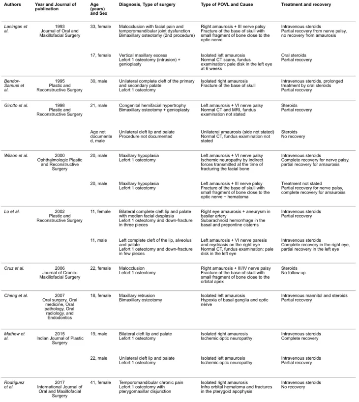
Orthognathic surgery is now recognized as a safe procedure (5,6) with several minor complica-tions: infection, excessive bleeding, bad split, tooth and inferior alveolar nerve injury (5,10). Se-vere complications such as condylar resorption, osteonecrosis, cerebrovascular accident, and carotid-cavernous sinus fistula were reported in the literature (5). Blindness after Le Fort I os-teotomy is an uncommon but serious complication, which can be isolated or associated with others nerve injuries (5,10,13). One of the most common cause is PION. The few cases that have been described in the literature are summarized in Table 1. The exact mechanism of amaurosis is not clearly identified so that its prevention remains difficult.
Table 1. Literature review of POVL after orthognathic surgery
Authors Year and Journal of
publication Age (years)
and Sex
Diagnosis, Type of surgery Type of POVL and Cause Treatment and recovery
Laningan et
al. Journal of Oral and 1993 Maxillofacial Surgery
33, female Malocclusion with facial pain and temporomandibular joint dysfunction Bimaxillary osteotomy (2nd procedure)
Right amaurosis + III nerve palsy Fracture of the base of skull with small fragment of bone close to the optic nerve
Intravenous steroids
Partial recovery from nerve palsy, no recovery from amaurosis
17, female Vertical maxillary excess Lefort 1 osteotomy (intrusion) + genioplasty
Isolated left amaurosis Normal CT scans, fundus examination: pale disk in the left eye at 6 weeks Oral steroids Partial recovery Bendor-Samuel et al. 1995 Plastic and Reconstructive Surgery
30, male Unilateral complete cleft of the primary and secondary palate
Lefort 1 osteotomy
Isolated right amaurosis
Fracture of the base of skull Intravenous steroids, prolonged treatment by oral steroids Partial recovery
Girotto et al. 1998 Plastic and Reconstructive Surgery
21, male Congenital hemifacial hypertrophy
Bimaxillary osteotomy + genioplasty Left amaurosis + VI nerve palsy Normal CT and MRI, fundus examination not stated
Steroids Partial recovery
Age not documente d, male
Unilateral cleft lip and palate
Procedure not documented Unilateral amaurosis (side not stated) Normal CT, fundus examination not stated Steroids No recovery Wilson et al. 2000 Ophthalmologic Plastic and Reconstructive Surgery
20, male Maxillary hypoplasia
Lefort 1 osteotomy Left amaurosis + VI nerve palsy Ischemic neuropathy by indirect forces transmitted at the time of fracturing the facial bone
Intravenous steroids
Complete recovery for nerve palsy, partial recovery for amaurosis
20, male Maxillary hypoplasia
Lefort 1 osteotomy Left amaurosis + III nerve palsy Fracture of the base of skull with small fragment of bone close to the optic nerve + hematoma
Treatment not stated Partial recovery for nerve palsy, complete recovery for amaurosis
Lo et al. 2002 Plastic and Reconstructive Surgery
11, female Bilateral complete cleft lip and palate with median facial dysplasia Lefort 1 osteotomy and down-fracture in three pieces
Right eye amaurosis + aneurysm in basilar artery
Subarachnoid hemorrhage in the basal and prepontine cisterns
Intravenous steroids Partial recovery
11, male Left complete cleft of the lip, alveolus and palate
Lefort 1 osteotomy and down-fracture in few pieces
Left amaurosis + VI nerve paresis and mydriasis on the right eye Normal CT, fundus examination: pale disk in the left eye
Intravenous steroids
Complete recovery in the right eye, partial recovery in the left eye
Cruz et al. 2006 Journal of Cranio-Maxillofacial Surgery
22, female Malocclusion
Lefort 1 osteotomy Right amaurosis + III/IV nerve palsy Fracture of the base of skull with small fragment of bone close to the orbital apex
Steroids No follow up
Cheng et al. 2007 Oral surgery, Oral
medicine, Oral pathology, Oral radiology, and Endodontics
18, female Maxillary retrusion
Bimaxillary osteotomy Isolated left amaurosis Hypoxia of basal ganglia and optic nerve
Intravenous mannitol and steroids Partial recovery
Mathew et
al. Indian Journal of Plastic 2015 Surgery
19, male Bilateral cleft lip and palate
Lefort 1 osteotomy Isolated right amaurosis Ischemic optic neuropathy Intravenous steroids Complete recovery
22, male Unilateral cleft lip and palate
Lefort 1 osteotomy Isolated left amaurosis Ischemic optic neuropathy Intravenous steroids Partial recovery
Rodriguez
et al. International Journal of 2017 Oral and Maxillofacial
Surgery
41, female Temporomandibular chronic pain Lefort 1 osteotomy with pterygomaxillar disjunction
Isolated right amaurosis Infra orbital hematoma and fractures in the pterygoid apophysis
Intravenous steroids No recovery
In the above-mentioned case, the patient had no cardiovascular disease detected in the preope-rative evaluation; however, as he presented with high arterial pressure before and after the sur-gery, undiagnosed hypertension could be suspected. Also, he presented with intense bradycar-dia after induction and a high variability in blood pressure. He immebradycar-diately complained about left amaurosis associated with left ophthalmic nerve (V1) hypoesthesia after bimaxillary orthognathic
surgery. CT scan, MRI, and initial fundus examination did not present abnormalities. The six-month examination revealed disc pallor. The recovery of the visual defect and the nerve injury were partial. During the surgery, the patient suffered from significant blood loss despite prolon-ged hypotension and received a significant quantity of fluids (colloids and crystalloids). These different factors may induce PION involving decreased visual acuity.
Causes of POVL and factors risk of ION
Classical surgical causes of POVL in orthognathic surgery are direct injuries of the visual sys-tem by irradiated fractures of the skull base or by bone fragment displacement close to the optic nerve (7,9–11). Irradiated fractures of the skull base could be explained by incomplete pterygo-maxillary disjunction or primary Le Fort I osteotomy with presence of secondary bone callus (7). Indirect trauma of the optic nerve can also cause POVL and can be explained by transmitted
cerebral venous pressure (prone spine surgeries, surgeries in steep Trendelenburg position, or bilateral head and neck surgical procedures), exceeding procedure duration (an average dura-tion of 6.5 h [range: 2–12 h]), amount of blood loss (mean loss, 44.7 % [range: 10–200%] of es-timated blood volume) and the need for intraoperative blood pressure support with vasoactive agents (3,4,16). Another accepted risk factor is intraoperative prolonged hypotension. The 2006 report from the ASAs’ POVL registry did not find any definitive causal link between hypotension and ION, although many patients experienced intraoperative hypotension. However, ASA ap-proved the induction of intentional prolonged hypotension in patients without preoperative chro-nic hypertension. (3,4,15). This relative hypoperfusion can be increased by perioperative he-morrhage and blood replacement by colloids or crystalloid solutions to maintain intravascular volume. These factors are responsible for decreased oxygen delivery to the optic nerve (16). The percentage of colloid used for non-blood replacement was inversely related to the odds ra-tio of ION incidence (3,4). Obviously, several ION risk factors were combined in our case: in-tense bradycardia with preoperative hypertension, massive blood loss and fluid replacement.
Theoretical pathophysiology
PION is characterized by ischemic optic nerve injury often on the posterior part of the optic nerve (several millimeters anterior to the optic canal). Compared with the anterior optic nerve, this area is vulnerable to hypoperfusion because of the minimal overlap of blood supply in these watershed areas. However, it is unclear if the optic nerve ischemia is caused by edema forma-tion and compression of the small vessels, high interstitial pressure causing direct injury or ve-nous hemorrhage or infarction.
Hypotension, hypovolemia, and anemia decrease oxygen delivery to the optic nerve. Lee et al. studied the lone and combined implication of these three factors on the blood flow of optic nerve in pig models. They showed that isolated hypotension, anemia, or hypovolemia did not signifi-cantly modify the blood flow, but the combination of hypovolemia and hypotension resulted in significant reduction in optic nerve blood flow (17). Anemia may only be a surrogate marker for low oncotic pressure. Higher blood loss is also frequently associated with intermittent reduction in cardiac output and perfusion (16).
Excessive volume resuscitation resulting in hypervolemia may alter the venous outflow orbital parameters, resulting in orbital compartment syndrome (3,4). Fluid may accumulate in the lami-na cribrosa compressing the axons of this region. Replacement with colloids causes less reduc-tion in oncotic pressure and theoretically reduces the risk of edema.
In the case presented here, the patient was suffering from intense bradycardia secondary to the anaesthetic induction. Bradycardia can be explained by the action of propofol, which may in-duce vasomotor paralysis resulting in hypotension and bradycardia. This is particularly true in
This way to explain the PION aetiology is more attractive than the possibility of vascular throm-bosis or surgical complication. However, because the parient was a smoking 50 year-old man, the risk that he suffered from carotid artery disease and that he embolized an atheromatous ca-rotid plaque during surgery has to be considered. This plaque embolization could occlude vaso vasorum and induce the PION. The preoperative clinical check up and the orbital MRI did not find any arguments for this as well as the CT exam for the PION surgical origin option. This, despite the fact that the patient underwent two procedures on the maxilla and that a bone callus may also explain elevated forces transmitted during the maxillary down fracture potentially par-ticipating in the optic nerve injury.
Management of PION
No treatment has been found effective to recover or to improve visual loss after orthognathic surgery (18). Awareness of risk factors and appropriate precautions are important for preven-tion. Particularly for frail patients with preoperative high blood pressure, a careful consideration should be given to avoid prolonged arterial hypotension, excessive fluid replacement by colloids or crystalloids and massive hemodilution. The Practice Advisory on Peri-operative Blindness of the ASA recommends the use of colloids during prolonged surgery, and perioperative blood transfusion is generally not required for hemoglobin values >8.0 g/dL (3,4).
Many studies have shown the benefit of therapies that aim to decrease intraorbital inflammation or swelling; including optic-sheath fenestration, systemic corticosteroids, or attempts to restore blood flow by modulating the coagulation cascade (anticoagulants, thrombolytic agents, and
CONCLUSION
POVL after orthognathic surgery is a severe complication. Surgical and anaesthetic causes may have a role. However, although not all mechanisms are known, some predisposing risk factors have been identified and need to be taken into account. Patients in whom prolonged proce-dures, substantial blood loss, or both are anticipated need to be informed that there is a pos-sible risk of perioperative visual loss.
REFERENCES
1. Epstein NE. Perioperative visual loss following prone spinal surgery: A review. Surg Neu-rol Int. 2016;7(Suppl 13):S347‑60
2. Hayreh SS. Ischemic optic neuropathies - where are we now? Graefes Arch Clin Exp Ophthalmol Albrecht Von Graefes Arch Klin Exp Ophthalmol. août 2013;251(8):1873‑84
3. American Society of Anesthesiologists. Task Force on Perioperative Blindness: Practice advisory for perioperative visual loss associated with spine surgery: a report by the American Society of Anesthesiologists Task Force on Perioperative Blindness. Anesthesiology. juin 2006;104(6):1319‑28
4. American Society of Anesthesiologists. Task Force on Perioperative Visual Loss: Practice advisory for perioperative visual loss associated with spine surgery: an updated report by the American Society of Anesthesiologists Task Force on Perioperative Visual Loss. Anesthesiology. févr 2012;116(2):274‑85
5. Steel BJ, Cope MR. Unusual and rare complications of orthognathic surgery: a literature review. J Oral Maxillofac Surg Off J Am Assoc Oral Maxillofac Surg. juill 2012;70(7):1678‑91 6. Robl MT, Farrell BB, Tucker MR. Complications in orthognathic surgery: a report of 1,000 cases. Oral Maxillofac Surg Clin N Am. nov 2014;26(4):599‑609
9. Cruz AAV, dos Santos AC. Blindness after Le Fort I osteotomy: a possible complication
associated with pterygomaxillary separation. J Cranio-Maxillo-fac Surg Off Publ Eur Assoc Cra-nio-Maxillo-fac Surg. juin 2006;34(4):210‑6
10. Wilson MW, Maheshwari P, Stokes K, et al. Secondary fractures of Le Fort I osteotomy. Ophthal Plast Reconstr Surg. juill 2000;16(4):258‑70
11. Bendor-Samuel R, Chen YR, Chen PK. Unusual complications of the Le Fort I osteotomy. Plast Reconstr Surg. nov 1995;96(6):1289‑96; discussion 1297
12. Girotto JA, Davidson J, Wheatly M, et al. Blindness as a complication of Le Fort osteoto-mies: role of atypical fracture patterns and distortion of the optic canal. Plast Reconstr Surg. oct 1998;102(5):1409‑21; discussion 1422‑3
13. Lo L-J, Hung K-F, Chen Y-R. Blindness as a complication of Le Fort I osteotomy for maxillary distraction. Plast Reconstr Surg. févr 2002;109(2):688‑98; discussion 699‑700
14. Cheng H-C, Chi L-H, Wu J-Y, et al. Blindness and basal ganglia hypoxia as a complica-tion of Le Fort I osteotomy attributable to hypoplasia of the internal carotid artery: a case report. Oral Surg Oral Med Oral Pathol Oral Radiol Endod. juill 2007;104(1):e27‑33
15. Mathew P, Adenwalla HS, Narayanan PV, et al. A report of 2 patients with transient blind-ness following Le Fort I osteotomy and a review of past reported cases. Indian J Plast Surg Off Publ Assoc Plast Surg India. déc 2015;48(3):297‑300
16. Roth S. Perioperative visual loss: what do we know, what can we do? Br J Anaesth. déc 2009;103 Suppl 1:i31‑40
17. Lee LA, Deem S, Glenny RW, et al. Effects of anemia and hypotension on porcine optic nerve blood flow and oxygen delivery. Anesthesiology. mai 2008;108(5):864‑72
18. Lee LA, Newman NJ, Wagner TA, et al. Postoperative ischemic optic neuropathy. Spine. 20 avr 2010;35(9 Suppl):S105‑16
DISCUSSION GÉNÉRALE
La chirurgie orthognatique est maintenant reconnue comme étant une procédure sure avec des complications mineures (infections, saignements, fracture irradiée, lésion du nerf alvéolaire infé-rieur, lésion radiculo-dentaire,…), mais également parfois plus sévères (résorption condylienne, ostéonécrose, accident vasculaire cérébral, fistule carotidocaverneuse,…). Une cécité post chi-rurgie orthognatique peut être isolé ou associé à une autre atteinte nerveuse. La cause la plus fréquente est la NIOP. L’ensemble des cas référencés dans la littérature est résumé dans le
ta-bleau 1.
Causes de cécité post opératoire et facteurs de risques de neuropathie ischémique op-tique
Les causes chirurgicales les plus classiques sont les traumatises direct des voies optiques, soit par une fracture irradiée à la base du crane, soit par la présence d’une esquille osseuse au contact du nerf optique. Les fractures irradiés à la base du crâne peuvent être secondaire à une
démie, tabagisme actif, obésité, anémie, syndrome d’apnée du sommeil obstructif, facteurs d’hypercoagulabilité, facteurs de risque d’artériopathie. Une diminution de la pression
veineuse intracérabrale prolongée (chirurgie rachidienne, position de Tredelenburg prolongée, chirurgie tête et cou bilatérale) étaient également incriminée, ainsi que la durée excessive de l’intervention, des pertes sanguines per-opératoire importantes, l’utilisation d’agents vasoactifs, et l’hypotension per-opératoire prolongée. Associé à un remplissage par solutions collides ou cristalloides, cette relative hypoperfusion pouvait entrainer des phénomènes ischémiques sur le nerf optique.
Physiopathologie théorique
La NOIP se caractérise par l’atteinte ischémique de la partie postérieure du nerf optique (quelques millimètres en avant du canal optique). Cette zone est plus vulnérable à l’hypoperfu-sion que la partie antérieure du nerf optique, qui est une zone d’anastomose entre l’artère cen-trale de la rétine et les artères ciliaires postérieures. Cependant, on ignore si l'ischémie du nerf optique est causée par un œdème et une compression des petits vaisseaux, une pression in-terstitielle élevée entraînant une lésion directe, une hémorragie veineuse ou un infarctus.
L'hypotension, l'hypovolémie et l'anémie réduisent l'apport d'oxygène au nerf optique. Lee et al. ont étudié l'implication isolée et combinée de ces trois facteurs dans le flux sanguin du nerf op-tique chez le porc. Ils ont montré qu'une hypotension, une anémie ou une hypovolémie isolée ne modifiait pas de manière significative le débit sanguin, contrairement à l'association de
l'hy-associée à une réduction intermittente du débit cardiaque et de la perfusion.
Un remplissage excessif entraînant une hypervolémie peut modifier les paramètres orbitaux de sortie veineuse, entraînant un syndrome du compartiment orbital. Le remplacement par des col-loïdes entraîne une réduction moins importante de la pression oncotique et réduit théorique-ment le risque d'œdème.
Dans le cas présenté ici, le patient souffrait d'une bradycardie intense secondaire à l'induction anesthésique. La bradycardie peut s'expliquer par l'action du propofol, qui peut induire une pa-ralysie vasomotrice entraînant une hypotension et une bradycardie. Cela est particulièrement vrai dans les cas d'hypertension préexistante non contrôlée. Cette bradycardie intense peut être un facteur prédictif d'une mauvaise réponse à l'hypotension prolongée. L'association de l'hyper-tension artérielle préopératoire, de l'hypol'hyper-tension prolongée peropératoire, des saignements massifs et du remplissage par des cristalloïdes a présenté une combinaison de facteurs qui ont entraîné une diminution du débit sanguin du nerf optique, ce qui a entraîné une NOIP gauche.
interventions sur le maxillaire et qu’un cal osseux puisse également expliquer les forces élevées transmises lors de la fracture du maxillaire pouvant participer à la lésion du nerf optique.
Gestion de la NOIP
Aucun traitement n’a amélioré la baisse d’acuité visuelle de manière significative. La connais-sance des facteurs de risque et leur prévention sont importantes. En particulier chez les pa-tients fragiles souffrant d'hypertension artérielle préopératoire, il convient de veiller soigneuse-ment à éviter une hypotension artérielle prolongée, un remplissage excessif par colloïdes ou cristalloïdes et une hémodilution massive. Les conseils de bonne pratique de l’ASA recom-mandent l'utilisation de colloïdes au cours d'une intervention chirurgicale prolongée, et une transfusion sanguine périopératoire n'est généralement pas nécessaire pour des valeurs d'hé-moglobine> 8,0 g / dL.
De nombreuses études ont montré l'intérêt de thérapies visant à réduire l'inflammation ou le l’oedème intra-orbitaire; y compris les corticostéroïdes systémiques ou les tentatives de restau-ration du flux sanguin en modulant la cascade de la coagulation (anticoagulants, agents throm-bolytiques et antiagrégantps plaquettaires) et divers agents pharmacologiques tels que l'acéta-zolamide et les diurétiques.
CONCLUSION GÉNÉRALE
La cécité post opératoire après une chirurgie orthognathique est une complication grave. Les causes chirurgicales et anesthésiques peuvent jouer un rôle. Cependant, bien que tous les mé-canismes ne soient pas connus, certains facteurs de risque prédisposants ont été identifiés et doivent être pris en compte. Les patients chez lesquels des procédures prolongées, une perte sanguine importante, ou les deux sont attendues doivent être informés du risque possible de perte visuelle périopératoire.
Date de Soutenance : 14 décembre 2018 Titre de la Thèse :
Cécité post opératoire: Description d’un cas d’amaurose unilatérale après chirurgie orthognatique
Thèse - Médecine - Lille 2018
Cadre de classement : Chirurgie Maxillo Faciale et Stomatologie
DES + spécialité : Chirurgie Générale + Chirurgie Maxillo Faciale et Stomatologie Mots-clés : Chirurgie orthognatique, Hypotension prolongée, Anesthésie générale,
Neuropathie optique ischémique postérieure
Résumé : Introduction
Dans le cadre de la chirurgie orthognatique, une cécité post opératoire peut être la conséquence du geste chirurgical lui même ou de multiples facteurs induit par l’anesthésie générale. Néanmoins, les mécanismes physiopathologiques exactes sont encore mal connus. L’objectif est ici de présenter un cas de cécité post chirurgie orthognatique suivit d’une revue de la littérature sur le sujet, relativement rare.
Description de cas
Nous décrivons ici le cas d’un patient qui présentait une amaurose unilatérale avec paresthésie frontale homolatérale dans les suites immédiates d’une ostéotomie bimaxillaire. Il souffrait très probablement d’une hypertension non diagnostiquée, et en per-opératoire, il a souffert d’une intense bradycardie, ainsi que de pertes sanguines importantes, ayant nécessité un remplissage vasculaire par des solutions colloïdes et cristalloïdes. L’ensemble du bilan post opératoire concluait à une neuropathie optique ischémique postérieure (NOIP).
Discussion-Conclusion
Au travers d’une revue systématique de la littérature, nous reprenons les différentes causes chirurgicales et anesthésiques des cécités post chirurgie orthognatique, en essayant de détailler les mécanismes physiopathologiques amenant au NOIP. Plusieurs antécédents et facteurs de risque sont identifiés, et doivent être pris en compte lors du bilan pré opératoire.
Composition du Jury : Président :
Monsieur le Professeur FERRI Joël Assesseurs :
Monsieur le Professeur RAOUL Gwenaël Monsieur le Docteur NICOT Romain Monsieur le Docteur LEJEUNE Vincent Directeur de Thèse :