This is an author-deposited version published in : http://oatao.univ-toulouse.fr/ Eprints ID : 1180
To cite this version :
Mehanna, Maha and Basséguy, Régine and Delia, Marie-Line and Bergel, Alain ( 2009) Role of direct microbial electron transfer in corrosion of steels. Electrochemistry Communications, vol. 11 (n° 3). pp. 568-571. ISSN 1388-2481
O
pen
A
rchive
T
OULOUSE
A
rchive
O
uverte (
OATAO
)
OATAO is an open access repository that collects the work of Toulouse researchers and makes it freely available over the web where possible.
To link to this article : DOI: 10.1016/j.elecom.2008.12.019 URL : http://dx.doi.org/10.1016/j.elecom.2008.12.019
Role of direct microbial electron transfer in corrosion of steels
Maha Mehanna*, Regine Basseguy, Marie-Line Delia & Alain Bergel
Laboratoire de Génie Chimique, CNRS – Université de Toulouse, 5 rue Paulin Talabot, 31106 Toulouse, France
Abstract
It has recently been discovered that many microbial species have the capacity to connect their
metabolism to solid electrodes, directly exchanging electrons with them through
membrane-bound redox compounds, nevertheless such a direct electron transfer pathway has been
evoked rarely in the domain of microbial corrosion. Here was evidenced for the first time that
the bacterium Geobacter sulfurreducens is able to increase the free potential of 304L stainless
steel up to 443 mV in only a few hours, which represents a drastic increase in the corrosion
risk. In contrast, when the bacterial cells form a locally well-established biofilm, pitting
potentials were delayed towards positive values. The microscopy pictures confirmed an
intimate correlation between the zones where pitting occurred and the local settlement of
cells. Geobacter species must now be considered as key players in the mechanisms of
corrosion.
*Corresponding author. Tel: +33534615252; Fax: +33534615253.
Keywords : Microbial corrosion; Geobacter sulfurreducens; Direct electron transfer; 304L
stainless steel.
1. Introduction
Microbial corrosion concerns a broad variety of natural and industrial environments, in
which microbial biodiversity is extremely wide. Nevertheless, until now, only sulphate
reducing bacteria (SRB) have been acknowledged to play an obvious role in corrosion [1]. It
is generally agreed that microbial corrosion of iron alloys in anaerobic environments is mainly
due to the catalysis of a cathodic reduction of proton/water:
2H+ + 2e- Æ H2 or 2H2O + 2e- Æ H2 + 2OH- (1)
SRBs act via the metabolic production of sulphide ions:
SO42- + 4H2O + 8e- Æ S2- + 8OH- (2)
which form iron sulphide deposits that catalyses the proton/water cathodic reduction on the
material surface [2]. Actually, the mechanisms of anaerobic biocorrosion are more complex
than this raw scheme and remain difficult to decipher. The consumption of hydrogen by SRBs
cannot have a direct effect on the corrosion rate,, because reaction 2 can be decomposed first
into the Volmer reaction:
M + H2O + e- Ù M-Hads + OH- (3)
(where M represents a metallic site) followed by either Tafel reaction:
2M-Hads Ù 2M + H2 (4)
M-Hads + H2O + e- Ù M + H2 + OH- (5)
And both Tafel and Heyrovsky reactions are rate-limiting on iron alloy surfaces. Consumption
of the hydrogen produced cannot enhance them. Nevertheless, SRBs certainly take advantage
of the hydrogen produced by the corrosion process (reaction 1), using it as electron donor,
which promotes the production of sulphides. Moreover, the enzyme hydrogenase produced by
SRBs can adsorb on steel surfaces and catalyse proton reduction[3,4], and the presence of
phosphate buffer in laboratory experiments can introduce a supplementary cathodic reaction
[5]. Finally, although SRBs are the predominant subject of academic works, recent studies
have demonstrated that biocorrosion can also occur beneath biofilms where SRBs are not
predominant[6]. New pathways still need to be deciphered.
In recent years, more and more bacteria have been shown to be able to oxidise organic
matter and to transfer the electrons produced directly to solid electrodes[7,8]. Such bacteria
can completely oxidise organic electron donors (e.g. acetates, sugars) to carbon dioxide by
using a solid electrode as electron acceptor [9]. Bacteria implement different strategies to
achieve direct electron transfer: direct contact with the electrode surface established through
membrane-bound redox compounds[10] (e.g. c-type cytochromes) or cell-to-cell networking
through conductive nanowires[11]. Some bacteria can also produce soluble electron carriers
[12]. One of the most studied bacteria, Geobacter sulfurreducens, is also able to implement
cathodic reactions, extracting the electrons required for its metabolism directly from the
surface of a cathode [13,14]. Up to now, the implication of direct electron transfer between
material surfaces and microorganisms has been evoked only once in the framework of
biocorrosion, with Desulfobacterium-like and Methanobacterium-like isolates extracted from
The purpose of this work was to check the possible relevance of this newly discovered
mechanism in biocorrosion by monitoring the electrochemical behaviour of 304L stainless
steel in the presence of Geobacter sulfurreducens cells.
2. Experimental
Geobacter sulfurreducens strain PCA (ATCC 51573) purchased from DSMZ was
grown in the standard medium [14] that contained 10mM sodium acetate (electron donor) and
25mM sodium fumarate (electron acceptor). The bacteria were incubated for five days at
30°C. Electrochemical experiments were carried out in electrochemical reactors under
continuous N2/CO2 (80/20) bubbling, at 30°C for optimum bacteria growth. The reactors were
filled with 0.5L solution identical to the culture medium but with less acetate (5mM). The
bacteria (5% vol/vol. i.e. 142 000 CFU.mL-1) were injected into the reactors after 24 hours.
Working electrodes were 2cm diameter cylinders made of 304L stainless steel and embedded
in resin. Connections were made through titanium wire protected with resin. Coupons were
polished using P120-P800 grit SiC papers and cleaned with ethanol followed by thorough
rinsing in distilled water. Electrochemical measurements were performed using a
multipotentiostat (VMP-Bio-Logic) with Ag/AgCl reference electrode and a platinum grid as
counter electrode. Tafel plots were recorded before inoculation (around 21h), two days after
(around 71h) and at the end of the experiment (around 237h) by scanning the potential from
Eoc-100mV to Eoc +200mV, and from Eoc-100mV to Eoc +350mV for the last one.
At the end of the experiment, electrodes were removed from the reactors and stained with
acridine orange (0.03% w/w). Scanning electron microscopy (SEM) pictures were taken with
a LEO 435 VP-Carl Zeiss SMT. Epifluorescence microscopy was performed using Carl Zeiss
monochrome, digital camera (Evolution VF). Images were treated with Image-Pro Plus 5.0
software.
3. Results and discussion
Cells were first cultured in bulk solution according to the standard procedure, with 10
mM acetate as electron donor and 25 mM fumarate as electron acceptor. The culture was then
used to inoculate electrochemical cells which contained the 304L coupons in the culture
medium, but with 5mM acetate instead of 10mM. The concentration of the electron donor was
lowered in the aim of forcing the microbial cells to search for a supplementary electron source
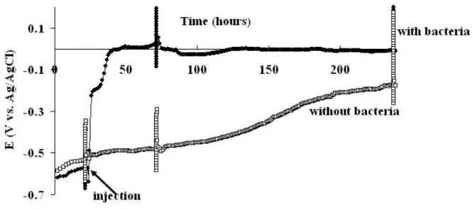
on the steel surface. The experiment, repeated seven times, gave reproducible results (Fig. 1).
The open circuit potential (Eoc) increased quickly during the three hours following injection of
the bacteria, up to ΔEoc=305+22 mV (average and standard deviation from 7 experiments). It continued to increase slowly for the next twenty hours, up to ΔEoc=443+51 mV. In control experiments, injection of the sterile culture medium did not cause any Eoc increase.
Tafel plots were recorded at various times by scanning the potential around Eoc. In the
absence of bacteria (Fig. 2A), the Ecorr values given by the Tafel plots were significantly more
negative than the Eoc values. In this case, the sole cathodic reaction consisted of the reduction
of protons (reaction 2), which had a very low concentration (6.310-8 M at pH 7.2). H+
depletion in the diffusion layer that was provoked by the potential scan logically decreased
the cathodic current and consequently shifted Ecorr towards negative potential values. In the
absence of bacteria Ecorr was controlled by the mass transfer limitation of the cathode reaction.
In the presence of the bacteria (Fig. 2B-C) there was less than 70mV difference between Eoc
and Ecorr. It can be concluded that the cathodic reaction was no longer controlled by the
reaction that was less sensitive to mass transfer. The anodic part was not significantly
modified by the presence of G.sulfurreducens during the first few days, confirming that the
abrupt increase of Eoc observed during the early hours was due to the modification of the
cathodic part. The Tafel plot recorded at the end of the experiments showed a clear oxidation
wave at potential values above 0.03 V vs. Ag/AgCl, which was not observed on the Tafel
plots recorded only 2 days after inoculation. This wave was due to the oxidation of acetate
catalysed by the biofilm. As already observed on polarised electrodes, it was confirmed here
that acetate oxidation occurred only with well-established biofilms[16].
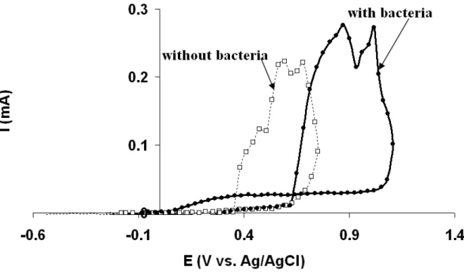
Pitting curves recorded at the end of the experiments indicated that the presence of
G.sulfurreducens shifted the pitting potential (Epit) from 840+80 mV vs. Ag/AgCl (without
bacteria) to 1009+20 mV vs. Ag/AgCl (Fig. 3). In the presence of bacteria, the abnormally
high anodic current, with regard to traditional pitting curves, that was recorded during the
scan in the positive direction was due to the biofilm-catalysed oxidation of acetate, as
observed on the Tafel plot (Fig. 2C). At high potential values, G.sulfurreducens had a clear
protective effect, due to the electrons provided to the material by the biofilm-catalysed
oxidation of acetate.
SEM micrography showed that the pits were deeper in the presence of bacteria. This is
relevant with the higher hysteresis effect observed on the repassivation curves (Fig. 3). In the
presence of bacteria, pitting occurred at higher potential values and resulted in higher
propagation currents. The epifluorescence microscopy pictures recorded after the pitting
curves showed that deep pits formed predominantly in zones where the biofilm was dense
(Fig. 4A), while zones free from pits revealed only scattered microbial settlement (Fig. 4B).
Two different hypotheses can explain this observation: i) bacteria preferentially colonise the
areas that are the most sensitive to further corrosion attacks, for instance because of local
promotes corrosion in its vicinity. Whatever are the hypothesis, microbial settlement and
pitting zones, showed intimate local correlation.
4. Conclusion
G.sulfurreducens revealed here as a main player in electron transfer between 304L
stainless steel and the surrounding medium. Experiments performed at open circuit
demonstrated that, in a medium with low electron donor concentration, G.sulfurreducens can
extract electrons from steel, causing a fast potential increase up to 443 mV, which drastically
increases corrosion risk. This reaction was due to the fast electron transfer already observed
between electrodes and G.sulfurreducens cells as soon as they settle on the electrode surface
[16,17]. In contrast, the catalysis of acetate oxidation that occurred at high potential values
only with well-developed biofilms delayed the occurrence of pitting. In this case the biofilm
revealed a protective effect. The corrosive/protective action of G.sulfurreducens on steel
surfaces depends strongly on the potential range and the age of the biofilm. From a
fundamental point of view, it has been demonstrated here that the mechanism of direct
microbial electron transfer can be of crucial importance in anaerobic biocorrosion. Moreover,
several Geobacter species have been shown to be able to achieve direct electron transfer with
solid electrodes and Geobacter species are abundant in soils, sediments and other natural
environments. Practically, these species should now be considered as possible main
contributors to biocorrosion, particularly when buried equipment is concerned.
References
2. W. Lee, Z. Lewandowski, W.A. Hamilton, Biofouling. 8 (1995) 165.
3. R.D. Bryant, W.J. Jansen, J. Boivin, E.J. Laishley, W. Costerton, Appl. Environ.
Microbiol. 57 (1991) 2804.
4. S. Da Silva, R. Basseguy, A. Bergel, Bioelectrochem. 56 (2002) 77.
5. L. De Silva Muñoz, A. Bergel, R. Basseguy, Corrosion Sci. 49 (2007) 3988.
6. M.A. Lopez, F.J.Z. Diaz de la Serna, J. Jan-Roblero, J.M. Romero, C.
Hernandez-Rodriguez, FEMS Microbiol. Ecol. 58 (2006) 45.
7. D.R. Lovley, Nature Rev. Microbiol. 7 (2006) 497.
8. K. Rabaey, W. Verstraete, Trends Biotechnol. 23 (2005) 291.
9. A. Esteve-Núñez, M. Rothermich, M. Sharma, D.R. Lovley, Environ. Microbiol. 7
(2005) 641.
10. D.E. Holmes, S.K. Chaudhuri, K.P. Nevin, T. Mehta, B.A. Methé, A. Liu, J.E. Ward,
T.L. Woodard, J. Webster, D.R. Lovley, Environ. Microbiol. 8 (2006) 1805.
11. G. Reguera, K.D. McCarthy, T. Mehta, J.S. Nicoll. M.T. Tuominen, D.R. Lovley,
Nature. 435 (2005) 1098.
12. E. Marsili, D.B. Baron, I.D. Shikhare, D. Coursolle, J.A. Gralnick, D.R. Bond, PNAS.
105 (2008) 3968.
13. K.B. Gregory, D.R. Bond, D.R. Lovley, Environ Microbiol. 6 (2004) 596.
14. C. Dumas, R. Basseguy, A. Bergel, Electrochim. Acta, 53 (2008) 2494.
15. H.T. Dinh, J. Kuever, M. Mubmann, A.W. Hassel, M. Stratmann, F. Widdel, Letters
Nature. 427 (2004) 829.
16. C. Dumas, R. Basseguy, A. Bergel, Electrochim. Acta. 53 (2008) 5235.
Figure 1 Open circuit potential versus time in the absence and presence of G. sulfurreducens.
Fluctuations in potential that appeared around 21h, 71h and 237h were due to Tafel plot recording.
Figure 2 Tafel plots performed from -100mV / Eoc to + 200mV / Eoc (scan rate 0.5mVs-1) A, two days after injection of sterile medium. B, with G. sulfurreducens two days after inoculation (142 000 CFU.mL-1). C, from -100mV / Eoc to + 350mV / Eoc at t =237h with G.
Figure 3 Polarisation curves (scan rate 0.5 mVs-1); scan was reversed when 0.1 mA was reached, performed at t = 240h in the absence and presence of G. sulfurreducens.
Figure 4 Epifluoresence microscopy of 304L stainless steel in the presence of G. sulfurreducens: A in the vicinity of a pit, and B in a zone free from pits (magnification 100x).