Direction des bibliothèques
AVIS
Ce document a été numérisé par la Division de la gestion des documents et des archives de l’Université de Montréal.
L’auteur a autorisé l’Université de Montréal à reproduire et diffuser, en totalité ou en partie, par quelque moyen que ce soit et sur quelque support que ce soit, et exclusivement à des fins non lucratives d’enseignement et de recherche, des copies de ce mémoire ou de cette thèse.
L’auteur et les coauteurs le cas échéant conservent la propriété du droit d’auteur et des droits moraux qui protègent ce document. Ni la thèse ou le mémoire, ni des extraits substantiels de ce document, ne doivent être imprimés ou autrement reproduits sans l’autorisation de l’auteur.
Afin de se conformer à la Loi canadienne sur la protection des renseignements personnels, quelques formulaires secondaires, coordonnées ou signatures intégrées au texte ont pu être enlevés de ce document. Bien que cela ait pu affecter la pagination, il n’y a aucun contenu manquant.
NOTICE
This document was digitized by the Records Management & Archives Division of Université de Montréal.
The author of this thesis or dissertation has granted a nonexclusive license allowing Université de Montréal to reproduce and publish the document, in part or in whole, and in any format, solely for noncommercial educational and research purposes.
The author and co-authors if applicable retain copyright ownership and moral rights in this document. Neither the whole thesis or dissertation, nor substantial extracts from it, may be printed or otherwise reproduced without the author’s permission.
In compliance with the Canadian Privacy Act some supporting forms, contact information or signatures may have been removed from the document. While this may affect the document page count, it does not represent any loss of content from the document.
Université de Montréal
V ASCULAR AND MORPHOLOGICAL CHANGES OF
THE OPTIC NERVE HEAD FOLLOWING
THERAPEUTIC INTRAOClJLAR PRESSURE
REDUCTION IN OPEN ANGLE GLAUCOMA AND
OCULAR HYPERTENSION
par
ALI S. HAFEZ
Département d'ophtalmologie Faculté des études supérieures
Thèse présentée à la Faculté des études supérieures en vue de l'obtention du grade de Philosophi<e doctor (Ph. D.)
en Sciences Biomédicales
Montréal, Québec, Canada
août 2007
& études fJ
"0 ~e !I \ doc t 0 (,1.061:
© Ali S. Hafez, 2007
§ 0 ,. ~ (J~ ç Grade conféré ÇI .... ~
Ill;;
II compter du ~ <:.. l.I.. <Pl')<JI
o
1 MAI 2008Université de Montréal
Faculté des études supérieures
Cette thèse intitulée :
11
V ASCULAR AND MORPHOLOGICAL CHANGES OF
THE OP TIC NERVE HEAD FOLLOWING
THERAPEUTIC INTRAOClJLAR PRESSURE
REDUCTION IN OPEN ANGLE GLAUCOMA AND
OCULAR HYPERTENSION
présentée par :
ALI S. HAFEZ
a été évaluée par un jury composé des personnes suivantes:
Leonard Levin MD PhD, président-rapporteur Mark R. Lesk MSc MD, directeur de recherche
Helene Kergoat OD PhD, membre du jury Nabil Saheb MD, examinateur externe
111
RÉSUMÉ
But de l'étude :
Évaluer les changements vasculaires et morphologiques de la tête du nerf optique (TNO) et de la rétine péripapillaire après thérapie visant à réduire la pression intraoculaire (PlO) chez les patients atteints de glaucome à angle ouvert (GAO) ou d'hypertension oculaire (HTO). La présence et l'étendue des changements au niveau du débit sanguin et de la topographie ont été corrélées avec des paramètres cliniques tel que le rapport excavation/papille, la vasospasticité périphérique et l'épaisseur cornéenne centrale.
Patients et Méthodes:
Vingt patients avec GAO non contrôlés, vingt patients avec HTO non contrôlé et 20 sujets normaux ont été recrutés pour l'évaluation de la fiabilité de la technique et les mesures de référence du débit sanguin.
Pour les patients avec GAO et HTO, la réduction de la PlO a été obtenue par thérapie chirurgicale, laser ou médicamenteuse. Tous les patients ont obtenu une réduction de la PlO de plus de 20% et un suivi minimum de 4 semaines.
La perfusion de la TNO et de la rétine péri papillaire a été évaluée avant et après réduction de la PlO utilisant un logiciel nommé «scanning laser doppler flowmetry (SLDF) full-field perfusion analysis» et permettant d'analyser les images obtenues avec le Heidelberg Retina Flowmeter (HRF). Le débit sanguin moyen a été obtenu à partir de 5 images successives de perfusion. Les mesures topographiques ont été recueillies avant et après réduction de la PlO utilisant le Heidelberg Retina tomograph
lV
(HRT). La topographie moyen a été obtenu a été à partir de 3 images successives et les différences topographiques ont été calculées. De plus, le débit sanguin au niveau d'un doigt a été déterminé grâce au Transonic laser Doppler Flowmeter après immersion dans l'eau froide. L'épaisseur cornéenne centrale a aussi fait l'objet d'une évaluation grâce au pachymètre à ultrasons. La stabilité du champ visuel a été examinée chez une cohorte de patients avec GAO et HTO sur une période de 4 ans en utilisant les critères adaptés de Hodapp, Parrish et Anderson.
Des tests statistiques ont été pratiqués en utilisant l'analyse de variance à 1 facteur et le test T pour données appariées. Les corrélations ont été évaluées grâce au coefficient de Pearson.
Résultats:
Le logiciel d'analyse «full-field perfusion» a peris d'obtenir des données intra- et inter-session hautement reproductibles chez les patients glaucomateux avec une fiabilité
~.99 pour l'anneau neurorétinien (ANR) et ~.87 pour la rétine péripapillaire.
Les patients avec GAO avant traitement avaient un débit sanguin significativement plus faible que celui des patients avec HTO et des sujets normaux (P=O.OOI). Chez les patients avec HTO, le débit sanguin de l'ANR etait inversement corrélé au rapport excavation/papille (P=O.039).
Bénéficiant d'un % de réduction similaire de la PlO (37% pour le groupe GAO versus
33% pour le groupe HTO), le débit sanguin de l'ANR s'est amélioré de 67% chez les patients avec GAO (P;;O.OOI) comparé à 7.5% pour ceux avec HTO (p=0.41). Chez ces derniers, l'amélioration du débit sanguin de ANR etait limité aux sujets vasospastiques
v
(P=O.OOl). Une corrélation négative et significative (P=O.003) a aussi été trouve entre les changements du débit sanguin dans l' ANR et le débit sanguin maximum au niveau du doigt chez les patients avec GAO contrairement aux patients avec HTO.
Les changements topographiques moyens de la TNO après réduction de la PlO ne différaient pas entre les groupes GAO et HTO étudiés (P;;::0.439, ANOV A). Les patients avec les cornées les plus minces présentaient une réduction nettement plus importante de la profondeur moyenne de l'excavation (P=O.OO3) et de la profondeur maximum de l'excavation (P=Ü.020) avec, en revanche, de petites améliorations du débit sanguin de l'ANR comparées aux patients ayant les cornées les plus épaisses (P=O.04).
Les patients avec une perte progressive du champ visuel ont été trouvés plus vasospastiques (P=O.OO6) et montraient une plus grande diminution plus importante de la profondeur maximum de l'excavation (P=O.OO5). lis ont aussi des cornées plus minces et une augmentation moins importante du débit sanguin de l'ANR (P;;::O.31).
Conclusion:
Cette étude est en accord avec des articles publiés précédemment et mettant en évidence, en l'absence de thérapie oculaire antihypertensive, un débit sanguin diminué chez les patients avec GAO comparés aux patients avec HTO et aux sujets normaux. Les patients avec HTO présentant un plus grand rapport excavation/papille ont aussi montré des débits sanguins de l'ANR plus faibles que ceux présentant des rapports cup/disk plus petits. Cette étude démontre que, pour un pourcentage similaire de réduction de la PlO, le débit sanguin de l'ANR chez les sujets glaucomateux s'améliore alors qu'il reste stable chez les patients présentant une hypertonie oculaire. Considérant
VI
ces derniers, de plus grandes améliorations du débit sanguin de l' ANR ont été observées chez les sujets vasospastiques que chez les sujets non vasospastiques.
Aucun changement topographique de la TNO ne montre une différence statistique significative entre les deux groupes étudiés avant et après thérapie oculaire antihypertensive. Cependant, les patients avec GAO et HTO présentant les cornées les plus minces ont montré un déplacement plus important de la lame criblée comparés à ceux ayant les cornées les plus épaisses.
L'évaluation de la stabilité visuelle à long terme d'une cohorte pilote de nos patients avant la baisse de PlO initiale montre que les patients glaucomateux présentant une progression étaient plus vasospastiques et présentaient une lame crible plus compliante comparés aux sujets avec un déficit stable.
Ces résultats démontrent l'existence d'un débit sanguin de la TNO altéré dans le glaucome et établissent des liens entre la neuropathie optique glaucomateuse et des facteurs tels que l'autorégulation vasculaire, la vasospasticité périphérique, l'épaisseur cornéenne centrale et la compliance biomécanique de la tête du nerf optique. '
Mots Clés
Glaucome tête du nerf optique débit sanguin pression intraoculaire topographie -vasospasticité - épaisseur cornéenne centrale - autorégulation.
VIl
ABSTRACT
Purpose:
To evaluate the vascular and morphologie changes of the optic nerve head (ONH) and peripapillary retina following therapeutic intraocular pressure (IOP) reduction in open angle glaucoma (OAG) and ocular hypertension (OHT).
The presence and extent of blood flow changes and topographic changes were correlated with clinieal parameters such as cup/disc ratio as weIl as peripheral vasospasm and central corneal thickness. A correlation between the ONH changes and long term visual field stability was also studied.
Patients and Methods:
Twenty uncontrolled OAG patients, 20 uncontrolled OHT patients and 20 normal volunteers were recruited for assessment of the reliability of the technique and for baseline blood flow measurements.
For both OAG and OHT groups, IOP reduction was achieved by medieal, laser or surgical therapy. AlI patients had IOP reduction more than 20% and a minimum of 4 weeks followup.
ONH and peripapillary retinal perfusion was assessed before and after IOP reduction using scanning laser Doppler flowmetry (SLDF) full-field perfusion image analysis of Heidelberg Retina Flowmetry (HRF) images. Mean flow values were derived from five consecutive perfusion images. Scanning laser topographie measurements were performed before and after IOP reduction using the Heidelberg Retina Tomograph (HRT). Mean topography of 3 consecutive images was obtained and topography
Vlll
differences were computed. Patients also underwent finger blood flow measurement using the Transonic laser Doppler flowmetry after cold water immersion, while central corneal thickness was determined using an ultrasound pachymeter. Visual field stability was monitored in a cohort of the OAG and OHT patients over a period of 4 years using modified Hodapp-Anderson-Parrish criteria.
Statistical evaluations were performed using one-way analysis of variance and two-tailed distribution paired T-test. Correlations were assessed using Pearson's coefficient.
ResuIts:
SLDF full-field perfusion analysis was found to be highly reproducible in glaucoma patients both within and between sessions with a reliability of~O.99 in the neuroretinal rim and ~O.87 in the peripapillary retina.
For baseline flow, OAG patients had significantly lower blood flow in the ONH compared with OHT patients and normal volunteers (P=O.OOl). Among patients with OHT, neuroretinal rim blood flow was inversely correlated to increased CID ratio (P=O.039).
Following similar % IOP reduction (37% in OAG versus 33% in OHT), ONH neuroretinal rim blood flow improved by 67% in OAG (P=O.OOl) compared to 7.5% in OHT (P=0.41). In OHTs, improvement in neuroretinal rim blood flow was limited to vasospastic subjects (P=O.Ol). A significant negative correlation (P=O.003) was also found between rim blood flow change and maximum fmger blood flow in OAG patients but not in OHTs.
IX
Mean change in ONH topographie al parameters following therapeutic IOP reduction
did not differ between the two study groups (P~0.439, ANOV A). Patients with thinner corneas had greater reductions in mean cup depth (P=O.003) and maximum cup depth (P=O.020) and smaller improvements in neuroretinal rim blood flow compared to those with thicker corneas (P=O.04).
Patients with progressive visual field changes were found to be vasospastic (P=O.006) and showed shallowing of maximum cup depth (P=O.005). They also had thinner corne as and smaller increases in neuroretinal rim blood flow (P~O.31).
Conclusion:
The study confirms previous reports as regards decreased baseline ONH blood flow in OAG patients compared to OHT patients and normal subjects. Ocular hypertensives with larger cup/disk ratios were also shown to have lower neuroretinal rim blood flow compared to those with smaller CID ratios. The study demonstrates that following a similar percentage of therapeutic IOP reduction, blood flow improved in the
neuroretinal rim of the ONH in glaucoma patients while it remained stable in ocular hypertensives. Greater improvements in rim blood flow were observed in vasospastic than in non-vasospastic ocular hypertensives.
None of the ONH topographie changes between the two study groups showed a statistically significant difference following therapeutic IOP reduction. However, OAG
and OHT patients· with thinner corneas showed greater forward displacement of the lamina cribrosa compared to those with thicker corneas.
x
Assessment of long-term visual stability in a pilot cohort of our subjects showed that at the time of initial IOP reduction, progressive glaucoma patients were more vasospastic and demonstrated a more compliant lamina cribrosa compared to stable glaucoma patients.
The results demonstrate the role of defective ONH blood flow in glaucoma and correlate glaucomatous optic neuropathy to factors such as autoregulation, central comeal thickness and peripheral vasospasm. The results also shed light on the mechanical properties of the ONH following sustained IOP reductions in a real-life context. Comparing these vascular and morphologic changes in both OAG and OHT
,
helps the assessment of long-term visual stability.
KeyWords
glaucoma optic nerve head blood flow intraocular pressure topography -vasospasticity - central comeal thickness - autoregulation.
Xl
TABLE OF CONTENTS
Chapter 1.
Introduction and Review of Literature
1
~ Anatomy and Blood Supply of the Optic Nerve Head2
•
Anatomy of the Lamina Cribrosa3
•
General Anatomy of the ONH Blood Supply5
•
Microvasculature of Specifie ONH Regions6
•
Variations in Blood Supply of the ONH8
•
Venous Drainage of the ONH 10~ Background and Definition of Glaucoma
14
~ Raie of Intraocular Pressure and Ocular Blood Flow
in the Pathogenesis of Glaucoma
- 22
•
Findings of Ocular Blood Flow Studiesin Glaucoma and their Interpretation
24
•
Potential Mechanisms of Ocular BloodFlow Reduction in Glaucoma Patients
28
•
Current Evidence of Abnormal Ocular BloodFlow in Glaucoma
32
~ Techniques Used for Evaluation of Ocular Blood Flow
55
A. Color Doppler Imaging
55
B. Pulsatile Ocular Blood Flow
&
Fundus Pulsation Amplitude58
C. Fluorescein and Indocyanine Green Angiography61
D. Laser Doppler Velocimetry
64
E. Laser Doppler Flowmetry
65
F. Scanning Laser Doppler Flowmetry
68
G. Laser Speckle Technique
71
H. Retinal Vessel Analyser
72
1.
Canon Laser Blood Flowmeter74
J. Blue Field Entoptics 77
K.
Peripheral Blood Flow78
L.
Animal Experimental Methods81
~ Technical Aspects of Scanning Laser Doppler Flowmetry
• Scanning Laser Doppler Flowmetry • Confocal Scanning Laser Ophthalmoscopy
Chapter 2.
Objectives and Study Design
Xll
95
106
121
~ Principal Research Objective and Hypothesis 122
~ Specifie Research Objectives and Hypotheses 122
~ Study Design 125
A. Patients 125
B. Inclusion Criteria and Patient Definitions 125
C. Exclusion Criteria 127
D. Study Procedures 127
E. Clinical Management & Timing of Post-IOP Reduction Tests 130
F. Data Management 131
G. Power and Sample Size 131
H. Ethical Considerations 132
1. Study limitations 132
J. Funding and disclosure 133
Chapter 3.
Reproducibility of Retinal and Optic Nerve
Head Perfusion Measurements using Scanning
Laser Doppler Flowmetry
• Introduction
• Patients and Methods • Results
• Tables and Figures • Discussion
• References
Chapter 4.
Evaluation of Optic Nerve Head and Peripapillary
Retinal Blood Flow in Glaucoma Patients,
Ocular Hypertensives and Normal Subjects
• Introduction
• Patients and Methods • Results
• Tables and Figures • Discussion • References
135
139
143149
153 161 167172
176 178183
190195
202X111
Chapter 5.
Changes in Optic Nerve Head Blood Flow after
Therapeutic Intraocular Pressure Reduction in
Glaucoma Patients and Ocular Hypertensives
210
•
Introduction215
•
Patients and Methods218
•
Results223
•
Tables and Figures229
•
Discussion237
•
References244
Chapter 6.
Correlation between Finger Blood Flow and
Changes in Optic Nerve Head Blood Flow following
Therapeutic Intraocular Pressure Reduction
254
•
Introduction258
•
Patients and Methods261
•
Results264
•
Tables and Figures268
•
Discussion272
•
References279
Chapter 7.
Relationship between Central Corneal Tl1ickness
and Changes of Optic Nerve Head Topography
and Blood Flow following IOP Reduction in
Open Angle Glaucoma and Ocular Hypertension
285
•
Introduction288
•
Patients and Methods290
•
Results292
•
Tables and Figures295
•
Discussion301
•
References304
Chapter 8.
Changes in Optic Nerve Head Topography following
Therapeutic Intraocular Pressure Reduction in
Glaucoma Patients and Ocular Hypertensives
309
•
Introduction313
•
Patients and Methods316
• Tables and Figures • Discussion
• References
Chapter 9.
Relationship between Glaucomatous Visual Field
Progression, Vasospasticity and Changes in Optic Nerve
Head Topography and Slood Flow at the time of
XIV
323
330
337
InitiallOP Reduction: A Prospective Pilot Study
343
• Introduction
• Patients and Methods • Results
• Tables and Figures • Discussion
• References
Chapter 10. Conclusion and Future Prospects
Appendices
Appendix 1 : Presentation of results Appendix Il : Accord des co-auteursAppendix III: PDF copies of published papers
347
350
356
361
367
373
382
LIST OF FIGURES
Figure 1.1 Scanning electron microscopie analysis of a normal human 4 ONH following trypsin digestion.
Figure 1.2 Schematic representation ofblood supply of the optic nerve. 6 Figure 1.3 Microvascular corrosion cast of human optic nerve showing 8
posterior ciliary arteries and pial arteries.
Figure 1.4 Ocular vascular findings in glaucoma. 26
Figure 1.5 Systemic vascular findings in glaucoma. 26
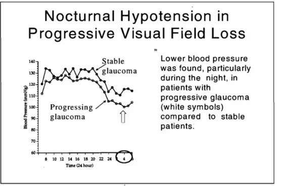
Figure 1.6 Noctumal hypotension in progressive visual field loss. 31 Figure 1.7 Correlations between IOP and visual field me an defect MD 41
in two distinct OAG populations.
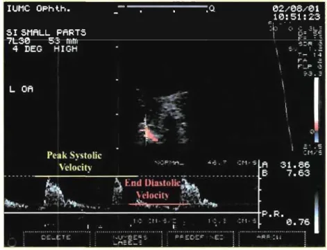
Figure 1.8 Color Doppler imaging showing peak systolic and end- 56 diastolic velocities in the ophthalmic artery.
Figure 1.9 Pulsatile ocular blood flowmeter. 59
Figure 1.10 Fluorescein fundus angiogram of the optic nerve head and 62 peripapillary retina.
Figure 1.11 Tracing of Laser Doppler Flowmetry for neuroretinal rim 66 blood flow in a normal subject.
Figure 1.12 Heidelberg Retinal Flowmeter. 69
Figure 1.13 Retinal Vessel Analyzer. 73
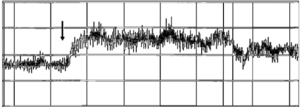
Figure 1.14 CLBF measurement of the inferior temporal retinal artery. 76 Figure 1.15 Tracing of Laser Doppler flowmetery (LDF) in a vasospastic 80
patient. And a nonvasospastic patient.
Figure 1.16 Doppler effect is caused by a frequency shift of waves 97 reflected by moving red blood cells (RBC).
Figure 1.17 Interference of the two reflected frequencies creates a 97 repetitive oscillation of light intensity or a "beat".
Figure 1.18 Fast Fourier Transform eonverts the intensity values to a 98 power spectrum.
Figure 1.19 HRF perfusion map and SLDF full-field perfusion image 102 analysis of the neuroretinal rim area.
Figure 1.20 Figure 1.20: HRT analysis showing stereometrie parameters 108 of the ONH (upper right) and topographie image (upper left).
Figure 3.1 SLDF full-field perfusion analysis. 160
Figure 4.1 Seattergrams of SLDF blood flow measurements for the 193 neuroretinal rim of the ONH in the OAG group, OHT group
and NOR group.
Figure 4.2 SLDF blood flow measurements for the neuroretinal rim of 194 the ONH in the OHT group as divided into two equal
subgroups based on CID ratio.
Figure 5.1 Flow image of temporal peripapillary retinal area using the 234 SLDF full-field perfusion analysis.
Figure 5.2 SLDF measurements for the parameter flow in the OAG 235 group before and after therapeutie IOP reduetion.
Figure 5.3 SLDF measurements for the parameter flow in the OHT 236 group before and after therapeutie IOP reduetion.
Figure 6.1 Finger Doppler of Vasospastie (Top) and Non-vasospastie 270 (Bottom) Patient.
Figure 6.2 Correlation between Flow Max (maximum [mger blood 271 flow) and Change in Neuroretinal Rim Blood Flow in OAG
and OHT patients.
Figure 7.1 Change in Topographie Parameters following IOP reduetion 299 for all patients (eombined OHT+OAG patients)
Figure 7.2 Change in neuroretinal rim blood flow following IOP 300 reduetion for eombined OAG+OHT patients and for OAG
and OHT subgroups.
Figure 8.1 Model of Topographieal ONH changes with IOP reduetion. 329 Figure 10.1 Proposed relationship between vaseular and meehanieal 387
factors and progression of glaueomatous optie neuropathy.
Table 3.1 Table 3.2 Table 3.3 Table 3.4 Table 3.5 Table 3.6 Table 3.7 Table 4.1 Table 4.2 Table 4.3 Table 5.1
LIST OF TABLES
Intraocular Pressure, CuplDisc Ratio and Ocular Perfusion Pressure in "Group G" and "Group N".
Intrasession Coefficients of Reliability (R) and 95% Confidence Intervals (CI.) for "Group G" (N=20) as calculated for one image (RI), mean of three images (R3) and mean of five images (RS).
Intrasession Coefficients of Reliability (R) and 95% Confidence Intervals (CI.) for "Group N" (N=20) as calculated for one image (RI), mean of three images (R3) and mean of five images (RS).
Intrasession variability of five images as measured by mean
(± SD) coefficients of variation according to parameter, location of measurement and study group.
Intersession Coefficients of Reliability (R) and 95% Confidence Intervals (CI.) for "Group G" (N=20) as calculated for one image (RI), mean of 3 images (R3) and mean of 5 images CR5).
Intersession Coefficients of Reliability (R) and 95% Confidence Intervals (CI.) for "Group N" (N=20) as calculated for one image (RI), mean of 3 images (R3) and mean of 5 images (R5).
Intersession variability of the mean (± SD) of five images as measured by mean percent change according to parameter, location of measurement and study group.
Patient Characteristics of the 3 study groups OAG, OHT and NOR.
SLDF Flow Measurements of the neuroretinal rim of ONH, temporal peripapillary retina and nasal peripapillary retina of the OAG, OHT and NOR groups.
Correlation between RIM, NAS and TEM blood flow versus various clinical parameters.
Characteristics of OAG and OHT groups.
xvii 153 154 155 156 157 158 159 190 191 192 229
Table 5.2 Table 5.3 Table 5.4 Table 5.5 Table 6.1 Table 6.2 Table 7.1 Table 7.2 Table 7.3 Table 7.4 Table 8.1 Table 8.2 Table 8.3 Table 8.4 Table 8.5 Table 8.6 Table 9.1
SLDF Perfusion Measurements in OAG and OHT Groups. Charaeteristies of Post IOP-Matehed Subgroups.
Charaeteristies of Age-Matehed Subgroups.
Mean Rim Z-Coordinate Change Relative to Reference Plane.
Patient Charaeteristies ID Vasospastie Group and
Non-vasospastie Group.
Changes in neuroretinal rim blood flow in vasospastie versus nonvasospastie OAG and OHT patients.
Charaeteristies of eombined OAG and OHT patients based on median CCT
Change in Topographie Parameters following sustained IOP reduetion among patients with OAG and OHT.
Change in Topographieal Parameters following sustained IOP reduetion among OAG and OHT patients.
Changes in Rim Blood Flow following sustained IOP reduetion.
Patient Charaeteristies in the OAG and OHT Groups. Patient Charaeteristies (eontinued).
Baseline optie dise topographie parameters in OAG patients and OHT patients.
Changes in optie dise topographie parameters after sustained IOP reduetion in OAG patients.
Changes in optie dise topographie parameters after sustained IOP reduetion in OHT patients.
Changes of topographie parameters in OAG patients versus OHT patients following sustained IOP reduetion.
Data of study sample when grouped as progressed and non-progressed. XVlll 230 231 232 233 268 269 295 296 297 298 323 324 325 326
327
328 361Table 9.2
Table 9.3
Table 9.4
Table 9.5
Table 9.6
Baseline topographie parameters of the study sample (progressed and non-progressed groups).
Post-IOP reduction topographie parameters of the study sample (progressed and non-progressed groups).
Change of topographie parameters (Post-IOP minus baseline) in the progressed and non-progressed groups. Optic nerve head blood flow data in progressed and non-progressed groups at baseline, follow-up, unit change and %
change.
Finger blood flow data for the progressed and non-progressed groups.
xix
362
363
364
365
366
ACE AFFPIA ANOVA AU CCT CID ratio CDI CLBF CRA CSLO CSM DBP DSFS EDV FA FPA GON HRF HRT HTG HVC ICG
LIST OF ABREVIATIONS
Angiotensin Converting EnzymeAutomatie Full-Field Perfusion Image Analysis Analysis of Variance
Arbitrary Units
Central Comeal Thiekness Cup to Dise ratio
Color Doppler Imaging
Cannon Laser Blood Flowmeter Central Retinal Artery
Confoeal Seanning Laser Ophthalmoseopy Cup Shape Measure
Diastolie Blood Pressure
Doppler Shift Frequeney Speetrum End Diastolie Veloeity
Fluoreseein Angiography Fundus Pulsation Amplitude Glaueomatous Optie neuropathy Heidelberg Retinal Flowmeter Heidelberg Retinal Tomograph High Tension Glaueoma Height Variation Contour Indocyanine Green
xxi
IOP Intraocular Pressure
LDF Laser Doppler Flowmetry
LDV· Laser Doppler Velocimetry
MD Mean Defect
NTG Normal Tension Glaucoma
OA Ophthalmic Artery
OAG Open Angle Glaucoma
OBF Ocular Blood Flow
OCT Optical Coherence Tomography
OHT Ocular Hypertension
ONH Optic Nerve Head
OPP Ocular Perfusion Pressure
PCA Posterior Ciliary Artery
PCV Peak Systolic Velocity
POBF Pulsatile Ocular Blood Flow
RBC Red Blood Cell
RGC Retinal Ganglion Cell
RI Resistivity Index
RNFL Retinal Nerve Fiber Layer
RNFLA Retinal Nerve Fiber Layer Area
RNFLT Retinal Nerve Fiber Layer Thickness
ROI Region of Interest
xxii
RVA Retinal Vessel Analyzer
SBP Systolic Blood Pressure
SD Standard Deviation
SLDF Scanning Laser Doppler Flowmetry
SPCAs Short Posterior Ciliary Arteries
TPU Tissue Perfusion Units
XXIII
Dedication
To my father and to the memory of my mother who taught me
the first step to success ... persistence ...
To my wife Nora for her endless help, support, patience and
motivation ...
And to the most precious gift in my Iife ... my two sons
Omar and Ziad ....
XXIV
ACKNOWLEDGEMENTS
No research endeavor is ever carried out in solitude. This work was possible through the efforts of a number of persons and institutions to whom 1 am extremely grateful.
1 deeply appreciate the support, guidance and encouragement that my supervisor and mentor Dr. Mark R. Lesk has given me in the production of this work and throughout
my career. ... Without his help the completion of this thesis would not have been possible .... His continuous support and desire for perfection greatly encouraged and motivated me .... and his friendship will always be cherished ....
My sincerest gratitude also goes to Regina Bizzarro OD for providing meticulous technical assistance since the beginning of my studies and throughout my PhD ... but above aIl for her kindness, sincerity and concern ...
1 would also like to thank my colleagues at the Glaucoma Research Laboratory at Maisonneuve-Rosemont Research Centre: Denise Descovich MD, Micheline Deschênes PhD, Marcelo Wajszilber MD, Fawzia Djafiri MD MSc, and Demos Papamatheakis MD for their assistance at different stages of this work .... also to Murielle Bégin, Lucille Cliché RN and Bonnie May OT for their encouragement and support.
1 am also very grateful to Drs. Michelle Rivard and Miguel Chagnon for their valuable efforts in providing the statistical analyses.
xxv
My deepest appreciation goes also to the staff members of the Department of Ophthalmology at University of Montreal, for helping me throughout the course of my fellowship and for supporting me establish my career in Canada.
Last but not least. ... my gratitude goes to my mentors and professors at the Department of Ophthalmology, Cairo University and the Research Institute of Ophthalmology in Cairo, Egypt for helping me be who 1 am .... and for giving me the chance to reach new horizons beyond my dreams ....
1
Ali Hafez - Vascular and Morphological Changes of the Op tic Nerve Head
Chapter 1
Introduction and Review of Literature
Ali
s.
Hafez, MD PhDDepartment of Ophthalmology, University of Montreal and Maisonneuve-Rosemont Hospital Research Center, Montréal, Québec, Canada
2 Ali Hafez - Vascular and Morphological Changes of the Optic Nerve Head
ANATOMY AND BLOOD SUPPL
y
OF THE OPTIC
NERVEHEAD
The optic nerve head (ONH), also known cIinicaIly as the optic di sc or papilla, forms the point of exit for the retinal ganglion cell (RGC) axons through the scIeraI canal. It is composed primarily of neural fibers (1.2-1.5 million retinal ganglion cell axons), glial ceIls, extracellular matrix supportive tissue and vascular elements (1-7). The ONH is delineated from the adjacent peripapillary tissue by a scIeraI rim of connective tissue, the border tissue of Eischnig (8). The diameter of the ONH and anterior portion of the optic nerve is approximately 1.5 mm (9).
The ONH may be divided conveniently into four anatomic regions, from front to back (10-16):
A. Surface nerve fiber layer: this region is continuous with the nerve fiber layer of the retina. It is composed of the non-myelinated axons of the RGCs in transition from the superficial retina to the neuronal component of the optic nerve.
B. Prelaminar region: this is the region between the surface nerve fiber layer and the lamina cribrosa, at the level of the choroid and outer retina. It consists of nerve fibers arranged in bundles, sUITounded by glial tissue septa and astrocytes.
C. Lamina cribrosa region: lies adjacent to the scIera, and provides the main support for the optic nerve as it exits the eye and penetrates the scIeraI coat.
3 Ali Hafez - Vascular and Morphological Changes ofthe Optic Nerve Head
D. Retrolaminar region: this region lies immediately posterior to the lamina cribrosa.
It is marked by the beginning ofaxonal myelination and is surrounded by the leptomeninges of the central nervous system.
Differences among these four regions reflect the conditions to which the axons are exposed to as they pass through the ONH. These differences incIude axon myelination posterior to the lamina cribrosa, sources of blood supply, and the change in tissue pressure from intraocular pressure to that of the cerebrospinal fluid.
Anatomy of the Lamina Cribrosa
The lamina cribrosa is a complex collagenous, relatively elastic structure that consists of a series of fenestrated sheets of connective tissue (approximately 10) and provides the main support for the axons of the optic nerve (1,10-12). The sheets of the lamina cribrosa span the scIeraI opening at the back of the eye inserting into the outer half of the scIera. They are arranged in a series of para lle1 stacked plates.
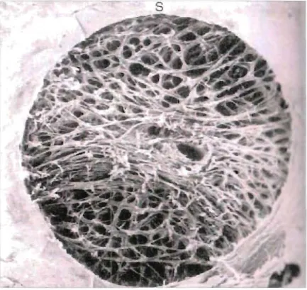
Each of these sheets contains fenestrations or pores that are vertically aligned to allow the passage of the neural elements of the optic nerve. Central pores allow transit of the central retinal artery and central retinal vein. In humans, the pores of the lamina cribrosa are histologically larger and fewer superiorly and inferiorly and the laminar sheets are thinner when compared to the nasal and temporal aspects ofthe optic nerve (17) (Figure 1.1). This correlates with the reported preferential loss of axons at the superior and inferior poles of the ONH in glaucomatous optic neuropathy (GON). Enlargement of the laminar pores and alteration of the shape of cup both peripherally and posteriorly are
Ali HarC7 - Vascular and Mnrphol0gicaJ Change\ (lI' the Ortie Nerve Head
L'haracteri\ric \igns ul' glaucOll1éll(lU'-, ortic neuropatily ( Il), ClauCUll1dLOlIS changes arc
thus not only charactcril.ed hy neural li\\ue los\, but al\o hy connective tissue change\ rc\u!ting in disturtion and poslcrior h0wing of the lamina eribms;\.
Figure l, !I!: Scanning clcctron microscopic analy.\i.\ of a normal human ONH folJowing
tryp\in dige\ti n, Thinner and les\ dense laminar \heet\ arc no[ed in the \uperior and
inl'erior region of the ONH, Courte\y Dr H. Quigley
Lamina l'ribms:l shects are Clll11lillsed primarily of collagen il.S weil as extracellular rnatrix componcnts including clastin. !ilIllinin and fibroneclin, ln 'Klults. laminar shects
PO\SCSS large arnounts of" type· 1 collagen requircd for rigidity and resist:mce to
ddu'-mation as weIl as lyrc-3 collagen and elastill l'eguil'cd l'(lr elasticity and cornplial1ce (18.19), Such COmrUSiliol1 wilh hoth \uppurtive and eJastic elements is
5
Ali Hafez - Vascular and Morphological Changes of the Optic Nerve Head
thought to have an impact on the behavior of the lamina in response to IOP elevation or fluctuation. Laminar sheets are also lined with astrocytes which pro vide an interface between the sheets and the nerve fiber bundles as weIl as provide metabolic support for the axons (20).
General Anatomy of the ONH Blood Supply
The arterial supply of the ONH is derived entirely from branches of the ophthalmic artery, a branch of the internaI carotid artery (13-16).
Typically between 2-4 posterior ciliary arteries arise from the ophthalmic artery in the posterior orbit and course anteriorly before dividing into approximately 10-20 short posterior ciliary arteries. Often the posterior ciliary arteries separate into a medial and lateral group before branching into short posterior ciliary arteries. The short posterior ciliary arteries penetrate the sclera surrounding the optic nerve. They anastomose to form a circle approximately 100-300 microns posterior to the suprachoroidal space, called the arterial circle of Zinn-Haller (21). This circle supplies the peripapillary choroid, as weIl as most of the anterior optic nerve.
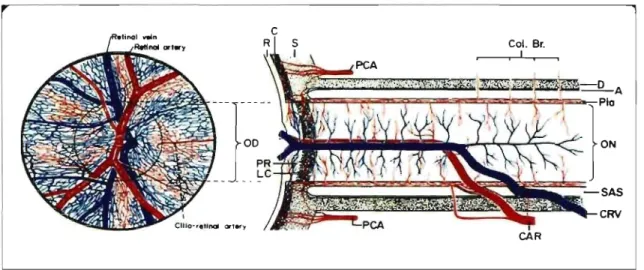
The central retinal artery (CRA), also a branch of the ophthalmic artery, penetrates the optic nerve 10-15 mm behind the globe but has few if any intraneural branches apart from a small branch within the retrolaminar region (Figure 1.2).
6 Ali Hafez - Vascular and Morphological Changes of the Optic Nerve Head
PCA Col. Br. 1 ON SAS CRV
Figure 1.2: Schematic representation ofblood supply of the optic nerve. Reprinted from Hayreh SS: Trans Am Acad Ophthalmol Otolaryngol 1974; 78:240-254. CAR: central retinal artery, CRY: central retinal vien, PCA: posterior ciliary artery, LC: lamina cribrosa, PR: prelaminar region, ON: optic nerve, R: retina, C: choroid S: scIera, 00: optic disc, 0: Dura, A: Arachnoid.
Microvasculature of specifie ONH regions
The blood supply of the optic nerve head can best be discussed under its four regions, from front to back (13-16) (Figure 1.3):
A. Surface nerve fiber layer: This layer is mostly supplied by recurrent retinal arterioles from the CRA and its branches. As the CRA emerges from the optic nerve, it branches into a superior and inferior trunk. From these major trunks as weIl as from the more distal branches, small arterioles supply the surface nerve fiber layer of the optic nerve and peripapillary retina. The cilioretinal artery, when present, usually supplies the corresponding sector of the surface layer.
B. Prelaminar region: This region is supplied by branches from the short PCAs (posterior ciliary arteries) either directly or through the arterial circle of
Zinn-7
Ali Hafez - Vascular and Morphological Changes of the Optic Nerve Head
Haller. Sorne investigators have reported that branches from the short PCAs may course through the choroid and suppl y the prelaminar region (22). The CRA provides no branches in this area.
C. Lamina cribrosa region: This region is similarly supplied by branches from the short PCAs either directly or through the arterial circle of Zinn-Haller. The CRA gives no branches in the lamina. Blood vessels in the lamina cribrosa are 10-20 microns in diameter, are situated in the fibrous septa and form a dense capillary plexus which makes this part of the ONH highly vascular.
D. Retrolaminar region: This region is also supplied by branches from the short PCAs as weIl as by the pial branches originating from the CRA before it pierces the retrobulbar optic nerve.
In surnrnary, except for the branches of the surface nerve fiber layer that arise fromthe CRA, the occasional pial branches of the CRA and the minor contribution from the choroidal vasculature to the prelaminar and laminar regions, the principal arterial suppl y to the ONH is derived from the short posterior ciliary arteries.
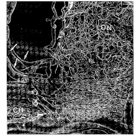
Corrosion casts and histological studies on enucleated human eyes (23) (Figure 1.3) demonstrated the capillary bed of the ONH as one continuous network, cornrnunicating anteriorly with the capillary network of the retina and posteriorI y with that of the rest of the optic nerve. The CRA contribution to the surface nerve fiber layer becomes intertwined with the short PCA contribution to the more posterior regions of the ONH.
8 Ali Hafez - Vascular and Morphological Changes of the Optic Nerve Head
Such anastomosis of capillaries could be viewed as a protective mechanism against ischemia.
Figure 1.3: Microvascular corrosion cast ofhuman optic nerve showing posterior ciliary arteries (arrows) and pial arteries (arrowheads). ON: optic nèrve, L: laminar region, RL: retrolaminar region, PL: prelaminar region, CH choroid. Reprinted from Onda EO, Cioffi GA et al. Am J Ophthalmol1995; 120: 92
Variations in Blood Supply of the ONH
The vascular anatomy of the ONH has been extensively studied. Yet the preCise microvasculature ofthis region remains difficult to as certain because of the small vessel caliber, the comp1ex three-dimensional architecture and the relative inaccessibility of
9
Ali Hafez - Vascular and Morphological Changes of the Optic Nerve Head
the tissues. Inter-individual variation in anatomic details also has led to confusion and controversy.
The existence of the arterial circle of Zinn-HaIler, the contribution of the peripapillary choroid to the ONH circulation and the longitudinal anastomosis of capillaries throughout the regions of the ONH are aIl examples of the persisting debates.
Inter-individual variations in the anatomical distribution and blood flow patterns of PCAs were described by Hayreh and coworkers (14) and could be summarized as:
1. Variations in the number of PCAs and their subdivisions: Prior to entering the sclera, the main PCAs (ranging from 1 to 5 PCAs arising from the ophthalmic artery) subdivide into multiple branches which consist of two subgroups: short and long PCAs. The short PCAs in turn con si st of two subgroups: paraoptic and distal. Evidence suggests that the ONH is mostly supplied by the paraoptic short PCAs
(24).
2. Variations in the area of supply of each PCA: In vivo experimental and clinical fluorescein angiographic studies have shown marked variations in the area of supply of each PCA. Studies have also revealed a segmental distribution with no anastomoses between the adjacent segments (25) and a marked inter-individu al as weIl as interocular variation in the areas supplied by each PCA (26).
10
Ali Hafez - Vascular and Morphological Changes of the Optic Nerve Head
3. Variations in location of watershed zones in the PCA vascular bed: In vivo studies have shown watershed zones in the border between the territories of distribution of PCAs (27). In the event of a drop in ocular perfusion pressure, the watershed zones become most vulnerable to ischemia. The location of the PCA watershed zone in relation to the ONH may be an important factor in determining the site and severity of glaucomatous optic neuropathy.
Venous Drainage of the ONH
The venous drainage from the ONH is by the central retinal vein and its tributaries (4). In the prelaminar region the ONH also has connections with the peripapillary choroid. In the laminar and retrolaminar regions, sorne venousdrainage may also be via pial veins and ultimately into the central retinal vein as it exits the optic nerve.
Il Ali Hafez - Vascular and Morphological Changes of the Optic Nerve Head
REFERENCES
01. Hemandez MR, Lua XX, Igoe F, Neufeld AH. Extracellular matrix of the human lamina cribrosa. Am J Ophthalmol1987; 104: 567.
02. Kronfeld PC. Normal variations of the optic disc as observed by conventiona1 ophthalmoscopy and their anatomic correlations. Trans Am Acad Ophthal Otol 1976; 81: 214.
03. Hayreh SS. Anatomy and physiology of the optic nerve head. Trans Am Acad Ophthal Otol1974; 78: 240.
04. Liebermann MF, Maumenee AE, Green WR. Histo10gic studies of the vascu1ature ofthe anterior optic nerve. Am J Ophthalmol1976; 82: 405.
05. Anderson DR, Hoyt WF: U1trastructure of intraorbita1 portion of human and monkey optic nerve. Arch Ophthalmol1969; 82:506.
06. Anderson DR: Ultrastructure of the optic nerve head. Arch Ophthalmol 1970; 83:63.
07. Minckler DS: Correlations between anatomic features and axonal transport in primate optic nerve head. Trans Am Ophthalmol Soc 1986; 84:429.
08. E1schnig A. Der Normal Sehnerveneintritt des menschlichen Auges, Denkschriften der Mathematisch-Naturwissenschaftliche Classe der Kaiserlichen Akademie der Wissenschaften in Wien 1901; 70:219-303.
09. Jonas JB, Gusek GC, Guggenmoos-Holtzmann l, Naumann GOH. Size of the optic nerve scieraI canal and comparison with intravitreal determination of optic disc dimensions. Graefe's Arch Ophthal1988; 226:213.
12 Ali Hafez - Vascu1ar and Morpho1ogica1 Changes of the Optic Nerve Head
10. Anderson DR. Ultrastucture of human and monkey lamina cribrosa and optic nerve head. Arch Ophtha1mol1969, 82:800.
Il. Quig1ey HA, Addicks EM. Regional differences in the structure of the lamina cribrosa and their relation to glaucomatous optic nerve damage. Arch Ophtha1mo1 1981; 99: 137.
12. Radius RL, Gonza1es M. Anatomy at the lamina cribrosa in human eyes. Arch Ophtha1mol1981; 99:2159.
13. Hayreh SS. The optic nerve head circulation in health and disease. Exp Eye Res 1995; 61: 259-272.
14. Hayreh SS. B100d supp1y of the optic nerve head. Ophtha1mo1ogica 1996; 210: 285-295.
15. Hayreh SS. B100d supp1y of the optic nerve head and its ro1e in optic atrophy, glaucoma and oedema of the optic dise. Br J Ophtha1mol1969; 53:721.
16. Cioffi GA, Van Buskirk EM. Vascu1ature anatomy of the optic nerve. In: Ritch R, Shields MR, Krupin T, eds. The G1aucomas. St Louis: CV Mosby; 1994: 8.
17. Quig1ey H, Addicks E. Regional differences in the'structure of the lamina cribrosa and their relation to glaucomatous optic nerve damage. Arch Ophthalmo1 1981; 99:137-143.
18. Hemandez M, Neufe1d A. The extracellu1ar matrix of the optic nerve. In Drance S, Neufe1d A, Van Buskirk E, eds. Pharmaco1ogy of the G1aucomas. Baltimore: Williams and Wilkins; 1992: 236-252.
13 Ali Hafez - Vascular and Morphologica1 Changes of the Optic Nerve Head
19. Morrison J, L' Hernault N, Jerdan J, Quigley H. Ultrastructural location of extracellular matrix componentsin the optic nerve head. Arch Ophthalmol 1989;
107:123-129.
20. Elkington A. The structure of the lamina cribrosa of the human eye: an immunocytochemical and e1ectron microscopie study. Eye 1990; 4: 42-57.
21. Zinn IG. Descriptio anatomica ocu1i humani. Gottingen, Abrami Vandenhoeck; 1755: 216-7.
22. Cioffi GA, Wang L. Optic nerve b100d flow in glaucoma. Sernin Ophtha1mo1 1999; 14:164-170.
23. Onda EO, Cioffi GA, Bacon DR, Van Buskirk EM. Microvascu1ature of the human optic nerve. Am J Ophtha1mo11995; 120: 92.
24. Ducournau D. Systematisation vasculaire de la choroide. Lyon, Association Corporative des Etudiants en Medecine de Lyon 1979; 17-22.
25. Hayreh SS. Segmental nature of the choroidal vasculature. Br J Ophthalmol1975; 59: 631-648.
26. Hayreh SS. Inter-individua1 variation in b100d supply of the optic nerve head: its importance in various ischemic disorders of the optic nerve head and glaucoma, low tension glaucoma and allied disorders. Doc Ophthalmol1985; 59: 217-246. 27. Hayreh SS. In vivo choroidal circulation and its watershed zones. Eye 1990;
14
Ali Hafez - Vascular and Morphological Changes of the Optic Nerve Head
BACKGROUND AND DEFINITION OF GLAUCOMA
Glaucoma is the leading cause of irreversible blindness worldwide (1), affecting an estimated 67 million individuals of whom approximately 10 percent are bilaterally blind. It is the number two cause of blindness and visual handicap in Canada affecting 1 % of the Canadian population (2). The prevalence of glaucoma rises dramatically with age increasing exponentially particularly after the age of 40 (3). While the prevalence of glaucoma in Caucasians between 40 and 50 years is around 0.9%, it increases to 2.1 % in patients over 80 years old (4) and to 8.8% in the elderly population of African descent (5).
Primary open angle glaucoma is by far the most common form of the disease in North America. Primary open angle glaucoma is defined as a chronic generally bilateral and often asymmetrical disease which is characterized in at least one eye by all of the following (6-9):
1. Evidence of glaucomatous optic nerve damage from either or both of the following:
a. appearance of the disc or retinal nerve fiber layer (e.g. thinning or notching ofthe disc rim, progressive change, nerve fiber layer defects)
b. characteristic abnormalities in the visual field (e.g. arcuate defect, nasal step, paracentral scotoma, generalized depression) in the absence of other causes 2. Adult onset
15 Ali Hafez -Vascular and Morphological Changes of the Optic Nerve Head
4. Absence of known other causes of open angle glaucoma
Elevated lOP remains a major risk factor for OAG, with an exponential increase in disease prevalence and incidence associated with increasing levels of lOP (9). Recent evidence from several multi-centered studies support the role of pressure lowering in glaucoma therapy (10-16).
Elevated lOP alone, however, cannot explain the pathogenesis of glaucoma. Numerous studies have demonstrated that as many as 50% of newly diagnosed cases of glaucoma have lOP within normallimits, and that a good percentage of these will never develop elevated lOP (normotensive glaucoma). Furthermore, up to 95% of individuals identified with' lOP above normallevels do not have glaucoma, and will not develop the disease overtime, even when left untreated (ocular hypertension). lOP reduction however remains the only proven method of reducing the number of individuals who will eventually develop glaucoma after four years from 9.5% to 4.4% (17).
Since the dominant risk factor for the development and progression of glaucoma is lOP, management of glaucoma involves reduction of lOP to a target level by medical, laser or surgical intervention. The target lOP is established by reducing the maximal untreated lOP by either 30% or to 21 mmHg whichever is lower. Additional lOP reduction could be indicated for eyes with advanced cupping and visual field loss.
/::1
Treated patients are thus observed for progression of cupping and field changes which if~ occurs is an indication for additional or alternative intervention to lower lOP (1'8).16
Ali Hafez - Vascular and Morphologicai Changes of the Optic Nerve Head
Conversely, several other risk factors other than IOP have been identified in glaucoma (19-24). Sorne of these risk factors have been related to decreased ocular perfusion specifical1y chronic ischemia, atherosclerosis, vasospasm or defective autoregulation. Others have been related to abnormalities in comeal, scleral and ONH structure, specifically altered lamina cribrosa compliance in response to fluctuation of IOP.
Until recently,. there had been a lack of noninvasive techniques to routinely and precisely assess the integrity of the vascular perfusion of the ONH and peripapillary retina, areas where most of the damage associated with glaucoma typically occurs. Recent technical advances have enabled noninvasive and reliable quantification of ultra-fine details of ONH topography (via confocal scanning laser ophthalmoscopy) and blood flow (via scanning laser Doppler flowmetry).
Using such techniques, a large body of epidemiologic, clinical and experimental evidence suggest that defective ocular blood flow is associated with glaucoma and involved in its pathogenesis (25-32). Substantial in vivo and ex vivo clinical studies also suggest that pressure-sensitive changes to the mechanical properties of the ONH and specifically to the lamina cribrosa have been invoked as contributing to glaucomatous optic neuropathy (33-38).
To date, the precise nature of such ONH biomechanical changes and the related defects in ocular blood flow remain unclear.
17 Ali Hafez Vascular and Morphological Changes ofthe Optic Nerve Head
REFERENCES
01. Quigley HA. Number ofpeople with glaucoma worldwide. Br J Ophthalmol1996; 80:389-393.
02. Klien BEK, Klien R, Sponsel WE, Franke T, Cantor LB, Martone J, Menage MJ. Prevalence of glaucoma: The Beaver Dam Eye Study. Ophthalmology 1992; 99: 1499-1504.
03. Mitchell P, Smith W, Attebo K, Healey PR. Prevalence of open angle glaucoma in
Australia : The Blue Mountains Eye Study. Ophthalmology 1996; 103:1661-69. 04. Tielsch JM, Summer A, Katz J, et al. Racial variations in the prevalence of
primary open angle glaucoma. The Baltimore Eye Survey. JAMA 1991; 266:369-374.
05. Mason RP, Kosoko 0, Wilson MR, et al. National survey of the preva]ence and risk factors of glaucoma in St. Lucia, West Indies. Part 1. Prevalence fmdings. Ophthalmology 1989; 96: 1363-68.
06. American Academy of OphthaJmology. Preferred practice pattern of primary open-angle glaucoma. San Francisco, Calif: American Academy of Ophthalmology 1996.
07. Van Buskirk M, Cioffi GA. Glaucomatous optic neuropathy. Am J Ophthalmol 1992; 113: 447-452.
08. Drance SM. Bowman Lecture. Glaucoma - Changing Concepts. Eye 1992; 6:337-345.
18 Ali Hafez - Vascular and Morphological Changes ofthe Optic Nerve Head
09. Graham PA. Epidemiology of simple glaucoma and ocular hypertension. Br J Ophthalmol1972; 56: 223-229.
10. Sommer A, Tielsch JM, Katz J, et al. Relationship between intraocular pressure and primary open angle glaucoma among white and black Americans. The Baltimore Eye Survey. Arch Ophthalmoll991; 109:1090-1095.
11. The Advanced Glaucoma Intervention Study (AGIS): 7.The relationship between control of intraocular pressure and visual field deterioration. The AGIS Investigators. Am J Ophthalmol. 2000; 130(4): 429-40.
12. Gordon MO, Beiser JA, Brandt JD, Heuer DK, et al. The Ocular Hypertension Treatment Study: baseline factors that predict the onset of primary open-angle glaucoma. Arch Ophthalmol. 2002; 120(6): 714-20.
13. Anderson DR, Drance SM, Schulzer M and Collaborative Normal-Tension Glaucoma Study Group. Natural history of normal-tension glaucoma. Ophthalmology 2001; 108(2): 247-53.
14. Dielemans l, Vingerling JR, Wolfs RC, et al. The prevalence of primary open angle glaucoma in a population based study in The Netherlands. The Rotterdam Study. Ophthalmology 1994; 101:1851-55.
15. Armaly MF. Lessons to be learned from the Collaborative Normal-Tension Glaucoma Study. SUry Ophthalmo11980; 25:139-144.
16. Leske MC, Connell AM, Wu SY, Hyman LG, Schachat AP. Risk factors for open angle glaucoma. The Barbados Eye Study. Arch Ophthalmo11995; 113:916-924.
19 Ali Hafez - Vascular and Morphological Changes of the Optic Nerve Head
17. Kass MA, Heuer DK, Higginbotham EJ, et al. The Ocular Hypertension Treatment Study: a randomized trial determines that topical ocular hypotensive medication delays or prevents the onset of primary open-angle glaucoma. Arch Ophthalmol2002; 120:701-713.
18. Ritch R, Shields B, Krupin T. Chronic open-angle glaucoma: Treatment Overview. In The Glaucomas 1996; 1509-1510.
19. Hollows FC, Graham PA. Intraocular pressure, glaucoma and glaucoma suspects in a defined population. Br J Ophthalmol1996; 50: 570-86.
20. Hart W Jr, Yablonski M, Kass MA, et al. Multivariate analysis of the risk of glaucomatou8 field 108s. Arch Ophthalmol1979; 97: 1455-8.
21. Quigley HA, Enger C, Katz J, et al. Risk factors for the development of glaucomatous field loss in ocular hypertension. Arch Ophthalmol 1994; 112:644-9.
22. Wilson MR, Hertzmark E, Walker AM, et al. A case control study ofrisk factors in open angle glaucoma. Arch Ophthalmo 1 1987; 105: 1066-71.
23. Shin DH, Becker B, Kolker AE. Family history in primary open angle glaucoma. Arch Ophthalmol1977; 95: 598-600.
24. Tielsch JM, Katz J, Sommer A, et al. Family history and risk of primary open angle glaucoma. The Baltimore eye survey. Arch Ophthalmol1994; 112: 69-73. 25. Werner ER Treatment of progressive normal-tension glaucoma. J Glaucoma
20 Ali Hafez - Vascular and Morphological Changes of the Optic Nerve Head
26. Burgoyne CF, Quigley HA, Thomson HW, Vitale S, Varma R. Early Changes in Optic Disc Compliance and Surface Position in Experimental Glaucoma. Ophthalmology 1995;102:1800-1809.
27. Burgoyne CF, Quigley HA, Thomson HW, Vitale S, Varma R. Measurement of Optic Disc Compliance by Digitized Image Analysis in the Normal Monkey Eye.Ophthalmology 1995; 102:1790-1799.
28. Levy NS, Crapps EE, Bonney RC. Displacement of the Optic Nerve Head: Response to Acute Intraocular Pressure elevation in Primate Eyes. Arch Ophthalmol1981; 99: 2199-2174.
29. Levy NS, Crapps EE. Displacement of Optic Nerve Head in Response to Short-term Intraocular Pressure Elevation in enucleated Hu·man Eyes. Arch Ophthalmol 1984; 102:782-786.
30. Jonas JB, Gareis 0, Naumann GOH. Optic disc topography and short-term
increase in intra-ocular pressure. Graefe's Arch Clin Exp Ophthalmol 1990; 228:524-527.
31. Yan DB, Coloma FM, Metheetrairut A, Trope GE, Heathcote JG, Ethier CR. Deformation of the lamina cribrosa by elevated intraocular pressure. British Journal ofOphthalmology 1994; 78: 643-648.
32. Minckler DS, Spaeth GL. Optic Nerve Damage III Glaucoma. Survey of
21 Ali Hafez - Vascular and Morphological Changes ofthe Optic Nerve Head
33. Levy NS, Crapps EE. Displacement of the optic nerve head in response to short-term intraocular pressure elevation in human eyes. Arch Ophthalmol 1984;
102:782-6.
34. Yan DB, Flanagan JG, Farra T. Study ofregional deformation of the optic nerve head using scanning laser tomography. Curr Eye Res 1998; 17: 903-16.
35. Yan DB, Coloma FM, Metheetrairut A, et al. Deformation of the lamina cribrosa by elevated intraocular pressure. Br J Ophthalmol1994; 78:643-8.
36. Coleman AL, Quigley HA, Vitale S, et al. Displacment of the optic nerve head by acute changes in intraocular pressure in monkey eyes. Ophthalmology 1991; 98:35-40.
37. Zeimer RC, Ogura Y. The relation between glaucomatous damage and optic nerve head mechanical compliance. Arch Ophthalmoll989; 107: 1232-4.
38. Bourgoyne CF, Quigley HA, Thompson HW. Early changes ID optic dise
compliance and surface position in experimental glaucoma. Ophthalmology 1995; 102: 1800-1809
22 Ali Hafez - Vascular and Morphological Changes of the Optic Nerve Head
ROLE OF INTRAOCULAR PRESSlTRE AND OCULAR
BLOOD
FLOW
IN
THE
PATHOGENESIS
OF
GLAUCOMA
Although the clinical picture of glaucoma is well described, the exact mechanism leading to this specific type of damage to the op tic nerve head (ONH) is not yet clear. As previously mentioned it is generally accepted that the mechanism of damage in glaucoma is almost certainly multifactorial (1).
While elevated IOP remains the risk factor most commonly associated with glaucomatous optic neuropathy (GON), numerous other variables involved in the development and progression of glaucoma have been identified (2-7). Vascular risk factors in particular have been extensively studied (8,9). These include systemic blood pressure alterations (10-12), diabetes (13,14), reduced ocular blood flow (OBF) (15-18) and vasospasm (19-24).
Conventionally, two theories have been presented for the pathogenesis of glaucoma, pressure (25) and vascular (26):
A- Pressure theory, introduced by Muller, supposes that GON is a direct consequence of elevated IOP, damaging the lamina cribrosa and neural axons.
B- Vascular theory, suggested by von Jaeger, considers GON as a consequence of insufficient blood supply to the ONH due to either elevated IOP or to other risk factors reducing OBF.
23 Ali Hafez - Vascular and Morphological Changes of the Optic Nerve Head
Both theories are not mutually exclusive. In fact, they are believed to act in synergism, but discussing them separately helps to demonstrate their respective roles.
A- Role of Intraocular Pressure in Glaucoma
The pressure theory hypothesizes that e1evated IOP leads to e1ongation, stretching and collapse of the ONH tissues and in particular the lamina cribrosa. Plates of the lamina cribrosa demonstrate posterior rotation with misalignment of the laminar pores.
Axons of the retinal ganglion cells passing through the laminar pores can be damaged either directly by compression, kinking or tissue deformation or indirectly through disruption ofaxoplasmic transport (27,28). Retrograde axoplasmic transport is essential for the delivery of many substances necessary for the survival of the retinal ganglion cell bodies. Interruption of this process could trigger pathways that lead to retinal ganglion cell (RGC) death via apoptosis (28). Obstruction ofaxoplasmic transport at the level of the lamina cribrosa in response to elevated IOP has been demonstrated in the primate glaucoma model using radioactive tracers (29,30).
This pressure theory is consistent with studies showing that elevated IOP causes posterior bowing of the lamina cribrosa which can disrupt its organization. The hypothesis also correlates the characteristic pattern of optic nerve damage in glaucoma with the anatomy of the lamina cribrosa. The regions with the greatest damage in the superior and inferior poles of the ONH correspond to those areas of the lamina cribrosa that have the thinnest laminar beams or the least connective tissue density.
Both experimental as well as clinical studies have proven the role of IOP and the benefits of IOP lowering therapy in glaucoma. Yet therapeutic IOP reduction was
24 Ali Hafez - Vascular and Morphological Changes of the Optic Nerve Head
shown to improve the prognosis of patients but does not always stop progression of the disease. The existence of NTG on one hand and OHT on the other indicates that other factors might be involved in the pathogenesis of GON by rendering the eye more sensitive to IOP changes.
B- Role of Oeular Blood Flow in Glaueoma
The vascular theory proposes that glaucomatous optic neuropathy is due to insufficient perfusion of the ONH predominantly at the level of the lamina cribrosa resulting in ischemic damage. Defective autoregulation and abnormal vasospastic responses could account for differences in susceptibility to IOP-induced damage.
Findings of Oeular Blood Flow Studies ln Glaueoma and their
Interpretation
Investigations using epidemiologic, histological and non-invasive clinical techniques point to defective ocular blood flow as an important risk factor in glaucomatous optic neuropathy (15-18). Such hypoperfusion of the ONH was reported to be caused by atherosclerosis, vasospasm and vascular changes related to movement of the lamina cribrosa.
In general, studies have reported slower ocular blood flow velocities in glaucoma patients compared to normals. Blood flow velocities have been found to be lower in the
25 Ali Hafez - Vascular and Morphological Changes of the Optic Nerve Head
retina, ONH and choroid as well as in the retro-ocular vessels and in the peripheral circulation. The fact that the reduction of ocular blood flow has often been observed to precede the damage and that blood flow can also be reduced in other parts of the body of glaucoma patients, suggests that the hemodynamic alterations may at least partially be primary (31). AIso, given the probable variability in the vascular mechanisms, the observed variability in reduction ofblood flow is expected.
Studies have also shown that glaucoma patients are more likely to demonstrate ocular as well as systemic vascular changes compared to normal subjects. Such observations could well point to an underlying vascular mechanism for glaucomatous optic neuropathy (GON):
1- Ocular Vascular Changes in Glaucoma:
A number of ocular signs point indirectly to the fact that at least in sorne glaucoma patients, blood flow plays an important role. Changes in conjunctival capillaries (e.g. perilimbal aneurysms), localized constriction of peripapillary retinal arteries (32), increased prevalence of disc hemorrhages (33) preservation of nerve fibres around retinal vessels (34) and the possible significance of cilioretinal arteries (35) have all been described in glaucoma patients (Figure 1.4).
Studies have also shown that glaucoma patients are twice as likely to have crescent-shaped RPE and/or choroidal atrophic changes at the disc margin which might be attributed to ischemia (36).
Ali H· fez - Vascular and Morphologieal Chang s of the Optie N rve Head
Figure 1.4: Oeular vascular findings in glaueoma includiIFT Jisc hen orrhages. local ized
constriction ofpcripapillary artcries and peripapillary atroflhy.
Figure 1.5: Systemic vascular findings in glaucoma .c;howing a six-fold increase in the
prevalence f glaucorna in lhos with lowest ocular perfusion pressure. ourtesy Dr