Université de Montréal
Ocular rigidity: A previously unexplored risk factor in the pathophysiology of open-angle glaucoma
Assessment using a novel OCT-based measurement method
By
Diane Noël Sayah
Department of Ophthalmology, Faculty of Medicine
Thesis submitted in partial fulfillment of the requirements of the degree of Doctorate (PhD) in Vision Science, option Biology of Ocular Diseases
February 2020
Université de Montréal
Unité académique : Département d’Ophtalmologie, Faculté de Médecine
Cette thèse intitulée
La rigidité oculaire : Facteur de risque nouvellement considéré dans la physiopathologie du glaucome à angle ouvert
Investigation par l’entremise d’une nouvelle méthode de mesure basée sur l’imagerie par OCT Présentée par
Diane Noël Sayah
A été évaluée par un jury composé des personnes suivantes Langis Michaud Président-rapporteur Mark R Lesk Directeur de recherche Santiago Costantino Codirecteur Adriana Di Polo Membre du jury Cindy Hutnik Examinateur externe
Résumé
Le glaucome est la première cause de cécité irréversible dans le monde. Bien que sa pathogenèse demeure encore nébuleuse, les propriétés biomécaniques de l’œil sembleraient jouer un rôle important dans le développement et la progression de cette maladie. Il est stipulé que la rigidité oculaire (RO) est altérée au travers les divers stades de la maladie et qu’elle serait le facteur le plus influent sur la réponse du nerf optique aux variations de la pression intraoculaire (PIO) au sein du glaucome. Pour permettre l’investigation du rôle de la RO dans le glaucome primaire à angle ouvert (GPAO), la capacité de quantifier la RO in vivo par l’entremise d’une méthode fiable et non-invasive est essentielle. Une telle méthode n’est disponible que depuis 2015. Basée sur l'équation de Friedenwald, cette approche combine l'imagerie par tomographie par cohérence optique (TCO) et la segmentation choroïdienne automatisée afin de mesurer le changement de volume choroïdien pulsatile (ΔV), ainsi que la tonométrie dynamique de contour Pascal pour mesurer le changement de pression pulsatile correspondant.
L’objectif de cette thèse est d’évaluer la validité de cette méthode, et d’en faire usage afin d’investiguer le rôle de la RO dans les maladies oculaires, particulièrement le GPAO. Plus spécifiquement, cette thèse vise à : 1) améliorer la méthode proposée et évaluer sa validité ainsi que sa répétabilité, 2) investiguer l’association entre la RO et le dommage neuro-rétinien chez les patients glaucomateux, et ceux atteints d’un syndrome de vasospasticité, 3) évaluer l’association entre la RO et les paramètres biomécaniques de la cornée, 4) évaluer l’association entre la RO et les pics de PIO survenant suite aux thérapies par injections intravitréennes (IIV), afin de les prédire et de les prévenir chez les patients à haut risque, et 5) confirmer que la RO est réduite dans les yeux myopes.
D’abord, nous avons amélioré le modèle mathématique de l’œil utilisé pour dériver ΔV en le rendant plus précis anatomiquement et en tenant compte de la choroïde périphérique. Nous avons démontré la validité et la bonne répétabilité de cette méthodologie. Puis, nous avons effectué la mesure des coefficients de RO sur un large éventail de sujets sains et glaucomateux en utilisant notre méthode non-invasive, et avons démontré, pour la première fois, qu'une RO
basse est corrélée aux dommages glaucomateux. Les corrélations observées étaient comparables à celles obtenues avec des facteurs de risque reconnus tels que la PIO maximale. Une forte corrélation entre la RO et les dommages neuro-rétiniens a été observée chez les patients vasospastiques, mais pas chez ceux atteints d'une maladie vasculaire ischémique. Cela pourrait potentiellement indiquer une plus grande susceptibilité au glaucome due à la biomécanique oculaire chez les patients vasospastiques. Bien que les paramètres biomécaniques cornéens aient été largement adoptés dans la pratique clinique en tant que substitut pour la RO, propriété biomécanique globale de l'œil, nous avons démontré une association limitée entre la RO et ces paramètres, offrant une nouvelle perspective sur la relation entre les propriétés biomécaniques cornéennes et globales de l’œil. Seule une faible corrélation entre le facteur de résistance cornéenne et la RO demeure après ajustement pour les facteurs de confusion dans le groupe des patients glaucomateux. Ensuite, nous avons présenté un modèle pour prédire l'amplitude des pics de PIO après IIV à partir de la mesure non-invasive de la RO. Ceci est particulièrement utile pour les patients à haut risque atteints de maladies rétiniennes exsudatives et de glaucome qui nécessiteraient des IIV thérapeutiques, et pourrait permettre aux cliniciens d'ajuster ou de personnaliser le traitement pour éviter toute perte de vision additionnelle. Enfin, nous avons étudié les différences de RO entre les yeux myopes et les non-myopes en utilisant cette technique, et avons démontré une RO inférieure dans la myopie axiale, facteur de risque du GPAO. Dans l'ensemble, ces résultats contribuent à l’avancement des connaissances sur la physiopathologie du GPAO. Le développement de notre méthode permettra non seulement de mieux explorer le rôle de la RO dans les maladies oculaires, mais contribuera également à élucider les mécanismes et développer de nouveaux traitements ciblant la RO pour contrer la déficience visuelle liée à ces maladies.
Mots-clés : Rigidité oculaire, Biomécanique oculaire, Choroïde, Sclère, Pression intraoculaire,
Abstract
Glaucoma is the leading cause of irreversible blindness worldwide. While its pathogenesis is yet to be fully understood, the biomechanical properties of the eye are thought to be involved in the development and progression of this disease. Ocular rigidity (OR) is thought to be altered through disease processes and has been suggested to be the most influential factor on the optic nerve head’s response to variations in intraocular pressure (IOP) in glaucoma. To further investigate the role of OR in open-angle glaucoma (OAG) and other ocular diseases such as myopia, the ability to quantify OR in living human eyes using a reliable and non-invasive method is essential. Such a method has only become available in 2015. Based on the Friedenwald equation, the method uses time-lapse optical coherence tomography (OCT) imaging and automated choroidal segmentation to measure the pulsatile choroidal volume change (ΔV), and Pascal dynamic contour tonometry to measure the corresponding pulsatile pressure change.
The purpose of this thesis work was to assess the validity of the methodology, then use it to investigate the role of OR in ocular diseases, particularly in OAG. More specifically, the objectives were: 1) To improve the extrapolation of ΔV and evaluate the method’s validity and repeatability, 2) To investigate the association between OR and neuro-retinal damage in glaucomatous patients, as well as those with concomitant vasospasticity, 3) To evaluate the association between OR and corneal biomechanical parameters, 4) To assess the association between OR and IOP spikes following therapeutic intravitreal injections (IVIs), to predict and prevent them in high-risk patients, and 5) To confirm that OR is lower in myopia.
First, we improved the mathematical model of the eye used to derive ΔV by rendering it more anatomically accurate and accounting for the peripheral choroid. We also confirmed the validity and good repeatability of the method. We carried out the measurement of OR coefficients on a wide range of healthy and glaucomatous subjects using this non-invasive method, and were able to show, for the first time, that lower OR is correlated with more glaucomatous damage. The correlations observed were comparable to those obtained with recognized risk factors such as maximum IOP. A strong correlation between OR and neuro-retinal damage was found in patients
with concurrent vasospastic syndrome, but not in those with ischemic vascular disease. This could perhaps indicate a greater susceptibility to glaucoma due to ocular biomechanics in vasospastic patients. While corneal biomechanical parameters have been widely adopted in clinical practice as surrogate measurements for the eye’s overall biomechanical properties represented by OR, we have shown a limited association between these parameters, bringing new insight unto the relationship between corneal and global biomechanical properties. Only a weak correlation between the corneal resistance factor and OR remained in glaucomatous eyes after adjusting for confounding factors. In addition, we presented a model to predict the magnitude of IOP spikes following IVIs from the non-invasive measurement of OR. This is particularly useful for high-risk patients with exudative retinal diseases and glaucoma that require therapeutic IVIs, and could provide the clinician an opportunity to adjust or customize treatment to prevent further vision loss. Finally, we investigated OR differences between non-myopic and myopic eyes using this technique, and demonstrated lower OR in axial myopia, a risk factor for OAG. Overall, these findings provide new insights unto the pathophysiology of glaucomatous optic neuropathy. The development of our method will permit further investigation of the role of OR in ocular diseases, contributing to elucidate mechanisms and provide novel management options to counter vision impairment caused by these diseases.
Keywords : Ocular rigidity, Ocular biomechanics, Choroid, Sclera, Intraocular pressure, Glaucoma,
Table of Contents
Résumé ... 5 Abstract ... 7 Table of Contents ... 9 List of Tables ... 13 List of Figures ... 15 List of Abbreviations ... 19 Acknowledgements ... 23 Chapter 1 – Introduction ... 24Chapter 2 – Literature Review ... 29
2.1 Aqueous Humor Dynamics and Intraocular Pressure ... 29
2.1.1 Aqueous Humor Production and Drainage ... 29
2.1.2 Intraocular Pressure ... 30
2.1.3 Tonometry ... 31
2.2 Ocular Hemodynamics ... 33
2.2.1 Anatomy of the Ocular Vascular System ... 34
2.2.2 Blood Flow and Regulation ... 36
2.2.3 Blood Flow and Glaucoma ... 38
2.2.4 Blood Flow and Vasospastic Disorders ... 39
2.2.5 Blood Flow and Medication ... 40
2.2.6 Blood Flow and Systemic Factors ... 43
2.3 Ocular Biomechanics ... 45
2.3.1 Mechanics of the Cornea ... 46
2.3.2 Mechanics of the Globe, or Ocular Rigidity ... 49
2.3.3 Mechanics of the Lamina Cribrosa ... 55
Chapter 3 – Method Developed to Measure Ocular Rigidity ... 59
3.1 Pulsatile Ocular Volume Change ... 59
3.2 Pulsatile Intraocular Pressure Change ... 63
Chapter 4 – Method Improvement, Validation and Repeatability Assessment ... 65
Supplementary Findings ... 75
Chapter 5 – Lower Ocular Rigidity is Associated with Glaucomatous Neuro-Retinal Damage ... 77 Abstract ... 77 Introduction ... 78 Methods ... 79 Results ... 81 Discussion ... 84
Chapter 6 – Ocular Rigidity and Neuro-Retinal Damage in Vasospastic Patients: A Pilot Study ... 87 Abstract ... 87 Introduction ... 88 Methods ... 89 Results ... 90 Discussion ... 95
Chapter 7 – Limited Association between Ocular Rigidity and Corneal Biomechanical Parameters ... 97 Abstract ... 97 Introduction ... 98 Methods ... 99 Results ... 101 Discussion ... 104
Chapter 8 – Ocular Rigidity as a Predictor of IOP spikes following Therapeutic Intravitreal Injections ... 107
Published Article ... 108
Chapter 9 – Lower Ocular Rigidity in Myopia ... 113
Abstract ... 113 Introduction ... 114 Methods ... 114 Results ... 116 Discussion ... 118 Chapter 10 – Discussion ... 119
10.1 Synthesis ... 119
10.1.1 Method Validation and Improvement ... 119
10.1.2 Relevance of Ocular Rigidity in Disease ... 120
10.2 Future Work ... 122
References ... 125
List of Tables
Baseline characteristics of participants. ... 82 Comparison of the association between parameters of structural damage
in glaucoma and ocular rigidity, as well as with other known risk factors. Pearson correlation coefficients and significance values are shown (in bold,
if p < 0.05). ... 83 Partial correlation between ocular rigidity, rim area, GCC and RNFL
thicknesses in the superior, temporal and inferior quadrants. Pearson correlation coefficients adjusted for age, sex and ethnicity (and disc area for the correlation with the rim area) and significance values are shown (in
bold, if p < 0.05). ... 83 Baseline characteristics of participants in the vasospastic and
atherosclerotic groups. ... 92 Comparison of the association between ocular rigidity and structural
damage in glaucoma in the vasospastic group and atherosclerotic group. Spearman correlation coefficients and significance values are shown (in
bold, if p < 0.05). ... 93 Baseline characteristics of subjects in the healthy and OAG groups. ... 102 Average ocular rigidity, corneal hysteresis, corneal resistance factor and
central corneal thickness values in the healthy and OAG groups. ... 102 Pearson correlation coefficients and significance values between OR, CH,
CRF, CCT and other relevant variables for the healthy and OAG groups. ... 103 Pearson correlation coefficients adjusted for confounding variables (and
significance values) between OR, CH, CRF, CCT for the healthy and OAG
groups. ... 103 Baseline characteristics of participants in the healthy axial myopic group,
healthy non-myopic group, glaucomatous myopic group and glaucomatous
non-myopic group. ... 116
List of Figures
Figure 1. – Glaucomatous optic neuropathy is characterized by the progressive deformation and excavation of tissues at the optic nerve head. Thinning of the neuroretinal rim due to axonal loss, increased cupping and bowing of the lamina cribrosa are clinical hallmars of this disease. (Courtesy of Dr Lesk
and Dr Sayah.) ... 26 Figure 2. – Representation scheme of the biomechanical paradigm of glaucoma.
IOP-induced deformation, stress and strain produce alterations in physiological processes which ultimately lead to axonal loss and glaucomatous damage.
(Reprinted from Sigal et al. (15) with permission from Elsevier.) ... 27 Figure 3. – Schematic representation of the anterior segment of the human eye.
Arrows indicate aqueous humor flow pathways. Aqueous humor is formed by the ciliary processes, enters the posterior chamber, flows through the pupil into the anterior chamber, and exits at the chamber angle via the trabecular and uveoscleral routes. (Reprinted from Kaufman, Wiedman
(34), with permission from Springer.) ... 30 Figure 4. – Anatomy of the ocular vascular system. A) Cutaway drawing of the human
eye showing the major blood vessels supplying the retina, choroid and anterior segment. B) Cutaway drawing along the superior–inferior axis of the human eye through the optic nerve, showing the vascular supply in this location. C) Drawing showing the vasculature of the retina and choroid. (Drawings by Dave Schumick. Reprinted from Anand-Apte and Hollyfield
(77), with permission from Elsevier.) ... 35 Figure 5. – Ocular Response Analyzer signal showing the cornea’s response to
deformation by a rapid 30 ms air jet pulse. Infrared light (red signal) is emitted and detected by a photodetector after reflecting on the corneal surface. Following the air jet pulse, the cornea is applanated (green signal) and flattened; this corresponds to the inward applanation pressure (P1). It continues to move inward, becomes concave, then rebounds to reach a second flattened state at an outward applanation pressure (P2), and return to its original shape. Corneal hysteresis is defined as P1-P2. CRF corresponds to P1-kP2 where k is a constant derived empirically from
central corneal thickness. ... 49 Figure 6. – Behavior of the lamina cribrosa when subjected to low or high IOP. When
the sclera is compliant, increased IOP pulls the LC taut due to the expansion of the sclera and scleral canal. When the sclera is stiff, minimal scleral deformation occurs. Instead, the LC deforms posteriorly under the effect of
Figure 7. – Michelson interferometer. A laser light is shone and passes through a beam splitter. One beam travels to the sample (Mirror 1) and another to the reference arm (Mirror 2). Both light beams then travel to a photodetector. If they are detected simultaneously (ie. if the distance traveled by both
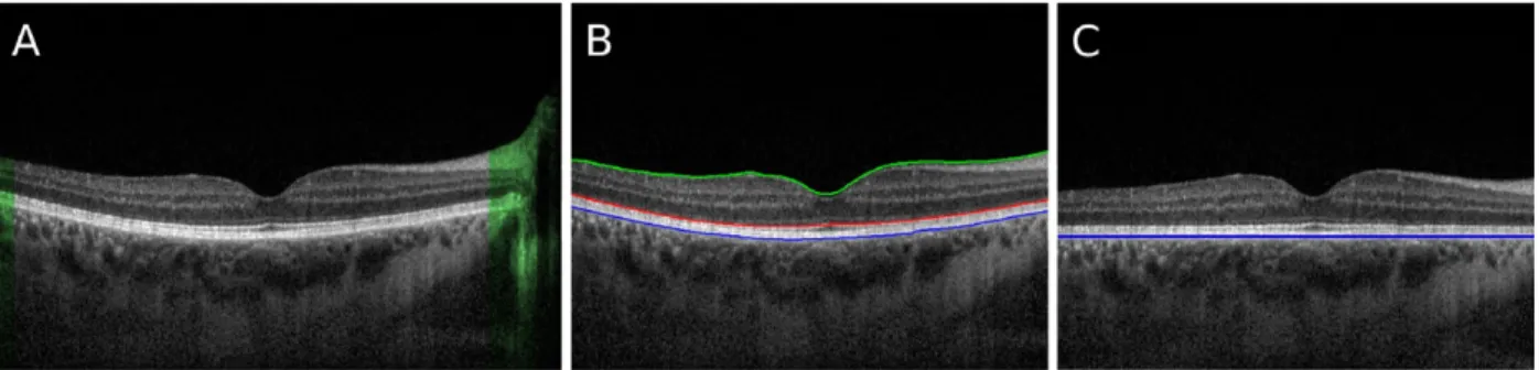
beams is the same), an interference pattern forms. ... 59 Figure 8. – Automated segmentation of retinal layers of interest from OCT images. The
image in A is a typical frame from the video series. A) A-scans where the choroid is absent (highlighted in green) are discarded from all frames. B) Segmentation of the outmost layers of the retina: RVI is indicated in green, anterior RPE in red, and posterior RPE in blue. C) The A-scans are shifted so that the blue layer appears flattened. (Reprinted from Beaton et al. (27)
with permission from The Optical Society.) ... 62 Figure 9. – Top) Heatmap showing pixels which are very likely to lie on a boundary,
including node locations (yellow) and the CSI (red line). The b-scan is flattened and the green dashed line shows the limit of 585 µm below the Bruch’s beyond which nodes are discarded. Bottom) The original b-scan overlaid with the RPE (blue), CSI (yellow) and the mean CT (red dotted line). (Reprinted from Beaton et al. (27) with permission from The Optical
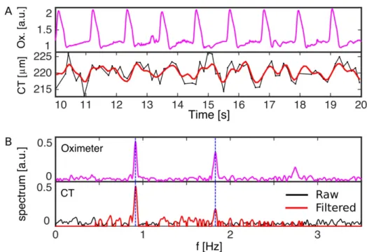
Society.) ... 62 Figure 10. – A) Frequency spectrum analysis of CT fluctuations in time, showing the
oximeter signal, raw fluctuations of CT versus time (black) and band-pass filtered CT signal (red). B) Frequency spectrum of the oximeter signal (top), and CT signal (bottom), where the offset component has been omitted. The filtered frequency band for the CT spectrum is shown in red. The dashed blue lines indicate the two first harmonics of the measured heart rate which are observed in both signals. (Reprinted from Beaton et al. (27) with
permission from The Optical Society.) ... 63 Figure 11. – Scatterplot showing the relationship between ocular rigidity and the
pulsatile ocular volume change in 260 subjects. ... 75 Figure 12. – Scatter plots showing significant correlations between ocular rigidity
coefficients and the A) neuro-retinal rim area (r=0.287, p=0.002; Rim Area=0.85+6.33*OR); B) minimum ganglion cell complex (GCC) thickness (r=0.304, p=0.002; GCC=60.92+262*OR); C) average retinal nerve fiber layer (RNFL) thickness (r=0.220, p=0.017; Average RNFL=74.36+202*OR); D) RNFL thickness in the inferior quadrant (r=0.255, p=0.007; Inferior
Quadrant RNFL=88.05+420*OR). ... 84 Figure 13. – Questionnaire developed to establish the presence of vasospasticity,
cardiovascular diseases, and other vascular risk factors. ... 91 Figure 14. – Relationship between ocular rigidity coefficients and neuro-retinal damage
parameters in the vasospastic group. Scatter plots showing significant correlations between ocular rigidity coefficients and the A) minimum
ganglion cell complex (GCC) thickness (r=0.681, p=0.030; GCC=56.51+440*OR); B) average retinal nerve fiber layer (RNFL) thickness (r=0.745, p=0.013; Average RNFL=61.42+729*OR); C) RNFL thickness in the temporal quadrant (r=0.772, p=0.009; Temporal Quadrant
RNFL=46.23+432*OR). ... 94 Figure 15. – Relationship between ocular rigidity coefficients and neuro-retinal damage
parameters in the atherosclerotic group. Scatter plots showing significant correlations between ocular rigidity coefficients and the A) minimum ganglion cell complex (GCC) thickness (r=0.219, p=0.282; GCC=60.81+206*OR); B) average retinal nerve fiber layer (RNFL) thickness (r=0.190, p=0.261; Average RNFL=74.77+182*OR); C) RNFL thickness in the temporal quadrant (r=0.179, p=0.319; Temporal Quadrant
RNFL=52.62+212*OR). ... 94 Figure 16. – Ocular rigidity differences between healthy axial myopic (0.015±0.007/μL)
and non-myopic (0.033±0.013/μL) eyes (p<0.001), as well as glaucomatous axial myopic (0.0189±0.007/μL) and non-myopic eyes (0.0282±0.014/μL) (p=0.020). Data is presented as mean of OR coefficients and 95%
confidence intervals. ... 117 Figure 17. – Linear regression plot showing a moderate positive correlation (r = -0.475,
p < 0.001) between the ocular rigidity (OR) coefficient and ocular axial length (AL) in 112 eyes. A significant regression equation was found: AL =
List of Abbreviations
AC: Anterior chamber
ACD: Anterior chamber depth AH: Aqueous humor
AL: Ocular axial length
AMD: Age-related macular degeneration BF: Blood flow
BM: Bruch’s membrane BP: Blood pressure
CCT: Central corneal thickness CH: Corneal hysteresis
CRA: Central retinal artery CRF: Corneal resistance factor CSI: Choroid-sclera interface CT: Choroidal thickness
ΔCT: Pulsatile choroidal thickness change DBP: Diastolic blood pressure
DCT: Dynamic contour tonometry EDI: Enhanced depth imaging ET-1: Endothelin-1
FAZ: Foveal avascular zone
GAT: Goldmann applanation tonometry HTN: Hypertension
IOP: Intraocular pressure
IOPcc: corneal-compensated IOP IOPg: Goldmann-correlated IOP IVI: Intravitreal injection
LC: Lamina cribrosa
MD: Visual field mean defect NCT: Non-contact tonometry NTG: Normal-tension glaucoma OAG: Open-angle glaucoma
OCT: Optical coherence tomography OHT: Ocular hypertension
ONH: Optic nerve head OPA: Ocular pulse amplitude OPP: Ocular perfusion pressure OR: Ocular rigidity
ORA: Ocular response analyzer POAG: Primary open-angle glaucoma PP: Perfusion pressure
PPS: Peripapillary sclera
PVD: Primary vascular dysregulation R: Radius
RGC: Retinal ganglion cell RNFL: Retinal nerve fiber layer RPE: Retinal pigmented epithelium RVI: Retina-vitreous interface SBP: Systolic blood pressure
SPCA: Short posterior ciliary arteries TM: Trabecular meshwork
Tmax: Maximum historic IOP ΔV: Pulsatile ocular volume change VF: Visual field
Acknowledgements
This thesis work would not have been possible without the support of many.
First, I would like to thank my thesis directors, Dr Mark Lesk and Dr Santiago Costantino. I am truly grateful to have had the opportunity to work with you and to have you as mentors. Your help, availability and constant guidance are truly recognized and appreciated. Thank you for challenging me to learn and grow as a clinician-scientist.
Thank you to all members of the thesis evaluation committee, Dr Cindy Hutnik, Dr Adriana Di Polo, Dr Michaud, for taking the time to review my thesis and provide valuable suggestions. I would like to thank the funding agencies, including the Fonds de Recherche du Québec – Santé (FRQS), the Ophthalmology Department of University of Montreal Research Fund (FROUM), and the Faculty of Graduate Studies of the University of Montreal for granting doctoral scholarship awards during my PhD studies.
To the members of the research laboratory at HMR, research is a team effort, and I couldn’t have done it without you. To all my colleagues and collaborators at HMR, as well as to my colleagues at the University of Montreal and School of Optometry, you have made this journey memorable! Finally, to my parents and siblings, you are partakers of this accomplishment. Your unwavering love and support have carried me through. Thank you!
Chapter 1 – Introduction
Glaucoma is the leading cause of irreversible blindness in the world (1). An insidious and unpredictable disease, glaucoma causes damage to the retinal ganglion cells (RGCs) that form the optic nerve and can remain asymptomatic until major irreversible visual loss has occurred. The clinical hallmark of this disease includes the progressive deformation and excavation of the tissues of the optic nerve head (2), as seen in Figure 1. Once detected, the disease’s progression rate cannot be anticipated. Furthermore, the pathogenesis of open-angle glaucoma (OAG), the main form of glaucoma, is poorly understood.
While the development of OAG was traditionally attributed to elevated intraocular pressure (IOP), the susceptibility of individual eyes to glaucomatous damage is variable. Nearly half of OAG patients have IOP within the normal range (3), going up to almost 90% of patients in some populations (4). In contrast, most patients with elevated IOP do not develop glaucoma (5). This suggests that factors other than IOP must also underlie the susceptibility of the optic nerve head (ONH) to glaucomatous injury.
The realization that a given IOP can result in very different strains at the ONH in different eyes led to an entire field of research known as ocular biomechanics. Central to this theory is the fact that the retinal axons that unite at the ONH to form the optic nerve leave the eye through the lamina cribrosa. The lamina is the major load-bearing tissue of the ONH and is accepted as both a site of discontinuity and weakness in the corneoscleral shell of the eye and as the most likely site of damage to ONH axons (6-10). Two main hypotheses have been proposed to explain the development of glaucomatous optic neuropathy, namely the mechanical and vascular theories (11). The mechanical theory postulates that elevated mechanical stresses and strain lead to axonal damage and loss of retinal ganglion cells (12-14). The ONH’s response to these biomechanical stimuli has been found to depend on eye-specific geometrical and material properties (Figure 2) (15). This is thought to determine an individual’s predisposition to develop OAG.
There is mounting evidence that the stiffness of the sclera, major contributor to the rigidity of the corneoscleral shell, is an important risk factor in the development and progression of glaucomatous optic neuropathy, perhaps more so than IOP (16). Despite numerous studies on the association between ocular rigidity (OR) and OAG in the last eighty years (17-23), this remains unclear, and competing hypotheses are highly debated (24). On one side, OR is thought to be higher in glaucomatous eyes, producing higher IOP fluctuations due to rigid ocular walls, and hence more deformation at the ONH. On the other side, OR is thought to be lower in early glaucoma, engendering axonal stretching and damage. According to this theory, increased OR would occur at later stages of the disease.
Challenges that researchers face when studying OR range from a plethora of confounding factors, both ocular and systemic, including ocular volume and shape, scleral thickness, choroidal blood volume, age and ethnicity, which can have an effect on OR (25). The difficulty in diagnosing glaucoma at the earliest phase of the disease is another obstacle. The lack of longitudinal studies to show whether OR contributes primarily to glaucoma or is altered due to the disease hinders our knowledge of this parameter. More importantly, the ability to quantify OR in living human eyes using a reliable and non-invasive method is essential to investigate the role of OR in OAG. Such a method has only recently become available thanks to developments in our laboratory. Based on the Friedenwald equation (26), the method uses video-rate optical coherence tomography (OCT) imaging and automated choroidal segmentation to measure the pulsatile choroidal volume change (ΔV), and Pascal dynamic contour tonometry to measure the corresponding pulsatile pressure change (27). It will be described in more detail in Chapter 3. The objectives of this thesis work were thus to validate the method developed to measure OR in
vivo, and to investigate the relevance of OR in ocular diseases, particularly in OAG. More
specifically, we sought to 1) To improve the extrapolation of ΔV and evaluate the method’s validity and repeatability, 2) To investigate the association between OR and neuro-retinal damage in glaucomatous patients, as well as those with concomitant vasospasticity, 3) To evaluate the association between OR and corneal biomechanical parameters, 4) To assess the association between OR and IOP spikes following therapeutic intravitreal injections, to predict and prevent them in high-risk patients, and 5) To confirm that OR is lower in myopia.
Before addressing this, the following chapter will briefly review ocular hydrodynamics (IOP and aqueous humor dynamics) and hemodynamics (blood flow), as these elements are involved in OR measurement. This will be followed by a review of the most prominent findings pertaining to ocular biomechanics, and will present the current evidence on the link between OR and OAG.
Figure 1. – Glaucomatous optic neuropathy is characterized by the progressive deformation and excavation of tissues at the optic nerve head. Thinning of the neuroretinal rim due to axonal
loss, increased cupping and bowing of the lamina cribrosa are clinical hallmarks of this disease. (Courtesy of Dr Lesk and Dr Sayah.)
Figure 2. – Representation scheme of the biomechanical paradigm of glaucoma. IOP-induced deformation, stress and strain produce alterations in physiological processes which ultimately lead to axonal loss and glaucomatous damage. (Reprinted from Sigal et al. (15)
Chapter 2 – Literature Review
2.1 Aqueous Humor Dynamics and Intraocular Pressure
The eye is a pressurized shell, filled with a colorless fluid of similar composition to plasma – the aqueous humor (AH). AH circulates in the anterior part of the eye, while the vitreous humor, or vitreous body, a gelatinous mass composed primarily of water (98-99.7%) and hyaluronic acid, is found in the retrolental space, with little moving fluid (28). In the eye, AH plays an important role in the nourishment and homeostasis of the cornea and lens, transparent and avascular structures that permit light transmission to the retina.
2.1.1 Aqueous Humor Production and Drainage
AH is continually produced and evacuated. The production of AH occurs in the ciliary processes (Figure 3). From the fenestrated capillaries, through the ciliary process stroma and a double-layered epithelium, it is in the non-pigmented epithelial cells of this double-layer that almost 90% of AH is formed through active secretion (29). AH is secreted into the posterior chamber, traverses the pupil and circulates in the anterior chamber (AC), following a convective flow pattern. This pattern is caused by the temperature difference between the iris (higher temperature) and the cornea (lower temperature) in the AC, producing an upward flow near the iris and a downward flow near the cornea (30).
The evacuation of AH occurs through two routes, the trabecular and uveoscleral pathways. The trabecular or conventional pathway is responsible for 85% of AH outflow from the eye. Its primary constituent is the trabecular meshwork (TM), composed of the uveal meshwork, corneoscleral meshwork and juxtacanalicular tissue, the latter providing the most resistance to AH outflow (31). Driven by the pressure gradient, AH flows out through the TM, across the inner wall of Schlemm’s canal, and drains in the collector channels, aqueous veins and episcleral veins (32, 33). When traveling through the uveoscleral pathway, AH flows through the uveal meshwork and anterior face of the ciliary muscle, and drains into the suprachoroidal space and the sclera (29).
Figure 3. – Schematic representation of the anterior segment of the human eye. Arrows indicate aqueous humor flow pathways. Aqueous humor is formed by the ciliary processes, enters the posterior chamber, flows through the pupil into the anterior chamber, and exits at the
chamber angle via the trabecular and uveoscleral routes. (Reprinted from Kaufman, Wiedman (34), with permission from Springer.)
2.1.2 Intraocular Pressure
At steady-state IOP, which is approximately 15 mmHg, the rate of AH production and evacuation are equal (35). In a healthy adult eye, this corresponds to an AH turnover rate of 2.75±0.63μl/min during daytime (36), or about 1-1.5% of the AC volume per minute (37). This state of equilibrium is essential in maintaining physiological processes as well as the shape of the eye.
Many factors are known to affect IOP. These include the circadian rhythm, heartbeat, respiration, exercise, posture, fluid intake, alcohol and cannabis use, as well as topical and systemic medications (38). Age and disease can also affect IOP. This is particularly true for glaucoma, in
which IOP holds an important role both as a risk factor and in its management. Increased resistance of aqueous outflow through the TM is a known risk factor for glaucoma and can cause ocular hypertension (5, 39, 40). From a biomechanical perspective, the stiffness of the TM is thought to increase in glaucoma (41, 42), although the mechanism through which this occurs is not well understood. However, in vivo measurement of TM stiffness can be challenging and their interpretation can be problematic as many agents, including topical glaucoma medications, can alter TM function and potentially TM stiffness (43). Currently, the only evidence-based treatment for OAG is to lower IOP, regardless of baseline IOP (44-48). IOP reduction can be carried out using pharmaceuticals, laser trabeculoplasty, surgical procedures, or a combination of these methods. In general, these methods reduce IOP by reducing AH production and/or increasing its evacuation. Some surgical procedures, such as trabeculectomy and drainage devices, create a new exit route to drain the AH and bring the IOP to lower levels.
2.1.3 Tonometry
Tonometry is a crucial part of an eye examination, especially for accurate measurement and management of IOP in glaucoma. Numerous methods have been developed to measure IOP in
vivo. The main tonometry techniques used in this thesis work, and those historically involved in
biomechanical measurements such as the rigidity of the eye, will be briefly described in this section.
One of the first and most commonly used modern device to measure IOP in the world was the Schiötz tonometer (49). As an indentation tonometer, it is based on the principle that a force or weight would produce less indentation in a harder object, or eye. Thus, the amount of indentation would enable IOP estimation, providing that less indentation would be indicative of elevated IOP. To measure IOP, the Schiötz’s curved footplate is set on the anesthetized cornea. Different weights, between 5 to 15 g, can be added to the plunger and used to indent the cornea. A table is then used to convert the scale reading to the IOP value in mmHg. One major caveat of the Schiötz is that it is highly dependent on ocular rigidity, leading to underestimation of IOP in eyes with lower rigidity and vice versa (50). Despite the development of updated conversion tables by Friedenwald to correct for OR (51, 52), this method was replaced by Goldmann applanation
tonometry (GAT) in the last quarter of the 20th century, rendering GAT the most used tonometer worldwide.
Applanation tonometry is based on the Imbert-Fick principle, which states that the pressure (P) inside a thin-walled sphere filled with liquid equals the force (F) necessary to flatten its surface divided by the flattened area (A), such that P=F/A (53). In GAT, the anesthetized cornea is flattened by a prism of 3.06 mm diameter (7.35 mm2). Using fluorescein dye to highlight the tear film, the IOP is determined by varying the applanation force to properly align the superior and inferior mires produced by the prism. Since the Imbert-Fick principles assumes a thin-walled sphere, it does not account for thickness and rigidity. This tends to lead to underestimation of the measured IOP using GAT in thin corneas, and vice versa (54-58). However, no accurate adjustment of GAT-IOP readings for CCT currently exists for individual eyes, as “true” IOP also depends on other biomechanical factors of the cornea which current nomograms do not account for (59, 60). Other applanation tonometers include non-contact tonometry (NCT), pneumotonometry and the Tono-Pen. NCT uses an air pulse of increasing intensity to applanate the cornea. The Ocular Response Analyzer (ORA), a newer type of NCT, measures the biomechanical response of the eye to the rapid air jet-induced deformation at the cornea (61). It yields the Goldmann-correlated IOP (IOPg) and the corneal-compensated IOP (IOPcc), which is less dependent on biomechanical properties, as well as the corneal hysteresis (CH) and corneal resistance factor (CRF). CH and CRF provide information on the viscoelasticity of the cornea and will be described in more detail in the upcoming section on the mechanics of the cornea. Another device based on NCT is the Corneal Visualization Scheimpflug Technology tonometer (Corvis ST). Using a high-speed Scheimpflug camera to visualize and measure corneal deformation in response to an air impulse, the biomechanical corrected IOP (bIOP) is measured, along with various other parameters that describe the viscoelastic properties of the cornea (62). Pneumotonometry uses a 5mm diameter piston with silicone tip and air pressure to flatten the cornea. When the tip and the cornea are both flattened, this corresponds to the IOP (49, 63). The Tono-Pen is a portable tonometer, with an applanating surface and a small plunger protruding slightly from its center. A strain gauge creates an electrical signal that provides an IOP reading when both the plunger and the surrounding surface applanate the anesthetized cornea (63). Each IOP measurement obtained
with this tonometer is an average of multiple IOP readings, and is recorded along with a reliability index ranging from ≤5% to >20%, with the lowest number indicating the highest reliability. Finally, dynamic contour tonometry (DCT) is a more recent technique that enables the measurement of IOP using the principle of contour matching or Pascal principle. By matching the contour of the cornea, the pressure inside and outside a sphere should be equal. A piezoresistive sensor at the center of the concave tip measures the IOP dynamically when in contact with the cornea (64). When force is exerted on the sensor as it comes in contact with the cornea, the silicon, of which it is composed, becomes more resistant to the small current passing through it. The alteration in resistance is detected and amplified by a wheatstone bridge and is converted to a change in pressure, according to a linear relationship. The three output variables from the Pascal DCT are a quality signal (Q=1 to 5; lower Q is better), the diastolic IOP and the ocular pulse amplitude (OPA), which corresponds to the difference in IOP between the systole and diastole. The IOP measurement from the Pascal DCT, as well as the IOPcc obtained using the ORA, were shown to be independent of central corneal thickness (CCT) and corneal biomechanical properties (65, 66).
2.2 Ocular Hemodynamics
In the human body, the eye is the only place where a direct view to the fundus and blood vessels is possible; it is thus a privileged site for the study of blood flow and its regulatory mechanisms. It is also the organ with the highest metabolism and as such requires a high but regulated blood supply (67, 68). While the measurement of blood flow can contribute to a better understanding of the vasogenic theory of glaucoma (69), it can also bring some insight unto the mechanical theory of this optic neuropathy. In this thesis work, this is relevant for two reasons: 1) the method we developed to estimate the ocular rigidity coefficient involves the measurement of pulsatile choroidal blood flow (as will be described in Chapter 3), and 2) pathological conditions and medications can alter ocular blood flow (70). A review of blood flow and its regulatory mechanisms is thus relevant as it can have an impact on the measurement of OR. The following subsections will focus on the effects of glaucoma, vasospastic disorders and medications on ocular blood flow.
2.2.1 Anatomy of the Ocular Vascular System
Two main vascular systems, retinal and uveal, irrigate the eye, as seen in Figure 4. All blood supply to the eye originates from the ophthalmic artery, which proceeds from the internal carotid artery. The ophthalmic artery is divided into multiple branches, including the central retinal artery (CRA), the anterior ciliary arteries and the short and long posterior ciliary arteries (71). The blood flows out of the eye primarily via the central retinal vein and vortex veins which then drain into the cavernous sinus (71).
The CRA supplies nourishment to the inner retinal layers, from the retinal nerve fiber layer (RNFL) to the outer plexiform layer through two non-fenestrated capillary networks located in the inner nuclear and inner plexiform layers of the retina. The presence of tight junctions between the endothelial cells of these arterioles contributes to the inner part of the blood-retinal barrier. The outer part of this barrier is found at the retinal pigment epithelium cells layer (71).
The retinal arterioles do not supply the foveal avascular zone (FAZ); the absence of superficial blood vessels in this region allows for optimal visual acuity. Rather, the blood input to the FAZ and the outer third of the retina comes from the choroid which proceeds from the short posterior ciliary arteries (SPCA) (72). The choroid represents 80-90% of the total ocular blood volume and flow (73). This characteristic of the choroid is important in the methodology we developed to estimate ocular rigidity. The choroid is essential in maintaining nourishment to the retina, one of the highest oxygen-consuming tissue in the human body (67). It is composed of five layers: Bruch's membrane (BM), the choriocapillaris, Haller's and Sattler's vascular layers, and the suprachoroidea (74). It is also thought to be involved in disease processes such as glaucoma, thermoregulation, secretion of growth factors, and emmetropization (eye growth) (75).
In addition to supplying the choroid, the SPCA form a circular ring called the circle of Zinn and Haller which supplies blood to the prelaminar and laminar portion of the optic nerve head (ONH). The SPCA may also give rise to cilioretinal arteries in about 27% of optic disks, especially in large disks (76). The anterior and long posterior ciliary arteries anastomose to form the major and minor arterial arcades in the anterior part of the eye, supplying nutrients and oxygen to the ciliary body and the iris, and playing a role in AH production (71).
Figure 4. – Anatomy of the ocular vascular system. A) Cutaway drawing of the human eye showing the major blood vessels supplying the retina, choroid and anterior segment. B) Cutaway drawing along the superior–inferior axis of the human eye through the optic nerve, showing
the vascular supply in this location. C) Drawing showing the vasculature of the retina and choroid. (Drawings by Dave Schumick. Reprinted from Anand-Apte and Hollyfield (77), with
2.2.2 Blood Flow and Regulation
Proper homeostasis requires a continuous flow of blood through the capillaries of the body. This flow is governed by the pressure gradient, or perfusion pressure (PP; in mmHg), between the vessel ends, and the resistance (R), or friction, of the circulating blood on the vessel walls. Blood flow (BF) is a measurement of volume in function of time, is described by the following equation (78, 79):
𝐵𝐹 = 𝑃𝑃 𝑅
In the eye, the mean PP corresponds to the difference between the mean blood pressure (BP) in the ophthalmic artery and the venous pressure as the blood exits the eye. Since IOP is considered to be almost equal to the venous pressure, the PP at the eye level is defined as follows:
𝑃𝑃 = 23 )𝐷𝐵𝑃 + 13 (𝑆𝐵𝑃 − 𝐷𝐵𝑃)1 − 𝐼𝑂𝑃
where DBP and SBP are the diastolic and systolic blood pressures respectively (79). This shows that ocular blood flow can be strongly influenced by IOP (80).
Knowing that the resistance to flow in a cylindrical pipe, such as blood vessels, depends on its radius (r), its length (L), the blood’s viscosity (h) and the inherent resistance to flow, these elements can be included in the Hagen-Poiseuille law, as follows (78, 79):
𝐵𝐹 = 𝑃𝑃 𝜋 𝑟! 8 𝐿 h
An alternative equation, Murray’s law (81) may be more adequate for microvascular beds (79):
𝐵𝐹 = 𝑘 (𝑟" 9𝜂)
where k is a constant that depends on the vessel’s length and the blood’s viscosity.
Another important element of homeostasis is the ability of the vascular system to regulate. Regulation is usually achieved with the influence of the autonomic nervous system and circulating hormones, as well as local autoregulation (82, 83). Autoregulation is the ability of local tissue to
maintain a constant blood flow despite perturbations such as variations in metabolic demand or in PP (84). In the first condition, the aim is to regulate oxygen and carbon dioxide concentration as well as adenosine, nitric oxide and endothelin-1 (ET-1) levels (82, 83). The second mechanism of autoregulation pertains to mechanical, or myogenic, influences (85, 86), such as changes in PP (or systemic BP or IOP), which can alter vessel diameter to modify the vascular resistance accordingly as shown by the Hagen-Poiseuille and Murray equations (79, 82).
In the eye, the retinal and choroidal systems possess various intrinsic differences (82, 87). Unlike the retinal vasculature, where the lack of autonomic innervation (88) and the blood-retinal barrier (87, 89) limits the effect, the choroid’s blood vessels are fenestrated and innervated, and can thus be affected by the body’s chemical messengers as well as the autonomic nervous system (82, 87). Another difference is their ability to autoregulate. The retinal vasculature possesses contractile elements, including several layers of smooth muscle cells and a high density of pericytes (82, 90, 91). Retinal blood flow can thus be maintained constant through autoregulation up to an IOP of about 30 mmHg (73, 92). At elevated IOP, retinal blood flow decreases. This may be relevant to glaucoma, considering that retinal circulation provides the inner retina and inner parts of the ONH (73). This ability to autoregulate is widely recognized for the retina, optic nerve, retrobulbar vessels (83, 87, 93-95), and the anterior uveal circulation (73). However it remains nebulous for the choroid, as it was thought not to be autoregulated (73) until recent evidence pointing to the contrary (96, 97).
Many experimental methods have been developed and have enabled the study of autoregulative responses. These methods often involve the alteration of OPP by inducing a significant change in systemic BP, a change in IOP, positional change, or through exercise, and measuring blood flow following perturbation (83). Experiments using flickering light stimuli were also designed to evaluate autoregulative responses of the ocular vasculature by increasing the neuronal activity of the eye, which increases the metabolic demand, and should ultimately lead to increased blood flow (98, 99).
Various studies investigating choroidal blood flow have shown that it remains somewhat constant with PP changes. Riva et al. developed a method based on laser doppler flowmetry to measure
the relative choroidal blood flow subfoveally (100). They found evidence of autoregulation in the choroid in response to acute PP changes due to isometric exercise (101). In their study, when PP increased by up to 67%, an elevation in vascular resistance in the choroid was suggested as the main mechanism for this autoregulation. Other studies showed similar findings by altering IOP or BP through exercise or different means, both in human and rabbit eyes (96, 102-105).
2.2.3 Blood Flow and Glaucoma
Blood flow dysregulation is an independent risk factor to various pathological conditions (70). In glaucoma, impaired autoregulation is thought to be present at all levels of the vascular system (83). While OAG is a multifactorial disease leading to RGC death and visual field loss, it is believed that autoregulative ability as well as OPP play an important role in the development of this condition (106-109).
Some examples of dysregulation include impaired response to posture-induced changes in OPP in the retinal vasculature in OAG patients compared to controls (110), and less retinal vessel reactivity (diameter change) to short-term IOP elevation in OAG than OHT eyes (111). Hafez et al. found markedly improved blood flow in the neuro-retinal rim following therapeutic IOP reduction in OAG eyes, as opposed to eyes with OHT (112). This may indicate that OAG patients have impaired autoregulation in the neuro-retinal rim, whereas OHT patients do not. Studies have also shown impaired autoregulation in the choroidal (113-116) as well as in the retrobulbar vasculature (117-119) in OAG. For example, improvement of choroidal blood flow was demonstrated following trabeculectomy, an IOP-lowering surgery, suggesting impaired choroidal autoregulation in glaucoma (115). A higher susceptibility to IOP-induced glaucomatous damage was also shown to occur in patients with vascular dysregulation, with visual field progression at lower levels of IOP (114). Numerous studies also show an association between reduced blood flow and glaucomatous damage or visual field loss (120-129), and disease progression (130). While the exact cause of vascular dysregulation in glaucoma is unknown, it could result from an inconsistent supply of oxygen to ocular tissue, or in other words, from alternating periods of hypoxia and reperfusion (131). This phenomenon causes oxidative damage to cells, and mitochondria, which are numerous in the optic nerve head (132) and also present in the
trabecular meshwork (133). Mechanical and ischemic stress is also thought to activate astrocytes, which can lead to reduced trophic functions, tissue remodeling and subsequent ONH excavation (134). The major causes of unstable blood flow and oxidative stress are elevated IOP which exceed the autoregulative capacities, or vasospastic disorders.
2.2.4 Blood Flow and Vasospastic Disorders
Vasospastic disorders are characterized by vascular dysregulation in the extremities, leading to vasospasm, an abnormal constriction of arteries (135). Common examples of vasospastic disorders include Raynaud’s disease and migraines. However vasospastic disorders can include a range of signs and symptoms, and may not all result in Raynaud-like manifestations. They can be secondary to a systemic disease or can present in otherwise healthy patients.
Primary vascular dysregulation (PVD) is characterized by the body’s abnormal response to stimuli such as temperature and emotional stress, leading to cold hands and/or feet. These symptoms are usually present after puberty but may improve with menopause, suggesting hormonal involvement in PVD. The use of certain medications such as calcium channel blockers which have vasodilatory properties, as well as gingko biloba supplements can have beneficial effects in vasospastic subjects (136, 137).
PVD is thought to be more common in thin women with type-A personalities (138). Several characteristics of PVD have been identified including a reduced feeling of thirst, low blood pressure, and increased ET-1 plasma levels (139). In terms of ocular manifestations, PVD can be associated with splinter hemorrhages, focal rim loss, dense central scotomas, as well as a dysregulation of blood flow to perfusion pressure changes (138, 140, 141). Vasospasticity is thus associated with glaucoma, particularly normal-tension glaucoma (NTG) (142-144).
While testing for ET-1 may be one way to test for vasospasticity, other objective tests exist. The most common include finger laser doppler flowmetry and nailfold capillaroscopy to evaluate the peripheral vascular response to temperature change (138).
There is some evidence that biomechanical stimuli may bring forth abnormal vascular response in vasospastic patients, which renders this condition particularly interesting in the context of this
thesis work. In a previous study, our group showed a greater improvement in rim blood flow following IOP reduction in vasospastic patients compared to non-vasospastic patients, indicating defective autoregulation in patients with vasospasm (145). Another study by Schulzer et al. (144) also demonstrated differences between vasospastic and non-vasospastic patients. More specifically, they showed that within a glaucomatous population, two distinct populations could be identified and each exhibited different characteristics. Interestingly, the group with vasospasticity had a high positive correlation between the mean defect (MD) index of visual field and the maximum IOP (Tmax), while the group with vascular disease, akin to atherosclerosis, showed no correlation between these variables.
2.2.5 Blood Flow and Medication
As discussed previously, reduced blood flow in glaucoma may have adverse effects on the progression of the disease (130, 146). While treatment aimed at lowering IOP is thought to improve OPP and blood flow, the effect of some medication, particularly some systemic hypertensive drugs, can lower the diastolic blood pressure, which in turn lowers the OPP. This can have a significant impact on blood flow regulation, and have a negative effect on glaucoma progression (147-149).
2.2.5.1 Blood Flow and Ocular Hypotensive Medication
Various IOP-lowering agents are commonly used to treat glaucoma. We will focus on the most common and current ones.
Prostaglandin analogues use generally shows an improved ocular blood flow. Most studies on latanoprost showed improved pulsatile ocular blood flow (150-156) as well as beneficial effects on the ONH circulation (157-159), including increased OPP at the optic disc (160). Unoprostone improved ONH circulation (161-165), although no improvement was found in vasospastic subjects (166). Unoprostone also exhibited an antagonistic effect of ET-1 by improving impaired choroidal blood flow (167, 168). Bimatoprost was shown to cause vasoconstriction of the ciliary arteries
in-vitro at high concentrations (169), tafluprost exhibited a vasodilatory effect in-in-vitro (170) and
travoprost did neither (171). Numerous studies with bimatoprost showed no effect on retrobulbar hemodynamics in glaucomatous eyes (172-176), whereas one study found a positive
effect in healthy eyes (177). However, travoprost mostly showed a positive effect on the retrobulbar vasculature (173, 177), as well as a sustained ONH circulation improvement (162). Topical clonidine, an alpha-2 receptor agonist, demonstrated a reduction in OPP in OAG patients (178), and brimonidine showed an increased ocular blood flow in glaucoma patients (179, 180). However, brimonidine has also been associated with decreased nocturnal OPP due to its lowering effect on BP overnight (181). Non-selective b-blockers, particularly timolol, have been associated with unchanged (182-186) or decreased (187-190) ocular blood flow as measured using various techniques. Betaxolol, a b1-selective adrenoceptor antagonist with a probable calcium-channel blocker action, has demonstrated a beneficial effect on blood flow at multiple levels in the eye, including the choroid and ONH circulation (191-194). Topical carbonic anhydrase inhibitors (CAI), dorzolamide and brinzolamide, have both shown enhancing effects on ocular blood flow and its regulation (148, 186, 195, 196). Pilocarpine, a parasympathomimetic vasodilator, displays no effect on ocular blood flow in most studies (197-200). A new class of IOP-lowering drug, rho kinase inhibitors, have been shown to have vasodilatory effects on the conjunctiva (201) and increase ocular blood flow (202).
2.2.5.2 Blood Flow and Systemic Medication
Few studies have been carried out to investigate the role of systemic medications on ocular blood flow. Even fewer have looked at the ocular effects of systemic drugs in glaucoma subjects (137). This section will present the effects of the most common and relevant medications on the eye, as found in the literature.
Systemic CAIs, namely acetazolamide and dorzolamide, have generally been shown to exert a vasodilatory effect on both the brain and the eye, showing increased retinal arterioles’ diameter and increased retinal, choroidal and ONH blood flow in the latter (203-206). The dilation of retinal capillaries in an animal model has been linked to decreased extracellular pH (207).
Calcium channel blockers, or antagonists, are among the most prescribed medications to lower blood pressure in hypertensive patients (208). Their mechanism of action involves the inhibition of calcium ions’ entry into cells which leads to smooth muscle relaxation and vasodilation. Centrally acting calcium channel blockers appear to increase blood flow in the eye, whereas
peripherally acting calcium channel blockers do not (137). This was shown in multiple reports demonstrating increased blood flow in the ONH and juxtapapillary retina, as well as in other ocular structures including the choroid (209-228). As mentioned previously, calcium channel blockers can also have positive effects on blood flow in vasospastic subjects compared with non-vasospastic subjects (229).
The renin-angiotensin system plays a key role in vasoconstriction, leading to high systemic BP (230). Two classes of medications act on this system to lower BP, namely the angiotensin-converting enzyme (ACE) inhibitors and the angiotensin II receptor blockers (ARB). In the eye, an increase in the velocity of blood flow in the CRA as well as in the posterior ciliary arteries in subjects suffering from essential hypertension treated with an ACE inhibitor was reported (231). The effect of losartan, an ARB, in normal subjects shows some effects on the retrobulbar and choroidal blood flow that can lead to an increased fundus pulse amplitude (232, 233).
Nitric oxide (NO) can lower IOP and increase blood flow through its vasodilatory role on smooth muscle cells, as well a potential autoregulatory effect on the retinal vasculature (234-236). Simvastatin, a drug used to treat dyslipidemia, leads to increased blood velocity and blood flow in the retinal vasculature (237). These effects may be due to increased plasma levels of NO triggered by the drug.
Cannabinoids have IOP-lowering effects as well as vasodilating properties which may be able to increase blood flow in the eye in theory (238, 239). However, they can also cause systemic BP reduction and tachycardia at the same dose required to effectively lower IOP, which may hinder the therapeutic effect of the drug on glaucoma by thus lowering the ocular blood flow (238, 240). Ginkgo biloba extract is thought to have numerous beneficial properties for the treatment of glaucoma. There is evidence for reduced blood viscosity and vasospasms (137), increased ocular blood flow (241, 242) and neuroprotective properties with ginkgo biloba leading to improved RGC survival (243, 244). Improved visual function was also demonstrated in some patients with NTG (245, 246).
Finally, estrogens and hormonal replacement therapy are thought to have neuroprotective properties (247) and were shown to improve ocular blood flow in both the retrobulbar and retinal circulation (137).
2.2.6 Blood Flow and Systemic Factors
Numerous other factors, including dietary and lifestyle choices as well as overall health can influence ocular blood flow. Some common factors, especially those which can be relevant to glaucoma, will be briefly reviewed in this section.
Caffeine increases blood pressure, decreases heart rate and causes vasoconstriction (248, 249). It was shown to decrease blood flow in the eye in healthy eyes (250-252) and increase IOP slightly, by about 1 mmHg, in glaucoma patients (253-255). The Blue Mountains Eye Study found that glaucoma patients who reported regular caffeine consumption had a higher average IOP (256), which may be explained by an increased AH production due to caffeine (257, 258). However, coffee consumption was not associated with POAG (259, 260).
Alcohol is a vasodilator and was shown to increase blood flow at the ONH level (261), but not in the retrobulbar circulation (262). Its acute consumption was also shown to lower IOP (261-263), and was not found to be a risk factor for POAG (264).
Interestingly, while cigarette smoking is thought to induce vasospasm and increase blood viscosity (265), some studies have instead shown increased cerebral and ocular blood flow in response to nicotine (266-269). This may be due to increased cerebral oxygen consumption in smokers (270). Cigarette smoking however was not found to be an important risk factor for POAG in a large prospective study (271).
Obesity has been associated with elevated IOP and reduced retrobulbar blood flow (272, 273), as well as reduced ocular pulse amplitude (274). Epidemiological studies have shown conflicting results regarding the risk of POAG in obese subjects, with some showing increased risk (275), while others decreased risk (276-278) or no associated risk (279, 280).
Obstructive sleep apnea syndrome (OSAS) is a disorder in which breathing can be interrupted multiple times during sleep. It is characterized by a relaxing of the muscles in the throat, which in
turn cause the upper airway to collapse (281). While the typical OSAS patient is thought to be a middle-aged, obese man who snores loudly, this is not always the case and should be considered when screening for this sleep disorder (282-284). OSAS was found to be associated with POAG, particularly NTG, (285-295). It is thought that a dysregulation of blood flow caused by repetitive hypoxic events as well as insufficient ONH perfusion and increased vascular resistance is probably at play in both of these conditions (285). Systemically, impaired coronary endothelial vasoreactivity (296) and decreased cerebral blood flow (297-302) was found in patients with sleep apnea. However, in the eye, blood flow was found to be unaltered in OSAS subjects compared with controls in most studies (303-306), except in one study where impaired autoregulation in the retrobulbar vasculature of OSAS subjects was reported (307).
Both low and high BP have been found to be associated with glaucoma. Numerous studies have reported an association between low DBP and low OPP and a higher prevalence and/or incidence of OAG (308-311). The Baltimore Eye Survey reported a six-fold increase in the prevalence of OAG in patients with the lowest OPP, suggesting altered ocular blood flow and autoregulation (311). The combination of nocturnal hypotension in patients with low systemic BP, or those with treated hypertension (HTN), and elevated IOP is a risk factor for glaucoma, leading to reduced OPP and ONH ischemia (106, 311-315). An association between HTN and OAG, as well as a correlation between SBP and IOP were also consistently found in several clinical studies, suggesting that chronic HTN may impair blood flow to the ONH (309, 316-321). However, this association was later attributed to the correlation between age and HTN (4). In the context of biomechanics which will be discussed in the following section, altering BP was suggested to have an effect on the pressure-volume relationship in rabbit eyes, by altering blood volume and choroidal blood flow (322). These findings were not corroborated in human studies in the range of clinically encountered BP (23, 323).
Diabetes mellitus is known to damage the endothelium and pericytes of blood vessels, leading to dysfunctional regulation of ocular blood flow in the early stages of the disease, followed by ischemia and vascular proliferation in the later stages of diabetic retinopathy (324). Findings regarding choroidal blood flow in diabetes using subfoveal laser Doppler flowmetry generally indicate a reduction (325, 326) and dysregulation (327) of choroidal blood flow in eyes with
diabetic retinopathy. Although diabetes was initially thought to be a risk factor for glaucoma (328-332), conflicting data suggests that diabetes may not be associated with glaucoma (278, 333-338).
2.3 Ocular Biomechanics
Biomechanics is a rapidly developing field, joining physics and biology, and bringing new insights into physiological and pathophysiological mechanisms. In a normal eye, various forces are constantly being exerted on its walls by surrounding or internal structures, including the extraocular muscles and ciliary muscle, during accommodation for example, as well as by fluids such as the AH and the vascular system during the cardiac cycle (80). Throughout these processes, the shape of the eye and position of its optical components is maintained to ensure optimal vision. In other cases, the mechanical properties of the eye can dictate its behaviour in response to different factors, including IOP, and lead to adverse morphological and functional changes. This is thought to be particularly true in diseases such as OAG, age-related macular degeneration (AMD) and myopia. Hence, understanding the structural and material properties of the eye could help elucidate the mechanisms underlying these potentially-blinding diseases, and in turn have a significant impact on clinical practice.
The following sections will present the main findings pertaining to the biomechanical properties of the eye in OAG, from the most anterior to the most posterior structures. They are adapted from a textbook chapter on Glaucoma and Ocular Rigidity that I co-authored (339). The outer coat of the eye is formed by the cornea and the sclera, two tough connective tissues that make up the corneoscleral shell. Posteriorly, the corneoscleral shell is pierced by the scleral canal through which the retinal ganglion cells’ (RGC) axons exit the eye on their way to the brain. The lamina cribrosa (LC), a specialized region of the sclera, spans the scleral canal. It is clear that remodeling of these tissues occurs in glaucoma (14, 340, 341), thus altering the mechanical environment of the optic nerve head (ONH). The properties of these structures will be reviewed with an emphasis on their relevance in glaucoma.
2.3.1 Mechanics of the Cornea
The cornea is the anterior extension of the sclera, and its viscoelastic properties and thickness contribute to the overall rigidity of the eye. The main biomechanical properties of the cornea which can be currently measured and studied in glaucoma are the central corneal thickness (CCT), corneal hysteresis (CH), corneal resistance factor (CRF), and many others, including the ORA waveform parameters and the Corvis ST parameters, that have been studied less extensively.
2.3.1.1 Central Corneal Thickness
CCT is most frequently measured using ultrasound pachymetry, optical pachymetry, Scheimpflug imaging or anterior segment optical coherence tomography (OCT) (342). Initially, CCT was used in the clinical management of glaucoma to correct IOP readings (343). This correction was later shown to be inadequate due to the absence of algorithm to accurately predict the true IOP corrected for CCT (344). The Ocular Hypertension Treatment Study (OHTS) was the first to demonstrate the importance of CCT as a predictor for the development of OAG (5). In this study, a CCT of 555 µm or less was associated with a three-fold increased risk of developing OAG. Further investigation has confirmed CCT to be an independent predictor for the development of POAG (5, 345) as well as a risk factor for the development of visual field (VF) loss in glaucoma patients (346). Several experiments were carried out to better understand the link between CCT and posterior structures of the eye in glaucoma. While this association remains unclear, due to the absence of correlation between CCT and laminar and scleral thicknesses (347, 348), a thinner cornea was suggested to be associated with a more compliant lamina cribrosa due to larger displacement of the LC with IOP reduction in eyes with lower CCT (349). Furthermore, an inverse relationship was found between CCT and optic disc size or area, perhaps indicating larger and more deformable optic discs with lower CCT (350). In a study involving non-invasive measurement of OR, a positive albeit weak correlation was also found between OR and CCT, indicating that subjects with a thinner cornea may have a more compliant sclera (23). In a similar clinical study, no relationship between OR and CCT was found, arguably due to low statistical power (351).
In subjects with no corneal pathology, CCT remains relatively stable. CCT was reportedly lower in subjects from African descent (AD) and Hispanics compared to Caucasians (352-355), although