J. Clin. Endocrinol. Metab. 2010 95:E373-E383 originally published online Aug 4, 2010; , doi: 10.1210/jc.2009-2556 Lauri A. Aaltonen and Albert Beckers
Günter K. Stalla, Anna Spada, Sabina Zacharieva, Jerome Bertherat, Thierry Brue, Vincent Bours, Philippe Chanson, Anne-Paule Gimenez-Roqueplo, Fergus J. Cameron, Françoise Borson-Chazot, Ian Holdaway, Sergio P. A. Toledo,
Halaby, Vinciane Corman, Marie-Thérèse Hagelstein, Jean-François Vanbellinghen, Gustavo Barcelos Barra, Saloranta, Wouter De Herder, Renato Cozzi, Mirtha Guitelman, Flavia Magri, Maria Stefania Lagonigro, Georges Longás, José Ignacio Labarta Aizpún, Marianthi Georgitsi, Ralf Paschke, Cristina Ronchi, Matti Valimaki, Carola
A. Toledo, Maria Isabel Sabaté, Chiara Villa, Marc Popelier, Roberto Salvatori, Juliet Jennings, Ángel Ferrandez Antoine Tabarin, Elisa Verrua, Eija Eloranta, Arnaud Murat, Outi Vierimaa, Pasi I. Salmela, Philippe Emy, Rodrigo
Montañana, Gerald Raverot, Robert J. Weil, Timo Sane, Dominique Maiter, Sebastian Neggers, Maria Yaneva, Luciana A. Naves, Tapani Ebeling, Auli Karhu, Antti Raappana, Laure Cazabat, Ernesto De Menis, Carmen Fajardo Adrian F. Daly, Maria A. Tichomirowa, Patrick Petrossians, Elina Heliövaara, Marie-Lise Jaffrain-Rea, Anne Barlier,
Mutations and Pituitary Adenomas: An International Collaborative Study
AIP
Society please go to: http://jcem.endojournals.org//subscriptions/
or any of the other journals published by The Endocrine
Journal of Clinical Endocrinology & Metabolism
To subscribe to
Clinical Characteristics and Therapeutic Responses in
Patients with Germ-Line AIP Mutations and Pituitary
Adenomas: An International Collaborative Study
Adrian F. Daly,* Maria A. Tichomirowa,* Patrick Petrossians,* Elina Helio¨vaara, Marie-Lise Jaffrain-Rea, Anne Barlier, Luciana A. Naves, Tapani Ebeling, Auli Karhu, Antti Raappana, Laure Cazabat, Ernesto De Menis, Carmen Fajardo Montan˜ana, Gerald Raverot, Robert J. Weil, Timo Sane, Dominique Maiter, Sebastian Neggers, Maria Yaneva, Antoine Tabarin, Elisa Verrua, Eija Eloranta, Arnaud Murat, Outi Vierimaa, Pasi I. Salmela, Philippe Emy, Rodrigo A. Toledo, Maria Isabel Sabate´, Chiara Villa, Marc Popelier, Roberto Salvatori, Juliet Jennings, A´ngel Ferrandez Longa´s,Jose´ Ignacio Labarta Aizpu´n, Marianthi Georgitsi, Ralf Paschke, Cristina Ronchi, Matti Valimaki, Carola Saloranta, Wouter De Herder, Renato Cozzi, Mirtha Guitelman, Flavia Magri, Maria Stefania Lagonigro, Georges Halaby, Vinciane Corman,
Marie-The´re`se Hagelstein, Jean-Franc¸ois Vanbellinghen, Gustavo Barcelos Barra,
Anne-Paule Gimenez-Roqueplo, Fergus J. Cameron, Franc¸oise Borson-Chazot, Ian Holdaway, Sergio P. A. Toledo, Gu¨nter K. Stalla, Anna Spada, Sabina Zacharieva, Jerome Bertherat, Thierry Brue, Vincent Bours, Philippe Chanson, Lauri A. Aaltonen, and Albert Beckers† Context: AIP mutations (AIPmut) give rise to a pituitary adenoma predisposition that occurs in familial isolated pituitary adenomas and less often in sporadic cases. The clinical and therapeutic features of AIPmut-associated pituitary adenomas have not been studied comprehensively. Objective: The objective of the study was to assess clinical/therapeutic characteristics of AIPmut pituitary adenomas.
Design: This study was an international, multicenter, retrospective case collection/database analysis. Setting: The study was conducted at 36 tertiary referral endocrine and clinical genetics departments. Patients: Patients included 96 patients with germline AIPmut and pituitary adenomas and 232 matched AIPmut-negative acromegaly controls.
Results: The AIPmut population was predominantly young and male (63.5%); first symptoms oc-curred as children/adolescents in 50%. At diagnosis, most tumors were macroadenomas (93.3%); extension and invasion was common. Somatotropinomas comprised 78.1% of the cohort; there
were also prolactinomas (n⫽ 13), nonsecreting adenomas (n ⫽ 7), and a TSH-secreting adenoma.
AIPmut somatotropinomas were larger (P⫽ 0.00026), with higher GH levels (P ⫽ 0.00068), more
frequent extension (P⫽ 0.018) and prolactin cosecretion (P ⫽ 0.00023), and occurred 2 decades
before controls (P⬍ 0.000001). Gigantism was more common in the AIPmut group (P ⬍ 0.000001).
AIPmut somatotropinoma patients underwent more surgical interventions (P⫽ 0.00069) and had
lower decreases in GH (P⫽ 0.00037) and IGF-I (P ⫽ 0.028) and less tumor shrinkage with
soma-tostatin analogs (P⬍ 0.00001) vs. controls. AIPmut prolactinomas occurred generally in young
males and frequently required surgery or radiotherapy.
Conclusions: AIPmut pituitary adenomas have clinical features that may negatively impact treat-ment efficacy. Predisposition for aggressive disease in young patients, often in a familial setting, suggests that earlier diagnosis of AIPmut pituitary adenomas may have clinical utility. (J Clin
Endocrinol Metab 95: E373–E383, 2010) ISSN Print 0021-972X ISSN Online 1945-7197
Printed in U.S.A.
Copyright © 2010 by The Endocrine Society
doi: 10.1210/jc.2009-2556 Received December 2, 2009. Accepted July 6, 2010. First Published Online August 4, 2010
* A.F.D., M.A.T., and P.P. contributed equally to this work. †Author Affiliations are shown at the bottom of the next page.
Abbreviations: AhR, Aryl hydrocarbon receptor; AIPmut, AIP mutations; CNC, Carney complex; FIPA, familial isolated pituitary adenomas; MEN, multiple endocrine neoplasia; SSA, somatostatin analog; ULN, limit of normal.
A d v a n c e s i n G e n e t i c s — E n d o c r i n e C a r e
P
ituitary adenomas occur relatively frequently and the prevalence of clinically apparent pituitary adenomas is one in 1064 –1289 of the general population (1, 2). Al-though almost universally benign, pituitary tumors are associated with a heavy clinical burden due to a combi-nation of local compressive symptoms, the systemic effects of hormonal hypersecretion, and the need for neurosur-gery, chronic medical therapy, or radiotherapy. Hence, the molecular pathophysiology underlying pituitary adenoma formation has been the subject of extensive research.Mutations in multiple oncogenes and tumor suppressor genes have been associated with a role in pituitary tumor-igenesis (3). The best characterized of these include gsp, PTTG, and MEG3 among others (3–5). These are gener-ally noted as somatic mutations in tumor specimens after surgery. In contrast, very few germline genetic mutations that are implicated in inherited pituitary tumor risk are known. Multiple endocrine neoplasia (MEN) type 1 and Carney complex (CNC) are the best-described familial pitu-itary tumor syndromes (6, 7). MEN4 is a newer, rare MEN1-like syndrome caused by germline mutations in the CDKN1B gene (8). MEN1 and CNC can be screened for genetically to identify at-risk carriers and potentially diag-nose tumors, pituitary or others, at an earlier stage. However, the molecular pathophysiology of pituitary adenomas is less clear in many other families, such as kindreds with familial isolated pituitary adenomas (FIPA) (9).
Recently interest has turned to the identification of new genes associated with familial pituitary adenomas. In 2006 Vierimaa et al. (10) reported that mutations in the
aryl hydrocarbon receptor interacting protein gene (AIP) conferred a pituitary adenoma predisposition in familial pituitary adenoma kindreds in Finland and Italy. Since then, extensive studies have identified many AIP mutations (AIPmut) in familial and sporadic pituitary adenomas (11–17). AIPmut account for 15% of FIPA kindreds (50% of those with homogeneous familial somatotropinomas) and are associated with somatotropinomas, prolactinomas, nonsecreting adenomas, and rare cases of Cushing disease (11, 12, 18).
To date, studies have concentrated largely on the issue of AIPmut prevalence among various patient populations. There have been indications of relatively aggressive dis-ease features in pituitary adenoma patients with AIPmut (10, 11, 17, 19), but clinical aspects have not been studied specifically in a standardized fashion. Therefore, we under-took a standardized, comprehensive analysis of a large in-ternational cohort of patients with AIPmut and pituitary adenomas to determine the demographic, hormonal, radio-logical, and therapeutic characteristics of these patients.
Subjects and Methods
This was an international collaborative study to determine the clinical characteristics and responses to therapy in patients with
AIPmut-associated pituitary adenomas. The collaboration
in-volved 36 centers in Belgium, Finland, France, Italy, Spain, Ger-many, Bulgaria, The Netherlands, Brazil, Argentina, the United States, Australia, New Zealand, and Lebanon. This study in-cluded pituitary adenoma patients without MEN1, MEN4, or
Departments of Endocrinology (A.F.D., M.A.T., P.P., A.Be.) and Molecular Genetics (M-T.H., J-F.V., V.B.), Centre Hospitalier Universitaire de Lie`ge, University of Lie`ge, 4000 Lie`ge, Belgium; Molecular and Cancer Biology Program and Department of Medical Genetics (E.H., A.K., M.G., L.A.A.), Biomedicum Helsinki, University of Helsinki, Helsinki, FI-00014 Finland; Department of Experimental Medicine (M.-L.J.-R.), University of L’Aquila, and Neuromed, Istituto di Ricovero e Cura a Carattere Scientifico, 86077 Pozzili, Italy; Laboratory of Biochemistry and Molecular Biology (A.Ba.), Centre Hospitalo Universitaire Conception, Centre de Recherche en Neurobiologie Neurophysiologie de Marseille, Unite´ Mixte de Recherche 6231 Centre National de la Recherche Scientifique, Universite´ de la Me´diterrane´e, 13385 Marseille, France; Division of Endocrinology (L.A.N.), University of Brasilia, 70910 Brasilia, Brazil; Departments of Medicine (T.E., E.E., P.I.S.), Otorhinolaryngology (A.R.), and Clinical Genetics (O.V.), University of Oulu and Oulu University Hospital, FIN-90029/FIN-90014 Oys, Finland; Institut National de la Sante´ et de la Recherche Me´dicale, Unite´ 567, De´partement d’Endocrinologie, Me´tabolisme, et Cancer (L.C., J.B.), Centre National de la Recherche Scientifique Unite´ Mixte de Recherche 8104, Institut Cochin, Universite´ Paris V, Faculte´ de Me´decine, Rene´ Descartes and Department of Endocrinology, Hoˆpital Cochin, Assistance Publique, Hoˆpitaux de Paris, 75475 Paris, France; Ospedale Generale Montebelluna (E.D.M.), 1 31044 Montebelluna, Italy; Department of Endocrinology (C.F.M.), Hospital Universitario de la Ribera, 46600 Alzira, Valencia, Spain; Department of Endocrinology (G.R., F.B.-C.), Centre Hospitalier Universitaire de Lyon, 69495 Lyon, France; Brain Tumor Institute and Department of Neurosurgery (R.J.W.), Cleveland Clinic, Cleveland, Ohio 44195; Department of Endocrinology (T.S., M.V., C.S.), Helsinki University Central Hospital, 00029 Helsinki, Finland; Department of Endocrinology (D.M.), St. Luc University Hospital, Universite´ Catholique de Louvain, B-1200 Brussels, Belgium; Section of Endocrinology (S.N., W.D.H.), Department of Internal Medicine, Erasmus Medical Centre, 3015 GD Rotterdam, The Netherlands; Clinical Center of Endocrinology and Gerontology (M.Y., S.Z.), Medical University, 1431, Sofia, Bulgaria; Department of Endocrinology (A.T.), Hoˆpital Haut Le´veˆque-Centre Hospitalier Universitaire de Bordeaux, 33600 Pessac, France; Unit of Endocrinology (E.V., C.R., A.S.), Fondazione Instituto di Ricovero e Cura a Carattere Scientifico Ospedaliera Maggiore Policlinico Mangiagalli Regina Elena, 20122 Milan, Italy; Department of Endocrinology (A.M.), Centre Hospitalier Universitaire de Nantes, 44093 Nantes, France; Department of Endocrinology (P.E.), Centre Hospitalier Regional, 45032 Orle´ans, France; Unidade de Endocrinologia Gene´tica Laboratório de Investigação Médica-25 (R.A.T., S.P.A.T.), Division of Endocrinology, Hospital das Clínicas da Faculdade de Medicina da Universidade de Sa˜o Paulo, Sa˜o Paulo 05403-900 SP, Brazil; Austral University Hospital (M.I.S.), 1629 Buenos Aires, Argentina; Department of Neuropathology and Neurosurgery (C.V.), CH Sainte Anne, Institut National de la Sante´ et de la Recherche Me´dicale Unite´ 984, Universite´ Paris Descartes, 75014 Paris, France; Unit of Internal Medicine and Endocrinology (C.V., F.M., S.L.), Fondazione Salvatore Maugeri Instituto di Ricovero e Cura a Carattere Scientifico, Istituto Superiore Prevenzione e Sicurezza sul Lavoro Laboratory for Endocrine Disruptors (S.L.) and Department of Endocrinology (C.V., F.M.), University of Pavia, 27100 Pavia, Italy; Neuroscience Institute (M.A.G.), Faculty of Medicine, University of Buenos Aires, 1428 Buenos Aires, Argentina; Department of Endocrinology (M.P.), Centre Hospitalier, Service Me´decine A, 78514 Rambouillet-cedex, France; Division of Endocrinology (R.S.), Johns Hopkins University School of Medicine, Baltimore, Maryland 21287; Department Of Endocrinology and Diabetes and Centre for Hormone Research (J.J., F.J.C.), The Murdoch Children’s Research Institute, The Royal Children’s Hospital, Parkville, Victoria 3052, Australia; Department of Pediatrics (A.F.L., J.I.L.A.), Hospital Infantil Miguel Servet, 50009 Zaragoza, Spain; Medical Department III (R.P.), Leipzig University, 04103 Leipzig, Germany; Division of Endocrinology (R.C.), Ospedale Niguarda, 20162 Milan, Italy; Department of Endocrinology and Metabolism (G.H.), Hoˆtel Dieu Hospital, 16-6830 Beirut, Lebanon; Department of Endocrinology (V.C.), Centre Hospitalier Regional de La Citadelle, 4000 Lie`ge, Belgium; Laboratorio Sabin (G.B.B.), 8000 Brasilia, Brazil; Department of Genetics (A.-P.G.-R.), Assistance Publique-Hoˆpitaux de Paris, Hoˆpital Europe´en Georges Pompidou, Department of Genetics, Universite´ Paris Descartes, Institut National de la Sante´ et de la Recherche Me´dicale Unite´ 770, Paris, France; Department of Endocrinology (I.H.), Greenlane Clinical Centre, 1051 Auckland, New Zealand; Department of Endocrinology (G.K.S.), Max Planck Institute of Psychiatry, 80804 Munich, Germany; Service d’Endocrinologie, Diabe`te, et Maladies Me´taboliques and Centre de Reference des Maladies Rares d’Origine Hypophysaires (T.B.), Hoˆpital de la Timone, 13385 Marseille, France; and Department of Endocrinology and Reproductive Diseases (P.C.), Assistance Publique-Hoˆpitaux de Paris, Hoˆpital de Biceˆtre and Universite´ Paris-Sud 11, Le Kremlin-Biceˆtre F-94276, France
CNC that were diagnosed with AIPmut from 2006 to 2009 and were originally diagnosed with pituitary adenomas between Jan-uary 1, 1970, and December 31, 2009. The only selection cri-terion was a willingness to undergo genetic studies after provi-sion of informed consent, and the population was not otherwise selected by uniform criteria such as age, sex, tumor type/char-acteristics, or responses to therapy (1727 patients consented to take part). AIP genetic studies were performed using leukocyte DNA extracted from peripheral blood as described by Vierimaa
et al. (10); multiplex ligation-dependent probe amplification
stud-ies were performed as described previously (13, 20). Normal pop-ulation genetic databases were assessed for the presence of AIP polymorphisms. All patients and controls (see below) provided in-formed written consent for genetic testing at their center in their local language, and the study was approved by the Ethics Commit-tee of the Centre Hospitalier Universitaire of Lie`ge.
Clinical and therapeutic data were collected de novo using standardized data collection under predefined criteria for all pa-tients at all participating study centers (see Supplemental Mate-rial, published on The Endocrine Society’s Journals Online web site at http://jcem.endojournals.org). Anonymized patient infor-mation on demographics, diagnosis, genetics, hormonal profiles at diagnosis, and radiological criteria were collected. Therapeu-tic responses for each patient after neurosurgery, somatostatin analog (SSA) therapy, radiotherapy, dopamine agonists, and pegvisomant were collected and tabulated. Long-term responses to therapy were collated for patients treated for 12 months or longer after initial treatment and included information on hor-monal, clinical, and radiological disease status; treatment mo-dalities used; and the presence of hypopituitarism. Tumor size was measured as the maximum diameter on computed tomog-raphy or magnetic resonance imaging and tumors were classified accordingly as microadenomas (⬍10 mm) or macroadenomas (ⱖ10 mm); giant adenomas were tumors measuring 40 mm or greater in maximum diameter. Information on extrasellar ex-tension and invasion of surrounding structures were also col-lected in all available instances from radiological reports or from surgical notes. Diagnosis of gigantism was verified in patients with current/previous evidence of abnormal, progressive, and excessively rapid growth velocity for age, a height greater than 2
SDscores above normal for their population (i.e.⬎ 95th percen-tile) and height greatly (⬎5 cm) in excess of the calculated mid-parental height in the absence of constitutional tall stature (21–23).
Long-term disease control criteria (ⱖ12 months of follow-up after therapy) were defined according to tumor type. In all cases tumor size had to be stable without growth or expansion. For patients with somatotropinomas, control at last follow-up was defined as the absence of clinical activity, an age/sex-appropriate IGF-I that was at the upper limit of normal (ULN) or less for the assay used and a valid random GH level less than 1 ng/ml at last follow-up. In prolactinoma cases, serum prolactin had to be at the ULN or less for the assay used. For nonsecreting-adenomas, disease control was defined as long-term tumor size stability; in thyrotropi-noma cases, patients had to be symptom free and have serum TSH, T4, and T3levels within normal limits.
Control population
Previous studies reported that AIPmut are predominantly as-sociated with somatotropinomas (10, 11, 12, 17, 19, 20). A suitable control population database of AIPmut-negative soma-totropinoma patients was developed de novo to compare
demo-graphic, clinical, and therapeutic features. The control database comprised 298 non-MEN1, non-CNC acromegaly patients from the collaborating study centers. All patients had normal germline
AIP gene sequences. Anonymized demographic, clinical, and
therapeutic data and long-term outcomes were collected on con-trol patients using identical criteria used for the AIPmut patients. To minimize potential bias due to variations in treatment prac-tice among centers and over time, the control group was stratified according to decade of diagnosis and geographic region (north-ern Europe, south(north-ern Europe/Mediterranean, North America, South America, and Oceania). Control patients were then ran-domly extracted to match the AIPmut group in terms of decade of diagnosis and geographic region to give a proportion of three or more control cases for each AIPmut case. This final stratified control group used for comparative purposes consisted of 232
AIPmut-negative somatotropinoma patients.
Predefined comparisons between the AIPmut and the control group were performed on the following disease and treatment characteristics: gender ratio, ages at diagnosis and at first symp-toms, tumor size and classification, proportion of patients with extrasellar extension and invasion, GH and IGF-I levels at base-line, prolactin cosecretion at basebase-line, treatment characteristics (number/type of surgery, use of radiotherapy, hormonal and ra-diological responses to medical therapies), proportions of pa-tients with controlled and active disease, disease control as a function of cumulative therapies, and frequency of hypopitu-itarism among patients with controlled and active disease.
Statistics
Continuous data were represented as medians and ranges. Because data were nonnormally distributed, comparisons were made using a nonparametric test (Wilcoxon’s signed rank test). For count data, values were placed in a contingency table and compared with a2
test. Where continuous data were plotted as density graphs, a kernel density approximation was computed using a Gaussian kernel, and bandwidth was calculated using Silverman’s rule of thumb. The kernel density was finally plotted as a continuous curve. All statistical analyses were performed using the R statistical package, version. 2.7.0 (R Development Core Team, R Foundation for Statistical Computing, Vienna, Austria; http://www.r-project.org).
Results
Study population
The study population comprised 96 patients with AIP-mut and pituitary adenomas. There were 43 separate AIP mutations; 54.2% of patients had mutations leading to premature stop codons causing protein truncation, whereas a further 31.3% had missense mutations. Most patients presented in FIPA kindreds (59.4%), 10.4% had a familial AIPmut and no known relatives with pituitary adenomas, and 29 patients (30.2%) were apparently spo-radic cases.
Clinical characteristics
Demographic and clinical features of the AIPmut co-hort and each tumor subgroup are shown in Table 1. The
population was predominantly male (63.5%) and the age at diagnosis was young. The median age at first symptoms of 18.0 yr indicates half the patients were children or ad-olescents at clinical onset. Tumors were overwhelmingly macroadenomas (93.3%), were large (12 were giant adeno-mas), and 56.3% had invaded local structures at diagnosis. No statistically significant differences existed between char-acteristics in male and female patients with AIPmut. Analyses by tumor type
Somatotropinomas
Somatotropinomas were the predominant tumor type associated with AIPmut with 75 patients (78.1% of the cohort); 34 separate mutations were noted. The AIPmut somatotropinoma group (Table 2) was mainly male (61.3%), and there was a significantly higher male to
fe-male ratio than controls (P⫽ 0.027). The median age at
first symptoms was 20.5 yr earlier in AIPmut
soma-totropinoma patients vs. controls (P ⬍ 0.000001); first
symptoms occurred as children or adolescents in 52.2% of the AIPmut cohort as compared with only 4.3% of con-trols (Fig. 1). Similarly, the AIPmut cohort was diagnosed
nearly 2 decades before mutation-negative controls (P⬍
0.000001; Fig. 1). Gigantism was significantly more
fre-quent in the AIPmut cohort than controls (P⬍ 0.000001);
all 24 patients with gigantism in the AIPmut group were males as compared with five of 15 patients with gigantism in the control group who were female.
Median maximum tumor diameter was larger (P ⫽
0.00026; Fig. 2), and the proportion of patients with mac-roadenomas was higher in the AIPmut group vs. controls
(P⫽ 0.026; Table 2); 9.3% of tumors were giant
adeno-mas in the AIPmut group as compared with 1.3% among controls. There was a higher frequency of extrasellar
ex-tension (P ⫽ 0.018) and a trend toward more frequent
invasion of local structures in the AIPmut cohort vs. con-trols. Larger tumor size in the AIPmut group was associ-ated with significantly higher median levels of GH at
di-agnosis than controls (P⫽ 0.00068; Fig. 3); median IGF-I
levels did not differ (P⫽ 0.48). Cosecretion of GH and
prolactin was nearly twice as frequent in the AIPmut
group as in controls (P⫽ 0.00023).
Treatment of patients was multimodal in 61.3 and 66.4% of the AIPmut cohort and control cases, respec-tively. The proportions of patients that received various combinations of different treatment modalities were the same in both groups. The median duration of follow-up after diagnosis was similar in the two groups [AIPmut: 9.0 yr (range 1.0 –38.5 yr); control: 9.5 yr (range 0.5–34.5 yr)]. Follow-up periods after diagnosis and after treatment did not differ between the AIPmut and control populations. TABLE 1. Demographics, tumor phenotypes and tumor characteristics in 96 patients with AIP mutations and pituitary adenomas All (n ⴝ 96) Somatotropinoma (n ⴝ 75) Prolactinoma (n ⴝ 13) Non-secreting adenoma (n ⴝ 7) TSH-secreting adenoma (n ⴝ 1) Sex (% male) 61 males/35 females (63.5%) 46 males/29 females (61.3%) 10 males/3 females (76.9%) 4 males/3 females (57.1%) 1 male/0 females (100%) Age at diagnosis (yr) 23.0 (8.0 –74.0) 22.5 (8.0 – 60.0) 22.0 (12.0 –39.0) 31.0 (12.0 –74.0) 39.0 Age at first symptoms (yr) 18.0 (4.0 – 67.0) 17.8 (4.0 –50.0) 18.0 (12.0 –39.0) 31.0 (12.0 –74.0) 39.0 Delay in diagnosis (yr) 2.0 (0.0 –19.0) 2.0 (0.0 –19.0) 0.0 (0.0 – 6.0) 0.0 (0.0 –7.0) 0.0 Maximum tumor diameter (mm) 25.0 (6.0 – 85.0) 22.5 (7.0 – 60.0) 31.0 (6.0 – 85.0) 27.5 (14.0 –35.0) 30.0 Macroadenoma (%) 93.3 93.1 92.3 100 100 Extrasellar extension (%) 79.5 65.1 91.7 85.7 0.0 Invasion (%) 56.3 51.7 69.2 57.1 0.0 Extrasellar extension was defined as clearly visible superior or lateral extension of the tumor beyond the sellar borders on radiological imaging or at surgery. Invasion was defined as radiological, surgical, or pathological evidence of the presence of pituitary tumor tissue invading or penetrating the structures forming the normal border of the pituitary gland. Age at diagnosis, age at first symptoms, delay in diagnosis, and maximum tumor diameter are presented as median (ranges).
Among 71 AIPmut somatotropinoma patients with more than 12 months of follow-up, control was achieved in 50 cases (70.4%) and acromegaly remained active in 21 cases (26.8%). The long-term disease control rate was higher in control patients (182 of 226; 80.5%), but this was not
sta-tistically significant (P⫽ 0.06). Among the patients with a
higher cumulative treatment burden (three or more distinct modalities), long-term disease control rates were signifi-cantly poorer in the AIPmut group vs. controls [15 of 27
(55.6%) vs. 63 of 76 (82.9%), respectively; P⫽ 0.01].
Similar proportions of patients had pituitary neurosur-gery in the AIPmut (87.3%) and control groups (80.5%); reoperation was significantly more frequent in the AIPmut
group than the controls (21.9 vs. 5.5%, respectively; P⫽
0.00069). There was a trend toward more frequent use of radiotherapy in the AIPmut group than in controls (41.4
vs. 24.7%, respectively; P⫽ 0.15). Percentage reductions
in GH and IGF-I were similar for primary, pre-, and post-operative SSA use within each group. In the AIPmut group
(n ⫽ 38), the median SSA-induced reductions in GH
[40.0% (range 0.0 –99.0%)] and IGF-I [47.4% (range 0.0 – 83.4%)] were significantly lower than those seen in the 164 control patients treated with long-term SSA [GH:
75.0% (range 0.0 –99.0%); P ⫽ 0.0004; IGF-I: 56.0%
(range 0.0 –100.0%); P⫽ 0.028)]. The median magnitude
of tumor shrinkage achieved with SSA was significantly higher in the control group [median 41.1% (range 0.0 – 95.0%)] vs. AIPmut patients [0.0% (range 0.0 –90.0%);
P⬍ 0.000001]. The disease control rates achieved with
SSA in the AIPmut and control groups, respectively, were as follows: primary treatment (one of six vs. 17 of 32); preoperative (one of six vs. six of 16) and postoperative (nine of 26 vs. 51 of 84). Concomitant radiotherapy use was similar among patients who were controlled vs. those
not controlled by SSA in the two groups. Four cases, all in the AIPmut group, had complete postoperative SSA re-sistance with increasing GH/IGF-I levels; tumor expan-sion during SSA therapy occurred in three of these cases. Unlike in the AIPmut group in which three of four patients were uncontrolled by pegvisomant therapy, all 19 control acromegaly patients who received pegvisomant had con-trolled IGF-I levels at follow-up.
The frequency of hypopituitarism was similar in the AIPmut and control groups (22.5 vs. 25.2%), but the AIP-mut group had a significantly higher number of deficient
axes than controls patients (P⬍ 0.000001).
Prolactinomas
There were 13 patients with AIPmut and prolactino-mas in the cohort, nine of whom have not been reported previously. Seven patients came from FIPA kindreds, two had familial mutations without other known affected fam-ily members, and four were apparently sporadic cases. Most patients were male (76.9%; Table 1). Patients had young median ages at first symptoms (18.0 yr) and diag-nosis (22.0 yr), and median prolactin levels at diagdiag-nosis were high (2520.0 ng/ml; range 74.0 – 60,000.0 ng/ml). Median maximum tumor diameter was large (31.0 mm; range 6.0 – 85.0 mm), 12 of 13 tumors were macroadeno-mas, 11 of these had extrasellar extension and nine were invasive at diagnosis.
All but one patient received primary dopamine agonist therapy, which was associated with reductions from baseline in prolactin of 50 –99%. Initial normalization of prolactin secretion occurred in five cases (maximum cabergoline dose 2.5 mg/wk); one further patient later developed secondary dopamine agonist resistance and tumor growth despite high-dose cabergoline (7 mg/wk). Two transsphenoidal surgeries TABLE 2. Comparisons between clinical characteristics at diagnosis of AIPmut-associated and non-AIPmut
somatotropinoma groups
Somatotropinoma group
AIPmut (nⴝ 75) Control (nⴝ 232) P value
Sex ratio (male/female) 1.6 0.87 0.027
Age at diagnosis (yr) 22.0 (8.0 – 60.0) 43.0 (16.0 –72.0) ⬍0.000001 Age at first symptoms (yr) 17.5 (4.0 –50.0) 38.0 (14.0 –70.0) ⬍0.000001 Maximum tumor diameter (mm) 22.5 (7.0 – 60.0) 16.0 (3.0 – 48.0) 0.00026
Macroadenoma 93.1 80.8 0.026
Extrasellar extension (%) 65.1 49.8 0.018
Invasion (%) 51.7 38.8 0.11
GH level at diagnosis (ng/ml) 28.5 (3.3–183.0) 17.4 (1.7–180.0) 0.00068 IGF-I level at diagnosis (% ULN) 217.0 (116.0 –1090.0) 210.5 (20.0 –550.0) 0.48 Prolactin cosecretion (%) 56.1 28.9 0.00023
Gigantism (%) 32.0 6.5 ⬍0.000001
Extrasellar extension was defined as clearly visible superior or lateral extension of the tumor beyond the sellar borders on radiological imaging or at surgery. Invasion was defined as radiological, surgical, or pathological evidence of the presence of pituitary tumor tissue invading or penetrating the structures forming the normal border of the pituitary gland. Age at diagnosis, age at first symptoms, delay in diagnosis, maximum tumor diameter, and GH and IGF-I levels at diagnosis are presented as median (ranges).
plus radiotherapy was needed to achieve disease control. Six patients (50%) were initially uncontrolled with dopamine agonists and underwent surgery, one of whom underwent three transsphenoidal and one transcranial interventions plus radiotherapy, whereas another two patients had two surgical interventions each. Radiotherapy was eventually un-dertaken in three operated patients. Long-term control of
prolactin secretion was achieved in eight of 13 patients (61.5%) and two patients developed hypopituitarism.
Nonsecreting adenomas
Seven nonsecreting pituitary adenomas occurred in pa-tients (four males, three females) with AIPmut, and all came from FIPA kindreds. The median age at diagnosis
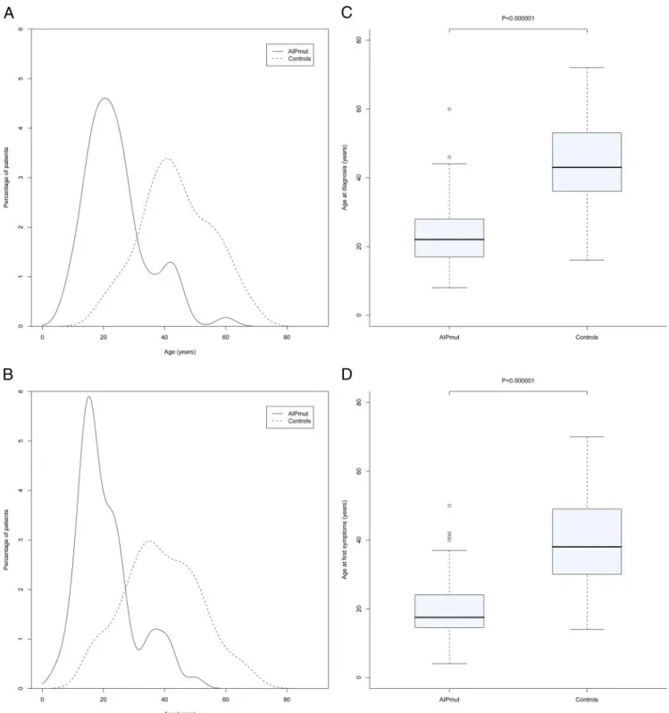
FIG. 1. Significantly younger age at diagnosis (A and C) and age at first symptoms (B and D) in somatotropinoma patients with AIPmut (n⫽ 71)
and control somatotropinoma patients (n⫽ 232). A and C, Frequency plot curves of ages at diagnosis and first symptoms for the two groups. B
and D, Box and whisker plots in which the box represents the 25th and 75th percentiles, and the dark line within the box is the median. The whiskers represent the extremes of data that lie one box length distance above and below the 25th and 75th centiles, respectively.
was younger than commonly described for this disease [31.0 (range 12.0 –74.0 yr)] (24). All tumors were mac-roadenomas, six had suprasellar extension and four were invasive at diagnosis. Two patients presented with pituitary
apoplexy. All patients had mildly elevated prolactin at diag-nosis, and in three patients who received dopamine agonists, two achieved normal prolactin (no tumor shrinkage). At diagnosis, one patient had hypogonadism and one had
FIG. 3. Significantly greater GH level at diagnosis in somatotropinoma patients with AIPmut (n⫽ 71) and control somatotropinoma patients (n ⫽
232). A, A frequency plot curve. B, Box and whisker plot in which the box represents the 25th and 75th percentiles, and the dark line within the box is the median. The whiskers represent the extremes of data that lie one box length distance above and below the 25th and 75th centiles, respectively.
FIG. 2. Significantly greater maximum tumor diameter in somatotropinoma patients with AIPmut (n⫽ 71) and control somatotropinoma patients
(n⫽ 232). A, A frequency plot curve. B, Box and whisker plot in which the box represents the 25th and 75th percentiles, and the dark line within
the box is the median. The whiskers represent the extremes of data that lie one box length distance above and below the 25th and 75th centiles, respectively.
hypofunction of the cortisol, thyroid, and gonadal axes, which did not resolve after therapy. Six patients under-went surgery and one patient who underunder-went a transcra-nial approach received radiotherapy due to a large rem-nant. Long-term control of tumor size was achieved in all cases.
TSH-secreting adenoma
A 39-yr-old male patient presented with a 6-month his-tory of tachycardia and breathing difficulties in
associa-tion with elevated T3and T4levels, a normal TSH level,
and had a noninvasive pituitary macroadenoma on mag-netic resonance imaging. No other hormonal abnormali-ties were noted at diagnosis. The patient twice underwent transsphenoidal surgery, but the tumor regrew on both occasions within less than 1 yr. A missense AIPmut (I257V) was discovered; family screening identified the same mutation in the unaffected mother and brother. Af-ter the second tumor recurrence, the patient was treated with octreotide long-acting repeatable 20 mg/month, which resulted in a hormonal normalization but no change in the residual tumor size.
Genotype-phenotype relationships
There were no statistical differences in terms of the clinical or therapeutic characteristics among patients with different types of AIPmut (truncating, frameshift muta-tions, missense mutamuta-tions, intronic mutamuta-tions, or in-frame deletions). The characteristics of the three most frequent
AIP mutations, Q14X (n ⫽ 13), R304X (n ⫽ 8), and
R271W (n⫽ 7), did not differ from the group as a whole.
Discussion
In this study we report the clinical and therapeutic features in 96 patients with germline AIPmut and anterior pitu-itary adenomas in the setting of FIPA and sporadic disease (10 –13, 16, 20, 39, 41, 47, and Supplemental Material Refs. 1 and 2); 41 patients are reported for the first time. This study is the first to apply standardized data collection methods to an extensive international AIPmut cohort to assess clinically relevant patient and disease characteris-tics, including responses to therapy.
The spectrum of anterior pituitary tumors associated with AIPmut now includes all clinical subtypes. Nearly 80% of patients with AIPmut present with somatotropi-nomas, and more than half cosecrete GH and prolactin. A third of somatotropinoma patients in the AIPmut group had gigantism. A further 13.5% of patients had prolacti-nomas, whereas nonsecreting pituitary adenomas are clearly also a feature of the AIPmut spectrum. The AIPmut TSH-secreting tumor is the first to be reported; because
these are rare tumors (⬍1% of all pituitary tumors), it
remains to be seen whether AIPmut is frequent in this setting (25). Cushing disease is a very rare association with AIPmut, with only two cases in the literature and none in the current series (12, 18).
The reason for the predominance of somatotropinomas among patients with AIPmut is unclear; however, this sub-group has specific features compared with a well-matched international AIPmut-negative control somatotropinoma group. AIPmut-associated somatotropinomas had first symptoms and were diagnosed 20 yr earlier as compared with controls and were significantly larger, more fre-quently extensive and had a greater frequency of prolactin hypersecretion. In addition, AIPmut status was also asso-ciated with significantly higher levels of GH secretion at baseline vs. controls. These features also appeared to im-pact the therapeutic responses, with poorer disease out-comes in the AIPmut group. Large, extensive, and invasive macroadenomas and high GH secretion are associated with a lower rate of control with primary neurosurgery; hence, the significantly higher rate of reoperation in the AIPmut cohort is not surprising (26). In addition, SSA therapy was associated with significantly lower decreases from baseline in GH and IGF-I in the AIPmut group, whereas tumor shrinkage was also significantly less pro-nounced than in controls. A trend toward more frequent use of radiotherapy and the failure of pegvisomant to con-trol IGF-I in three of four individuals in the AIPmut group (as compared with 19 of 19 controlled pegvisomant-treated sporadic patients) lends further evidence to AIP-mut patients forming a challenging part of the therapeutic spectrum in acromegaly. This is supported by the finding that AIPmut somatotropinoma patients who received
similarly high (ⱖ3) cumulative numbers of therapies had
significantly lower rates of long-term disease control as compared with the somatotropinoma control group. The reason for the poorer responses to SSA is not known and is a compelling topic for further study, particularly be-cause SSA receptor expression and the activity of vital determinants of SSA function like the ZAC1 (zinc finger protein which regulates apoptosis and cell cycle arrest) (27), in AIPmut somatotropinoma cells remain unknown. Practically it may be that large tumor size and the relatively poor SSA responses in such cases might warrant a tumor debulking approach to favor eventual control with SSA (28, 29).
Gigantism is an integral but very rare component of the acromegaly disease spectrum, with little more than 100 cases reported (30 –34). Gigantism may occur exception-ally in other conditions like MEN1, CNC, or McCune-Albright syndrome (30). In contrast, the current results suggest that gigantism is a frequent finding among patients
with AIPmut, with 32% of all AIPmut somatotropinomas having gigantism, which contrasts strongly with 6.5% among controls. This latter figure in the non-AIPmut con-trol group is itself suggestive that gigantism may not be as rare as previously thought. Gigantism occurred in a fa-milial setting in 63% of AIPmut cases in this series, al-though there were nine apparently sporadic giants. In con-trast, Leontiou et al. (17) found no AIPmut cases among seven sporadic giants, although gigantism did appear to occur frequently among their FIPA kindreds. The likely explanation for the high frequency of gigantism in the setting of AIPmut is due to the common features of large somatotropinomas secreting high levels of GH that be-come symptomatic predominantly before epiphyseal closure.
A pronounced gender imbalance was seen in the AIP-mut cohort with about two thirds of patients being male. This gender imbalance was marked in the prolactinoma group, which was 76.9% male. These patients had large tumors, half of which were not controlled by dopamine agonists, and some were difficult to control with multiple surgeries and radiotherapy. Male sex is known to be as-sociated with a higher rate of aggressive or treatment-resistant prolactinomas, and AIPmut status might explain a proportion of such cases (35, 36). Overall, the male preponderance among this series differs markedly from the pituitary disease characteristics in MEN1, in which the gender balance is reversed (69% female) (37). This differ-ence may be due to the fact that prolactinomas comprise 62% of pituitary tumors in MEN1 and are 2.5 times more frequent in women (37). Interestingly, prolactinomas in MEN1 patients are, like the AIPmut cases, also compar-atively difficult to treat. CNC is, in general, a disease with a strong female preponderance (63%) (38). Although ac-romegaly is a recognized phenotypic component of CNC, it is relatively uncommon, occurring in only 42 patients (12%) in the largest series, making valid comparisons with AIPmut patients difficult.
The penetrance of the pituitary adenoma predisposi-tion conferred by AIPmut remains an unresolved
ques-tion. Based on current figures (there are⬎100
asymptom-atic AIPmut carriers related to patients in this study), the penetrance of pituitary adenoma among FIPA kindreds with AIPmut is 15– 45%. This incomplete penetrance stands in direct contrast to MEN1 and CNC. Because most AIPmut-related pituitary adenomas (87.5%) present be-fore the age of 40 yr, many younger AIPmut carriers will require extended follow-up to definitively determine pen-etrance. Current penetrance rates suffer from various sources of bias, such as small kindreds with limited avail-ability for genetic and clinical evaluation, in addition to apparently sporadic patients in whom family AIP genetic
testing was not possible. True de novo sporadic cases have been identified (18). As demonstrated by large, apparently extensive multigenerational families in Finland, Italy, and elsewhere, mutation founders may have lived in the distant past (10, 39 – 41); this suggests that AIPmut status does not greatly impair biological fitness, unlike in more ag-gressive genetic tumor syndromes (42). It remains to be determined whether some AIPmut confer a lower disease penetrance than others.
Another important feature is the almost uniformly early age at onset and the rapid growth characteristics in AIPmut pituitary adenomas. The fact that more than half of patients present already with extensive pituitary mac-roadenomas as children or adolescents suggest that AIP-mut status confers a predisposition to rapid tumor growth, a point underlined by the short time from first symptoms to diagnosis (2.0 yr). It is unclear whether loss of the wild-type AIP allele in pituitary tissue itself leads to pituitary adenoma development or whether this somatic second hit permits rapid expansion of preexisting nests of abnormal cells (e.g. hyperplastic zones). It remains to be determined whether other modulating factors exist that can alter the development of pituitary adenomas among AIPmut car-riers. Furthermore, AIPmut-related disease in humans re-mains limited to a pituitary adenoma phenotype, which contrasts strongly with other genetic causes of pituitary adenomas (MEN1, MEN4, CNC), which tend to affect multiple tissues. Pituitary data from Ara9 knockout mice models have not yet been reported (43, 44). Few data are available on the molecular effects of AIPmut in the pi-tuitary itself, and it remains unclear whether the pri-mary mechanism governing tumorigenesis is via the aryl hydrocarbon receptor (AhR), down-regulation of AhR nuclear translocator (45), interactions with phosphodi-esterases (17), or via RET-survivin (46). Recent immu-nohistochemical data indicate that AIP expression is high in somatotropinomas and nonsecreting tumors (17, 47). In somatotropinomas, however, significantly lower AIP im-munostaining occurs in invasive as compared with noninva-sive cases. Furthermore, AIP immunostaining was abolished in only a minority of AIPmut pituitary adenomas (46). It may be that decreases in AIP immunostaining is a feature of ag-gressiveness in somatotropinomas, irrespective of mutation status.
Conclusions
AIPmut status is associated with the development of anterior pituitary adenomas, and all pituitary tumor phe-notypes have now been described, usually in a familial setting. AIPmut-related pituitary adenomas are generally large and expansive, and more than half are invasive at diagnosis. Patients are predominantly male and young,
with half of cases presenting during childhood or adoles-cence. Somatotropinomas are encountered most fre-quently (nearly 80%) and gigantism is notably frequent. AIPmut-associated somatotropinomas are significantly larger, more commonly extensive, occur at a younger age, secrete higher levels of GH, and have more frequent pro-lactin cosecretion than matched acromegalic patients without AIPmut. AIPmut somatotropinomas require re-peat surgery significantly more often than controls, whereas hormonal and tumor responses to SSA are sig-nificantly lower than controls; an increased risk of hypop-ituitarism is seen in the AIPmut cohort. AIPmut-related prolactinomas appear also to have aggressive and diffi-cult-to-treat clinical characteristics.
These results suggest that improving outcomes among AIPmut-associated pituitary tumors might require earlier diagnosis at the microadenoma or enclosed macroad-enoma stage. This adds impetus to exploring the most appropriate way to identify AIPmut patients, which might be aided by considering genetic screening only in FIPA kindreds and young patients with large tumors (48).
Acknowledgments
We acknowledge the Instituto and Laboratorio Sabin (Brasilia, Brazil) for laboratory assistance in Brazil.
Address all correspondence and requests for reprints to: Pro-fessor Albert Beckers, M.D., Ph.D., Centre Hospitalier Univer-sitaire de Lie`ge, University of Lie`ge, Domain UniverUniver-sitaire du Sart-Tilman, 4000 Lie`ge, Belgium. E-mail: albert.beckers@ chu.ulg.ac.be.
This work was supported by the Fonds d’Investissement pour la Recherche Scientifique du Centre Hospitalier Universitaire de Lie`ge, University of Lie`ge, Lie`ge, Belgium, and grants from the Finnish Cancer Societies, the Academy of Finland, and the Sigrid Juselius Foundation.
Disclosure Summary: None of the authors have any relevant disclosures.
References
1. Daly AF, Rixhon M, Adam C, Dempegioti A, Tichomirowa MA, Beckers A 2006 High prevalence of pituitary adenomas: a cross-sectional study in the province of Liege, Belgium. J Clin Endocrinol Metab 91:4769 – 4775
2. Fernandez A, Karavitaki N, Wass JA 2010 Prevalence of pituitary adenomas: a community-based, cross-sectional study in Banbury (Oxfordshire, UK). Clin Endocrinol (Oxf) 72:377–382
3. Asa SL, Ezzat S 2009 The pathogenesis of pituitary tumors. Annu Rev Pathol 4:97–126
4. Melmed S 2003 Mechanisms for pituitary tumorigenesis: the plastic pituitary. J Clin Invest 112:1603–1618
5. Gejman R, Batista DL, Zhong Y, Zhou Y, Zhang X, Swearingen B, Stratakis CA, Hedley-Whyte ET, Klibanski A 2008 Selective loss of MEG3 expression and intergenic differentially methylated region
hypermethylation in the MEG3/DLK1 locus in human clinically non-functioning pituitary adenomas. J Clin Endocrinol Metab 93: 4119 – 4125
6. Boikos SA, Stratakis CA 2006 Carney complex: pathology and mo-lecular genetics. Neuroendocrinology 83:189 –199
7. Marx SJ 2005 Molecular genetics of multiple endocrine neoplasia types 1 and 2. Nat Rev Cancer 5:367–375
8. Pellegata NS, Quintanilla-Martinez L, Siggelkow H, Samson E, Bink K, Ho¨fler H, Fend F, Graw J, Atkinson MJ 2006 Germ-line muta-tions in p27Kip1 cause a multiple endocrine neoplasia syndrome in rats and humans. Proc Natl Acad Sci USA 103:15558 –15563 9. Daly AF, Jaffrain-Rea ML, Ciccarelli A, Valdes-Socin H, Rohmer V,
Tamburrano G, Borson-Chazot C, Estour B, Ciccarelli E, Brue T, Ferolla P, Emy P, Colao A, De Menis E, Lecomte P, Penfornis F, Delemer B, Bertherat J, We´meau JL, De Herder W, Archambeaud F, Stevenaert A, Calender A, Murat A, Cavagnini F, Beckers A 2006 Clinical characterization of familial isolated pituitary adenomas. J Clin Endocrinol Metab 91:3316 –3323
10. Vierimaa O, Georgitsi M, Lehtonen R, Vahteristo P, Kokko A, Raitila A, Tuppurainen K, Ebeling TM, Salmela PI, Paschke R, Gu¨n-dogdu S, De Menis E, Ma¨kinen MJ, Launonen V, Karhu A, Aaltonen LA 2006 Pituitary adenoma predisposition caused by germline mu-tations in the AIP gene. Science 312:1228 –1230
11. Daly AF, Vanbellinghen JF, Khoo SK, Jaffrain-Rea ML, Naves LA, Guitelman MA, Murat A, Emy P, Gimenez-Roqueplo AP, Tamburrano G, Raverot G, Barlier A, De Herder W, Penfornis A, Ciccarelli E, Estour B, Lecomte P, Gatta B, Chabre O, Sabate´ MI, Bertagna X, Garcia Basavilbaso N, Stalldecker G, Colao A, Ferolla P, We´meau JL, Caron P, Sadoul JL, Oneto A, Archambeaud F, Calender A, Sinilnikova O, Montan˜ana CF, Cavagnini F, Hana V, Solano A, Delettieres D, Luccio-Camelo DC, Basso A, Rohmer V, Brue T, Bours V, Teh BT, Beckers A 2007 Aryl hydrocarbon receptor-in-teracting protein gene mutations in familial isolated pituitary ade-nomas: analysis in 73 families. J Clin Endocrinol Metab 92:1891– 1896
12. Georgitsi M, Raitila A, Karhu A, Tuppurainen K, Ma¨kinen MJ, Vierimaa O, Paschke R, Saeger W, van der Luijt RB, Sane T, Robledo M, De Menis E, Weil RJ, Wasik A, Zielinski G, Lucewicz O, Lubinski J, Launonen V, Vahteristo P, Aaltonen LA 2007 Mo-lecular diagnosis of pituitary adenoma predisposition caused by aryl hydrocarbon receptor-interacting protein gene mutations. Proc Natl Acad Sci USA 104:4101– 4105
13. Barlier A, Vanbellinghen JF, Daly AF, Silvy M, Jaffrain-Rea ML, Trouillas J, Tamagno G, Cazabat L, Bours V, Brue T, Enjalbert A, Beckers A 2007 Mutations in the aryl hydrocarbon receptor inter-acting protein gene are not highly prevalent among subjects with sporadic pituitary adenomas. J Clin Endocrinol Metab 92:1952– 1955
14. Iwata T, Yamada S, Mizusawa N, Golam H, Sano T, Yoshimoto K 2007 The aryl hydrocarbon receptor-interacting protein gene is rarely mutated in sporadic GH-secreting adenomas. Clin Endocrinol (Oxf) 66:499 –502
15. Yu R, Bonert V, Saporta I, Raffel LJ, Melmed S 2006 Aryl hydro-carbon receptor interacting protein variants in sporadic pituitary adenomas. J Clin Endocrinol Metab 91:5126 –5129
16. Toledo RA, Lourenco Jr DM, Liberman B, Cunha-Neto MB, Cavalcanti MG, Moyses CB, Toledo SP, Dahia PL 2007 Germline mutation in the aryl hydrocarbon receptor interacting protein gene in familial somatotropinoma. J Clin Endocrinol Metab 92:1934–1937 17. Leontiou CA, Gueorguiev M, van der Spuy J, Quinton R, Lolli F,
Hassan S, Chahal HS, Igreja SC, Jordan S, Rowe J, Stolbrink M, Christian HC, Wray J, Bishop-Bailey D, Berney DM, Wass JA, Popovic V, Ribeiro-Oliveira Jr A, Gadelha MR, Monson JP, Akker SA, Davis JR, Clayton RN, Yoshimoto K, Iwata T, Matsuno A, Eguchi K, Musat M, Flanagan D, Peters G, Bolger GB, Chapple JP, Frohman LA, Grossman AB, Korbonits M 2008 The role of the aryl hydrocarbon receptor-interacting protein gene in familial and spo-radic pituitary adenomas. J Clin Endocrinol Metab 93:2390 –2401
18. Stratakis CA, Tichomirowa MA, Boikos S, Azevedo MF, Lodish M, Martari M, Verma S, Daly AF, Raygada M, Keil MF, Papademetriou J, Drori-Herishanu L, Horvath A, Tsang KM, Nesterova M, Frank-lin S, VanbelFrank-linghen J-F, Bours V, Salvatori R, Beckers A 23 Feb-ruary 2010 The role of germline AIP, MEN1, PRKAR1A, CDKN1B and CDKN2C mutations in children and adolescents with pituitary adenoma. Clin Genet 10.1111/j.1399-0004.2010.01406.x 19. Cazabat L, Guillaud-Bataille M, Bertherat J, Raffin-Sanson ML
2009 Mutations of the gene for the aryl hydrocarbon receptor-in-teracting protein in pituitary adenomas. Horm Res 71:132–141 20. Georgitsi M, Helio¨vaara E, Paschke R, Kumar AV, Tischkowitz M,
Vierimaa O, Salmela P, Sane T, De Menis E, Cannavo` S, Gu¨ndogdu S, Lucassen A, Izatt L, Aylwin S, Bano G, Hodgson S, Koch CA, Karhu A, Aaltonen LA 2008 Large genomic deletions in AIP in pituitary adenoma predisposition. J Clin Endocrinol Metab 93:4146 – 4151
21. Nwosu BU, Lee MM 2008 Evaluation of short and tall stature in children. Am Fam Physician 78:597– 604
22. Kuczmarski RJ, Ogden CL, Guo SS, Grummer-Strawn LM, Flegal KM, Mei Z, Wei R, Curtin LR, Roche AF, Johnson CL. 2002 2000 CDC Growth charts for the United States: methods and development. Vital Health Stat 11 1–190 (http://www.cdc.gov/nchs/data/series/sr_11/
sr11_246.pdf)
23. World Health Organization Multicentre Growth Reference Study
Group. 2009 WHO child growth standards: growth velocity based
on weight, length and head circumference: methods and develop-ment. Geneva: World Health Organization (http://www.who.int/ childgrowth/standards/velocity/tr3_velocity_report.pdf)
24. Ferrante E, Ferraroni M, Castrignano` T, Menicatti L, Anagni M,
Reimondo G, Del Monte P, Bernasconi D, Loli P, Faustini-Fustini M, Borretta G, Terzolo M, Losa M, Morabito A, Spada A, Beck-Peccoz P, Lania AG 2006 Non-functioning pituitary adenoma
da-tabase: a useful resource to improve the clinical management of pituitary tumors. Eur J Endocrinol 155:823– 829
25. Beck-Peccoz P, Brucker-Davis F, Persani L, Smallridge RC, Weintraub
BD 1996 Thyrotropin-secreting pituitary tumors. Endocr Rev 17:610–
638
26. Buchfelder M, Schlaffer S 2009 Surgical treatment of pituitary ad-enomas. Best Pract Res Clin Endocrinol Metab 23:677– 692 27. Theodoropoulou M, Tichomirowa MA, Sievers C, Yassouridis A,
Arzberger T, Hougrand O, Deprez M, Daly AF, Petrossians P, Pagotto U, Beckers A, Stalla GK 2009 Tumor ZAC1 expression is
associated with the response to somatostatin analog therapy in pa-tients with acromegaly. Int J Cancer 125:2122–2126
28. Petrossians P, Borges-Martins L, Espinoza C, Daly A, Betea D,
Valdes-Socin H, Stevenaert A, Chanson P, Beckers A 2005 Gross
total resection or debulking of pituitary adenomas improves hor-monal control of acromegaly by somatostatin analogs. Eur J Endo-crinol 152:61– 66
29. Karavitaki N, Turner HE, Adams CB, Cudlip S, Byrne JV,
Fazal-Sanderson V, Rowlers S, Trainer PJ, Wass JA 2008 Surgical
debulk-ing of pituitary macroadenomas causdebulk-ing acromegaly improves con-trol by lanreotide. Clin Endocrinol (Oxf) 68:970 –975
30. Eugster EA, Pescovitz OH 1999 Gigantism. J Clin Endocrinol Metab 84:4379 – 4384
31. Rix M, Laurberg P, Hoejberg AS, Brock-Jacobsen B 2005 Pegviso-mant therapy in pituitary gigantism: successful treatment in a 12-year-old girl. Eur J Endocrinol 153:195–201
32. Mu¨ssig K, Gallwitz B, Honegger J, Strasburger CJ, Bidlingmaier M,
Machicao F, Bornemann A, Ranke MB, Ha¨ring HU, Petersenn S
2007 Pegvisomant treatment in gigantism caused by a growth hor-mone-secreting giant pituitary adenoma. Exp Clin Endocrinol Di-abetes 115:198 –202
33. Goldenberg N, Racine MS, Thomas P, Degnan B, Chandler W,
Barkan A 2008 Treatment of pituitary gigantism with the growth
hormone receptor antagonist pegvisomant. J Clin Endocrinol Metab 93:2953–2956
34. Schoof E, Do¨rr HG, Kiess W, Lu¨decke DK, Freitag E, Zindel V,
Rascher W, Do¨tsch J 2004 Five-year follow-up of a 13-year-old boy
with a pituitary adenoma Rausing gigantism— effect of octreotide therapy. Horm Res 61:184 –189
35. Colao A 2009 Pituitary tumours: the prolactinoma. Best Pract Res Clin Endocrinol Metab 23:575–596
36. Ciccarelli A, Daly AF, Beckers A 2005 The epidemiology of pro-lactinomas. Pituitary 8:3– 6
37. Verge`s B, Boureille F, Goudet P, Murat A, Beckers A, Sassolas G,
Cougard P, Chambe B, Montvernay C, Calender A 2002 Pituitary
disease in MEN type 1 (MEN1): data from the France-Belgium MEN1 multicenter study. J Clin Endocrinol Metab 87:457– 465 38. Bertherat J, Horvath A, Groussin L, Grabar S, Boikos S, Cazabat L,
Libe R, Rene´-Corail F, Stergiopoulos S, Bourdeau I, Bei T, Clauser E, Calender A, Kirschner LS, Bertagna X, Carney JA, Stratakis CA
2009 Mutations in regulatory subunit type 1A of cyclic adenosine 5⬘-monophosphate-dependent protein kinase (PRKAR1A): pheno-type analysis in 353 patients and 80 different genopheno-types. J Clin En-docrinol Metab 94:2085–2091
39. Jennings JE, Georgitsi M, Holdaway I, Daly AF, Tichomirowa M,
Beckers A, Aaltonen LA, Karhu A, Cameron FJ 2009 Aggressive
pituitary adenomas occurring in young patients in a large Polynesian kindred with a germline R271W mutation in the AIP gene. Eur J Endocrinol 161:799 – 804
40. Occhi G, Jaffrain-Rea ML, Trivellin G, Albiger N, Ceccato F, De
Menis E, Angelini M, Ferasin S, Beckers A, Mantero F, Scaroni C 30
March 2010 The R304X mutation of the aryl hydrocarbon receptor interacting protein gene in familial isolated pituitary adenomas: mu-tational hot-spot or founder effect? J Endocrinol Invest 10.3275/ 6956
41. Naves LA, Daly AF, Vanbellinghen JF, Casulari LA, Spilioti C,
Magalha˜es AV, Azevedo MF, Giacomini LA, Nascimento PP, Nunes RO, Rosa JW, Jaffrain-Rea ML, Bours V, Beckers A 2007 Variable
pathological and clinical features of a large Brazilian family harboring a mutation in the aryl hydrocarbon receptor-interacting protein gene. Eur J Endocrinol 157:383–391
42. Wang ZJ, Churchman M, Avizienyte E, McKeown C, Davies S,
Evans DG, Ferguson A, Ellis I, Xu WH, Yan ZY, Aaltonen LA, Tomlinson IP 1999 Germline mutations of the LKB1 (STK11) gene
in Peutz-Jeghers patients. J Med Genet 36:365–368
43. Lin BC, Nguyen LP, Walisser JA, Bradfield CA 2008 A hypomor-phic allele of aryl hydrocarbon receptor-associated protein-9 pro-duces a phenocopy of the AHR-null mouse. Mol Pharmacol 74: 1367–1371
44. Lin BC, Sullivan R, Lee Y, Moran S, Glover E, Bradfield CA 2007 Deletion of the aryl hydrocarbon receptor-associated protein 9 leads to cardiac malformation and embryonic lethality. J Biol Chem 282: 35924 –35932
45. Helio¨vaara E, Raitila A, Launonen V, Paetau A, Arola J, Lehtonen
H, Sane T, Weil RJ, Vierimaa O, Salmela P, Tuppurainen K, Ma¨kinen M, Aaltonen LA, Karhu A 2009 The expression of AIP-related
mole-cules in elucidation of cellular pathways in pituitary adenomas. Am J Pathol 175:2501–2507
46. Vargiolu M, Fusco D, Kurelac I, Dirnberger D, Baumeister R, Morra
I, Melcarne A, Rimondini R, Romeo G, Bonora E 2009 The tyrosine
kinase receptor RET interacts in vivo with aryl hydrocarbon recep-tor-interacting protein to alter surviving availability. J Clin Endo-crinol Metab 94:2571–2578
47. Jaffrain-Rea ML, Angelini M, Gargano D, Tichomirowa MA, Daly
AF, Vanbellinghen JF, D’Innocenzo E, Barlier A, Giangaspero F, Esposito V, Ventura L, Arcella A, Theodoropoulou M, Naves LA, Fajardo C, Zacharieva S, Rohmer V, Brue T, Gulino A, Cantore G, Alesse E, Beckers A 2009 Expression of aryl hydrocarbon receptor
(AHR) and AHR-interacting protein in pituitary adenomas: patho-logical and clinical implications. Endocr Relat Cancer 16:1029 – 1043
48. Beckers A, Daly AF 2007 The clinical, pathological, and genetic features of familial isolated pituitary adenomas. Eur J Endocrinol 157:371–382