The Veterinary Record, May 24, 2008
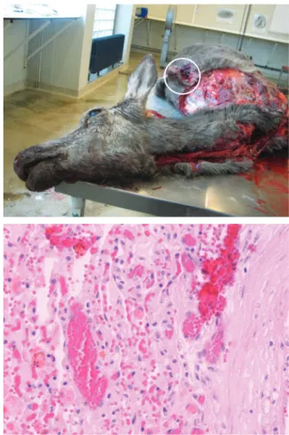
eter, were scattered throughout the lungs (Fig 1). On section, the masses were dark red and oozed blood. The mediastinal lymph nodes were not enlarged. A soft, invasive subcutaneous mass of approximately 15 cm in diameter on the left flank was dissected carefully (Fig 2). The mass appeared to protrude from the seventh rib, from which a 5 cm segment was missing due to bone lysis. Section of the mass revealed large areas of necrosis and haemorrhages. There were no abnormal findings elsewhere in the carcase. Samples of the rib, paracostal mass and lung nodules were taken and immediately fixed in 10 per cent phosphate-buffered formalin solution for approximately 48 hours before being routinely processed and stained with haematoxylin and eosin for light microscopic examination.
The paracostal mass and the pulmonary masses displayed a similar histopathological pattern (Fig 3). The pulmonary masses had conspicuous accumulations of blood, either fill-ing small clefts or giant cavernous channels, or freely dissect-ing the tissues (haemorrhages). The clefts or channels were clearly delineated by endothelial cells; some of them were visibly ruptured or thrombosed. The stroma interspersed between the channels and clefts was constituted of neoplastic cells that varied in size and shape but were usually elongated. The nuclei of these cells were round to ovoid, very hyper-chromatic and commonly displayed mitotic figures. All of the masses contained numerous macrophages filled with haemo-siderin. The paracostal mass also displayed very large areas of necrosis, with foci of neutrophilic accumulation. Osteoid formation was not observed in the paracostal and pulmonary masses, excluding the hypothesis of telangiectatic osteosar-coma (Gleiser and others 1981, Thompson and Pool 2002). The histological features were compatible with a diagnosis of cavernous haemangiosarcoma, a malignant tumour of vascu-lar endothelial cells. The primary tumour probably developed within the seventh left rib, then extended to the pleurae and the thoracic muscles, and finally disseminated as metastases in
Cavernous
haemangiosarcoma in
a free-living red deer
(Cervus elaphus)
F. Grégoire, B. Mousset, D. Hanrez,
D. Cassart, D. Desmecht, A. Linden
HAEMANGIOMAS and haemangiosarcomas are neoplasms of vascular endothelium that occur commonly in dogs and rarely in other domestic animals (Goldschmidt and Hendrick 2002). Haemangiomas are benign and are usually solitary masses in the dermis or subcutis; on the basis of the size of the vascular channels, these tumours are classified as cavern-ous or capillary haemangiomas (Hendrick and others 1998). Haemangiosarcoma is the malignant counterpart of haeman-gioma, and most commonly presents in dogs as a multi centric disease involving the right atrium, spleen, liver and lungs (Garzotto and Berg 2003, Smith 2003, Waters and Cooley 1998). Haemangiosarcomas are less frequently reported in cats than in dogs, and the lesions are usually observed in the skin of the head, distal limbs, lungs and paws (Moran and Suster 2001, Goldschmidt and Hendrick 2002, Yamagami and others 2006). In horses, neoplasia of vascular origin is also uncom-mon (Johnson and others 1988, Kennedy and Brown 1993, Berry 1999); haemangiosarcoma was recorded in only three of 1404 horses examined in a study by Stencel and Grotelueschen (1989). Haemangiosarcomas in horses have been reported in the vertebrae, spinal cord, oral cavity, tarsal sheath and frontal sinus (Southwood and others 2000). The few cases reported in cattle were located in the lungs and long bones (Guard and Wilkinson 1984), vertebrae (Zachary and others 1981), cuta-neous external nares (Queen and others 1992), muscles and extradural spinal cord (Sutton and McLennan 1982).
In wild ruminant species, only one case of haemangio-sarcoma has been described, in a 20-year-old female Père David’s deer raised at a zoo in South Korea (Yoon and others 1999). This short communication describes gross and micro-scopic pathological findings in a free-living red deer (Cervus
elaphus) with cavernous haemangiosarcoma.
The 14-year-old female red deer was found dead in March 2003 in the region of Bièvre in southern Belgium. Its age was determined based on standardised dental inspection (Mitchel 1963). At postmortem examination, numerous nodular,
well-demarcated, red-black, soft to firm masses, 5 to 30 mm in diam- Veterinary Record (2008)
162, 692-693 F. Grégoire, DVM, B. Mousset, BCh, D. Hanrez, BCh, D. Cassart, DVM, PhD, A. Linden, DVM, PhD, Department of Infectious Diseases, D. Desmecht, DVM, PhD, Department of Morphology and Pathology, Faculty of Veterinary Medicine, University of Liège, Sart Tilman B43, B-4000 Liège, Belgium
Correspondence to Dr Linden
FIG 1: Gross postmortem view showing the left lung covered with nodular red-black masses
FIG 2: Gross postmortem view showing the sectioned paracostal mass (circle)
FIG 3: Histological view of a red-black lung nodule showing the characteristic features of an haemangiosarcoma. Haematoxylin and eosin. x 400
S h o rt C o m mu n i c at i o n s
S h o rt C o m mu n i c at i o n s
The Veterinary Record, May 24, 2008
the lungs. To the authors’ knowledge, this is the first reported case of haemangiosarcoma in a non-captive cervid.
References
BERRY, S. (1999) Spinal cord compression secondary to hemangiosarcoma in a saddlebred stallion. Canadian Veterinary Journal 40, 886-887
GARZOTTO, C. & BERG, J. (2003) Musculoskeletal system. In Textbook of Small Animal Surgery. 3rd edn. Ed D. Slatter. Philadelphia, Saunders. pp 2460-2474 GLEISER, C. A., RAULSTON, G. L., JARDINE, J. H., CARPENTER, R. H. &
GRAY, K. M. (1981) Telangiectatic osteosarcoma in the dog. Veterinary Pathology 18, 396-398
GOLDSCHMIDT, M. H. & HENDRICK, M. J. (2002) Tumors of the skin and soft tissues. In Tumors in Domestic Animals. 4th edn. Ed D. J. Meuten. Ames, Iowa State Press. pp 45-118
GUARD, C. & WILKINSON, J. E. (1984) Hemangiosarcoma in a cow. Journal of the American Veterinary Medical Association 185, 789-790
HENDRICK, M. J., MAHAFFAY, E. A., MOORE, F. M., VOS, J. H. & WALDER, E. J. (1998) Tumors of vascular tissue. In Histological Classification of Mesenchymal Tumors of Skin and Soft Tissues of Domestic Animals. Vol II. Washington DC, Armed Forces Institute of Pathology. pp 15-33
JOHNSON, J. E., BEECH, J. & SAIK, J. E. (1988) Disseminated hemangio-sarcoma in a horse. Journal of the American Veterinary Medical Association
193, 1429-1431
KENNEDY, F. A. & BROWN, C. M. (1993) Vertebral angiosarcoma in a horse. Journal of Veterinary Diagnostic Investigations 5, 125-127
MITCHEL, B. (1963) Determination of age in Scottish red deer from growth levels in dental cement. Nature 198, 350-351
MORAN, C. A. & SUSTER, S. (2001) Tumors of the lung and pleura. In
Diagnostic Histopathology of Tumors. Vol 1. Ed C. D. M. Fletcher. London, Churchill Livingstone. pp 184-189
QUEEN, W. G., MASTERSON, M. A. & WEISBRODE, S. E. (1992) Hemangiosarcoma of the external naris in a cow. Journal of the American Veterinary Medical Association 201, 1411-1412
SMITH, A. N. (2003) Hemangiosarcoma in dogs and cats. Veterinary Clinics of North America: Small Animal Practice 33, 533-552
SOUTHWOOD, L. L., SCHOTT, H. C. II. & HENRY, C. J. (2000) Disseminated hemangiosarcoma in the horse: 35 cases. Journal of Veterinary Internal Medicine
14, 105-109
STENCEL, E. & GROTELUESCHEN, D. (1989) Hemangiosarcoma involving the frontal sinus of a horse. Equine Practice 11, 14-16
SUTTON, R. H. & MCLENNAN, E. G. (1982) Hemangiosarcoma in a cow. Veterinary Pathology 19, 456-458
THOMPSON, K. G. & POOL, R. R. (2002) Tumors of bones. In Tumors in Domestic Animals. 4th edn. Ed D. J. Meuten. Ames, Iowa State Press. pp 245-317
WATERS, D. J. & COOLEY, D. M. (1998) Skeletal neoplasms. In Cancer in Dogs and Cats: Medical and Surgical Managements. Ed W. B. Morrison. Baltimore, Williams & Wilkins. pp 639-654
YAMAGAMI, T., NOMURA, K., FUJITA, M., OZAKI, K., ORIMA, H. & NARAMA, I. (2006) Pulmonary intravascular hemangiosarcoma in a cat. Journal of Veterinary Medical Sciences 68, 731-733
YOON, B., KWEON, O., KWON, S., SHIN, N., SEO, I. & KIM, D. (1999) Concurrent multicentric hemangiosarcoma and ovarian teratoma in an aged Père David’s deer (Elaphus davidianus). Journal of Zoo and Wildlife Medicine
30, 456-458
ZACHARY, J. F., JONES, M. G. & WOLFF, W. A. (1981) Multicentric osseous hemangiosarcoma in a Chianina-Angus steer. Veterinary Pathology 18, 266-270