HAL Id: tel-00813418
https://tel.archives-ouvertes.fr/tel-00813418
Submitted on 15 Apr 2013
HAL is a multi-disciplinary open access
archive for the deposit and dissemination of
sci-entific research documents, whether they are
pub-lished or not. The documents may come from
teaching and research institutions in France or
abroad, or from public or private research centers.
L’archive ouverte pluridisciplinaire HAL, est
destinée au dépôt et à la diffusion de documents
scientifiques de niveau recherche, publiés ou non,
émanant des établissements d’enseignement et de
recherche français ou étrangers, des laboratoires
publics ou privés.
recognition by the curvature sensor ALPS (amphipathic
lipid packing sensor) : molecular dynamics studies
Paula Gonzalez-Rubio Garrido
To cite this version:
Paula Gonzalez-Rubio Garrido. New perspectives to undestand the lipid packing recognition by the
curvature sensor ALPS (amphipathic lipid packing sensor) : molecular dynamics studies. Genetics.
Université Pierre et Marie Curie - Paris VI, 2009. English. �NNT : 2009PA066441�. �tel-00813418�
Docteur de l’Université de Pierre et Marie curie
Nouvelles perspectives pour comprendre la reconnaissance des défauts
d’empaquetage lipidique par le senseur de courbure membranaire ALPS
(Amphipathic Lipid Packing Sensor): études par dynamique moléculaire.
Soutenue le 27.11.2009 devant le jury composé de : Rapporteurs: Monique Genest James Sturgis Examinateurs: Guillaume Drin Julie Ménétrey Germain Trugnan Directrice de thèse: Catherine Etchebest
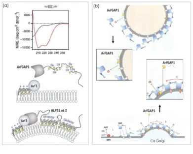
propriétés particulières a été identifiée récemment et il a été appelé ALPS (ArfGAP1 Amphipathic lipid packing sensor). Ce motif se plie en hélice au contact avec les membranes positivement courbées, sa face polaire est riche en serines/threonines et sa face hydrophobe contient plusieurs résidus aromatiques. Il a été montré que son interaction avec la membrane était indépendante des charges électrostatiques et en conséquence, un rôle prépondérant a été attribué aux résidus hydrophobes. En effet, le modèle actuel suggère que ces résidus hydrophobes soient les responsables de l’ancrage d’ALPS à la membrane en reconnaissant les défauts d’empaquetage lipidique. Nous avons réalisé des simulations par dynamique moléculaire du motif ALPS et une de ces mutants, qui expérimentalement compromet la reconnaissance de la courbure membranaire, liés à l’interface solvant/lipides des membranes de composition simple et mixte. Nous montrons que les particularités dans la séquence d’ALPS, exhibant un motif répétitif Bulky-small&polar-Bulky, lui permet de (1) avoir une considérable liberté conformationnelle et structurelle, (2) les serines /threonines participent dans des interactions hydrogènes importantes pour la reconnaissance de défauts d’empaquetage aux niveaux des têtes polaires et (3) les résidus aromatiques hydrophobes lui permettent d’explorer les défauts d’empaquetage au niveau des chaînes aliphatiques. Également, nous montrons que la déformabilité et flexibilité d’ALPS dépendent du contexte membranaire et donc des défauts d’empaquetage lipidique intrinsèques à la membrane. Finallement, nous proposons des nouvelles perspectives au niveau atomique de la reconnaissance des défauts d’empaquetage lipidique par ALPS où la déformabilité et flexibilité structurale ainis que les propriétés dynamiques et physiques de la membrane jouent un rôle prépondérant.
Dynamique Moléculaire ; Hélices amphipathiques ; Senseur de courbure ; Défauts d’empaquetage Lipidique ; ALPS ; Courbure membranaire ; ArfGAP1 ; Membrane simulations
face and hydrophobic aromatic residues rich hydrophobic face. Its membrane interaction depends on the recognition of defects in lipid packing and not in electrostatic charges. We performed molecular dynamics simulations of the amphipathic helical peptide ALPS, and a triple-mutant that experimentally compromises the sensitivity of ALPS to the membrane curvature. Both peptides were embedded at the water/lipid interface of explicit simple and mixed phosphatidylcholine membranes and simulations over 300 ns were run. In this thesis, we propose a novel atomistic view of ALPS curvature sensor lipid-packing recognition, where the dynamics and plasticity of both, ALPS and the membrane, act and adapt in a perfect synchony. We propose that ALPS is able to adapt to these inhomogeneities of the membrane thanks to the conformational deformability and structural flexibility that allow it to explore the lipid packing defects at the level of the polar heads and the acyl chains. This deformability is favor by a bulky-small&polar-bulky pattern that dispose the bulky hydrophobic residues and the small polar residues in a way that favors peptide-lipid interactions. Moreover we suggest that ALPS can induce adaptative dynamic response of the membrane that leads to a bilayer-coupling effect and a reciprocal orchestrated adaptation process. We show how the absence of lipid-packing defects avoid ALPS deformability and structural flexibility, affecting in consequence important intra-peptide and lipid-peptide interactions. Our results show that the deformability and structural flexibility of ALPS and the presence of lipid-packing defaults in the membrane are correlated. We also propose that the deformability is environment-dependent whereas the structural flexibility depends on the particularities of the sequence. Hence, ALPS plasticity must be of relevance for its curvature sensitivity. We advance that this could imply a concertated mechanism to recognize curved membranes. The partitioning of ALPS at the interfacial phosphate/glycerol level, suggest an adaptive interplay between the peptide-sequence geometrical and space restrictions, the lipids conformations and the physical forces that shape the membrane.
Key Words
Molecular dynamics ; Amphipathic helix ; Curvature sensor ; Lipid packing defects ; ALPS ; Membrane curvature ; ArfGAP1 ; Membrane simulations
2.1.2 Lipid packing and the membrane shape . . . 8
2.1.3 Membrane composition . . . 10
2.1.4 Lipid dynamics and membrane fluidity . . . 11
2.2 Membrane structure is a complex target . . . 13
2.2.1 Structure . . . 14
2.2.2 Partitioning . . . 15
2.2.2.1 Partitioning-folding-coupling mechanism . . . 17
2.3 A Glimpse of Membrane proteins . . . 18
2.3.1 Membrane-proteins determin membrane topology . . . 19
2.3.2 Membrane-proteins secondary structure . . . 21
2.4 Amphipathic alpha-helices . . . 23
2.4.1 Alpha-helix structure . . . 24
2.4.2 Amphipathic helices . . . 25
2.4.3 Amphipathic helices as mediators of the membrane interaction of amphitropic proteins and as modulators of bilayer physical properties . . . 26
3 Remodelling and sensing the membrane 29 3.1 Determinants of the membrane shape . . . 30
3.1.1 Membrane tension . . . 30
3.1.2 Spontaneous curvature . . . 30
3.2 Forces within the lipid bilayer . . . 32
3.3 Membrane bending . . . 33
3.4 Proteins that bend membranes . . . 33 vi
3.4.1 Proteins actin by the scaffold mechanism . . . 34
3.4.2 Proteins acting by the local spontaneous curvature mechanism . . . 36
3.4.2.1 The local spontaneous curvature mechanism . . . 36
3.4.2.2 The bilayer-coupling mechanism and other distorsions of the mem-branes attributed to amphiphatic helices . . . 37
3.4.3 Simultaneous operation of bending mechanisms . . . 38
3.5 The importance of sensing shape . . . 38
4 Regulation of vesicular transport by membrane curvature 40 4.1 The secretoy pathway and vesicle formation . . . 41
4.2 Membrane-curvature regulates traffic . . . 42
4.3 ALPS, a membrane curvature sensor . . . 43
4.3.1 ALPS discovery . . . 45
4.3.1.1 Amphipathic alpha helix structure . . . 45
4.3.1.2 Identification of ALPS2 . . . 45
4.3.2 Importance of hydrophobic residues on lipid packing recognition . . . 46
4.3.3 ALPS binding does not depend on charged electrostatic interactions . . . 47
4.3.4 Dependence on lipid composition . . . 49
4.4 Model of ALPS curvature recognition . . . 51
4.5 ALPS-like motifs are present in numerous proteins . . . 51
4.6 Main interests of the present work . . . 52
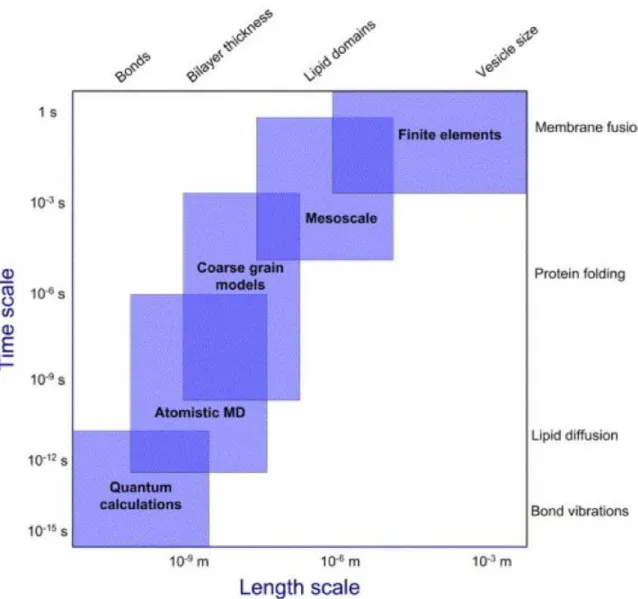
5 Molecular Dynamic Simulations 54 5.1 Simulations of membranes systems . . . 54
5.2 When to use simulations . . . 57
5.2.1 Simulation capabilities on membrane systems . . . 58
5.3 Force Fields . . . 59
5.3.1 Energies . . . 61
5.4 Principles of MD simulations . . . 63
5.4.1 Periodic conditions . . . 65
5.4.2 Pressure and temperature control . . . 66
5.4.3 Electrostatic interactions calculations . . . 67
5.5 System set up . . . 68
5.6 Simulations details . . . 72
5.7 Trajectory analysis . . . 72
5.7.1 Analysis of the lipid bilayer properties . . . 73
5.7.1.1 Bilayer thickness . . . 73
5.7.1.2 Order Parameter . . . 73
5.7.2 Analysis of the peptide properties . . . 74
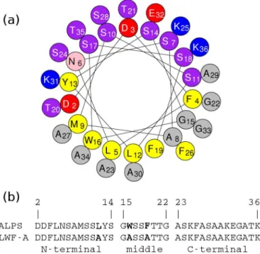
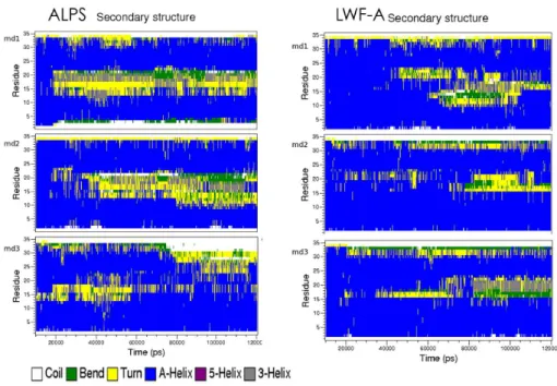
6.2 ALPS structure and conformational diversity . . . 79
6.2.1 ALPS secondary structure and deformation . . . 80
6.2.1.1 Mutant LWF-A limited deformation . . . 82
6.2.2 Characterization of helix deformations . . . 83
6.3 Peptide orientation relative to the membrane . . . 83
6.4 ALPS and LWF-A partitioning inside the membrane . . . 85
6.5 Lipid-peptide interactions . . . 89
6.6 Side chains flexibility and intra-peptide interactions . . . 92
6.7 Aromatic residues side-chain orientations with respect to the membrane . . . 94
6.8 Synchronized Intra-peptide and lipid-peptide interactions determine ALPS deformability 96 7 Membrane-peptide reciprocal adaptation 98 7.1 Lipid-lipid interactions . . . 98
7.2 Lipids diffusion . . . 98
7.3 Order parameter . . . 100
7.4 Bilayer thickness in the presence of the peptides . . . 102
8 Elucidating lipid-packing influence on ALPS structure 107 8.1 ALPS in DMPC and POPC membranes: Role of the acyl chains nature . . . 107
8.1.1 ALPS secondary structure and deformability in DMPC and POPC . . . 108
8.1.1.1 ALPS structure deformation and orientation . . . 108
8.1.2 Intra-peptide interactions and aromatic side-chains orientations with respect to the membrane . . . 110
8.1.3 Interfacial partitioning and orientation of ALPS in DMPC and POPC . . . 110
8.1.4 Response of DMPC and POPC membranes to ALPS presence . . . 112
8.1.4.1 Order Parameter . . . 113
8.1.4.2 Bilayer thickness . . . 114
8.1.4.3 Lipids diffusion . . . 115
8.2 ALPS in a DOPC-DOG membrane: Role of the size of polar heads . . . 116
8.2.2 Intra-peptide interactions and aromatic side-chains orientations with respect to
the membrane . . . 118
8.2.3 Interfacial partitioning and orientation of ALPS in DOPC-DOG . . . 120
8.2.4 Lipid-peptide interactions . . . 120
8.2.5 Response of DOPC-DOG membrane to ALPS presence . . . 123
8.2.5.1 Lipid-lipid interactions . . . 123
8.2.5.2 DOPC-DOG membrane lateral and transversal dynamics . . . 123
8.2.5.3 Order parameter . . . 124
8.3 Effect of the lipid-packing on ALPS deformability . . . 126
9 Novel atomistic view of ALPS curvature sensor lipid-packing recognition 128 9.1 Importance of the local 310 motif in ALPS helix deformation and adaptation to the membrane . . . 128
9.2 Dynamic lipid-packing defaults as mayor potentiators of ALPS deformability and adapt-ability . . . 130
9.3 ALPS sequence: an optimal synergy between polar and hydrophobic steric interactions 131 9.4 Sensing properties of the Bulky-small&polar-Bulky motif at the light of experiments . . 133
9.5 Bulky-small&polar-Bulky pattern in other ALPS-like motifs: ALPS the paradigm . . . 136
9.6 Delimiting ALPS sensor . . . 137
9.7 Understanding other ALPS mutants in the BssB context . . . 138
9.8 Conclusions . . . 140
9.8.1 Completing ALPS curvature sensing model . . . 141
9.9 Perspectives . . . 142
“When you are describing A shape, or sound, or tint; Don’t state the matter plainly, but put it in a hint; And learn to look at all things With a sort of mental squint” Lewis Carroll
The cell is the scenario of a complex interplay of biophysical and biochemical processes that make possible its survival. Cells interact with their environment in various ways, by secreting, for instance a great variety of molecules to modify their surroundings. They are also able to protect themselves by producing extracellular matrices or cell walls, and more importantly, they interact with other cells to share information, control cellular populations or to preserve multicellular organisms. Cells can be assigned to one of three domains based on their biophysical, biochemical and phylogenetic characteris-tics: Eukaryota, Archaea and Bacteria. These three types of cells are in constant interaction with their surroundings and the major cellular structure that make this possible is the plasmatic membrane that delimits individual cells. The nature of the lipid components of Eukaryota and Bacteria membrane is the same whereas Archaea membranes differ from the other life domains. These differences have been explained by two opposed hypothesis, in the first one Archaea would have completely replaced their plasmatic membrane by changing their lipid metabolic pathways (Lopez-Garcia and Moreira, 2004), having at some point a mixed membrane, in the second hypothesis the last universal common ances-tor (LUCA)(Delaye et al., 2005; Forterre et al., 2005; Jekely, 2006) was not yet delimited by a lipid membrane, and archaeal and bacterial/eukaryal membranes emerged independently afterwards (Koonin and Martin, 2005). Phylogenetic analyses of the enzymes involved in the lipid metabolic pathways of archaea and bacteria have showed that these life domains share highly divergent but homologous en-zymes. Researches have therefore confirmed that the plasmatic membranes have always been a crucial condition for life through its multiple shape manifestations (Fig.1.1).
During the second half of the 19th century, while Charles Darwin was preparing its masterpiece ’On the Origin of Species’, C. Naegeli and C. Cramer described for the first time the cell membranes, as essential barriers to maintain osmosis equilibrium in plant cells. Indeed, before the advent of modern cell biology, pioneered by Albert Claude in the 1940s, many biologists viewed the cell as a mere "bag of enzymes" or a "biochemical bog" filled with formless protoplasm and devoid of inner structure. These ideas started to change with the discovery, in the late 19th century, by light microscopy staining techniques, of internal organelles such as the chloroplast, the mitochondria and the Golgi apparatus. However, the real breakthrough was 60 years ago, when Albert Claude, Keith Porter and Ernest Fullam published the first picture of an intact cell taken with an electron microscope (Edidin, 2003). Since then, and thanks to the advances in electron microscopy during the last decades, we have been able to look inside the eukaryotic cell to find many specialized membrane-delimited compartments, as well as complex intracellular dynamic processes through which there is a constant flow of vesicles scaffolded by an internal cytoskeleton (Fig.1.1). Moreover the intricate scaffold constituted by the cytoskeleton and the vesicles transport, determines the global form and dynamics of the cell, the organelles and the endomembranes.
Figure 1.1: Membranes sculpt different shapes in nature.(a) Bacteria: Spirochetes sp, (b)Arquaea: Haloquadratum walsbyi, (c) Eukaryote: Neuron, (d) Chloroplast and its inner membranes (thilachoids), (e) Internal membranes in an eukaryot cell. In yellow the Golgi apparatus, in green the nuclear envelope and in bleu other endomembranes systems, (f) Plant cell showing endomembane systems such as vacuoles in yellow, mitochondria in red and nucleous in bleu. (Microscopy Photographs taken from the website http://www.denniskunkel.com/)
example the biconcave disc-like shape of erythrocytes guarantees the optimal surface-to-volume ratio that is necessary for fast oxygen exchange between hemoglobin and the outside medium.
The shape of all inner-cellular membrane compartments also depends on the different forms a membrane can adopt: tubes, vesicles, invaginations, protrusions. Organelles such as lysosomes and peroxisomes have a basic shape that is relatively spherical. Other organelles, on the other hand, have more complex shapes. Mitochondria and chloroplast have outer limiting membranes and complex networks of internal tubular membranes, cristae and thylacoides respectively. Golgi apparatus and endoplamic reticulum (ER) contain regions that form elaborate networks of interconnected cisternae, tubules and fenestrations which dynamic modulation (i.e budding and forming vesicles) is crucial for the secretory pathways2. During endocytosis, cells deform their membranes to engulf molecules such as
proteins that cannot pass through the membrane. This process can be macropinocytosis, caveolae, or receptor-mediated endocytosis (Doherty and McMahon, 2009). Endocytosis is also used to introduce other cells or viruses, by phagocitosis. During this precise cellular event the shape of the particle that will be internalized determines the deformation of the engulfing membrane. These membrane-dependent processes are tightly regulated by specific molecular mechanisms and depend, on their turn, on the membrane curvature and on the physcial forces that govern membrane dynamics (Veiga and Cossart, 2006).
The shape of most organelles is highly conserved across species. Moreover, different organelles can have subdomains that resemble to each other in shape and architecture. Electron tomography studies have shown tubules with similar diameters in numerous cell types (approximately 60 nm and 30 nm in diameter for ER (Voeltz et al., 2006)and inner mitochondrial membranes tubules (IMM), respectively). As the physical principles underlying shape formation and the sensing of this shapes (curvature-sensing) must be universal not matter the biological system, the inter-species organelle shape conservation indicates that specific shapes play an important role for the organelle proper functions (Voeltz, 2007). It has now been clear for a long time that a complex interplay of factors determines organelle morphology. However, so far, nobody knows how this is produced and how this is recognized. Are there any lipid-protein domains present exclusively in the Golgi apparatus, for instance, that would stabilize
1
Variable in shape
2
The secretory pathway is the process of exocytosis that involves a complex dynamic of endomembrane shape modu-lations to direct the secretory vesicles carring the cell products to the extracellular environement.
the strange fenestration and interconnection of the Golgi stacks? How the mechanical and biophysical properties of the membrane influence the shape deformation? What is the identity of those proteins involved in all these processes? How the membrane curvature is controlled and sensed for the correct feedback and distinction of multiple regions in the same organelles?
These are great times for the research on membrane-shape related processes. Our knowledge on structure and dynamics of the cell, organelles, membranes and proteins increases steadily along with the technical developments. For example, last-generation biophysical techniques allow to track single-molecule functions or manipulate membranes and molecular motors in synchrony (i.e pulling tubes from giant unilamellar vesicles (GUV) with kinesines (Roux et al., 2005; Sorre et al., 2009). Moreover, we have seen great advances in biochemistry and cell biology, which methods have become more sophisticated. The imaging techniques such as atomic force and electron micrography, X-ray diffraction, solid-state NMR, fluorescence resonance energy transfer and simple direct fluorescence measurements of probes in model membranes, have played a crucial role in the evolution of our understanding of many cellular or biochemical processes that, in many cases, have been explained at the atomic level. This last point have also been possible thanks to the increase in computational power, which incidentally have allowed to perform longer and realistic computer simulations using macromolecular complexes and membranes-protein assembles.
1.2
Outlook
Given this overview about the multiple membrane shapes and their importance in macro and micro scale biological processes, in the follow chapters I will develope a discourse to understand our study subjet : the curvature sensor Amphiphatic helical Lipid-Packing Sensor (ALPS).
The research about membrane-shape related processes is a new fascinating field. In order to explore and answer some of its fundamental questions, we must first accept that membrane curvature is gener-ated as a result of a complex interplay between membrane proteins, membrane lipids and the physical forces that modulate this multi-dimensional assembly. Just an integral perspective which includes the dynamics of the membranes and of the membrane-proteins will importantly contribute to understand this amazing natural setting of sensors, sculptors and malleable matter. In the following chapters you will find some of the most important details about theses actors in the membrane-shape related process. Among an increasing number of lipid-binding domains, that sculpt or sense the shape of the mem-brane, ALPS is a particular interesting case. Biochemical studies on these motifs have revealed the importance of the amphipathic helix, which potentially intercalates into the lipid bilayer to sense mem-brane curvature. A combination of bioinformatics with structural analyses has been identifying an increasing number of novel families of lipid-binding domains or potential candidates. Most of the stud-ies related to membrane-shape related proteins have been focus in the generation of the curvature and less attention has been put on the mechanism of curvature sensing. Thus, this fascinating subject remains less studied and less understood. In this thesis, we propose a novel atomistic view of ALPS curvature sensor lipid-packing recognition, where the dynamics and plasticity of both, ALPS and the
particular.
In chapter 3 Remodelling and sensing the membrane, I discuss the membrane mechanical and physical foces that contribute to shape the membranes. I also develope an overview of the state of the art regarding the mechanism of generation, regulation and sensing of the membrane curvature. I explain the differnt lipid-binding domains or proteins that scult or recognize the membrane shape by different but complementing mechanisms. As in chapter 2, I constantly underline the contribution of the Molecular dynamics simulatioons to understand these remodelling processes.
Chapter 4 Regulation of vesicular transport by membrane curvature, is dediated to explain the biological context of ALPS function, the secretory pathway. I make a resume of the most importante stages of the pathway and I focus on the role of ALPS sensor in the regulations of the vesicular transport. I also described the discovery of ALPS sensors and the research done about them in the recent years.
In Chapter 5 Molecular Dynamic Simulations, I resume the main principles in Molecualr Dynamic Simulations and I describe the methology use in this thesis. I also discuss about force fields and today simualtion capabilities in membrane systems.
Chapter 6 to 8 comprise all the results of my work. In chapter 6 I focus on the structural aspects of ALPS in comparison with a mutant that is inefficent as curvature sensor. In chapter 7, I explain all the effects induced in a DOPC membrane by ALPS. And in chapter 8, I evaluate the effect of different lipid-packing defaults in the structural properties of ALPS.
To conclude, in chapter 9 I discuss all my results. I propose a novel perspective to understand ALPS fuction and the posibility to extend my explanations to other membrane-curvature sensing contexts. I also propose further experiments and simulations.
Membranes are complex and
dynamic systems
Our first conception of the cell membranes is thanks to Hooke and his discovery of the plant cells in the 16th century. But our knowldge about these biological membranes rather goes back to the end of the 19th century when, when Hugo de Vries found that the cell membrane was permeable for ammonia and glycerol. Ten years later, Walther Nernst developed the theory of electrical potentials based on diffusion of ions in solution. During those same years, Charles Overton proposed for the first time that lipid membranes enclosed animal and plant cells. Moreover, he introduced the hypothesis in which the exchange of external Na+ for internal K+ ions was performed by an active membrane transport that
required metabolic energy, and it was not until 1930s when J.R. Danielli and H. Davson proposed that proteins formed also part of the cellular membrane (Campbell and Mitchell., 1999).
The membrane was for long time perceived as just a barrier which served as the support for mem-brane proteins. Nowadays, this vision has deeply evolved. Computer simulations have turned out to be particularly important to address key questions on this research area and to substantially change our old perceptions. This new era on membrane research began in 1985 with the first X-ray low-resolution structure of a membrane protein, the bacterial reaction center (1PRC, PDBcode), by M. Diesenhofer, R. Huber, H. Michel, and then after the determination of the bacterial K+ channel at the beginning of
this century by Doyle and MacKinnon (Doyle et al., 1998), the amount of structural information about membrane-proteins increase constantly. Computer simulations have taken advantages of these crystal-lographic structures in order to analyze the dynamics and behaviors of proteins inside the membrane at atomic level. For the time being, these researches have provided valuable insights on the role of membranes, not just as a support-based structure, but rather as a key active actor for protein functions. Although it is still difficult to obtain crystallographic structures of membrane proteins, the scientific community has overcome this specific handicap by increasing the interplay between solid-state NMR studies and computer simulations. Thanks to the constant growing in computer power and more efficient simulation softwares, today it is becoming possible to analyze and understand different membranes
membrane components and some of the membrane structural properties. Once these aspects introduced I will discuss in the next chapter about the forces that shape the membrane. It is almost impossible to cover here all the researches that have been performed on this vast subject, however I will develop in the following sections the most important findings that are relevant for the understanding of the scope of this thesis.
2.1
A glance at membrane lipids
Lipids, the main component of biological membranes, provide a complex, dynamic and active support for integral membrane proteins, and at the same time, represent a varied functional relevant surface for the interaction of soluble amphitropic1proteins. The large repertoire of lipids makes biological membranes a
highly diverse system depending on both, the type of cell and the inner-cellular organelle. Furthermore, the large diversity of lipid structures produces a broad spectrum of chemical and physical properties that influence protein functions and their organization. That is, the ability of lipids to form subdomains of unique composition provides a physical mechanism to compartmentalize proteins by confining them in a specific and reduced membrane microdomain or raft (Rajendran and Simons, 2005). These lipid-protein rafts could play an important role, for instance in protein regulation and efficient signal transductions. Lipid patches found in organelles may also form microdomains, whose distinct physiochemical properties are a mere reflect of both, the stereochemical and electrostatic characteristics of the lipids shaping these organelles (McIntosh, 2007). Therefore, the structure and chemical properties of cellular membranes and their microdomains are rather difficult to define, especially since these microdomains are highly dynamic (Risselada and Marrink, 2008; Niemela et al., 2009). The study of lipids and their links to cellular physiology and cellular pathology have become a major research target. Technological advances in mass spectrometry have boosted up these studies creating in recent times the field of “lipidomics” (Han and Gross, 2003)).
1
2.1.1
Lipids structure
The works performed by Overton showed for the first time, at the end of the 19th century, that phospholipids were the principal components of biological membranes (Edidin, 2003). Phospholipids are amphipathic2 molecules, which hydrophilic and hydrophobic moieties induce the formation of lipid
bilayers. I will detail this in the next sections, for the moment lets talk about their structure.
The basic structure of phospholipids consists of a headgroup or polarhead (HG) (the hydrophilic moiety) and two acyl chains (AC) (the hydrophobic moiety). The polarhead is composed of a glycerol molecule and one of the different derivatives of the phosphoric acid. The nature of these phospho-ric acid derivatives allows to differentiate and classify the phospholipids. Among some of the most abundant phospholipids we find the phosphatidylcholine (PC), the phosphatidylserine (PS), the phos-phatidylethanolamine (PE), the phosphatidylinositol (PI), and the phosphatidic acid (PA) (Fig.2.1(b)). Whereas PC and PE are neutral zwitterions, PS, PI, and PA bear a net negative charge. The acyl chains are derivatives of saturated and unsaturated fatty acids, such as palmitic acid and the oleic acid respectively (Fig.2.1(a)). Therefore, acyl chains can be also saturated or unsaturated, this nature can be useful to classify the lipids based on the global form of the lipid, the arrangements they can form, and their influence in the fluidity of the membrane. The most studied lipids in model membranes are the PC phospholipids, such as dimyristoylphosphatidylcholine (DMPC), dipalmitoylphosphatidilcholine (POPC) and dioleoylphosphatidilcholine (DOPC)3(Fig.2.1). As mention above, biological membranes
are built by a great diversity of lipids, other than phospholipids. For instance, cholesterol accumulates transiently in membranes and cause changes in the physical properties of the bilayer, influencing deeply the membrane fluidity. On their turn, diacylglycerols (DAG), which can be regrouped in restricted portions of the membrane and by doing this, they induce changes on the membrane curvature that facilitate membrane fission4and fusion5. I will discuss these aspects in detail in chapter 3.
2.1.2
Lipid packing and the membrane shape
Phospholipids, as many other lipids, are able to form spontaneous fluid crystalline structures, which are entropy favored. The resulting structure depends on the shape of the lipid molecules. Lipids associate in different arrangements that reduce the contacts between their hydrophobic part and the solvent. The organization that lipids may adopt depends on the relative size of their hydrophilic and hydrophobic
2
Or amphiphilic, pertains to a molecule containing both polar (water-soluble, hydrophilic) and nonpolar (water-soluble, hydrophobic) portions in its structure.
3
These are the lipids used on this work.
4
Membrane fission occurs in eukaryotic cells whenever a vesicle is produced or a larger subcellular compartment is divided into smaller discrete units. During endocytosis, cell membranes bud and then pinch off smaller sack-like "vesicles." This process is possible because the protein dynamin, forms a short "collar" of proteins around a bit of the membrane that has emerged from the "parent" membrane, and then squeezes it tight, cleanly separating the new "daughter" vesicle. Recent evidence suggests this fission event is promoted by enzymes that generate phosphatidic acid and thereby cause a distortion of the lipid bilayer.
5
Cellular membrane fusion is one of the most common ways for molecules to enter or exit cells, in processes such as fertilization and viral infection, for example. When two cells fuse together, their membranes come together at one location and create a connection between the cells that allows the exchange of material between them. The protein complex SNARE is implicated in this fusion proces. Eventually, the two membranes form one single, continuous membrane surrounding the contents of both cells.
Figure 2.1: Exemple of lipid structure (a) shows the acyl chains (one saturated (Palmitoyl) and the other one monounsaturated (Oleoyl) of palmitoyloleoylphosphadidylcholine, and its headgroup with the glycerol and the derivative of phosphatidic acid, phosphadidylcholine. (b) Representations of those derivatives that can be found as polar heads of the most common phospholipids in biologicla systems. moieties.
Gorter and Grendel, at the dawn of the 20th century, discovered that cell membranes are in fact structured by a lipid bilayer (Gortel and Grendel, 1925), but it was Robertson who, in the 1950s, argued that all cell membranes have the same common structure (Robertson, 1959). This phospholipid bilayer is arranged by two phospholipid leaflets that form a hydrophobic interior (the hydrophobic core [HC]), which is stabilized by the contacts between the hydrophobic acyl chains of the phospholipids. The phospholipids headgroups of each leaflet form the interior and exterior surfaces of the bilayer that are in contact with the aqueous environment.
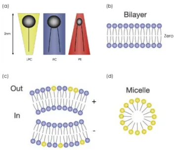
A useful measure that allows the distinction of lipid molecules, from the point of view of their organization, is the dimensionless packing parameter (PP) (or shape factor), which is defined as the ratio of the cross-section area of the headgroup (HG) to that of the acyl chain (AC), i.e. HG/AC (De Krujiff, 1985). Consequently, large values of the PP (HG/AC > 1) correspond to molecules of large head group section area and small acyl chain section, inversed conical shape. On the other hand, if HG/AC < 1 then both sections are close (this usually being the case of lipid molecules with two acyl chains as the POPC in Fig.2.1) and the molecules can be represented as a cylinder (Przestalski et al., 2000) (Fig.2.2). I will discuss this in more detail in chapter 3, for the moment keep in mind that these cylindrical lipids tend to form lamellar structures, which can additionally form lipid bilayers separated by water layers, as commonly occur in biological membranes. This particular observation has been use as a central argument to speculate that in the early stages of life, the spontaneous formation of lipid bilayers gave rise to the development of biological membranes (Tien, 2000). In some pioneering works, Marrink and co-workers have been recently able to simulate the spontaneous formation of bilayers. These works demonstrated that even processes that involved hundreds of lipids can be simulated obtaining realistic intermediate stages (de Vries et al., 2004a; Marrink et al., 2001).
Figure 2.2: Lipid shape determine the organization in bilayers. Bilayers can be asymmetric in lipid composition (Modified from (Janmey and Kinnunen, 2006a)).
2.1.3
Membrane composition
The chemical composition of the two leaflets of the lipid bilayer is different (Fig.2.2). For example, in eukaryotes, nearly only anionic lipids face towards the cytoplasm, whereas most lipids with large glycosylated headgroups are exposed to the extracellular environment. Differences in bilayer asymmetry between eukaryotic and prokaryotic membranes are essential for the activity of endogenous antimicrobial factors that break bacterial membranes but are harmless to eukaryotic cells ((Sevcsik et al., 2007; Sato and Feix, 2006).
The chemical composition of the bilayer affects its mechanical properties and conversely, the use of physical forces on membranes alters their chemical composition. The bilayer asymmetry is highly dynamic and different cell types as well as organelles and cells at different states of activity are likely to change the lipid distribution. Moreover, the bilayer trasversal asymmetry have important consequences in the spontaneous curvature of the membrane (Devaux, 2000). I will explain in more detail all these bilayer mechanical properties in chapter 3.
The formation of microdomains and the presence of mixed bilayers induce lateral asymmetry. In these bilayers, the differences in lipid shapes (cylinders, cones, etc.) between both bilayers create lipid mismatches, known as lipid packing defects. These packing inhomogeneities can also appear by an increase in the membrane curvature. This particular phenomenon will be also discussed in the following chapter. Complicated mixtures have proven to be difficult to simulate for the moment. However, there exist few exemples of simulations made on bilayer with a simple (2 or 3 different lipids) mixed lipid compostion. However they have been able to describe some important atomic feature related with the bilayer properties (Chiu et al., 2002; de Joannis et al., 2006b,a).
is very limited. The chemical structure of lipids influences their kinetic behavior: the presence and nature of the polar head, the length of the lipid, the degree of saturation or instauration of the acyl chains, etc. define and delimit the behavior that a certain lipid may adopt (Kusumi and Suzuki, 2005; Tristram-Nagle and Nagle, 2004; Marrink et al., 2009a). Moreover, the surrounding environment of the lipid molecules introduces additional parameters to this behavior and movement, such as the interaction of the polar head with the aqueous medium and ions, and with the membrane proteins, which also exert an influence on lipids.
Membrane fluidity is first of all determined by the lipid composition and depending on it the mem-brane will exhibit a particular lamellar fluid state known as lamellar phase. The lamellar phases can vary as a function of the lipid content or with the temperature, leading to different phases exemplified in Figure 2.3. This phases can be disordered or ordered depending on the orientations of the lipids acyl chains with respect to the normal of the bilayer. As an exemple, if the acyl chains are almost parallel to the bilayer normal the lamellar phase is ordered, this is the case of saturated acyl chains; the disorder in the bilayer can be induce by many factors (Feigenson, 2006). The biological membrane correspond to a lamellar liquid-disordered phase. In order to understand the dynamics of this biological relevant phase and the effect of lipid dynamics on membrane the global membrane structure, I will next explain these aspects in more detail.
Two main lipid movements are rotation and oscillation. Both of them are implicated in the lipid transport. Additionally, lipids display many other important movements for the membrane function, which has been assessed by MD simulations (Marrink et al., 2009a): the lateral diffusion6 in the plane
of the membrane, the transversal diffusion from one layer to the second one. This diffusion can be partial, concerning solely one fragment of the acylchains, or can be total, that is when the transversal diffusion concern the entire lipid that has passed from one leaflet to the opposite one (process known as flip-flop)7(Holthuis and Levine, 2005). The bilayer asymmetry discussed in section 2.1.3 is dependent
6
The lipids can difusse in a membrane covering an area from 0,1 to 1 microm2
s-1
in a cell membrane. However, lipid diffusion can vary in function of the lipid nature and the environment surrounding it.
7
In model membranes, the flip-flop is very slow for lipids with polar heads compared with lipids without polar heads (Bai & Pagano, 1997). For some membranes such as the reticulum endoplamic, the expansion of the membrane is very important and depends on the flip-flop of lipids at a high rate. This is possible thanks to the proteins called flipases (Buton et al. , 1996) that assure the transversal diffusion from one leaflet to another at a high rate. In the case of plasmatic membranes that have to mantain an asymmetric composition, the ATP-dependent flipases, make possible the traslocation of PS or PE phospholipids (Seigneuret & Devaux, 1984).
Figure 2.3: Liquid phases in biological membranes that determine the fluidity and order/disorder of the lipids. (Modified from (Veatch, 2008)
of this last process.
The range of phospholipid motions are correlated with the acyl chain orientation and movements. Lipid order inside membranes reflects their rotational motion and the average orientation of the acyl chains C-C bond with respect to the normal to the membrane which can be measured with the Order Parameter (see section 5.7.1.2)(Takaoka et al., 2000). For instance, if we consider a fully saturated phospholipid below the phase transition temperature, the acyl chains are in the extended all-trans conformation and are closely packed (ordered), in this case the range of motion is small. However, above the phase transition temperature, the chains contain a number of gauche configurations, which makes the packing of the chains looser and the range of motion of the acyl chains higher (disordered). The presence of different types of lipid molecules in a cell membrane decreases the order in the bilayer, and produces the different lamellar phases we just described (Feigenson, 2006) (Fig2.3).
Among other important factors that influence the lipids order are the volume and ratio of the polar head and the acyl chains, as well as the nature of the polar head, the saturation of the fatty acyl chains, and the number of trans and gauche positions of the C-C bonds in the chain. In saturated PC membranes, the increase of acyl chain lengths also increases their order but decreases their reorientation motion (Kusumi and Suzuki, 2005; Subczynski et al., 1992, 1993). The introduction of unsaturations in the PC acyl chains generates packing defects in the lipid bilayer and affects deeply the narrow lipid packing and the membrane order. For instance, the presence of a cis double bonds introduces a bend in the unsaturated acyl chain that would create some non-conformability between chains and increase chain disorder, leading to the lipid packing defects we discuss previously. Additionally, the presence of either cis or trans double bonds in the acyl chain would reduce the dynamics of the chain around the rigid double bonds.
Figure 2.4: (a) Singer & Nicholson Mosaic-fluid membrane model. (b) Engelman model of a membrane more mosaic than fluid. (Taken from (Engelman, 2005)).
influence in already highly ordered saturated membranes since it is capable to increase even more the membrane rigidity (Chiu et al., 2002; Pasenkiewicz-Gierula et al., 1990). On the other hand, the introduction of unsaturated acyl chains in these rigid membranes, as in any other membrane, increases greatly the fluidity and the disorder of the membrane. The “fluidizing” effect of unsaturated chains observed in biological membranes moderates and counteracts the “rigidifying” effect of cholesterol (as well as the effect of any other rigid-membrane modifiers).
The proteins embedded in the membrane can cause dramatic changes in this fluid states structure of the lipid bilayer, since the size, structure and composition of these proteins may restrict lipid movements or increase lipid disorder (Sanderson, 2005; Salnikov et al., 2009b,a). All these effects in the lipid bilayer are going to modify the order and the diffusion of each lipid. Moreover, these effects are specific of the lipid structure, its environment and the lapse of time the lipids spend in that environment. In order to clarify these dynamic aspects, I will discuss now the structure of the biological membrane. Remember nonetheless, that since the biological membrane is a mixture of many elements, its behavior is far from the ideal liquid structures we illustrate in Figure 2.3.
2.2
Membrane structure is a complex target
Almost forty years ago Singer and Nicholson proposed the first structural model for the biological membrane, the fluid-mosaic model (Singer and Nicolson, 1972). This first model proposed an initial vision of static proteins, which at low concentrations were embedded in the fluid membrane. The membrane, on the other hand, was not perturbed by the presence of proteins nor by its direct contact with the solvent. All the functional properties were attributed to the membrane-proteins embedded in it. Nowadays, this vision has become more complex. We now know that the biological membrane is a highly dynamic system whose components are in continuous change and movement. The membrane, by
it-self, is starting to be recognized as an important actor of many membrane-protein related functions. The plasmatic membrane is composed of regions showing different and specific structures and functions (Engelman, 2005) (Fig.2.4). The most important of these specialized membrane structures are the lipid rafts (Rajendran and Simons, 2005; McIntosh, 2007). These lipid specific regions modulate the membrane fluidity by restricting the directions lipids can follow (Kusumi and Suzuki, 2005). Moreover, the thickness of these membrane regions can also change according to the lipid composition and as a function of the length8of the proteins that are embedded in these membrane regions (Mitra et al., 2004).
The prevailing model suggests that the membrane is more mosaic than fluid (Engelman, 2005). This change of vision became possible thanks to the development of new methodologies and technologies such as the electron microscopy, NMR and the fluorescence spectroscopy. Finally, in recent times, the computer simulations have started to play an important role in the construction of these new perspectives of the biological membranes (Marrink et al., 2009a; Gumbart et al., 2005; Risselada and Marrink, 2009; Niemela et al., 2009).
2.2.1
Structure
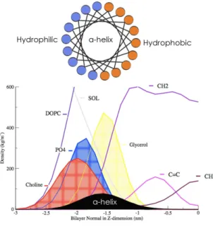
Since cellular membranes should remain in a fluid state for normal cell functions, it is exactly this fluid structure (Lα-phase) (Fig.2.3) of the bilayer that is relevant for the understanding of membrane-peptide interactions at a molecular level. Unfortunately, the disorder found in fluid bilayers prevents high atomic-resolution. Useful low-resolution structural information can nevertheless be obtained by diffraction methods using multilamellar bilayers (liquid crystals), which have been dispersed in water or placed on surfaces, and whose structure along the bilayer normal is highly periodic. This one-dimensional crystallinity shows a distribution of matter along the bilayer normal that can be hence determined from combined X-ray and neutron diffraction measurements ((White SH, 1996). The dynamic structure of the fluid (Lα-phase ) bilayer that results from such measurements is a collection of time-averaged spatial distributions (Gaussians) of the principal structural groups of the lipid (carbonyls, phosphates, etc.) and the water projected to the normal axis of the bilayer plane (Wiener and White, 1991a,b) (Fig.2.5) ). Using this technique White and co-workers determined for the first time the structure of a DOPC bilayer (Wiener et al., 1991). Inspired by these works, Tristram-Nagle and colleagues refined and completed the structural properties of DOPC (and other phospholipids) bilayers fully hydrated (Tristram-Nagle et al., 1998). This structure, which computer simulations can reproduce (Berger et al., 1997; Feller, 2007; Tieleman et al., 2006; Martinez-Seara et al., 2008a,b), revealed several features of great importance (Fig.2.5):
1. The DOPC bilayer has a highly disordered structure. This great amount of disorder is revealed by the width of the probability densities.
2. The crosssectional area per lipid is ~72 Å2.
8
Known as hydrophobic mismatch. Membrane hydrophobic mismatch is the difference between the hydrophobic length of α helices of the integral proteins and the hydrophobic thickness of the membrane they span. Under the consideration of energy requirement, in order to avoid unfavorable exposure of hydrophobic surfaces to a hydrophilic environment, the hydrophobic length of the integral proteins is supposed to be approximately equal to the hydrophobic bilayer thickness.
Figure 2.5: DOPC bilayer structure density profile obtained by X-ray Scattering (modified from 3. The optimal membrane thickness depends on the chain length, the degree of saturation and the
angle of tilt of the acyl chain within the membrane. The thcikness of the hydrophobic core (HC) is around 20 Å and the combined thickness of the interfacial regions (those covered by the density distribution of the waters of hydration) is around 30 Å thickness of the entire bilayer. The thickness of a single interface (15 Å) can easily accommodate unfolded and folded polypeptide chains such as an amphipathic α-helix with diameter ~10 Å (typical of TM helices in membrane proteins).
4. The interface is chemically heterogeneous; they can have multiple possibilities for non-covalent interactions with peptides. Since these interfaces are the sites where proteins have their first membrane contact (typical of interfacial amphipathic helices in membrane-proteins as we will discuss in section 2.4), they are especially important in the folding and insertion of peripheral proteins or non-constitutive membrane proteins such as toxins.
5. The interface, besides being chemically heterogeneous, is also regions where dramatic changes in polarity occur over small distances.
2.2.2
Partitioning
How the small molecules can pass through the complex structure of the membrane? How the proteins fold and insert in the membrane? These are two of the most studied questions in the membrane field. The answers are not clear since the ability of molecules to cross the membrane or to get embedded in it
depend on their partitioning behavior a fascinating and difficult problem. Therefore, and before I talk about membrane proteins, I would like to mention some aspects about the partitioning .
Many essential biomolecules such as water, oxygen, and carbon dioxide move across the cell mem-brane by passive diffusion (Bemporad et al., 2004; Xiang and Anderson, 1998a,b; Marrink et al., 1996; Tieleman, 2006) Additionally, many drug molecules are also capable to enter the cell through pas-sive diffusion across the cell membrane. This means that their partitioning within lipid bilayers has important implications for their pharmacokinetics (Pohorille et al., 1998). Finally, the protein folding adopted inside a membrane is determined by the partitioning behavior of the amino acid side chains (Ulmschneider and Ulmschneider, 2008; Ulmschneider, 2009; White et al., 1998; White and Wimley, 1999; Wimley et al., 1998, 1996b,a; MacCallum et al., 2008, 2007), based on the thermodynamics of lipid-side chain interactions. The partitioning has, in consequence, different energetic costs.
The structure of the bilayer and the gradient of polarity in the direction normal to the membrane, govern the behavior of polar and charged residues when they are moved into the hydrocarbon core by a large range of opposing tendencies, including hydrogen bonding, hydrophobic effects, and the energetic costs of membrane deformation and transfer of charges and dipoles in a low dielectric environment (White, 1994; White and Wimley, 1998; White, 2007). Numerous experimental studies have addressed the question of partitioning of amino acid side chains in lipid bilayers with significant success, finding unexpected behaviors for all polar and charged amino acids when they are confronted to the large range of opposing tendencies (White, 2007). However, most of the results from these experiments have limited spatial resolution. In particular, it is difficult to control the local environment of a side chain due to the inhomogeneous nature and the deformability of the bilayer. These two factors also prevent the prediction of side chain partitioning behaviors.
White and co-workers (Wimley and White, 1996) determined some hydrophobic scales for whole-residues by a combination of experiments that determined the free energies of transfer for each amino acid in POPC membranes (and n-octanol)9. These scales are essential for understanding the energetics
of protein-bilayer interactions. The most important feature of White and co-workers practical scales is that they decided to include in their calculations the contributions of the peptide backbone bonds in the free-energies of the different transfers, instead of limiting their research to just the side-chains. By including whole-residues, these data have become of great value to understand some partitioning issues and have turned out to be crucial to perform more realistic peptide-membrane simulations.
The atomistic computer simulations may provide a level of detail that is not accessible with other kind of experiments. Consistently, molecular dynamics simulations can give a complementary view of side-chain partitioning to experimental macroscopic measurements, and to elaborate predictions. Some simulations of the designed pentapeptides used by White and co-workers (Aliste and Tieleman, 2005; Ahumada et al., 2003) and of side-chains in bilayers (MacCallum et al., 2007, 2008) have provided a molecular interpretation of the thermodynamic measurements that form the basis of the hydrophobicity scales (Wimley and White, 1996; Wimley et al., 1996b,a) (Fig.2.6 ). On the other hand, the partitioning of side chain between water and hydrophobic solvents has been used to test and parameterize the force
9
Many experiemental and simulation research in membrane partitioning sometimes use hydrophobic solvents that mimic the HC of the membrane. In these environments, is not possible to asses the effect of the interface.
Figure 2.6: Transfer free energies calculated by MD simulations (right) and an example snapshot of a side-chain inside the bilayer :acyl chains in grey, phosphate level (bleu), glycerol level (orange) and water (red).
field used in molecular dynamics (Tieleman et al., 2006; MacCallum et al., 2007; MacCallum and Tieleman, 2003; Oostenbrink et al., 2004). Finally, several computational studies have also addressed the distribution of specific small molecules in lipid bilayers, finding the basic atomic characteristics of this phenomenon highly dependent on bilayer properties and solute volume, size, and cross-sectional area (Bemporad et al., 2004; MacCallum et al., 2008; MacCallum and Tieleman, 2006; Marrink et al., 1996; Norman and Nymeyer, 2006). Marrink and co-workers have recently simulated the insertion of an entire peptide in the membrane, and demonstrate that even with long times of simulations, this process, highly energetically expensive, cannot be succesfully simulated (Yesylevskyy et al., 2009). 2.2.2.1 Partitioning-folding-coupling mechanism
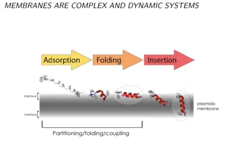
In order to get inside the membrane bilayer, a single peptide pass througth different stages: (i) parti-tioning of an unfolded protein fragment chain into the bilayer interface, (ii) the formation of secondary structure in the interface, (iii) insertion of the secondary structure element across the membrane, fol-lowed by the association of the secondary structure elements within the membrane (Fig.2.7).
The folding of membrane-proteins in the bilayer environment is a difficult experimental and simu-lation problem, some studies in both fields have addressed this question indirectly, traditionally using small peptides that are unfolded in aqueous solution but are fully structured upon partitioning into the interface. White and co-workers have called this process partitioning-folding coupling mechanism, based on their works with an amphipathic helical peptide, the antimicrobial melittin (Hristova et al., 2001). They suggest then that the stages (i) and (ii) are coupled. They established that the energetic cost of transferring an unfolded state (a virtual unfolded state calculated from the hydrophobicity scales) to the interface environment was higher than transferring a folded state. Therefore, the partitioning-folding coupling implies that secondary structure formation is driven by lipid-peptide interactions and the sub-sequent H-bond formation that accompanies the partitioning of the peptides. Numerous studies have studied the thermodynamics of this process (Seelig, 2004) and some MD simulations have addressed the question of membrane folding and insertion with results that have provided some promising insights
Figure 2.7: Insertion of a peptide in the bilayer showing the partitioning-folding coupling process (Mod-ified from (Ulmschneider and Ulmschneider, 2008)
about this complicated problem (Chipot et al., 1999; Tieleman et al., 2001a; Ulmschneider and Ulm-schneider, 2008; Yesylevskyy et al., 2009). It is just recently that Ulmschneider (UlmUlm-schneider, 2009) has achieved the folding of a TM peptide in explicit model membranes using high temperatures though. In eukaryots, the energetic cost of folding and translocating an entier membrane-protein is counteract by the translocon machinary10. I consider now apropiated to talk about the membrane-proteins and the
different interactions they can establish with the peptides. In particular, I will discuss the amphipathic α-helix, which is a conspicuos membrane-binding motif, that we already slightly mentioned in this last section.
2.3
A Glimpse of Membrane proteins
Membrane proteins represent 30% of the Open Reading Frames (ORFs) identified in complete sequenced genomes (Stevens and Arkin, 2000), which makes more than 10,000 protein molecules. From the last sections and given this number, it is possible toi infer that our current understanding on the structure of membrane protein and protein-lipid interactions is rather limited. There are two mayor reason for this: one, the difficulties to obtain high-resolution structures of membrane proteins, and two, the disordered nature of the lipid environment with its strong gradients of density, chain order, and polarity. Even if the structures of membrane proteins has increased steadily in the PDB database during the last years, they remain under-represented, and in many cases, only the soluble sub-domains are known. This technical difficulty to obtain the membrane-embedded protein sub-domains has become a serious challenge for the molecular dynamics and structural prediction sciences. Nowadays, the available structures of membrane-embedded protein sub-domains are modeled in silico inside artificial membranes, in order to evaluate the role of lipid dynamics in the protein function and structure (Appelt et al., 2005; Blood et al., 2008;
10
The translocon (commonly known as a translocator or translocation channel) is the complex of proteins associated with the translocation of nascent polypeptides across membranes.
Membrane proteins are classified in two groups, depending on their localization in the membrane: integral proteins, those inserted in the bilayer, and peripheral proteins, those that establish transitive interactions with the membrane using both electrostatic and hydrophobic interactions or thanks to membrane-protein partners. According to the interactions the membrane proteins establish with the membrane, two main categories of membrane-protein interaction can be distinguished (Sanderson, 2005)(Fig.2.8):
• The polytopic interaction (i.e. transmembrane (TM) proteins) typical of integral proteins • The monotopic interaction (i.e non-TM proteins) typical of proteins that are only inserted in one
leaflet of the membrane, and peripheral proteins.
Polytopic transmembrane interactions are typical of those proteins that span the membrane one or many times. These proteins can be divided in two groups based on the two more frequent secondary structure elements that conform them: the α-helix TM interaction, with one or more hydrophobic helices spanning the membrane (from 1 to more than 20 helices) (Ubarretxena-Belandia and Engelman, 2001) and the β-sheets TM interaction, a succession of anti-parallel β-stands (8 to 22 strands) forming a β-barrel (Schulz, 2002) (Fig. 2.8). The α-helix and β-sheets of TMs are in contact with each other, with the bilayer hydrophobic core, the bilayer interface, and, of course, the water. This is the reason of the importance of non-random amino acid distributions on those secondary structures.
As in soluble proteins, the interiors of polytopic proteins are comprised of internally H-bonded α-helix and/or β-sheets (Fig.2.8). Moreover, the major portions of their masses are buried inside the HC of the membrane, and are arranged in such a manner that allows their outer surfaces to face acyl chains that conform the HC(Schulz, 2002; Ubarretxena-Belandia and Engelman, 2001). Although the average hydrophobicity of the membrane proteins interior is the same compared to soluble proteins, the amino acids of the outer surfaces are more hydrophobic (Samatey et al., 1995). The outside of an integral membrane protein is lipid-exposed. Its sub-domains are rich in hydrophobic amino acids, which are entirely matched by the lipid tails that make up about 40% of the thickness of the bilayer.
The average lengths of the TM α-helix and β-strands are greater than those observed in soluble proteins, so the 20 Å thick bilayer HC core can be spanned: α-helix are generally longer than 20 amino acids (1.5 Å /residue), and β-strands are longer than 10 amino acids (3.3 Å /residue). The TM
Figure 2.8: Schematics of the topology of the membrane depending on membrane-proteins interactions.(a-c) Correspond to integral proteins with polytopic interactions (a and b) with α-helices and (c) with β-sheets as the main secondary structures present. (d-g) Correspond to monotopyc inter-actions and they represent groups 1,2,3 and 4 described in the text, respectively.
proteins helices can tilt in order to make their length match the bilayer thickness, this is known as the hydrophobic-mismatch (Ozdirekcan et al., 2007; de Planque and Killian, 2003; Sparr et al., 2005; Debret et al., 2008). At the same time, the bilayer thickness can change in the proximities of the proteins as we will discuss in the next chapter. Because of the length and the highly non-polar character of TM helices, hydropathy plots (Engelman et al., 1986; White, 1994) have proven to be useful and remarkably accurate for predicting the topology of α-helical transmembrane proteins.
In turn, monotopic non-transmembrane protein-membrane interactions are characteristic of proteins that get in contact with only one leaflet of the lipid bilayer. They can be divided in four groups Lomize 2007 (Fig. 2.8):
• The first group includes those proteins that bind the membrane thanks to post-translational modification. Here, a covalent bond between the protein and a hydrophobic component is formed; most of the time this component is the Glycosyl-Phosphatidyl-Inositol (GPI) (Sangiorgio et al., 2004) or prenylations in the proteins (Resh, 2006).
• The second group consists in proteins that interact with the membrane thanks to hydrophobic loops in their surfaces. This segments, can insert deeply into the membrane interface. (i.e. cardiotoxine (Efremov et al., 2004))
• The third group corresponds to proteins that display electrostatic protein-membrane interactions. For example, the annexine V is capable to interact with the membrane thanks to a salt bridge formed between a Ca++ ion, the protein and the phospholipids negative charges (Mukhopadhyay
and Cho, 1996).
• The fourth group includes proteins that interact with the membrane thanks to one or many interfacial amphipatic α-helices, which remain parallel to the plane of the membrane (IPM helix for In-Plane Membrane ) (Sapay, 2006). Sapay and Gautier have develop new approaches in order
interactions. That is, the polypeptidic chains folds in order to expose the polar residues toward the polar environments (solvent or headgroups), forming the polar domains, and keeping the hydrophobic residues in the interior of the protein or exposed to the hydrophobic environment (the acyl chains), forming the hydrophobic domains (Ubarretxena-Belandia and Engelman, 2001; Samatey et al., 1995; Schulz, 2002).
The most predominant secondary structures in transmembrane proteins are α-helix and β-sheets. Some crystallographic structures of membrane proteins as well as numerous molecular dynamics simu-lations have started to highlight the importance of flexibility on these structures for the correct function of membrane proteins (Debret et al., 2008; Stelzer et al., 2008; Deupi et al., 2009; MS and H., 2000). Several experimental and simulation studies have analyzed the propensities of different amino acids to form particular secondary structures in solution (Petukhov et al., 1999, 2002; Ramirez-Alvarado et al., 1999; Viguera and Serrano, 1999; Chakrabartty et al., 1994, 1993b,a, 1991; Pace and Scholtz, 1998). Hydrogen bonds play a role in stabilizing the α-helix and β-barrel conformation. However, the size and charges of sidechains are also important factors. This turned out to be relevant in the description of different aminoacid propensities to form helices in an aqueous environment. Different amino-acid sequences have different propensities to form α-helical structure. Met, Ala, Leu, uncharged Glu, and Lys, have especially high helix-forming propensities, whereas Pro, Gly and Asp have poor helix-forming propensities. Pro tends to break or kink helices because it cannot form a hydrogen bond (having no amide hydrogen), and because its sidechain interferes sterically. However, Pro is often seen as the first residue of α-helix, presumably due to its structural rigidity. In contrast, Gly, which also tends to disrupt helices, has a high conformational flexibility that becomes entropically too expensive to stay in the relatively constrained α-helical structure. Ser and Thr have also been identified as helix breakers or mostly in loop regions since they introduce flexibility to the soluble proteins. Other residues such as Val and Ile (well known as β-sheets formers) also break the α-helices .
The folding and packing of the membrane proteins is a major issue. I already discuss the influence of the partitioning in the folding. This is correlated with the individual amino acids propensities to form specific secondary structures. Thus, it is not surprising, that the structural propensities of different residues in the membranes are not yet well defined. Some clues have been revealed though. The experimentalists have tried to understand the propensities for a particular folding by examining its secondary structure stability and oligomerization. Some of these studies have been performed using
Figure 2.9: (a) Frequencies of residues in TM proteins. (b) Propensities to form alpha helices in hydrophobic and aqueous environment (Adapted from (Liu and Deber, 1998b)).
transmembrane synthetic peptides in hydrophobic environments, such as micelles and vesicles (Li and Deber, 1994a), whereas others has been made in bulk-hydrophobic solutions (Liu and Deber, 1998b) (Fig. 2.9 ). These works have shown that, in contrast to their conformational preferences in water, the helical proclivity of the non-charged amino acids inside membranes is governed by their side chain hydrophobicity, and by the hydropathy of the local peptide segments in which the residues reside (Li and Deber, 1994b; Li et al., 1996; Liu and Deber, 1998b).
Gly, and β-branched residues, such as Ile, and Val (Li and Deber, 1992a) seem to have an environment-dependent role for the support and modulation of helices. In those examples, the helical propensity of hydrophobic segments with different composition, decrease in the following order: Ala-Leu-rich> Gly-Leu-rich> Gly-Ile(Val)-rich. These results suggest that these residues may provide, partially, the struc-tural basis for conformational transitions within or adjacent to membrane domains (Landolt-Marticorena et al., 1993). Indeed, it has been shown that the lipid hydrophobic effects can contribute to dimer sta-bility and the affinity of motif-mediated helix-helix interactions to define helix-helix interfaces (Johnson et al., 2006).
On the other hand, the ability of all naturally occurring amino acids to form a turn when placed in the middle of a transmembrane helix has recently been measured (M. et al., 1999). The observed rank order for turn-stabilizing tendencies are Asn=Arg=Pro (1.7)>Asp=Glu=His=Lys=Gln=(1.6)>Gly (1.3)>Ser=Trp (0.7)>Cys=Ile=Tyr (0.6)>Ala=Met=Val (0.5)>Leu=Phe=Thr (0.4). Moreover sta-tistical analysis have confirmed that clearly, there are two sets of residues with either high (≥1.3) or low (≤0.7) turn propensity. Charged or polar residues induce a turn (≥1.3), whereas hydrophobic residues plus Ser, Thr, and Cys remain α-helical (≤0.7) (Ballesteros et al., 2000). Furthermore, it has been
Other studies have highlighted that cation-pi interactions, particularly between Lys and Trp, Tyr, or Phe, as well as weakly polar interactions between pairs of aromatic residues, significantly enhance the strength of oligomerization of these hydrophobic helices. The contribution of these forces to the tertiary structure formation in designed transmembrane segments suggests that similar forces may also be a significant factor in the folding and stability of native membrane proteins (Johnson et al., 2007). Deber and co-workers proposed that the high frequency of occurrence in membranes of residues such as Leu, Val, Ile, and Phe derives not only from their hydrophobicity but also from their intrinsic propensity to form the α-helical conformation in the non-polar environments of membranes (Liu and Deber, 1998a), since the helical propensity for individual amino acids is correlated with non-random occurring frequency in protein TM helices (Landolt-Marticorena et al., 1993).
In brief, all that has been discussed until now gives the first notions to reconstruct the scenario for the action of the membrane curvature sensor ALPS, our study subject. The specific features of ALPS will be discuss in detail in chapter 4, for the moment, is important to remind that ALPS is an interfacial amphipathic α-helix, which structure depends on its partitioning. The individual propensities of its amino acids to form an α-helix, most also be taken in account. However, less is known about this matter at the interface due to its chemical heterogeneougenisity. Hence, its is crucial to consider that many non-covalent interactions are taking place and the structural and dynamic properties of the membrane will have an impact on them.
2.4
Amphipathic alpha-helices
This type of interaction to the membrane was discoverd more recently that the other monotopic binding interactions. The first amphipathic helix motif was identified in the Penicillin Binding protein 5 (PBP5) in 1986 (M et al., 1986), which coincidently, contributes to maintain the shape of the cell in E.coli (Nelson and Young, 2001). The first crystallographic structure of this kind of motif was from the protein Prostaglandin H2 synthase-1 in 1994 (D et al., 1994). Since then, many exemples of this binding motif have been identified. The amphiphatic helix is a conspicuous motif in proteins that bind to the membranes. It can be found for instance in many small GTPAses (Sar1, Arp1, Arf1, etc) (Farsad and De Camilli, 2003) or in some of their activators (as ArfGAP1 (Bigay et al., 2003)). It can be present in the sterol transporter Kes1p and nucleoporin Nup13, and the golgin GMAP-210 (Drin et al., 2007),