Université de Sherbrooke
ROLE OF THE HUMAN CHYMASE (CMA1) IN THE CONVERSION OF BIG-ENDOTHELIN-1 TO BIG-ENDOTHELIN-1 (1-31)
By Walid Semaan
Departement of Pharmacology-Physiology
Thesis presented to the Faculty of medicine and health sciences in order to obtain the diploma
maître ès sciences (M.Sc.) in pharmacology
May 2016
Members of the Jury: Pedro D’Orleans-Juste, Department of Pharmacology-Physiology; Ghassan Bkaily, Department of Anatomy and Cell Biology; Jean-Bernard Denault, Department of
Pharmacology-Physiology; Sheela Ramanathan, Department of Pediatrics, Division of Immunology
RÉSUMÉ
Rôle de la chymase humaine (CMA 1) dans la conversion de la big-endothéline-1 en endothéline-1 (1-31)
Par Walid Semaan
Programme de pharmacologie
Mémoire présenté à la Faculté de médecine et des sciences de la santé en vue de l’obtention du diplôme de maître ès sciences (M.Sc.) en pharmacologie, Faculté de médecine et des sciences de la
santé, Université de Sherbrooke, Sherbrooke, Québec, Canada, J1H 5N4
La voie de conversion de Big ET-1 en ET-1, chymase dépendante a été établie in vitro. Ce n'est que récemment, en 2009 que notre groupe a démontré que la conversion de Big ET-1 en ET-1 (1-31) peut avoir lieu in vivo chez la souris (Simard et al., 2009), sachant que ET-1 (1-31) est convertie en ET-1 via NEP in vivo (Fecteau et al., 2005). En plus, en 2013, notre laboratoire a démontré que la mMCP-4, l'analogue murin de la chymase humaine, produit l'ET-1 (1-31) à partir du précurseur Big ET-1 (Houde et al., 2013). Jusqu'a présent, dans la littérature, on ne trouve pas de caractérisations spécifiques de chymases (humaine ou murine) recombinantes. En fait, le groupe de Murakami, en 1995, a publié une étude caractérisant, d'une façon chymostatin dépendante, la CMA1 (chymase humaine) en utilisant l'Angiotensine I comme substrat (Murakami et al., 1995). Cependant, le chymostatin est un inhibiteur non-spécifique de la chymase. Il a été démontré que le chymostatin peut inhiber l'élastase, une enzyme pouvant convertir l'Angiotensine I en Angiotensine II (Becari et al., 2005). Basé sur ces observations, l'hypothèse formulée dans la présente étude est que la CMA1 recombinante ou extraite des cellules LUVA (lignée humaine de mastocytes) ou des fractions solubles des aortes humaines convertit la Big ET-1 en ET-1 (1-31) d'une façon TY-51469 (un inhibiteur spécifique de la chymase) sensible. Dans un deuxième volet, on a étudié la cinétique enzymatique de CMA1 en vers le substrat Big ET-1 et Ang I. L’affinité de CMA1 contre la Big ET-1 était plus grande comparé à l’Ang I (KM Big ET-1 : 12.55 μM et Ang I : 37.53 μM). Cependant CMA1 était plus efficace dans le clivage de l’Ang I comparé à la Big ET-1 (Kcat/KM Big ET-1 : 6.57 x 10⁻⁵ μM-1.s-1 et Ang I : 1.8 x 10⁻⁴ μM-1.s-1). Dans un troisième volet impliquant des expériences in vivo, l’effet presseur de la Big ET-1, l’ET-1 et l’Ang I a été testé chez des souris conscientes mMCP-4 KO comparé à des souris de type sauvage. L’augmentation de la pression artérielle moyenne a été plus importante chez les souris de type sauvage après l’administration de Big ET-1 que chez les souris mMCP-4 KO. Cet effet n’a pas été observé après l’administration d’ET-1 et/ou d’Ang I ce qui explique le rôle de la chymase dans l’effet de la conversion de Big ET-1 en ET-1 (1-31).
Mots clés : Chymase, Enzymes recombinantes, Spectrométrie de masse, radio-télémétrie, analyse in silico
ABSTRACT
Role of human chymase (CMA 1) in the conversion of big-endothelin-1 to endothelin-1 (1-31)
by Walid Semaan Program of Pharmacology
Thesis submitted to the Faculty of Medicine and Health Sciences for Masters of Science (M.Sc.) in Pharmacology, Faculty of Medicine and Health Sciences, Université de Sherbrooke, Sherbrooke,
Quebec, Canada, J1H 5N4
The chymase-dependant pathway responsible for converting Big ET-1 to ET-1 was established in vitro. It has only been recently, in 2009, that our group demonstrated that the conversion of Big ET-1 to ET-1 (1-31) can occur in vivo in mice (Simard et al., 2009), knowing that ET-1 (1-31) is converted to ET-1 via NEP in vivo (Fecteau et al., 2005). In addition, our laboratory demonstrated in 2013 that mMCP-4, the murine analogue of human chymase, produces ET-1 (1-31) from the Big ET-1 precursor (Houde et al. 2013).
Thus far, in the literature, there are no specific characterizations of recombinant chymases (human or murine). In fact, the group of Murakami published in 1995 a study characterizing the CMA1 (human chymase) in a chymostatin-dependent fashion, using Angiotensin I as a substrate (Murakami et al., 1995). However, chymostatin is a non-specific inhibitor of chymase. It has been shown that chymostatin can inhibit elastase, an enzyme that can convert Angiotensin I to Angiotensin II (Becari et al., 2005).
Based on these observations, the proposed hypothesis in the present study suggests that recombinant as well as extracted CMA1 from LUVA (human mast cell line), in addition to soluble fractions of human aortas, convert Big ET-1 into ET-1 (1-31 ) in a TY-51469 (a chymase-specific inhibitor) sensitive manner.
In a second component, we studied the enzyme kinetics of CMA1 with regard to the Big ET-1 and Ang I substrate. The affinity of CMA1 against Big ET-1 was greater compared to Ang I (KM Big ET- 1: 12.55 μM and Ang I: 37.53 μM). However, CMA1 was more effective in cleaving Ang I compared to Big ET-1 (Kcat / KM Big ET-1: 6.57 x 10⁻⁵ μM-1.s-1 and Ang I: 1.8 x 10⁻⁴ ΜM-1.s-1).
In a third component involving in vivo experiments, the pressor effects of Big ET-1, ET-1 and Ang I were tested in conscious mMCP-4 KO mice compared to wild-type mice. The increase in mean arterial pressure after administration of Big ET-1 was greater in wild-type mice compared to mMCP-4 KO mice. This effect was not observed after administration of ET-1 and / or Ang I.
TABLE OF CONTENT
RÉSUMÉ ………...iii
ABSTRACT………...iv
TABLE OF CONTENT ……….v
LIST OF FIGURES ………..vii
LIST OF TABLES ………..viii
LIST OF ABBREVIATIONS AND SYMBOLS………...ix
I- INTRODUCTION ………….………. 1
1.1 MAST CELLS DEVELOPMENT ………... 1
1.2 MAST CELL DEPENDENT DISEASES ………2
1.3 MAST CELLS IN CARDIOVASCULAR DISEASES………..…. 3
1.4 MAST CELL PROTEASES ……….... 3
1.5 CHYMASE SYNTHESIS ………..…. 4
1.6 CHYMASE SUBSTRATES ………...…. 7
1.7 THE ROLE OF CHYMASE IN PHYSIOLOGY AND PATHOPHYSIOLOGY ….10 1.7.1 Chymase in wound healing ………. 10
1.7.2 Chymase in cardiovascular diseases and Atherosclerosis ……….. 10
1.7.3 Chymase in metabolic diseases and diabetes mellitus ……… 11
1.8 THE ROLE OF CHYMASE IN THE CONVERSION OF ANGIOTENSIN I TO ANGIOTENSIN II ………..………. 12
1.9 THE ROLE OF ANGIOTENSIN II IN PHYSIOLOGY AND PATHOPHYSIOLOGY ………... 12
1.10 ANGIOTENSIN RECEPTORS ………... 13
1.11 ENDOTHELIN-1 ………. 16
1.12 THE ROLE OF ENDOTHELIN-1 IN PHYSIOLOGY AND PATHOPHYSIOLOGY ………..…. 16
1.13 BIOSYNTHESIS OF ENDOTHELIN-1 ………. 18
1.14 CLASSICAL PATHWAY ………... 18
1.17 ENDOTHELIN-1 (1-31) ……….…. 21
1.18 ENDOTHELIN RECEPTORS ……….... 21
1.19 ETA ………... 21
1.20 ETB ………... 22
1.21 CHYMASE INHIBITORS AND CLINICAL RELEVANCE ………….……..…. 24
1.22 AIM OF THE STUDY AND TARGETED OBJECTIVES ...………. 26
II- ARTICLE…..…..………...…….. 28 2.1 TITLE PAGE ………. 29 2.2 AFFILIATIONS ………...…. 30 ARITCLE ………...……….. 31 DISCUSSION ……….………...……….…. 62 CONCLUSION………..73 ACKNOWLEDGEMENTS………...74 REFERENCES ………...………. 75
LIST OF FIGURES
Figure 1. Structure of CMA1 …………..…….……….. 6 Figure 2. AT1R signaling pathway after activation by Ang II …..……….. 15 Figure 3. Schematization of the synthesis of ET-1 via the classical pathway and the alternative
pathway ……….………..………. 20 Figure 4. ETA and ETB signaling pathway after activation by ET-1 ……….... 23
LIST OF TABLES
Table I. Chymase substrates modified from Pejler et al. 2007 ………..……….….. 8 Table II. Diseases and pathologies which ET-1 is involved in ……….... 17 Table III. Chymase inhibitors and their therapeutic potential.……….………..…….. 25 Table IV. Enzyme Kinetics of rmMCP-4 and rCMA1 against Big ET-1 and Ang I ………..…. 69 Table V. Enzyme Kinetics of mMCP-4 against big ET-1 and Ang I compared to the literature..70
LIST OF ABBREVIATIONS
AAA Abdominal aortic aneurysms ACE Angiotensin converting enzyme ADH antidiuretic hormone
Ala Alanine AMC 7-amino-4-methylcoumarin Ang I Angiotensin I Ang II Angiotensin II Apo A I Apolipoprotein A I Apo B Apolipoprotein B Apo E Apolipoprotein E Arg Arginine Asn Asparagine
Asp Aspartic acid (Aspartate) AT1R Angiotensin type 1 receptor bFGF Basic fibroblast growth factor Big ET-1 Big endothelin-1
BMCP Bipotent Basophil/Mast cell progenitors
C1 Complement 1
C3a Complement 3 a
Ca2+ Calcium
CMA1 Human chymase
CMP Common myeloid progenitor
CPA Carboxypeptidase A
CTAP-III Connective tissue activating peptide- III CTMC Connective tissue mast cells
DAG Diacylglycerol
DM Diabetes Mellitus
DMI Diabetes Mellitus type I DMII Diabetes Mellitus type II DPPI Dypeptidyl Peptidase I
ECE Endothelin Converting Enzyme ECM Extracellular matrix
EDTA Ethylenediaminetetraacetic acid eNOS Endothelial Nitric Oxide synthase
ET-1 Endothelin-1
ET-2 Endothelin-2
ET-3 Endothelin-3
ETA Endothelin A receptor ETB Endothelin B receptor
Gly Glycine
GMP Granulocyte/Monocyte progenitor GPCR G protein coupled receptors HDL3 High Density Lipoprotein 3
His Histidine
HTN Hypertension
IBS Irritable bowel syndrome
IgE Immunoglobulin E
IL Interleukin
Ile Isoleucine
IP3 inositol-1,4,5-triphosphate
Kcat Productivity constant of Michaelis KM Michaelis constant
KO Knockout
LC-MS Liquid chromatography–mass spectrometry LDL Low Density Lipoprotein
Leu Leucine
MC Mast cells
MCT Mast cells with tryptase only granules
MCTC Mast cells with tryptase and chymase granules MEP Megakaryocyte/Erythrocyte progenitor
MLC Myosin light chain
MLCK Myosin light chain kinase mMCP-4 Mouse mast cell protease- 4 MMPs Matrix metalloproteases
MPP Multipotent hematopoietic progenitor NEP Neutral endopeptidase
NO Nitric Oxide
PGE2 Prostaglandin E2
Phe Phenylalanine
PI3K Phosphatidylinositol-4,5-bisphosphate 3-kinase PIP2 Phosphatidylinositol 4,5-bisphosphate
PKC Protein kinase C
PLC Phospholipase C
PMSF Phenylmethylsulfonyl fluoride
Pro Proline
RAS Renin-Angiotensin system SBTI Sterol Biosynthesis inhibitor SCF Stem cell factor
Ser Serine
SMC Smooth muscle cells SR Sarcoplasmic Reticulum
Suc Succynate
TGF-β1 Transforming growth factor beta-1
TIMP-1 Tissue inhibitor of metalloproteinase-1 TLR Toll-like receptors
Trp Tryptophan
Tyr Tyrosine
Val Valine
VIP Vasoactive intestinal peptide VSMC Vascular smooth muscle cells WAT White adipose tissue
I- INTRODUCTION
1.1 Mast cells development
Mast Cells (MC) are immune cells maturing from Mast cell-committed progenitor (MCcP) and originating from hematopoietic progenitors in the bone marrow .The multipotent hematopoietic progenitor stem cells (MPP) develop into common myeloid progenitor (CMP) and common lymphoid progenitor (CLP) (Kondo et al., 1997; Akashi et al., 2000).CMP will further divide into Megakaryocyte/Erythrocyte progenitor (MEP) and Granulocyte/Monocyte progenitor (GMP).The lineage origin of Mast cell-committed progenitor was a debate since the literature shows conflicting data. It was known for a certain time that they belong to the CMP/GMP lineage (Suda et al., 1983). However in 2005, Chen and collaborators concluded that it could be directly derived from MPP. On the other hand, Franco and collaborators found that MCcP are closer to the MEP lineage relying on gene expression profiling of cells (Franco et al., 2010). In contrast, Arinobu and colleagues verified that MCcP belonged to GMP lineage (Arinobu et al., 2005). Moreover, Bipotent Basophil/Mast cell progenitors (BMCP) were identified in the spleen of C57BL mice (Arinobu, 2005; Iwasaki, 2006; Qi et al., 2013); these progenitors were isolated and were capable of developing basophils and MC within the GMP fraction in the bone marrow (Qi et al., 2013). When put together, the data suggests that within the GMP pathway, bipotent progenitors give rise to the MCcP. These MCcP circulate in the blood and are home to tissues where they will mature into MC. MCcP express several markers and receptor such as CD34, CD13, c-kit, stem cell factor (SCF) and the IgE receptor FcƐRI just like the mature MC; however they are less granulated than MC. Homing of the MCcP is triggered by several cytokines and mediator such as IL-3 and SCF (Kawakami and Galli, 2002).
The maturation of MCcP give rise to two types of MC, the MCT and MCTC depending on the type of tissue they are maturing in (Irani et al., 1986; Kitamura et al., 1989; Metcalfe et al., 1997; Kawakami and Galli, 2002). The MCT are present essentially in the mucosa of the gastrointestinal tract and the bronchi. Their granules contain mostly Tryptase. On the other hand, the MCTC are present in the skin, lymph nodes and submucosa of the gastrointestinal tract. Their
1989; Metcalfe et al., 1997). It has been shown that MCTC are similar to murine connective tissue MC while MCT resemble mucosal MC. (Miyazaki et al., 2006; Pejler et al., 2010).
1.2 Mast Cell dependent diseases
MC have been known for their role in allergy (Williams and Galli,2000). Once activated, by Immunoglobulin E (IgE) binding on their Fcε receptor, they will degranulate and thus release hormonal mediators and proteases, triggering an allergic reaction and resulting in sustained inflammation (Galli,1993; He and Shi, 2013).
In addition to their pivotal role in allergy, mast cells are key players in mediating the immune response. Due to their Fcγ receptors I and III and toll-like receptors (TLR) types 2 and 4, they sense the microenvironment thus guiding the innate and acquired immunity (Frossi et al., 2004; Mekori et al., 2000; Okumura et al., 2003). For example, MC-deficient mice are not protected against sepsis secondary to bacterial peritoneal infection (Schneider et al., 2007). Given this role, their implication in inflammatory, immune and autoimmune diseases becomes consequential. It has, for example been shown that MC have an involvement in Rheumatoid arthritis, inflammatory bowel disease, metabolic diseases, atopic dermatitis, idiopathic pulmonary fibrosis, liver fibrosis and psoriasis.
In addition, their function is becoming clearer in neoplasms such as gynecological neoplasms and prostate cancer. MC are associated with tumor progression and cancer cells invasion in prostate cancer (Li et al., 2015; Johansson et al., 2010). Moreover, new studies are presenting MC as a target for immunotherapy in prostate cancer (Olford and Marshall, 2014). Furthermore, their implication in vascular and cardiovascular diseases is of significant importance especially regarding atherosclerosis, plaque erosion and abdominal aortic aneurysms (AAA) (Bot et al., 2008; Bot and Biessen, 2011).
1.3 Mast Cells in Cardiovascular diseases
In the 1950’s, MC were thought to have a protective role in atherosclerosis (Constantinides, 1953; Cairns and Constantinides, 1954) since their number was found to be inversely correlated with the disease. Myocardial tissue from atherosclerotic patients showed a reduced number of mast cells compared to healthy subjects (Constantinides, 1953; Cairns and Constantinides, 1954). However in the following years the tendency shifted more towards the proatherogenic function of Mast cells. Later studies showed that the number of MC increased in the intima and adventitia of atherosclerotic plaques with the progression of the disease. (Laine et al., 1999; Kaartinen et
al., 1994a; 1994b; Kovanen et al., 1995). These results were further confirmed to be true in
human aorta, coronary arteries and carotid arteries (Atkinson et al., 1994; Jeziorska et al., 1997). Both types of Mast cells were found to be present in the plaque the MCTand MCTC (Kaartinen
et al., 1994b). In addition, Mast cell granules were found to be ingested by foam cells and
smooth muscle cells suggesting a role for MC in plaque expansion (Kaartinen et al., 1995). Furthermore, some studies showed that MC expressing bFGF an angiogenic factor, were co-localized along with intraplaque neovessels. Indeed, the group of Kaartinen hypothesized in 1995 that with the release of histamine, the neovessels could bleed causing intraplaque hemorrhage and therefore plaque destabilization (Kamat et al., 1987Kaartinen et al., 1995; Lappalainen et al., 2004).
1.4 Mast Cell Proteases
The numerous physiological and pathological roles of MC are suggested to be dependent on their granule content. The MC degranulation can be initiated by various stimuli such as Fc receptor mediated activation- in allergic reactions- (Genovese et al., 2000), complement receptor mediated activation (Nilsson et al., 1996) or Toll-like receptor activation. MC activation enhances de novo synthesis of cytokines, prostaglandins (PGE2, PGD2) and Leukotrienes (Galli
et al., 2005). Once degranulated, MC release several mediators such as histamine (known role in
MC granules contain several proteases such as matrix metalloproteases (MMPs) (Baram et al., 2001), cathepsin D, C, and E (Dragonetti et al., 2000;Henningsson et al., 2005;Wolters et al., 2000), Chymase, Tryptase and Carboxypeptidase A (MC-CPA); of which the last three are known to be specific. Tryptases and chymases belong to theserine protease class, while MC-CPA is a zinc-dependent metalloprotease. These proteases have different substrate specificities. Tryptases have a Trypsin-like activity; Chymases have a Chymotrypsin like activity; whereas MC-CPA cleave at the C-terminal of peptides. It is believed that these MC proteases, especially the chymases and tryptases are at the basis of the role of MC in cardiovascular and metabolic diseases (Sun et al., 2011; Yang et al., 2008; Lutgens et al., 2006).
1.5 Chymase Synthesis
The chymase, a mast cell protease found in the granules, is synthesized as a preproenzyme. The preproenzyme has a signal on its N terminal responsible for the guidance of the peptide to the endoplasmic reticulum lumen (for review see Nakano et al., 1997; Watts et al., 2007; Pejler et
al., 2010 and Takai et al., 2010). Although there is no consensus regarding the biosynthesis of
the active chymase from prepro-chymase (Nakano et al., 1997; Watts et al., 2007; Pejler et al., 2010 and Takai et al., 2010), it is postulated that this signal peptide is 2 amino acids (aa) long. Once cleaved, the preproenzyme will give rise to a proenzyme that will also be cleaved on the N-terminal site leading to the active enzyme. It is noteworthy that the active enzyme itself is stored in the granules unlike other known zymogens. The prochymase is constituted of 226 aa. An additional 2 aa on the N-terminal will be cleaved (from the prochymase) to lead to the active chymase (Caughey et al., 1991; Huang et al., 1991; Serafin et al., 1991; Urata et al., 1991). Dypeptidyl Peptidase I (DPPI) or cathepsin C is believed to be the enzyme responsible for this cleavage. A paramount role for heparin has been demonstrated in this cleavage (Murakami et al., 1995; McEuen et al., 1998).The N-terminal of the proenzyme is attached to a region on the proenzyme which makes it inaccessible for cleavage by DPPI. Heparin will cause a conformational change in the prochymase, by binding to a heparin binding site on the proenzyme, exposing the N-terminal to DPPI. The cleavage can thus occur and activation of the enzyme takes place (Murakami et al., 1995; McEuen et al., 1998). In a DPPI Knockout (KO) mouse model, there was a failure in generating active chymase in connective tissue MC,
demonstrating further the importance of DPPI in the synthesis of active chymase (Wolters et al., 2001).
Once liberated from the granules, the active chymase will be bound to the extracellular matrix (ECM). In the ECM, endogenous chymase inhibitors (such as α1-antitrypsin, α2 antichymotrypsin, α 2-macroglobulin, and eglin C) block the activity of the chymase. However, the fact that it will be bound to heparin renders it resistant to the endogenous inhibitors and preserves its activity for several weeks (Lindstedt et al., 2001).
In Humans, a single chymase has been identified to date, the CMA1. Despite the fact that it is an α-chymase, it shows similarities in proteolytic activities to the mouse mast cell protease 4 (mMCP-4) which is a β-chymase present in murine connective tissue MC (Wu et al., 2005; Andersson et al., 2008; Urata et al., 1990).
Figure1. Structure of CMA1: CMA1 and PMSF-bound inhibitor (in grey)- complex. Catalytic residues are shown in ball-and-stick representation: His66 in purple, Asp110 in pink and Ser203 in orange. α-helices are shown in red and β-pleated sheets are shown in green. (MEROPS database)
1.6 Chymase substrates
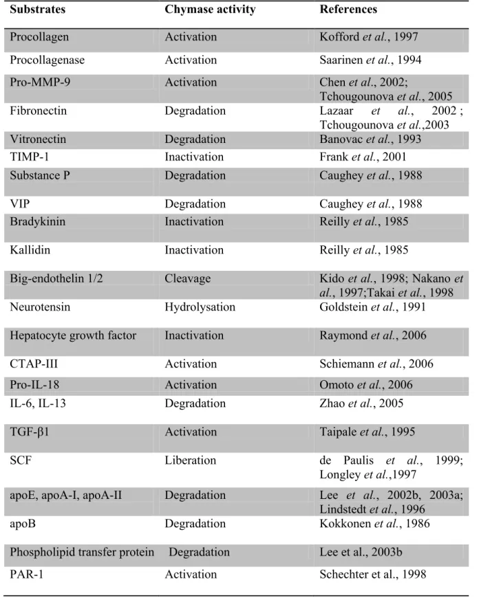
Chymase plays an important role in physiology and pathophysiology. This role is attained due to its broad cleavage specificity which explains its ability to process a large number of proteins/peptides. In table I, some chymase substrates are listed. It is important to note that some of these substrates were identified in vitro either by direct incubation or by the use of chymase inhibitors; other substrates were identified in vivo in mice models with chymase Knock-out (KO) genes.
Table I. Chymase substrates modified from Pejler et al. 2007
Substrates Chymase activity References
Procollagen Activation Kofford et al., 1997
Procollagenase Activation Saarinen et al., 1994
Pro-MMP-9 Activation Chen et al., 2002;
Tchougounova et al., 2005
Fibronectin Degradation Lazaar et al., 2002 ;
Tchougounova et al.,2003
Vitronectin Degradation Banovac et al., 1993
TIMP-1 Inactivation Frank et al., 2001
Substance P Degradation Caughey et al., 1988
VIP Degradation Caughey et al., 1988
Bradykinin Inactivation Reilly et al., 1985
Kallidin Inactivation Reilly et al., 1985
Big-endothelin 1/2 Cleavage Kido et al., 1998; Nakano et
al., 1997;Takai et al., 1998
Neurotensin Hydrolysation Goldstein et al., 1991
Hepatocyte growth factor Inactivation Raymond et al., 2006
CTAP-III Activation Schiemann et al., 2006
Pro-IL-18 Activation Omoto et al., 2006
IL-6, IL-13 Degradation Zhao et al., 2005
TGF-β1 Activation Taipale et al., 1995
SCF Liberation de Paulis et al., 1999;
Longley et al.,1997
apoE, apoA-I, apoA-II Degradation Lee et al., 2002b, 2003a; Lindstedt et al., 1996
apoB Degradation Kokkonen et al., 1986
Phospholipid transfer protein Degradation Lee et al., 2003b
Table I. Chymase substrates modified from Pejler et al. 2007 continued
Substrates Chymase activity References
C3a Degradation Gervasoni et al., 1986;
Kajita and Hugli, 1991
Albumin Degradation Raymond et al., 2003
Occludin Degradation Scudamore et al., 1998
C1 inhibitor Inactivation Schoenberger et al., 1989
Ang I Processing and cleavage Urata et al., 1990
1.7 The Role of Chymase in Physiology and Pathophysiology 1.7.1 Chymase in wound healing
Wound healing comprises three major stages: the inflammatory phase, the proliferative phase and the remodeling phase (Schilling 1976). Chymase has a role in all stages. It has a paramount role in ECM regulation in direct and indirect ways which is a key step in the inflammatory phase of wound healing (Nishikori et al., 1998; Noli and Miolo 2001, 2010; Younan et al., 2010). This is expected given the abundance of CTMC in the connective tissue (Irani et al., 1986; Kitamura
et al., 1989; Metcalfe et al., 1997; Kawakami et al., 2002). Chymase is responsible for the
degradation of fibronectin and Vitronectin, both of which are components of the ECM (Lazaar et
al., 2002; Tchougounova et al., 2003; Banovac et al., 1993).
On the other hand, in the proliferative and remodeling phases, chymase stimulates fibroblasts by releasing TGF-β1 causing ECM deposition (Lindstedt et al., 2001). In addition, chymase was shown to contribute to angiogenesis and vascular growth, in granulation tissue (Norrby et al., 1986). Furthermore, chymase was demonstrated to cleave precollagen leading to fibril formation (Kofford et al., 1997).
1.7.2 Chymase in cardiovascular diseases and Atherosclerosis
Atherosclerosis is an inflammatory disease of the arteries. As we have mentioned earlier, MC have been shown to be involved in this disease. More evidence has demonstrated a role for chymase in atherosclerosis (Bot et al., 2015).
Chymase can affect SMC directly and indirectly. It indirectly regulates SMC differentiation, migration and proliferation by the activation of TGF-β1 (Otsuka et al., 2006). It can also directly induce their apoptosis (Leskinen et al., 2001) which explains the thinning of the aortic wall media in atherosclerosis. Chymase can also inhibit collagen synthesis and induce the apoptosis of endothelial cells by degrading Vitronectin and Fibronectin of the ECM (Heikkilä et al., 2008) and/or via TGF-β1 which will further cause endothelial dysfunction.
Chymase has also a role in activating the metalloprotease Pro-MMP-9 which has been implicated in atherosclerosis (Wågsäter et al., 2011).
Within the atheroma, chymase is involved in the proteolysis of LDL, the step preceding foam cell formation (Lee et al., 1992). In addition, by degrading ApoE, ApoAI and HDL3, it inhibits the efflux of cholesterol from foam cells, the process that will maintain the presence of foam cells in atherosclerotic plaques (Lee et al., 1992, 1999, 2002 a, 2002 b; Lindstedt L et al., 1996). Finally, higher chymase levels were found in the serum of patients who suffered from a myocardial infarction or unstable angina compared to individuals without coronary artery disease (Xiang et al., 2011).
1.7.3 Chymase in Metabolic diseases and Diabetes Mellitus
Mast cells have been shown to be involved in the pathogenesis of metabolic diseases such as Obesity, Diabetes Mellitus (DM), type I (DM I) and II (DM II) and in complications of DM. Studies have shown that MC are present in a larger amount in white adipose tissue (WAT) of obese patients compared to lean subjects (Tanaka et al., 2011; Liu et al., 2009).
Mast cell deficient mice gained less body weight, had less adipose tissue inflammation and had improved glucose intolerance (Liu et al., 2009).
In a more specific way, chymase has been linked to metabolic diseases and DM. In fact, in a hamster model of DM I there was an increase in blood glucose levels, pancreatic chymase and Ang II formation; all of which were decreased with the inhibition of chymase by TY-51469 (Maeda et al., 2010; Takai et al., 2009).
On the other hand, chymase levels were measured in the blood of patients suffering from pre-diabetes and DM II and were shown to be higher than chymase levels in the blood of controls (with normal blood glucose levels) (Wang et al., 2011).
DM complications, such as nephropathies and retinopathies can be detrimental. Although it has not been directly linked to these complications, chymase has been reported to have an unfavorable role in these processes. Chymase levels were elevated in diabetic nephropathy. These levels were associated with glomerulosclerosis and tubulointerstitial fibrosis (Ritz, 2003) and diabetic vascular diseases secondary to Ang II formation (Koka et al., 2006). Furthermore, once activated by the chymase, pro-MMP-9 is believed to be active in diabetic nephropathy and
1.8 The role of Chymase in the conversion of Angiotensin I to Angiotensin II
The role of chymase in Ang II synthesis is well documented (for review see Takai et al., 2010 and Pejler et al., 2010). As mentioned in table I, Ang I is a substrate for chymase (Urata et al., 1990). Chymase was found to cleave Ang I and form Ang II. Ang I is a peptide constituted of ten aa Asp1-Arg2-Val3-Tyr4-Ile5-His6-Pro7-Phe8-His9-Leu10, chymase is responsible for the cleavage of the Phe8-His9 bond to form Ang II, an eight aa peptide (Reilly et al., 1982; Urata et
al., 1990).
It is well known that Ang II is a product of the activation of the Renin-Angiotensin system (RAS). The conventional conversion of Ang I to Ang II is done via Angiotensin converting enzyme (ACE). RAS is activated, in normal physiology, when the body senses a decrease in blood flow to the kidneys (decreased intratrarenal pressure), which implies a decrease in blood pressure. Ang II will be formed and will cause a vasoconstriction leading to restoration of normal blood pressure. This is believed to occur in the blood stream.
However this is not the only pathway, since ACE inhibitors could not inhibit totally the production of Ang II (Padmanabhan et al., 1999; Wolny et al., 1997) and in an ACE KO model of mice, the formation of Ang II was not totally repressed either (Wei et al., 2002). The alternative route, involving the chymase was shown to occur mainly in the tissues (as opposed to bloodstream) form Ang II in the tissues where Ang II plays a major role in Pathophysiology. 1.9 The role of Angiotensin II in physiology and Pathophysiology
As mentioned in the previous paragraph, Ang II plays a key role in physiology and Pathophysiology and works on several systems.
The main system we are interested in here is the cardiovascular system where Ang II can stimulate cardiac remodeling and hypertrophy as well as vascular hypertrophy (Humma and Terra, 2002; Mehta and Griendling, 2007). It also causes constriction of the resistance vessels which will increase the systemic vascular resistance hence increasing the arterial pressure (Humma and Terra, 2002).
Ang II also affects the renal system. It stimulates the release of aldosterone in the adrenal cortex causing an increase in sodium reabsorption and water retention; besides the stimulation of
Vasopressin or antidiuretic hormone (ADH) release will further increase fluid retention in the body (Humma and Terra, 2002).
On another level, Ang II impacts the nervous system by facilitating Norepinephrine release and inhibiting its reuptake on sympathetic synapses (Humma and Terra, 2002).
In more details, Ang II affects all the cells in the cardiovascular system. With excess production of Ang II, growth, hypertrophy and migration of vascular smooth muscle cells (VSMC) will take place. In addition, endothelial dysfunction will occur and there will be an increase in the expression of the adhesion molecules. Moreover, with cardiac remodeling, electrophysiological conduction will be altered (Mehta and Griendling, 2007).
These changes in physiology secondary to excess Ang II production implies that this peptide has a supreme role in myocardial infarction, arrhythmias, strokes, diabetic vascular diseases and congestive heart failure (Schieffer et al., 2000 ; Mehta and Griendling, 2007)
1.10 Angiotensin receptors
Most physiologic and pathophysiologic effects of Ang II are mediated by its receptor angiotensin type 1 receptor (AT1R). Other receptors also exist, such as angiotensin type 2, 3 and 4, however our focus will be on AT1R since it is responsible for most of the cardiovascular diseases related to the renin-angiotensin system (RAS). AT1R is a seven-membrane G protein-coupled receptor. It is composed of 359 aa. The receptor is widely distributed in the body: It is present on tissues of the heart, vessels, kidneys, adrenals, liver, lungs and brain (Griendling, Lassegue and Alexander, 1996).
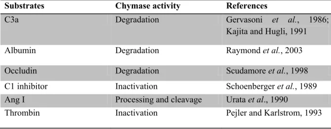
As shown in Fig. 2, once Ang II binds to AT1R, the Gαq/11 and Gα12/13 protein cascade will be activated (Ushio-Fukai et al., 1998). The Gαq/11 will activate the phospholipase C (PLC) which will cleave the Phosphatidylinositol 4,5-bisphosphate (PIP2) into diacylglycerol (DAG) and inositol-1,4,5-triphosphate (IP3). IP3 will increase calcium levels in the cytoplasm by binding to its receptor on the sarcoplasmic reticulum and opening a channel facilitating calcium efflux. Calcium released from the sarcoplasmic reticulum activates the myosin light chain kinase
between actin and myosin leading to SMC contraction (Yan et al., 2003). On the other hand, DAG will activate protein kinase C (PKC) which will phosphorylates the Sodium/Hydrogen (Na+/H+) exchange and will act as an effector in the Ras/Raf/MEK/ERK pathway (Yan et al., 2003).
Figure 2 AT1R signaling pathway after activation by Ang II. PLC: Phospholipase C, PIP2: Phosphatidylinositol 4,5-bisphosphate, DAG:Diacylglycerol, IP3: Inositol-1,4,5-triphosphate, SR: Sarcoplasmic Reticulum, Ca2+: Calcium, CaM: Calmodulin, MLC: Myosin light chain, MLCK: Myosin light chain kinase, MLC-P: Phosphorylated myosin light chain, PKC: Protein Kinase C.(Modified from Ushio-Fukai et al., 1998)
1.11 Endothelin-1
As mentioned in table I, Big endothelin-1 (Big ET-1) has been shown in vitro to be a substrate for the chymase (Kido et al., 1998; Nakano et al., 1997; Takai et al., 1998). The end product of the processing and cleavage of the Big ET-1 is endothelin-1 (ET-1).
ET-1 is a potent vasoconstrictor constituted of 21 aa. It was first isolated in 1988 from porcine aortic endothelial cells by Yanagisawa (Yanagisawa et al., 1988). Three isomers of endothelin exist 1, 2 and 3 (Inoue et al., 1989). Each peptide is encoded by a different gene. ET-1 is produced by endothelial cells, mainly the vascular endothelium (Masaki, 2000; Simonson and Dunn, 1990). ET-2 is produced by the renal medulla and is implicated mainly in the vascular function in the kidneys; ET-3 was found to be present in nerve endings and implicated in neurotransmission (Waeber et al., 1990; Spyer et al., 1991).
1.12 The role of Endothelin-1 in Physiology and Pathophysiology
ET-1 has several important physiologic actions in the embryologic and adult life. In fact, a repression of the ET-1 gene in mice caused their deaths from cardiovascular and craniofacial anomalies leading to respiratory failure minutes after their delivery (Kurihara et al., 1994; Yanagisawa et al., 1998). In addition, ET-1 has a key role in maintaining the basal vascular tone (Masaki, 2000). It is also involved in sodium excretion from the renal tubules (Hirata et al., 1988, Murray et al., 2008). It is also involved in bronchoconstriction, sputum production and MC degranulation (Rubanyi and Polokoff, 1994; Murray et al., 2008).
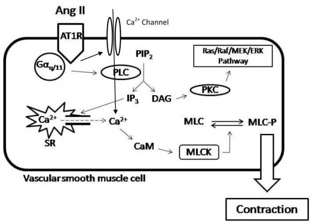
On the other hand, ET-1 is involved in several diseases and pathologies. Table II summarizes most of them.
Table II. Diseases and pathologies which ET-1 is involved in
Conditions References
Atherosclerosis Attina et al., 2005; Ihling et al., 2001
HTN Dhaun et al., 2008
Pulmonary HTN Attina et al., 2005
Metabolic syndrome Weil et al., 2011
IBS
Congestive Heart failure Wei et al., 1994; Kiowski et al., 1995; Pacher et al., 1996
Cancer metastasis Grant et al., 2003; Said and Theodorescu, 2012
1.13 Biosynthesis of Endothelin-1
The first product of the ET-1 gene is Pre-proendothelin, a peptide constituted of 212 aa. This peptide will be processed by a carboxypeptidase to form proendothelin which is the precursor of the Big ET-1. Furin, an enzyme of the subtilisin family will cleave the proendothelin further to generate Big ET-1(Blais et al., 2002; D’Orleans-Juste et al., 2003).
1.14 Classical Pathway
Big ET-1 is found in the peripheral circulation. It has some vasoconstrictive capacities however once converted to ET-1 via the Endothelin Converting Enzyme (ECE), the product, ET-1, has a much higher- 140 times higher- vasoconstrictive potency (Rubanyi and Polokoff, 1994).
1.15 Endothelin Converting Enzyme
The ECE cleaves the bond between Trp 21 and Val 22 of the Big 1 to generate ET-1(McMahon et al., 1991; D'Orleans-Juste et al., 2003). The ECE is a Zinc-dependent Metalloendopeptidase localized in several cell types such as endothelial cells, SMC, cardiomyocytes and macrophages (Hioki et al., 1991; Hisaki et al., 1993; Takahashi et al., 1995; Barnes et al., 1997; Barnes and Turner, 1999; Korth et al., 1999). Three isoforms of ECE have been identified, the ECE-1, ECE-2 and ECE-3 (Xu et al., 1994; Shimada et al., 1994 ; Maguire
et al., 1997; Schweizer et al., 1997; Fukuchi and Giaid, 1998; Kobayashi et al., 1998; Rossi et al., 1999).
ECE-1 and ECE-2 were shown to generate ET-1 from Big ET-1 (Emoto and Yanagisawa, 1995), however ECE-1 is believed to be more involved physiologically since ECE-2 maximal activity occurs at a more acidic pH (pH= 5.5) (Emoto and Yanagisawa, 1999). Furthermore, ECE-1 binds and process several substrates such as bradykinin, substance P, Ang I and insulin, with different affinities (Hoang et al., 1997; Johnson et al., 1999).
Four isoforms of ECE-1 as well as of ECE-2 have been identified: ECE-1a, ECE-1b, ECE-1c and ECE-1d; ECE-2a-1, ECE-2a-2, ECE-2b-1 and ECE-2b-2 (Shimada et al., 1995; Schweizer et
al., 1997; Valdenaire et al., 1999; Ikeda et al., 2002). The isoforms differ in their N terminal
sequences which dictate their cellular location. 1.16 Alternative pathway
The ECE dependent pathway does not seem to be the sole pathway leading to the formation of ET-1. It has been shown that in embryos of mice whose ECE-1 and ECE-2 genes were KO, the production of ET-1 was not completely inhibited; but was only decreased by 33% (Yanagisawa
et al., 2000). This suggests that other pathways involved in the production of ET-1 exist,
independent of ECE. Even though the role of chymase in Ang II biosynthesis is well covered in the literature (for review see Takai et al., 2010 and Pejler et al., 2010), less is reported concerning the role of chymase in ET-1 synthesis (for review see Nakano et al., 1997 and Watts
et al., 2007). One of the enzymes able to cleave Big ET-1, as mentioned in table I, is the
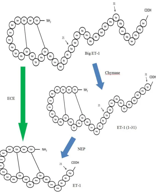
chymase. It has been shown that the latter enzyme can cleave the Big ET-1 at the bond Tyr 31-Gly 32 leading to an intermediate peptide formed of 31 aa, the ET-1 (1-31). This ET-1 (1-31) will be further processed by the neutral endopeptidase (NEP) which cleaves the Trp 21- Val 22 bond to form ET-1 (Hanson et al., 1997; Nakano et al., 1997); as shown in fig. 3. The NEP is a ubiquitous membrane bound metalloendopeptidase responsible not only for the generation of ET-1 from ET-1 (1-31) but has a role in the degradation of ET-1 (Vijavaraghavan et al., 1990; Turner and Tanzawa, 1997).
Figure 3: Schematization of the synthesis of ET-1 via the classical pathway (green arrow) and the alternative pathway (blue arrows). (Modified according to Goto et al., 1996, Hanson et al., 1997 and Nakano et al., 1997)
1.17 Endothelin-1 (1-31)
There has not been a clear conclusion in the literature on whether ET-1 (1-31) acts directly on the endothelin receptors or it has to be cleaved into ET-1 to achieve its activity. Many in vitro studies have shown that ET-1 (1-31) can act as an agonist on the endothelin receptors. For example, the group of Maguire showed in 2001 that ET-1 (1-31) has vasoconstrictive properties when activating the endothelin receptors in a human mammary artery (Maguire et al., 2001). Some studies have concluded that ET-1 (1-31) is a selective agonist for one of the endothelin receptors, the endothelin A receptor (ETA) (Mazzochi et al., 2000), whereas other studies have shown that ET-1 (1-31) is an agonist of both endothelin receptor, ETA and endothelin B (ETB). On the other hand, other studies indicated that ET-1 (1-31) needs to be processed by the NEP to generate ET-1 which will activate the receptors (Hayasaki-Kajiwara et al., 1999).
It is important to note that the studies mentioned in the previous paragraph were conducted in
vitro. A closer look to the studies performed on animals in vivo shows a consensus on the
necessity of the conversion of the ET-1 (1-31) to ET-1 by the NEP to get its functionality in vivo (Fecteau et al., 2005; Simard et al., 2009).
1.18 Endothelin receptors
There are two known receptors for the endothelin, ETA and ETB. They are G protein coupled receptors (GPCR) that have seven transmembrane domains. These receptors are coupled to Gq/11. They share about 50 % of identical sequence with the main differences existing in the N-terminal (Ogawa et al., 1991; Rubanyi and Polokoff, 1994; Murray et al., 2008). ETA is localized mainly on the VSMC whereas ETB is localized on the VSMC along with the endothelial cells (Hosoda et
al., 1991; Fan et al., 2000; Giannessi et al., 2001). 1.19 ETA receptor
The binding of ET-1 to ETA will activate the receptor. Similar to the AT1 receptor, stimulation of ETA receptor will activate the protein Gq/11 and activate phospholipase C (PLC) which will hydrolyze phosphatidylinositol-4,5-bisphosphate (PIP2) into inositol-1,4,5-triphosphate (IP3) and diacylglycerol (DAG). In addition, activation of both ETA and AT1 receptors induces
Alvarenga et al., 2016). Both types of receptors induce increase of intracellular calcium via stimulation of L- and R-type calcium channels as well as release of calcium from the endoplasmic reticulum (Bkaily et al., 2005, 2011; Simonson and Dunn, 1990; Giannessi et al., 2001; Becker et al., 2009). Little is known concerning the differences in signaling and biological effects between ETA and AT1 receptors activation.
1.20 ETB receptor
As mentioned earlier and shown in fig. 4, ETB is present on VSMC and endothelial cells. The receptors present on VSMC will activate Gq and Giwill cause an increase in intracellular calcium, in a cascade similar to the activation to ETA which will cause a contraction of the VSMC and hence a vasoconstriction. However, the activation of ETB present on the endothelial cells will cause an increase in Nitric Oxide (NO) and prostacyclin causing a relaxation of the VSMC hence a vasodilation (Giannessi et al., 2001; Attina et al., 2005; Murray et al., 2008).
Figure 4: ETA and ETB signaling pathway after activation by ET-1. PLC: Phospholipase C, PIP2: Phosphatidylinositol 4,5-bisphosphate, DAG:Diacylglycerol, IP3: Inositol-1,4,5-triphosphate, SR: Sarcoplasmic Reticulum, Ca2+: Calcium, CaM: Calmodulin, MLC: Myosin light chain, MLCK: Myosin light chain kinase, MLC-P: Phosphorylated myosin light chain, PKC: Protein Kinase C, NO: Nitric Oxide, PI3K: Phosphatidylinositol-4,5-bisphosphate 3-kinase, PIP3: Phosphatidylinositol (3,4,5)-trisphosphate, eNOS: Endothelial Nitric Oxide synthase. (Modified from Ushio-Fukai et al., 1998)
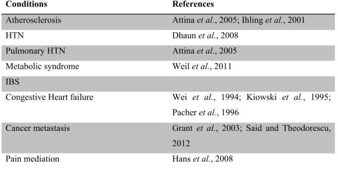
1.21 Chymase inhibitors and clinical relevance
Many chymase inhibitors are currently in clinical trials, some are currently in phase two such as SUN 13834 (Ogata et al., 2011). Table III summarizes some of the chymase inhibitors and their potential clinical relevance.
Table III. Chymase inhibitors and their therapeutic potential
Chymase inhibitors Therapeutic potential Reference
SUN-C8257 Atherosclerois, pulmonary
fibrosis, skin disease Doggrell, 2008
SUN13834 Atopic dermatitis Ogata et al., 2011
BCEAB Cardiac diseases Doggrell, 2008
Compound 17 Cardiac diseases, asthma Doggrell, 2008
NK 3201 Cardiac diseases Doggrell, 2008
TEI-ES48 Cardiac diseases Hoshino et al., 2003
RO5066852 Atherosclerosis Bot et al., 2011
JNJ-10311795 Anti-inflammatory De Garavilla et al., 2005
Suc-Val-Pro-Phe (OPh)2 Cardiac adhesions Soga et al., 2004
Y-40613 Atopic dermatitis Akahoshi et al., 2001; Imada
et al., 2002
TY-51463 Cardiac diseases, liver
fibrosis, gastroentesitinal diseases, diabetes mellitus
Oyamada et al., 2011; Komeda et al., 2010 ; Kakimoto et al., 2010 ; Takai
et al., 2009
Chymostatin Glaucoma, chorioretinal,
1.22 Aim of the study and targeted objectives
Since the establishment of the chymase dependent route of conversion of Big ET-1, the production of ET-1 was not demonstrated to occur in vivo up until recently in 2009, when our laboratory has shown that the conversion of Big ET-1 into ET-1 (1-31) occurs in mice (Simard et
al., 2009); knowing that ET-1 (1-31) is converted to ET-1 via NEP in vivo (Fecteau et al., 2005).
In addition in 2013, our laboratory has demonstrated that the mMCP-4, which is the murine analog of the human chymase, can generate ET-1 (1-31) from the Big ET-1 in vitro and in vivo (Houde et al., 2013). However, no information is available concerning the ability of recombinant chymases (murine or human) to cleave Big ET-1. In fact the literature shows a chymostatin dependent characterization of CMA1 in regards to generation of Ang II from Ang I (Murakami
et al., 1995). Chymostatin is a general chymotrypsin-like protease inhibitor, not specific to
chymase. It was shown to inhibit elastase II as well, an enzyme involved as well in the production of Ang II from its precursor Ang I (Becari et al., 2005).
Based on these observations, we hypothesized in this study that the CMA1, whether recombinant, extracted from the LUVA cells (human mast cell line) or in the soluble fractions of human aortas would generate ET-1 (1-31) from Big ET-1 in a chymase inhibitor - sensitive manner.
In a second aim, we also characterized the kinetic enzymatic activity of CMA1 towards its substrate Big ET-1.
In order to achieve these aims, we propose the following objectives:
To validate that the recombinant human chymase (CMA1) converts Big ET-1 into ET-1 (1-31)
To verify if CMA1 has a role in the degradation of ET-1 or ET-1 (1-31)
To determine the Kinetic constants of CMA1 towards a fluorogenic substrate, Big ET-1 and Ang I
To verify if the chymase extracted from the LUVA cells (human Mast cell line) converts the Big ET-1 into ET-1 (1-31)
To verify if CMA1 extracted from soluble fractions of healthy human aortas would generate ET-1 (1-31) from the precursor Big ET-1
2.1 TITLE PAGE
Title: Chymase inhibitor-sensitive synthesis of endothelin-1 (1–31) by recombinant mouse mast cell protease 4 and human chymase.
Authors:Walid Semaan, Louisane Desbiens, Martin Houde, Julie Labonté, Hugo Gagnon, Daisuke Yamamoto, Shinji Takai, Tanya Laidlaw, Ghassan Bkaily, Adel Schwertani, Gunnar Pejler, Christine Levesque, Roxane Desjardins, Robert Day, Pedro D’Orléans-Juste
Status of the article: Published in 2015 in Biochemical Pharmacology doi: 10.1016/j.bcp.2015.02.001
Foreword: On the experimental level, I have executed the fluorescence experiments with recombinant chymases, murine Mast cells extracted from peritoneal lavages and LUVA cells. I have also executed the experiments using the HPLC. Moreover I have prepared the LC-MS/MS conversion experiments with recombinant chymases and with human aortas. I have analyzed the results and plotted the graphs using Graphpad. The article was written under the supervision of Drs. Pedro D’Orleans- Juste and Ghassan Bkaily. All the authors have read and revised the article.
2.2 Affiliations:
Department of Pharmacology, Université de Sherbrooke, 3001, 12e Avenue Nord, Sherbrooke, QC, Canada J1H 5N4
Phenoswitch Bioscience Inc., 3001, 12e Avenue Nord, Sherbrooke, QC, Canada J1H 5N4 Biomedical Computation Center, Osaka Medical College, 2–7 Daigakumachi, Takatsuki 569-0801, Osaka Prefecture, Japan
Department of Pharmacology, Osaka Medical College, Takatsuki, Osaka Prefecture, Japan Department of Medicine, Brigham and Women’s Hospital, Harvard University, 75 Francis St, Boston, MA 02115, United States
Department of Anatomy and Cell Biology, Université de Sherbrooke, Sherbrooke, QC, Canada Division of Cardiology, McGill University, 1650 Avenue Cedar, Montreal, QC, Canada H3G 1A4
Department of Anatomy, Physiology and Biochemistry, Swedish University of Agricultural Sciences, Anatomi och fysiologi, Biokemi, Box 575, BMC B9 plan4, Dag Hammarskjo¨lds v-Husarg, 751 23 and Uppsala University, Department of Medical Biochemistry and Microbiology, Uppsala, Sweden
Department of Surgery, Division of Urology, Université de Sherbrooke, Sherbrooke, QC, Canada
ABSTRACT
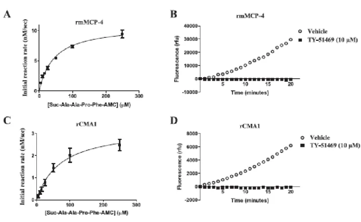
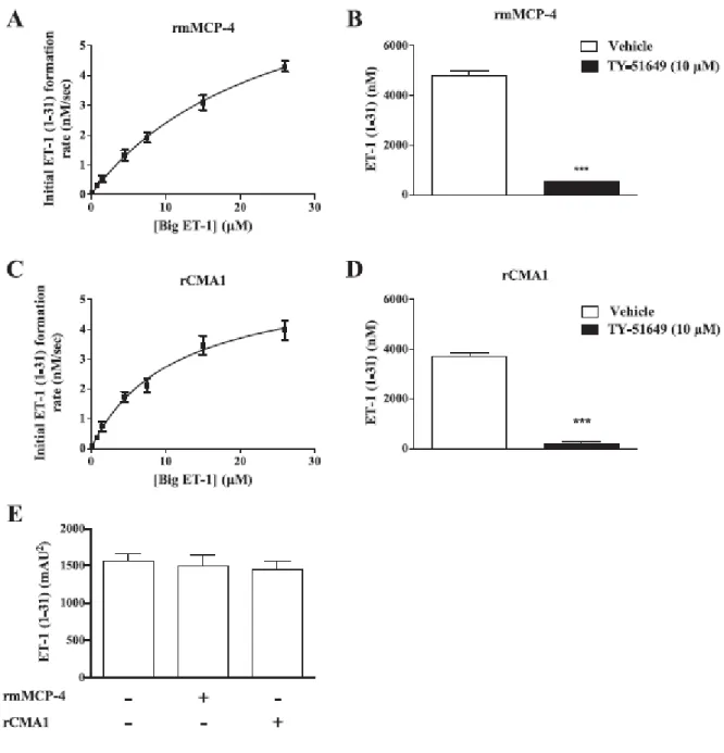
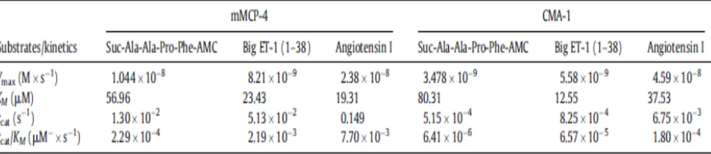
Important structural differences imply that human and mouse mast cell chymases may differ with respect to their enzymatic properties. We compared in this study the catalytic efficiencies of recombinant human chymase (rCMA1) and its functional murine homologue recombinant mouse mast cell protease-4 (rmMCP-4) toward a fluorogenic chymase substrate (Suc-Ala-Ala-Pro-Phe-7-amino-4-methylcoumarin (AMC) and by their ability to convert Big-endothelin (ET)-1 into ET-1 (1–31) using a LC/MS/MS system. Activities toward a fluorogenic substrate (Suc-Leu-Leu-Val-Tyr-AMC) and Big ET-1 were also measured in extracts from mouse peritoneal mast cells, LUVA human mast cell-like cells and human aortas. The specificity of these activities was assessed with the chymase inhibitor TY-51469 (2-[4-(5-fluoro-3- methylbenzo[b]thiophen-2-yl) sulfonamido-3-methanesulfonyl-phenyl]thiazole-4-carboxylic acid). For similar affinities, rmMCP-4 showed a higher activity toward the fluorogenic substrate and a higher ability to process Big ET-1 as compared to recombinant CMA1 (chymase activity (kcat/KM in µM-1s-1): 2.29 x 10⁻⁴ vs. 6.41 x 10⁻⁶; ET-1 (1–31) production: 2.19 x 10⁻³ vs. 6.57 x 10⁻⁵), and both of these activities of mouse and human chymase were sensitive to TY-51469. Furthermore, extracts from mouse peritoneal mast cells, LUVA cells and human aorta homogenates contained processing activities toward the fluorogenic chymase substrate as well as Big ET-1, all of which were sensitive to TY-51469. Finally, the pressor responses to Big ET-1 but not to ET-1 were significantly reduced in conscious and free moving mMCP-4 KO mice when compared to wild type congeners. Our results suggest that both mouse and human chymases have potent ET-1 (1– 31)-producing abilities, with the murine isoform being more efficient.
Keywords: Chymase, Recombinant enzymes, Mass spectrometry, Radiotelemetry, In silico analysis
1. Introduction
Chymases are serine proteases released by activated mast cells, involved in tissue repair and inflammatory processes such as wound-healing/fibrosis [1], cardiac remodelling and angiogenesis [2,3]. In humans and rodents, two types of mast cells have been identified. Human mast cells positive for both chymase and tryptase (MCTC) are similar to murine connective tissue mast cells (CTMC) while those positive only for tryptase (MCT) resemble murine mucosal mast cells (MMC) [4,5]. To date, a single chymase, a-chymase or CMA1 [6] has been identified in humans, expressed in MCTC. In mice, mouse mast cell protease 4 (mMCP-4), predicted as a rodent b-chymase from its deduced amino acid sequence, shows angiotensin II (Ang II)- forming properties, CTMC localization and serglycin storage dependence [7] similar to those afforded by CMA1 [6]. Importantly, like the a-chymase CMA1 [8], mMCP-4 does not share the preferential b-chymase Tyr4-Ile5 cleaving activity on Ang-II that mMCP-1 and rat mast cell protease 1 (rMCP-1, the mMCP-4 rat homolog) possess, making the mouse a more representative model than that of the rat to study human-like Ang-II formation [9]. mMCP-4 plays a protective role in a mouse model of cerebral trauma [10], yet is detrimental in bleomycin-induced lung inflammation and immune complex-induced glomerulonephritis [11,12].
The potent vasopressor peptide endothelin-1 (ET-1) on the other hand, is generated from a larger 38 amino acid precursor Big-endothelin-1 (Big ET-1) via the hydrolytic activity of an endothelin-converting enzyme (ECE) [13]. Besides, other proteases are also involved in the overall production of mature ET-1. Among those, chymase derived from human purified pulmonary tissue cleaves the Tyr31–Gly32 bond of Big ET-1 (1–38) to yield ET- 1 (1–31) [14]. Our group later reported that ET-1 (1–31) requires a further neutral endopeptidase (neprilysin, NEP)-dependent hydrolysis of the Trp21–Val22 bond to produce mature ET-1 in vivo [15]. Whether mMCP-4 is also involved in the in vivo synthesis of endothelins remained unexplored until we recently reported that this particular chymase isoform converts Big ET-1 to ET-1 (1–31) and subsequently to ET-1 in a study using anesthetised mMCP-4-/- mice [16].
Human chymase generated by a recombinant approach produces chymostatin-sensitive Ang II from Ang I, with a KM of 59 µM [17]. Chymostatin however, a general chymotrypsin-like protease inhibitor, is much less specific than newer generation chymase inhibitors such as
TY-51469 [18]. In addition, the comparative capacities of recombinant mMCP-4 and CMA1 to generate ET-1 (1–31) have not been assessed.
Based on our previous reports on the chymase-dependent conversion of the precursor Big ET-1 to ET-1 (1–31) in the anesthetised mouse model in vivo [16,19], we hypothesized that recombinant or mast cell-extracted mMCP-4 as well as CMA1, would generate the 31-amino acid intermediate in a TY 51469- sensitive fashion.
The first principal aim of this study was therefore to compare, by using recombinant mMCP-4 and its human counterpart CMA1, the capacity of murine and human chymases to generate ET-1 (1– 31). A second aim was to assess the ET-1 (1–31)-producing capacities of chymases derived from mouse (peritoneal mast cells) and human mast cells (LUVA cells, [20]) as well as the role of chymase in the production of ET-1 (1–31) by human aortic biopsies.
Our data show that the murine mMCP-4 and the human CMA1 generate ET-1 (1–31) from the precursor Big ET-1 via recombinant enzymes as well as in cellular or tissue extracts of mouse and human origin.
2. Materials and methods
2.1. Drugs and chemicals
Phosphate buffered saline (PBS) pH 7.4, ammonium hydroxide, bovine serum albumin (BSA), 2-(N-morpholino)ethanesulfonic acid (MES), pluronic F-68, formic acid (FA), trifluoroacetic acid (TFA), and N-ethylmaleimide (NEM) were obtained from Sigma-Aldrich (Oak- ville, ON, Canada). The StemPro-34 SFM culture medium was purchased from Invitrogen (Carlsbad, CA, USA). The RPMI-1640 and I-Max (IPL-41) culture media, fetal bovine serum (FBS) and the antibiotics hygromycin B and penicillin were obtained from Wisent (Montreal, QC, Canada). Dithiotreitol (DTT), dimethyl sulfoxide (DMSO) and HPLC-grade acetonitrile (ACN) were obtained from Fisher Scientific (Ottawa, ON, Canada). Murine active cathepsin C and recombinant CMA1 were obtained from R&D Systems (Minneapolis, MN, USA). Heparin was purchased from LEO Pharma A/S, (Ballerup, Denmark). Triton X-100 was obtained from ICN Biochemical (Aurora, OH, USA). Suc-Ala-Ala-Pro-Phe-7-amino-4-methylcoumarin (AMC), Suc-Leu-Leu-Val-Tyr-AMC and ET-1 (1–31) were obtained from Peptide Institute (Osaka, Japan), Big ET-1, Ang-I, Ang-II and Pro11- DAla12-Ang-I were obtained from American Peptide Company (Sunnyvale, CA, USA), ET-1 was obtained from Tocris Bioscience (Bristol, UK) and (13C6)Leu6-ET-1 was obtained from Bachem (Bubenford, Switzerland). Suc-Ala-Ala-Pro-Phe-chloromethylketone (CMK) was obtained from MP Biomedicals (Santa Ana, CA, USA). Ketamine was obtained from Bioniche (Belleville, ON, Canada), xylazine from Bimeda (Cambridge, ON, Canada) and buprenorphine from Reckitt Benckiser Healthcare (Slough, United Kingdom). Finally, TY-51469 (2-[4-(5-fluoro-3-methylbenzo[b]thiophen-2-yl)sulfona- mido-3-methanesulfonylphenyl]-thiazole-4-carboxylic acid) was graciously provided by Toa Eiyo Ltd. (Osaka, Japan).
2.2. Expression, purification, activation and titration of recombinant mMCP-4 and CMA1 2.2.1. Expression
The vector pAc5.1 (Life Technologies, Burlington, ON, Canada) containing the recombinant DNA of pro-mMCP-4 or pro-CMA1, with poly-histidine and V5 tags, was co-transfected with the selection vector pCoHygro (Life Technologies) into S2 drosophila cells. The S2 cells were grown in I-Max culture medium IPL-41 complemented by 10% of fetal bovine serum (FBS) and hygromycin B (300 mg/ml). The cells were scaled up and finally suspended in serum-free culture medium containing 1% pluronic F-68 at a concentration of 2 x 10⁶ cells/ml.
2.2.2. Purification
The crude extract sample containing the secretion of the drosophila cells was concentrated by ultrafiltration on an Ultracell Microcon 10 kDa filter (EMD Millipore, Billerica, MA, USA). This sample was then purified on a nickel affinity column by FPLC with an imidazole gradient (up to 250 mM) for isolation of poly-histidine tag positive samples. Those samples were then put on a Superdex200 26/60 size exclusion column (GE Life Sciences, CA, USA) and the V5-positive fractions (determined by Western blot, data not shown) were pooled and frozen.
2.2.3. Activation
The recombinant enzymes were thawed and diluted to a concentration of 20 mg/ml in maturation buffer (50 mM MES, 0.1% BSA, pH 5.5,). Active murine cathepsin C was diluted to 0.481 ng/ml in cathepsin C buffer (50 mM MES, 50 mM NaCl, 5 mM DTT, pH 5.5). Activation was performed by adding equal volumes of recombinant chymase and cathepsin C, adding 50 µg/ml heparinand incubating 1 h at room temperature. Chymase activation was stopped with NEM (3 mM) and diluted with assay buffer (20 mM Tris, 2 M KCl, 0.02% Triton X-100 (replaced with (0.1%) BSA for Ang- I assays), pH 9.0) to bring the recombinant chymase concentration to 2 mg/ml, and 5 min was afforded to completely stop the cathepsin C-dependent reaction.
2.2.4. Titration
The activated recombinant enzymes (0.025 ng/ml rmMCP-4 or 0.25 ng/ml rCMA1) were
inhibitory substrate Suc-Ala-Ala-Pro-Phe-chloromethylketone (CMK). When cleaved, the CMK compound covalently binds to the active site of recombinant chymase and blocks it. After this incubation, the fluorogenic substrate Suc-Ala-Ala-Pro-Phe-AMC was added and the chymase-like activity was analyzed as described in the main body of the article. The threshold of total inhibition of chymase activity was interpreted as the number of active sites in the recombinant chymases preparations and used as the molar concentration of enzymes for determination of their kinetics.
2.3. Animals
C57Bl/6J mice were purchased from Charles River (Montreal, QC, Canada) and housed in our facilities. Genitor mMCP-4 KO mice [2] were bred in our facilities. All animals were kept at constant room temperature (23ºC) and humidity (78%) under a controlled light/dark cycle (6:00 AM–6:00 PM), with standard chow and tap water available ad libitum. Animal care and experiments were approved by the Ethics Committee on Animal Research of the University of Sherbrooke following the Canadian Council on Animal Care guidelines and the Guide for the Care and Use of Laboratory Animals of the United States National Institutes of Health. All experiments on mice were performed on newly sacrificed animals, except for telemetric hemodynamic recording performed on live animals. The mice underwent general anesthesia, by the intramuscular administration of ketamine/xylazine (87/13 mg/kg). Complete anesthesia was assumed when no withdrawing reflex was found during pressure on any paw of the mouse. Anesthetized mice were killed by cervical dislocation.
2.4. Mast cell preparation
Mouse peritoneal mast cells preparations were used, as they are readily available, fully mature connective tissue resident mast cells containing their full complement of granule proteases without being primed with cytokines [21]. 5 ml of isolation buffer (phosphate buffer solution containing 1 mg/ml of BSA and 37.5 U/ml heparin, pH 7.4) was introduced into the peritoneal cavity of mice after peritoneal skin removal, then collected after 1 min of peritoneal massage and
centrifuged (200 x g, 5 min). In another series of experiments, mast cell-like LUVA cells were maintained in a StemPro1-34 SFM solution at a cell density of 5 x 10⁵cells/ml, in the absence of additional growth factors. The suspension was divided into 2 equal volumes of 30 ml and centrifuged (200 x g, 5 min). Treatment of LUVA and mouse mast cell pellets was similar thereafter. The supernatant was discarded and the mast cell rich pellet was suspended in RPMI-1640 medium (enriched with penicillin (100 U/ml), 2 mM L-glutamine and BSA (1 mg/ml)) and incubated for 1 h at 37 ºC. The suspension was centrifuged for 5 min at 200 x g, the supernatant was discarded and the pellet was suspended in isolation buffer. The cells were counted according to the Moore and James method [22] and adjusted to 2 x 10⁵mast cells/ml in PBS pH 8. The cells were lysed through sonication and then centrifuged at 200 x g for 5 min. The resulting pellet was washed thrice in 0.1 M PBS pH 8.0 by further centrifugation cycles.
2.5. Human aortas preparation
The human aortas were collected from middle-aged brain-deceased individuals with no histological signs of atherosclerosis. The tissues were weighed and grinded in PBS on ice by a tissue homogenizer (Polytron, ultra-turax T8, IKA, Wilmington, NC, USA) for 30 s. Centrifugation for 20 min at 25,000 x g at 4 ºC took place then the soluble fractions corresponding to the supernatant were collected and frozen at -80 ºC.
2.6. Specific chymase activity in vitro
Activated rmMCP-4 (0.025 ng/ml), activated rCMA-1 (0.25 ng/ml) or mast cell extracts (from 4 x 10⁵ peritoneal mouse mast cells or LUVA cells) were incubated in a 96 well plate. Increasing concentrations of the non-fluorescent substrates Suc-Ala-Ala-Pro- Phe-7-amino-4-methylcoumarin (AMC) (for recombinant enzymes) or Suc-Leu-Leu-Val-Tyr-AMC (for mast cell and tissue extracts) were added and the fluorescence AMC-forming activity, as chymase activity, was then measured with a fluorescence spectrophotometer (λex: 370 nm; λem: 460 nm) for 20 min (Molecular Devices, Sunnyvale, CA). Another series of experiments was performed
chymase inhibitor TY-51469 (10–50 µM). To determine enzyme kinetics, the recombinant enzyme active sites were titrated with the inhibitor substrate Suc-Ala-Ala-Pro-Phe-CMK.
2.7. In vitro conversion of Big ET-1 to ET-1 (1–31) and Ang-I to Ang-II 2.7.1. Recombinant enzymes
Big ET-1 and Ang-I dilutions were prepared in 0.1 M PBS pH 8.0. The recombinant enzymes (28.57 ng/ml activated rmMCP-4 or 1428 ng/ml activated rCMA1) were incubated at 37 ºC with Big ET-1 (0.15, 0.75, 1.5, 4.5, 7.5, 15 and 26 µM) or Ang-I (1.5625, 3.125, 6.25, 12.5, 25, 50 and 100 µM) for 20 min, after which the reactions were stopped with an equal volume of water:acetonitrile:dimethylsulfoxide:formic acid (H2O:ACN:DMSO:FA) mix (73:20:6:1) containing (13C6)Leu6-ET-1 (100 µg/ml) or a H2O:ACN:FA mix (76:20:4) containing Pro11-DAla12-Ang-I (176 ng/ml) (peptides as internal standards). In another series of experiments, 16.7 µM of Big ET-1 was incubated in the presence of TY-51469 (10 µM) and the reaction was stopped as described above. Samples were diluted 1:10 with the same stop solution without the internal standard before LC–MS/MS analysis. Kinetics parameters were calculated using Prism Software (GraphPad, La Jolla, CA, USA).
2.7.2. Mast cell and aortic extracts
In another series of experiments, the soluble fractions of 9 x 10⁵ WT peritoneal mouse mast cells, 4 x 10⁶ LUVA cells or the soluble fraction of the human aortas (adjusted to 2.2 mg/ml of protein) were pretreated with vehicle or TY-51469 (10 µM) and then incubated with Big ET-1 at 37 ºC at concentrations of 5, 5 and 13 µM, respectively, for 20 min. The reactions were stopped in with the stop solution described above containing (13C6) Leu6-ET-1 (50 ng/ml) and subsequently processed by solid phase extraction (SPE) before LC-MC/MS analysis, albeit the stop solution for the human aorta assay was ACN:DMSO:FA (88:6:6).
2.8. LC–MS/MS quantification of Big ET-1, ET-1 (1–31) and ET-1 2.8.1. Sample preparation
For in vitro conversion by recombinant enzymes, samples were analyzed directly. For analysis of samples from masts cells or human aortic tissue, SPE was performed consisting of a polymeric mixed mode strong cation exchange 1 ml cartridge containing 30 mg of sorbent (Phenomenex, Strata-X-C, Torrance, CA, USA).Briefly samples were pre-treated with 2% FA and loaded on the SPE column, samples were next washed once with 60% methanol containing 2% FA and eluted with 2 x 700 µl of 75% ACN containing 10% ammonium hydroxide.
2.8.2. System description
Analysis was performed by LC–MS/MS on TripleTOF 5600 mass spectrometer (ABSciex, Foster City, CA, USA) equipped with DuoSpray source. Samples were introduced to the electrospray ionization (ESI) source in a 50 µm ESI probe using a microLC200 system equipped with a 50 mm x 500 µm HALO C18 2.7 µm column.
2.8.3. System conditions
Chromatography was performed with a gradient of water containing 0.2 % formic acid and 3% DMSO (A) and ACN containing 0.2% formic acid 3% DMSO (B) (without DMSO for Ang-II determination). For endothelin peptides analysis, column temperature was set at 50 ºC. A 4 min gradient was run at 40 µl/min. It consisted of the following steps: hold at 10% B from 0 to 0.5 min, 10% B to 75% from 0.5 min to 2.7 min, hold at 100% B from 2.9 to 3.4 min and equilibration from 3.5 to 4 min at 10% B. Source parameters where the following: curtain gaz was set at 28, gaz 1 was set at 17, gaz 2 was set at 28, ion source voltage was set at 5500 and ESI probe temperature was set at 375. For Ang-II analysis, a 2.5 min gradient was run at 40 µl/min. It consisted of the following steps: hold at 15% from 0 to 0.4 min, 15% B to 100% from 0.4 min to 1.4 min, hold at 100% B from 1.4 to 2.9 min and equilibration from 2 to 2.5 min at 15% B. Source parameters where the following: curtain gaz was set at 27, gaz 1 was set at 25, gaz 2 was set at 20, ion source voltage was set at 4900 and ESI probe temperature was set at 400.