UNIVERSITÉ DE SHERBROOKE
Régulation de l’apoptose des lymphocytes T par GIMAP5 (GTPase of
Immune Associated Nucleotide Binding Protein 5)
Par
Xi-Lin Chen
Département de pédiatrie, programme d’immunologie
Thèse présentée à la Faculté de médecine et des sciences de la santé en vue de l’obtention du grade de philosophiae doctor (Ph.D) en Immunologie
Sherbrooke, Québec, CANADA Le 01 Mai 2015
Membres du jury d’évaluation
Dre Sheela Ramanathan, directrice, Département de pédiatrie, Service d’immunologie-Allergologie
Dre Jana Stankova, co-directrice, Département de pédiatrie, Service d’immunologie-Allergologie Dr Subburaj Ilangumaran, Département de pédiatrie, Service d’immunologie-Allergologie
Dr Guylain Boulay, Département de pharmacologie
Dr Woong-Kyung Suh, Programme de biologie moléculaire, Université de Montréal
Résumé
Régulation de l’apoptose des lymphocytes T par GIMAP5 (GTPase of
Immune Associated Nucleotide Binding Protein 5)
Par Xi-Lin Chen
Département de pédiatrie, programme d’immunologie,
Thèse présentée à la Faculté de médecine et des sciences de la santé en vue de l’obtention du diplôme de doctorante (Ph.D.) en immunologie
Faculté de médecine et des sciences de la santé, Université de Sherbrooke, Sherbrooke, Québec, Canada, J1H 5N4
La survie à long terme des lymphocytes T en état de repos est essentielle pour maintenir leurs nombres dans les organes lymphoïdes secondaires. Le récepteur antigénique des cellules T (TCR) en contact avec les peptides du soi / CMH et en synergie avec l'IL-7 induit des signaux anti-apoptotiques pour favoriser la survie des cellules T. Ces stimuli extrinsèques sont également impliqués dans le métabolisme et la survie des cellules T grâce à la régulation de plusieurs voies de signalisation dont la voie phosphatidyl-inositol-3 kinase (PI3K) /AKT.
Chez la souris et chez le rat, la perte de l’activité de GIMAP5 (GTPase of Immune Associated Nucleotide Binding Protein 5), provoque une lymphopénie T périphérique en raison de la mort spontanée des cellules T. Le mécanisme sous-jacent responsable de la fonction de survie de GIMAP5 dans les lymphocytes T reste largement inconnu. Nous avons observé que les cellules de rats déficients en GIMAP5, après stimulation par complexe TCR, montrent un afflux de calcium (Ca2+) réduit provenant du milieu extracellulaire. Dans cette thèse, J’ai caractérisé le mécanisme d’action de GIMAP5 dans la régulation de l'homéostasie du Ca2+, ainsi que les voies de signalisation modulées par GIMAP5 pour faciliter la survie des cellules T.
Tout d'abord, j’ai étudié si GIMAP5 empêche l’apoptose des lymphocytes T en affectant la capacité des mitochondries à réguler la concentration du Ca2+, ce qui est nécessaire pour soutenir l’influx de Ca2+. J’ai trouvé que l’accumulation du Ca2+ mitochondrial après l’entrée capacitive de Ca2+ est défectueuse dans les cellules T de rat déficientes en Gimap5. La disruption des microtubules, mais pas du cytosquelette d'actine, abroge la séquestration du Ca2+ mitochondrial dans les cellules T primaires de rat, mais pas dans les cellules T déficientes en Gimap5. J’ai observé que les cellules T provennant de souris deficientes en Gimap5 demontrent une dimunition de l’éntrée de Ca2+. De plus, la proliferation des cellules T deficientes en Gimap5 est diminué suite à la stimulation du TCR. En outre, la phosphorylation de STAT5 induit par l'IL-7 est diminuée dans les cellules T CD4+ de souris déficientes en Gimap5. Également, la perte de
Gimap5 aboutit à une activation accrue de la cible mammalienne de la rapamycine (mTOR),
indépendamment de la protéine phosphatase 2A (PP2A) ou de la protéine kinase activée par l'AMP (AMPK). Au lieu de cela, l'activation constitutive de la voie PI3K contribue à une forte activation spontanée de mTOR.
Collectivement, la fonction de survie de GIMAP5 dans les lymphocytes T peut être liée à la régulation de différentes voies de signalisation. GIMAP5 facilite la fonction, microtubule dépendant, des mitochondries dans leurs actions de régulation du Ca2+ après l’entrée capacitive de Ca2+. GIMAP5 est nécessaire pour intégrer les signaux de survie produites suite à l'activation du TCR et de l’IL-7R, qui pourrait être associée à la régulation de l'activité PI3K / AKT / mTOR.
Mots clés: GIMAP5, lymphocytes T, TCR, IL-7, le métabolisme, flux de calcium, mitochondrie, microtubules, STAT5, signalisation TCR proximale, PI3K / Akt / mTOR
Summary
Regulation of T Lymphocytes Apoptosis by GIMAP5 (GTPase of Immune
Associated Nucleotide Binding Protein 5)
By Xi-Lin Chen
Immunology Program, Department of Pediatrics
Thesis presented at the Faculty of Medicine and Health Science to obtain Doctor of Philosophy (Ph.D.) degree in Immunology
Faculty of Medicine and Health Science, University of Sherbrooke, Sherbrooke, Québec, Canada, J1H 5N4
Long-term survival of T lymphocytes in a quiescent state is essential to maintain their cell numbers in secondary lymphoid organs. Interaction of the T cell antigen receptor (TCR) with self-peptide/MHC synergizes with IL-7-induced anti-apoptotic signals to promote T cell survival. These extrinsic stimuli are also implicated in T cell metabolism and survival by regulating several signaling pathways including the phosphatidyl-inositol-3 kinase (PI3K)/Akt pathway.
In mice and in rats, loss of functional GTPase of the immune associated nucleotide binding protein 5 (GIMAP5) causes peripheral T lymphopenia due to spontaneous death of T cells. The underlying mechanism responsible for the pro-survival function of GIMAP5 in T lymphocytes remains largely unknown. Previous work form our laboratory has shown that T cells from GIMAP5-deficient rats show reduced influx of calcium (Ca2+) from the extracellular milieu following stimulation of the TCR complex. In this thesis, I characterized the mechanism by which GIMAP5 regulates Ca2+ homeostasis, and elucidated the signaling pathways modulated by GIMAP5 to facilitate the survival of T cells.
Firstly, I investigated if GIMAP5 prevents apoptotic death of T lymphocytes by affecting the Ca2+ buffering capacity of mitochondria, which is required for sustained Ca2+ influx via the plasma membrane channels. I observed that mitochondrial Ca2+ accumulation following capacitative Ca2+ entry is defective in T cells from Gimap5 deficient rats. Disruption of microtubules, but not the actin cytoskeleton, abrogated Ca2+ sequestration by mitochondria in T cells from control but not Gimap5 deficient mice. Similarly, mice lacking functional GIMAP5 displayed defective T cell development and Ca2+ influx. Furthermore, I observed that the proximal signaling events following TCR stimulation was reduced and was accompanied by defective proliferation in T cells from Gimap5 deficient mice. Additionally, IL-7 induced STAT5 phosphorylation was decreased in CD4+ T cells from Gimap5 deficient mice. I also showed that loss of functional Gimap5 results in increased basal activation of mammalian target of rapamycin (mTOR), independent of protein phosphatase 2A (PP2A) or AMP-activated protein kinase (AMPK). Instead, the constitutive activation the PI3K pathway contributed to the high mTOR activation.
Collectively, my observations suggest that the pro-survival function of GIMAP5 in T-lymphocytes may be linked to the regulation of diverse signaling pathways in a context dependent manner. GIMAP5 also facilitates microtubule-dependent mitochondrial buffering of Ca2+ following capacitative entry. GIMAP5 is required to integrate the survival signals generated following activation through TCR and IL-7R.
Key words: GIMAP5, T-lymphocyte, TCR, IL-7, metabolism, calcium flux, mitochondrial, microtubule, STAT5, proximal TCR signaling, PI3K/Akt/mTOR
TABLE OF CONTENTS:
Résumé ………..………. ii
Summary…………..………. iv
Table of Contents……….. vi
List of Figures……… viii
List of Tables …………..……….. x
List of Abbreviations…………..……….. xi
Chapter I: Introduction and Rationale…………..……… 1
1.1. Apoptotic cell death during T cell development and homeostasis ……… 1
1.2. Intrinsic and extrinsic pathways of T cell apoptosis ……….. 4
1.3. GTPase of the Immune Associated Protein 5 (GIMAP5) and T cell survival ……. 6
1.3.1. GIMAP family members …………..………... 6
1.3.2. Dysregulated apoptosis of T cells due to the mutation of the Gimap5 gene………. 7
1.3.3. Expression of GIMAP5 in lymphocytes…………..………... 10
1.3.4. Structure and subcellular location of GIMAP5……..………. 11
1.4. Role of GIMAP5 in cellular calcium (Ca2+) homeostasis ………... 13 1.4.1. Role of mitochondria in Ca2+ homeostasis in T lymphocytes………... 1.4.2. Role of GIMAP5 in cellular Ca2+ homeostasis………... 1.5. Survival signals for T lymphocytes ………... 1.5.1.TCR tuning and signaling pathways involved in the maintenance of naïve T cells survival ………...………... 1.5.2. IL-7 acts as a survival factor at various stage of T cell development……… 1.5.3. Metabolic pathways regulate T cell survival in the quiescent state………... 1.6. Hypothesis and objectives of this thesis………..…… (i) GIMAP5 prevents apoptotic death of T lymphocytes by regulating Ca2+
homeostasis at the level of mitochondria
(ii) GIMAP5 may interfere with IL-7 and TCR- dependent survival pathways
(iii) GIMAP5 enforces quiescence of T cells by repressing aberrant activation of TCR downstream metabolic signaling pathways
Chapter II: First article……… GTPase of the immune-associated nucleotide-binding protein 5 (GIMAP5)
regulates calcium influx in T-lymphocytes by promoting mitochondrial calcium accumulation.
Chapter III: Second article……… GIMAP5 is required for integrating survival signals in T cells
ii iv vi viii x xi 1 1 4 6 6 7 10 11 13 13 16 19 19 21 23 28 30 61
Chapter IV: Third article……….. GIMAP5 deficiency is associated with an increase in the spontaneous activity
of PI3K/AKT in T lymphocytes.
Chapter V: Discussions……… -Role of Gimap5 in Ca homeostasis ……….. -Role of Gimap5 in integrating survival signals in T cells……… - Gimap5 inhibits activation of PI3K/AKT/mTOR pathway in T cells ……… Chapter VI: Conclusions……… Acknowledgement……… References……… 82 109 111 111 114 116 119 120
LIST OF FIGURES
CHAPTER IFigure 1-1 T cell development.
Figure 1-2 Schematic representation of Gimap5 mutation. Figure 1-3 Ca2+ signaling in T cells.
Figure 1-4 Reduced Ca2+ entry in Gimap5lyp/lyp T cells does not result from impaired opening of the CRAC channel.
Figure 1-5 TCR and IL-7 signaling pathways involved in Naïve T cell survival in a quiescent state.
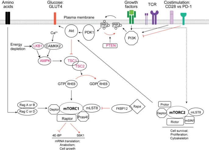
Figure 1-6 IL-7 does not prevent the loss of ΔΨm and apoptosis in Gimap5lyp/lyp CD4+ T cells. Figure 1-7 The mTORC network and key regulatory factors.
CHAPTER II
Figure 1 GIMAP5 deficiency impairs mitochondrial Ca2+ uptake in T-cells
Figure 2 Overexpression of Myc–GIMAP5 promotes mitochondrial Ca2+ accumulation Figure 3 Impaired accumulation of [Ca2+]m is not an intrinsic mitochondrial defect in Gimap5lyp/lyp T-cells
Figure 4 Disruption of microtubules impairs mitochondrial Ca2+ uptake in wild-type but not in Gimap5lyp/lyp T-cells
Figure 5 GIMAP5-mediated mitochondrial Ca2+ accumulation in non-T-cells is dependent on both microtubules and actin cytoskeleton
Figure 6 Cytoskeletal reorganization at the immunological synapse is not affected in
Gimap5lyp/lyp T-cells
Figure 7 Overexpressed GIMAP5 co-localizes with proteins involved in mitochondrial movement.
CHAPTER III
Figure 1 Gimap5 deficiency results in disrupted T cell development.
Figure 2 Increased frequency of macrophages in mononuclear cells isolated from the liver of Gimap5sph/sph mice.
Figure 3 Gimap5 deficiency results in the absence of proliferative response to cognate antigen in CD4+ T cells.
Figure 4 TCR-induced proximal signaling is decreased in Gimap5 deficient T cells.
Figure 5 Mouse T cells lacking a functional GIMAP5 display defective Ca2+ flux response. Figure 6 IL-7 induced STAT5 phosphorylation is down regulated in Gimap5-deficient T cells.
CHAPTER IV
Figure 1 GIMAP5 regulates the activity of mTORC1.
Figure 2 The inhibition of GIMAP5 on mTORC1 activity is independent of AMPK. Figure 3 Mitochondrial function is normal in Gimap5 deficient CD4+ T cells. Figure 4 GIMAP5 inhibits mTORC1 signaling in a PP2A-independent manner. Figure 5 Gimap5 mutation results in constitutive activation of AKT.
Figure 6 Aberrant activation of PI3K contributes to hyper-activated AKT in Gimap5 deficient CD4+ T cells.
CHAPTER V
Figure 5-1 Gimap5 mutation results LAT-RasGRP independent ERK activation. CHAPTER VI
TABLES
CHAPTER ITable 1-1 Comparison of Gimap family members
ABBREVIATIONS
Ag APC ATP Apaf-1 AIG1 AMPK BrdU Bax Bim Bad BB-DP CTL CD95L Ca2+ CamKK Cavβ3 CRAC DN DP DISC ER ENU ETC EGTA 4E-BP1 eIF4E GAP GIMAP H2O2 HSC HIF1 IL-7R IL-2 IAN ITAMs IP3 JAK1/3 KLF4 LAT LKB1 MHC ∆ψm mTOR Antigen Antigen-presenting cells Adenosine triphophate Apoptotic-protease-activating factor 1 Avirulence protein-induced gene AMP activated protein kinase BromodeoxyuridineBcl-2-associated X protein
Bcl-2-interacting mediator of cell death Bcl-2 antagonist of cell death
Diabetes-Prone BioBreeding Cytotoxic T lymphocytes CD95 ligand
Calcium
Calmodulin-dependent protein kinase kinase Voltage-gated calcium channels
Ca2+ release-activated Ca2+ channel Double-negative
Double positive
Death-inducing signaling complex Endoplasmic reticulum
N-ethyl-N-nitrosourea Electron transport chain
Ethylene glycol tetraacetic acid 4 elongation factor-binding protein 1 Eukaryotic translation initiation factor GTPase-activating protein
GTPase of the immune associated protein Hydrogen peroxide
Hematopoietic stem cell Hypoxia-inducibel factor 1 Interleukin-7 receptor Interleukin-2
Immune-associated nucleotide-binding protein Immunoreceptor tyrosine-based activation motifs Inositol 1,4,5-trisphosphate
Janus kinase 1 and 3 Kruppel-like factor
Linker for activation of T-cells Liver kinase B1
Major histocompatibility
Mitochondrial transmembrane potential Mammalian target of rapamycin
xii Mcl-1 NK OMM OXPHOS O2·– PDK1 PTEN PHA PTP PTKs PTP PLC-1 PIP2 PKC PIP3 PI3K RTE ROS RHEB Raptor Rictor SLE SERCA SP SLP-76 STIM1 SOC Ser/Thr S6K1 STAT5 SCID SGK-1 TCR TH TNFR TCA TAK1 Tob1 TG TSC1/2 WASP
Myeloid cell leukaemia sequence 1 Natural killer
Outer mitochondrial membrane Qxidative phosphorylation Superoxide anion
Phosphoinositide-dependent kinase-1 Phosphatase and tensin homologue Phytohemagglutinin
Permeability transition pore Protein tyrosine kinases Permeability transition pore Phospholipase C-gamma 1
Phosphatidylinositol-3,4-bisphosphate Protein kinase C
Phosphatidylinositol (3,4,5)-trisphosphate Phosphatidylinositol 3-kinase
Recent thymic emigrants Reactive oxygen species Homologue enriched in brain
Regulatory-associated protein of mTOR Rapamycin-independent companion of mTOR Systemic lupus erythematosus
Sarcoplasmic reticulum Ca2+-ATPase Single positive
SH2 domain-containing leukocyte protein-76 Stromal interaction molecule 1
Store-operated Ca2+ channel Serine/threonine
Ribosomal S6 kinase
Signal transducer and activator of transcription 5
Severe combined immunodeficiency
Serum and glucocorticoid-inducible kinase 1 T-cell receptor T helper TNF receptor Tricarboxylic acid TGF-activated kinase-1 Transducer of ErbB2-1 Thapsigargin
Tuberous sclerosis 1 and 2 complex Wiskott-Aldrich syndrome protein
CHAPTER 1
Background:
1.1 Apoptotic cell death during T cell development and homeostasis
The hematopoietic stem cell (HSC) residing in the bone marrow is capable of differentiating into the lymphoid and myeloid progenitors (Kondo et al., 2003). Lymphoid progenitors give rise to T, B, and natural killer (NK) cells, while the myeloid progenitors can differentiate into megakaryocytes and erythrocytes as well as granulocytes and macrophages (Weissman, 2000). Among them, lymphoid progenitors that migrate to the thymus continue their development to give rise to T cell progenitors. These T cell progenitors undergo distinct developmental programs in the thymus before differentiating into mature T cells (Zuniga-Pflucker and Lenardo, 1996). Early thymic progenitors that lack expression of T-cell receptor (TCR), CD4 and CD8, are termed double-negative (DN) thymocytes. DN cells are comprised of four fractions (DN1 through DN4), which can be identified by the differential expression of CD25, CD44 and CD117 (Godfrey et al., 1993). As thymocytes develop through the DN2 to DN4 stages, the expressed β chain of the TCR (TCRβ) is associated with the precursor-α chain of the TCR (pre-TCRα) and form the pre-TCR complex on the cell surface (von Boehmer, 2005). These cells undergo substantial cell proliferation during the transition from DN4 to CD4+ CD8+ double positive (DP) stage (Hoffman et al., 1996). The DP thymocytes recombine the genes that generate the TCRα, allowing TCRα to assemble with TCR-β to form the complete αβ TCR (Manolios et al., 1991). The αβ TCR+ DP thymocytes then engage self-peptides presented by class I and II major histocompatibility complex (MHC) molecules expressed on thymic stromal cells, initiating effective maturation (positive selection), a process that allows only cells with appropriate, intermediate level of TCR signaling to differentiate into CD8+ or CD4+ single positive (SP) thymocytes. DP thymocytes that bind to MHC class I-restricted TCR differentiate into CD8+ SP cells, whereas MHC class II-restricted cells differentiate into CD4 SP cells. These SP cells are then ready for export as mature naïve CD4+ or CD8+ T lymphocytes to secondary lymphoid organs (referred to as the periphery), that includes the spleen, the lymph nodes, and mucosal-associated lymphoid tissues (Figure 1-1) (Stefanova et al., 2002). Mature lymphocytes recirculate between the blood stream and the peripheral lymphoid organs until they encounter their specific antigen.
Figure 1-1: T cell development.
Lymphoid progenitors that have developed from HSCs in the bone marrow migrate to the thymus. During thymic development, T cell precursors proliferate, rearrange their antigen receptor genes, develop their specific T cell markers, including TCR, CD3, CD4 or CD8, and eventually give rise to the mature T cell. During this process, thymocytes undergo thymic positive and negative selection that result in the apoptotic death of most cells and shape the mature T cell repertoire. (adapted from (Germain, 2002))
Within the thymus, more than 95% of the developing thymocytes that fail to complete the necessary differentiation steps die of apoptosis. Apoptosis is programmed cell death executed by highly orchestrated cellular machinery in response to various physiological and pathological stimuli (Kataoka and Tsuruo, 1996; Kerr et al., 1972). The characteristic features of apoptosis include nuclear and cytoplasmic condensation, the formation of cell fragments and apoptotic bodies. These apoptotic bodies are eventually internalized and cleared by phagocytes to avoid inflammation and damage to surrounding cells (Earnshaw et al., 1999). At first, DN thymocytes that fail to rearrange a TCR gene undergo apoptosis due to the absence of pre-TCR signaling (Falk et al., 2001; Mombaerts et al., 1992). During positive selection, a majority of DP thymocytes die by delayed apoptosis (neglect) because their TCRs fail to recognize self-MHC molecules (Bouillet et al., 1999; Rathmell et al., 2002). However, DP thymocytes with very strong affinity towards self-MHC will die from acute apoptosis (negative selection), as these cells can be potentially autoreactive (Bouillet et al., 2002; Villunger et al., 2004).
In the periphery, T cells are maintained at very stable numbersdespite periodic expansion and contraction during an immune response (Badovinac et al., 2002; Sprent and Tough, 1994; Tough and Sprent, 1994; Van Parijs and Abbas, 1998). A careful balance of generation, survival, proliferation, differentiation and death shapes the naïve T cell repertoire. Naïve T cells exist in a
resting, quiescent stage in the peripheral lymphoid organs. Continuous low-affinity interaction between the TCR and MHC:self-peptide complexes, as well as signals through the interleukin-7 receptor (IL-7R) maintain the survival of naïve T cells (Maraskovsky et al., 1997; Maraskovsky et al., 1996; Schluns et al., 2000; Schluns and Lefrancois, 2003; Tan et al., 2001). During an immune response, T cells are activated by foreign antigen (Ag) presented by antigen-presenting cells (APC) and gain the ability to enter sites of inflammation. These Ag-specific T cells proliferate and differentiate extensively. CD4+ T cells can become T helper 1 (TH1), TH2, TH17 cells or regulatory T (TReg) cells (Kidd, 2003; Nurieva and Chung, 2010). CD8+ T cells differentiate into cytotoxic T lymphocytes (CTLs) which kill infected cells (cellular immunity) (Bevan, 2004). Concomitant signaling through co-stimulatory molecules such as CD28 maintains the robustness of T cell response and provides survival signals by upregulating the anti-apoptotic molecules Bcl-2 and Bcl-xL (Boise et al., 1995). To maintain T cell homeostasis after clonal expansion and to avoid the development of autoimmunity and lymphomas (Krammer et al., 2007), activated T cells are removed once the antigen is cleared, while a fraction of the Ag-experienced T cells differentiate into long-lived memory cells (Kondrack et al., 2003; Ku et al., 2000; Sad and Krishnan, 2003; Sallusto et al., 2004). These memory T cells are resistant to death by apoptosis and respond rapidly to subsequent exposure to the same antigen (Krueger et al., 2003). Memory T cells constantly cycle in response to homeostatic pressures, without any significant increase in total T cell numbers (Tough and Sprent, 1994). Collectively, both induction and inhibition of apoptotic cell death programs are essential events in the physiology of T cells.
1.2 Intrinsic and extrinsic pathways of T cell apoptosis
T cells share with most other cell types the various pro-apoptotic and anti-apoptotic mechanisms that determine cell death or survival. The apoptotic pathways are broadly classified into ‘intrinsic’ and ‘extrinsic’ pathways. The intrinsic pathway is initiated by the mitochondria in response to a wide range of death stimuli, including activators of tumor suppressor proteins (such as p53) and oncogenes (c-Myc), DNA damage, chemotherapeutic agents, endoplasmic reticulum (ER) stress, gamma radiation and growth factor deprivation (Harris and Thompson, 2000; Lorenzo and Susin, 2004). All of these stimuli cause changes in the integrity and opening of the permeability transition pore (PTP) complex at contact sites between the inner and outer
mitochondrial membrane (Mattson and Kroemer, 2003). In vitro, PTP opens under conditions of oxidative stress, high Ca2+or low adenosine triphophate (ATP) concentrations, thereby allowing the low-molecular weight solutes to diffuse across the inner membrane leading to mitochondrial swelling and permeabilization of the outer mitochondrial membrane (OMM) (Bernardi, 1999). During apoptosis, permeabilization of OMM occurs as an irreversible step that results in mitochondrial transmembrane potential (∆ψm) dissipation and the release of pro-apoptotic proteins including cytochrome c, from the intermembrane space into the cytosol (Scorrano and Korsmeyer, 2003). This mitochondrial compartmentalization of cytochrome c is regulated by the Bcl-2 family, which includes anti-apoptotic (Bcl-2 like proteins, such as Bcl-2 and Bcl-xL) or pro-apoptotic proteins like Bax and Bak. Cytochrome c binds and activates cytoplasmic apoptotic-protease-activating factor 1 (Apaf-1), which recruits and activates the initiator pro-caspase-9 (Hengartner, 2000; Lorenzo and Susin, 2004), leading to the activation of effector caspases such as caspase-3, caspase-6 and caspase-7. Activation of the caspase cascade, the family of cysteine proteases, results in the cleavage of a broad spectrum of target proteins including regulatory proteins, structural components, and DNase inhibitors (Nunez et al., 1998; Rathmell and Thompson, 1999) and eventually to cell death (Mathiasen and Jaattela, 2002). The extrinsic pathway is initiated by the cell-surface death receptors containing death domains following their engagement by their ligands such as tumor-necrosis factor (TNF), TNF-related apoptosis-inducing ligand (TRAIL) and CD95 ligand (CD95L; also known as FASL). The interaction between cell death receptors and their ligands results in the formation of a large protein complex called death-inducing signaling complex (DISC) at the cell membrane (Krammer, 2000). Formation of the DISC results in the activation of caspase-8 and pro-caspase-10, which are initiator caspases in the extrinsic pathways of apoptosis. At later stages, the extrinsic apoptotic pathway converges on the mitochondria at the level of the effector caspases (Opferman and Korsmeyer, 2003; Plas et al., 2002). Thus, mitochondria play a pivotal role in both intrinsic and extrinsic pathways of apoptosis (Ferri and Kroemer, 2001; Green and Kroemer, 2004). Most of the activated T cells are eliminated through the extrinsic death pathway, refered to as ‘activation-induced cell death’. In this process, re-stimulation of activated T cells through the TCR in the absence of appropriate co-stimulatory signals leads to the engagement of cell death receptors such as CD95 and TNFR, resulting in cell death (Tibbetts et al., 2003).
1.3 GTPase of the Immune Associated Protein 5 (GIMAP5) and T cell survival
1.3.1 GIMAP family members
GIMAP, previously known as immune-associated nucleotide-binding proteins (IAN) is a family of putative small GTP-or/and ATP binding proteins that are conserved among vertebrates and higher plants. All Gimap genes are clustered within a short single locus in the genome. The human GIMAP cluster is located on chromosome 7 and contains seven functional genes and one pseudogene. The murine Gimap cluster is located on chromosome 6 with eight functional genes and one pseudogene. The seven rat Gimap genes are clustered on chromosome 4. All GIMAP family members have an avirulence protein-induced gene (AIG1) domain containing a GTP-binding motif. This GTP-GTP-binding domain comprises the five G motifs G1, G2, G3, G4, and G5, which are characteristic of GTP/GDP binding proteins. Only some GIMAPs have been shown to bind GDP/GTP or to hydrolyze GTP. GIMAP4 is the first member reported to bind GDP and GTP and exhibit GTPase activity (Cambot et al., 2002). Further studies have shown GIMAP2 and GIMAP5 bind GTP with high affinity but can not hydrolyze it on their own (Schwefel et al., 2010b). However, GIMAP7 can stimulate its own GTPase activity and enhance GTP hydrolysis of GIMAP2 (Schwefel and Daumke, 2011). Besides the GTPase domain, all GIMAP proteins contain one to three putative coiled-coil domains that are involved in protein-protein interactions. In addition, GIMAP1, GIMAP3, and GIMAP5 contain a transmembrane hydrophobic domain at the C-terminus that has been shown to mediate membrane anchoring. The GIMAP family is highly expressed in cells of the immune system, and is tightly associated with the development and maintenance of lymphocytes (Filen and Lahesmaa, 2010). GIMAP proteins are distributed in different subcellular organelles and membranes (Table 1-1). However, a defined phenotype has been attributed only to the founding member, GIMAP5 (Hernandez-Hoyos et al., 1999; Lang et al., 2004; Moralejo et al., 2003; Nitta et al., 2006; Pandarpurkar et al., 2003; Sandal et al., 2003; Zenz et al., 2004).
Table 1-1: Comparison of Gimap family members.
1.3.2 Dysregulated apoptosis of T cells due to the mutation of the Gimap5 gene
Several studies have linked GIMAP5 to the regulation of apoptosis. The first evidence for
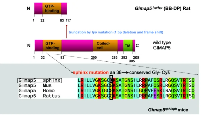
this function of GIMAP5 came from studies on Bio-Breeding Diabetes-Prone (BB-DP) strain of rats that develop spontaneous autoimmune type 1 diabetes (Elder and Maclaren, 1983; Jackson et al., 1981; Poussier et al., 1982). Even though the thymic cellularity in BB-DP rats is normal and DN, DP and SP thymocytes show normal distribution, these rats display a 5-10 fold reduction in the number of CD4+ TCR + T cells and a virtual absence of CD8+ TCR + T cells in secondary lymphoid organs compared to non-lymphopenic, diabetes-resistant BB-DR rats (Elder and Maclaren, 1983; Jackson et al., 1981; Poussier et al., 1982; Ramanathan and Poussier, 2001). The cell survival defect is confined to T cells in BB-DP rats. B cells are relatively unaffected as shown by normal B cell numbers and T independent antibody responses (Guttmann et al., 1983; Lang et al., 2004; Pandarpurkar et al., 2003). The genetic defect that underlies the lymphopenia in BB-DP rats has been mapped to the lyp locus on chromosome 4 (Jacob et al., 1992). The lyp allele arises from a single base pair deletion causing a frameshift mutation within the GTPase domain of the Gimap5 gene, resulting in a truncated protein lacking 223 amino acids at the C-terminus that are replaced by 19 other residues (Hornum et al., 2002; MacMurray et al.,2002)(Figure 1-2).
Thymus grafts or reconstitution with T cell depleted bone marrow cells between BB-DP and BB-DR rats has shown that the T lymphopenia results from an intrinsic defect in T-cell precursors (Francfort et al., 1985). The half-life of mature T cells that have recently emigrated from the thymus to the periphery is markedly lower in BB-DP rats than in control rats (3 days versus 15 days) (Groen et al., 1995; Hernandez-Hoyos et al., 1999; Ramanathan et al., 1998; Ramanathan and Poussier, 2001; Zadeh et al., 1996). This accelerated death of recent thymic emigrants (RTE) in BB-DP rats most likely contributes to the reduction in T cell numbers. Following thymectomy, the size of the peripheral T cell pool drops by 80% in BB-DP rats within 5 days while it remains relatively stable in control rats (Sarkar et al., 1994). This phenomenon is due to either short life span or reduced proliferative capacity of peripheral T cells. Furthermore, by in vivo bromodeoxyuridine (BrdU) labeling it has been shown that BB-DP rats maintain a small pool of cycling T cells, possibly due to homeostatic signals via TCR and cytokine receptors (Ramanathan and Poussier, 2001). These cells exhibit a partial activation phenotype characterized by downregulation of CD62L, and increased expression of MHC class I, MHC class II, and CD28 (Lang et al., 2004). The partial activation phenotype appears to be due in part to activation of NF-κB through a MAPK-dependent pathway (Kupfer et al., 2007). However, pulse chase analysis with BrdU revealed that the cycling T cells have a reduced half-life once they enter the resting phase despite the high mitotic activity of these cells (Ramanathan et al., 1998). Consistent with the defective survival of RTE and peripheral T cells in vivo, CD4+ and CD8+ SP thymocytes and peripheral T cells from BB-DP rats were more prone to apoptosis when compared to BB-DR rats (Ramanathan and Poussier, 2001). Even though the BB-DP rat T cells die by apoptosis ex vivo, stimulation by mitogens such as Con A or anti-CD3 Ab induces proliferation, suggesting that activation rescues BB-DP rat T cells from apoptosis (Lau et al., 1998; Lee, 1994; Ramanathan et al., 1998). Collectively, these data suggest that Gimap5 is required to maintain T cells in a state of quiescence. Freshly isolated BB-DP T cells exhibit a loss of mitochondrial integrity suggesting a dysregulated mitochondrial apoptotic pathway in these cells (Keita et al., 2007). Furthermore, this survival defect is not due to any defect in the expression of Bcl-2 and Bcl-xL in thymocytes and peripheral T cell subsets (Ramanathan and Poussier, 2001). Moreover, spontaneous death of BB-DP rat T cells occurs by a caspase-independent mechanism, as broad-spectrum caspase inhibitors DEVD, ZVAD and BOC do not
delay the accelerated death in vitro.
Consistent with the pro-survival function of GIMAP5 in rat T lymphocytes, anti-apoptotic functions of human, rat and murine GIMAP5 have been demonstrated ex vivo. Expression of
Gimap5 mRNA is consistent and significantly increased after stimulation with apoptotic
cytokines in two different monocyte cell lines, which could relate GIMAP5 with the anti- apoptotic response (Hellquist et al., 2007). Overexpression of human GIMAP5 in Jurkat T cells confers protection against apoptosis induced by okadaic acid and -radiation, whereas it fails to prevent apoptosis induced by anti-Fas, TNF, staurosporine and some chemotherapeutic drugs (Sandal et al., 2003). However, thymocytes and peripheral T and B cells from Gimap5lyp/lyp rats do not show increased sensitivity to -radiation-induced apoptosis (Dion et al., 2005). siRNA mediated knockdown of mGIMAP5 increases apoptosis of IL-2-dependent 23-1-8 T cells caused by IL-2 withdrawal, which was markedly restored by the overexpression of Bcl-xL (Nitta et al., 2006). This is recapitulated by inhibition of hGimap5 expression by RNA interference in Jurkat cells resulting in mitochondrial dysfunction and a significant increase in apoptotic cells (Pandarpurkar et al., 2003). On the contrary, Dalberg has shown that a reduction of Gimap5 by RNA interference had no effect on apoptosis in Jurkat cells, but overexpression of Gimap5 results in apoptosis in Jurkat T cells as well as naive human T cells but not in activated human T cells. Surprisingly, over expression of rat wild type or the mutant BB-DP allele of rGIMAP5 in the rat T-cell line also induces spontaneous apoptosis of T cells (Dalberg et al., 2007). In mice, GIMAP5 was shown to be involved in the mitochondria-mediated apoptotic pathway as GIMAP5 interacts with members of the Bcl-2 family of anti-apoptotic proteins (Barnes et al., 2010; Chen et al., 2011; Nitta et al., 2006; Zenz et al., 2004). GIMAP5 gene polymorphisms are related to susceptibility to autoimmune disorders where apoptosis and lymphopenia are key features, such as type I diabetes and systemic lupus erythematosus (SLE), implicating the role of
Gimap5 in human diseases (Hellquist et al., 2007; Lim et al., 2009; Shin et al., 2007). However,
precise mechanisms underlying the anti-apoptotic function of GIMAP5 protein remain to be elucidated.
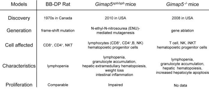
Recently, 2 different lines of Gimap5 deficient mice were generated to define the in vivo function of mouse Gimap5. Gimap5-/- mice were generated by selective genetic ablation of the translation initiation signal and the putative nucleotide-binding motif of the Gimap5 gene (Schulteis et al., 2008). Gimap5sph/sph mice were generated by N-ethyl-N-nitrosourea
(ENU)-induced germline mutant with a missense mutation in the P-loop of AIG1 domain of Gimap5 (Barnes et al., 2010) (Table 1-2). Unlike Gimap5-/- mice, the mRNA expression of Gimap5 in
Gimap5sph/sph mice remains unaffected. Similar to Gimap5 mutation in the rat, survival of peripheral T cells is impaired in these mice. In addition, B cells and NKT cells exhibit loss of immune homeostasis (Aksoylar et al., 2012; Barnes et al., 2010; Chen et al., 2011; Schulteis et al., 2008). Even though Gimap5-deficient CD4+ T cells acquire a partial activation phenotype, they fail to proliferate following stimulation of the TCR. In addition, dependent and T-independent B cell responses are abrogated. Moreover, Gimap5 deficient mice show defects in various hematopoietic cell types including an increase in HSCs but fewer lineage-committed hematopoietic progenitors. The HSC and progenitors undergo increased apoptosis and display impaired long-term repopulation capacity. All these observations indicate a loss of HSC quiescence (Barnes et al., 2010; Chen et al., 2011; Schulteis et al., 2008) (Table 1-2). These mice exhibit chronic hepatic extramedullary hematopoiesis with foci of hematopoietic cells associated with liver failure. In addition, Gimap5sph/sph mice develop spontaneous colitis, resulting the early morbidity and weight loss (Barnes et al., 2010). Collectively, these data indicate that Gimap5 is a critical regulator of lymphocyte homeostasis and hematopoiesis.
1.3.3 Expression of GIMAP5 in lymphocytes
GIMAP5, the founding member of the GIMAP family, is essentially expressed in cells of the immune system (Krucken et al., 2004; Liu et al., 2008). In rats, the expression of Gimap5 is significantly increased from the early DN to DP and SP transition. Studies have shown that DP thymocytes are reduced and DN thymocytes are increased in the congenic Gimap5-/- F344 rats. (Moralejo et al., 2003). The highest expression of rGimap5 was observed in peripheral T cells, whereas the expression of rGimap5 in B cells was lower than in different T cell subsets except DP cells (Dion et al., 2005). Murine Gimap5 is expressed abundantly in CD4+, CD8+ T cells and B cells of the spleen and lymph nodes, but not in myeloid cells or NK cells (Dion et al., 2005; Nitta et al., 2006). In the thymus, murine Gimap5 expression is moderate in pre-selected DP thymocytes but increases significantly during maturation of DP to CD4+ or CD8+ SP thymocytes upon TCR mediated positive selection (Dion et al., 2005; Nitta et al., 2006). The short hairpin RNA knockdown of mice Gimap5 in DN perturbs the generation of DP thymocytes and reduced the survival of mature T cells (Nitta and Takahama, 2007). Murine Gimap5 mRNA is also expressed in HSCs and hematopoietic progenitors (Chen et al., 2011). Human Gimap5 mRNA is highly expressed in T lymphocytes from spleen and lymph nodes, whereas weak signals can be detected in normal B lymphocytes (Krucken et al., 2004; Zenz et al., 2004). In contrast, human
Gimap5 mRNA expression is increased in certain chronic lymphocytic leukemias and leukemic
mantle cell lymphomas (Zenz et al., 2004). In primary human T cells, an increase in Gimap5 mRNA levels is observed during T cell activation with phytohemagglutinin (PHA) and IL-2 (Dalberg et al., 2007).
1.3.4 Structure and subcellular distribution of GIMAP5
Based on the phenotype of Gimap5-deficient rats and mice, it is believed that all the domains of GIMAP5 are required for proper functioning of GIMAP5 (Figure 1-2). The putative GTPase domain at the N-terminal is distantly related to RAS GTPases but is homologous to the AIG domain in plants (Hornum et al., 2002; MacMurray et al., 2002). GIMAP5 has been shown to bind GTP but fails to hydrolyze GTP (Schwefel et al., 2010b). X-ray crystallography study of GIMAP5 has indicated that the GTPase activity may require a GAP protein, whose identity is not known (Schwefel et al., 2010a). There are 2 coiled-coil domains in GIMAP5 that can function as oligomerization domains for structural proteins, transcription factors and motor proteins (Lupas
and Gruber, 2005). GIMAP5 also possesses a transmembrane hydrophobic domain at the C terminus, which may target it to intracellular membranes. Overexpressed GIMAP5 has been shown to localize to intracellular membrane fractions including the mitochondrial outer membrane, the ER and the centrosomal regions (Hernandez-Hoyos et al., 1999; Hornum et al., 2002; Lau et al., 1998; Zadeh et al., 1996). Consistent with these findings, in murine T cells, GIMAP5 was shown to interact with members of the Bcl-2 family including anti-apoptotic Bcl-2 and Bcl-xL and pro-apoptotic Bax, Bak, Bad and BimEL (Barnes et al., 2010; Chen et al., 2011; Nitta et al., 2006; Zenz et al., 2004), suggesting that Gimap5 plays a role in regulation of apoptosis via Bcl-2 family proteins. Accordingly, loss of GIMAP5 impairs mitochondrial membrane integrity (Pandarpurkar et al., 2003). Another study showed that GIMAP5 maintains ER homeostasis by inhibiting ER stress associated chaperones in T cells (Pino et al., 2009). Most of these experiments were done by overexpressing Gimap5 constructs carrying an epitope tag or EGFP fused to the C-terminus, which might interfere with membrane localization. In contrast, by using antiserum against rat GIMAP5 (rGIMAP5) raised in the rabbit and rGIMAP5 construct with a Myc epitope tag at the N-terminus, our lab have shown that endogenous GIMAP5 resides in a cellular compartment distinct from mitochondria and ER in T cells, and that neither endogenous rat GIMAP5 nor the overexpressed protein interacts with Bcl-2 (Keita et al., 2007). In addition, endogenous mouse and human GIMAP5 were found to localize to lysosomes and multivesicular compartments but not to ER or mitochondria (Wong et al., 2010), whose identities are also unknown. Despite a decade of efforts by several groups, there is a lack of consensus on the subcellular distribution of GIMAP5.
Figure 1-2. Schematic representation of Gimap5 mutation.
GIMAP5 is a 308-amino acid protein that contains an unconventional GTP-binding motif and a coiled-coil domain and a C-terminal hydrophobic domain. The Lyp locus frameshift mutation mapped to gene encoding Gimap5 results in a truncated protein in which the C-terminal 223 amino acids are replaced by 19 other residues. The sphinx mutation results in a glycine to cysteine substitution at position 38 of the AIG1 domain. A glycine at this position is conserved in all Gimap proteins.
1.4 Role of GIMAP5 in cellular calcium (Ca
2+) homeostasis
1.4.1 Role of mitochondria in Ca
2+homeostasis in T lymphocytes
Cytosolic calcium ([Ca2+]c) is an important regulator of various cellular functions ranging from fertilization to cell death (Berridge et al., 2000). In T lymphocytes, Ca2+signals are crucial for the proper activation, differentiation and effector functions (Berridge et al., 2000). The cytosolic Ca2+concentration in T cells is tightly regulated and varies between ~100 nM in resting cells to ~1µM following TCR stimulation. Engagement of the TCR results in transphosphorylation and proximal activation of the Src family of protein tyrosine kinases (PTKs) Lck and Fyn. Either or both Lck and Fyn phosphorylate immunoreceptor tyrosine-based activation motifs (ITAMs) on the CD3 chains of the TCR to provide binding sites for other PTKs including ZAP70. Once ZAP70 is activated, it phosphorylates the downstream substrates, linker for activation of T-Cells (LAT) and SH2 domain-containing leukocyte protein-76 (SLP-76), to
recruit many other signaling molecules including phospholipase C-gamma 1 (PLC-1) (Lin and Weiss, 2001). Activation of PLC-1 hydrolyzes phosphatidylinositol-4,5-bisphosphate (PIP2) to produce second messengers including inositol 1,4,5-trisphosphate (IP3) and 1,2-diacylglycerol. IP3 binds to its receptor IP3R on the ER and triggers Ca2+ release from the internal ER store. This Ca2+ store depletion can also be induced by thapsigargin (TG), the inhibitor of ATP-dependent Ca2+ pump called sarco/endoplasmic reticulum Ca2+-ATPase (SERCA) that pumps Ca2+ into the sarcoplasmic and ER (Thastrup et al., 1990). Store depletion results in a conformational change in the ER-localized stromal interaction molecule 1 (STIM1) protein (Liou et al., 2005; Roos et al., 2005). STIM1 is transported to the plasma membrane to associate with the ORAI proteins of the store-operated Ca2+ channel (SOC) called the Ca2+ release-activated Ca2+ channel (CRAC), inducing the opening of CRAC and Ca2+ entry from the extracellular milieu (Berridge, 1993; Hogan et al., 2010). Defects in ORAI proteins result in immunodeficiency as Ca2+ influx from the extracellular medium is completely absent in T lymphocytes (Feske et al., 2006). TCR stimulation by antigen induces sustained Ca2+ influx via CRAC channels leading to activation of Ca2+-dependent enzymes, such as calcineurin, and thereby transcription factors such as NFAT and NF-B that drive T cell activation and gene expression (Valitutti et al., 1995). In the absence of stimulation by nominal antigen, the interaction between non-stimulatory self peptides:MHC and TCR, which provides survival signals without inducing cell proliferation, is also capable of eliciting discernible Ca2+ response (Delon et al., 1998; Kondo et al., 2001; Revy et al., 2001; Takeda et al., 1996; Wei et al., 2007). Several studies have clearly established a critical function for mitochondria in cellular Ca2+ homeostasis (Contreras et al., 2010; Spat et al., 2008) (Figure 1-3). Following sustained Ca2+ entry via the plasma membrane CRAC channels, the rising concentration of [Ca2+]c activates the Ca2+ uniporter, which is a Ca2+-selective channel that spans the inner mitochondrial membrane. This induces a slow, membrane potential-driven uptake of Ca2+, which is released later via the Na+/ Ca2+ exchanger (Pfeiffer et al., 2001). This process ensures that Ca2+ entering via the CRAC channel does not cause a feedback inhibition of the CRAC channel activity (Hajnoczky et al., 2002; Hoth et al., 1997). Thus, intracellular Ca2+ is tightly controlled by the slow, non-saturable, non-linear buffering capacity of mitochondria, as they sequester [Ca2+]c following rapid Ca2+ entry and release Ca2+ slowly to sustain the [Ca2+]c level even after the cessation of Ca2+ inflow (Hoth et al., 1997). Ca2+ uptake by mitochondria is facilitated by the movement of mitochondria
along the T cell cytoskeleton including microtubules or actin filaments to sites of Ca2+ entry (Liu and Hajnoczky, 2009). It is known that actin filaments control cell morphology and plasticity, generating the mechanical force necessary for motility (Vicente-Manzanares and Sanchez-Madrid, 2004). The microtubular network is thought to regulate the polarized secretion of effector molecules by T cells and might contribute to migration and division (Sancho et al., 2002). Following T cell activation through APCs or crosslinking by immobilized anti-TCR antibodies, the sustained entry of Ca2+at the immunological synapse requires actin cytoskeleton dependent accumulation of mitochondria underneath the CRAC channels (Schwindling et al., 2010). However Ca2+ uptake by mitochondria following thapsigargin or soluble anti-CD3 antibody is mediated by the translocation of mitochondria on microtubules (Quintana et al., 2007). Collectively, these mechanisms sustain the Ca2+ influx for hours, which is required for the activation of transcription factors NFAT, AP1 and NF-kB that drive the transcription of interleukin-2 (IL-2) and other genes involved in T cell activation (Feske et al., 2001).
Of note, Ca2+ acts as a physiological stimulus for ATP synthesis (Balaban, 2002). It is known that naïve T cells utilize the catabolic metabolism to generate ATP through the tricarboxylic acid (TCA) cycle and oxidative phosphorylation (OXPHOS) in mitochondria. Uptake of Ca2+ by mitochondria directly activates key mitochondrial enzymes such as Ca2+ -dependent dehydrogenases of the TCA cycle, driving mitochondrial NADH production and ATP production by OXPHOS during early T cell activation (Tarasov et al., 2012). At the mitochondrial inner membrane, electrons from NADH and succinate are delivered to oxygen via the electron transport chain (ETC). The ETC (complexes I-IV) establish a H+ gradient and the energy produced by this electrochemical gradient is then utilized by complex V to generate ATP. During this process, reactive oxygen species (ROS) such as superoxide anion (O2·–) and hydrogen peroxide (H2O2) are accidentally generated by electron transfer (Baughman and Mootha, 2006). ROS play an important role in shaping T cell response by activating several transcription factors and enzymatic cascades (Devadas et al., 2002; Droge, 2002; Jackson et al., 2004). Studies have shown that mitochondrial ROS produced during OXPHOS is essential for T cell activation (Sena et al., 2013). T cells deficient for Uqcrfs1, a subunit of complex III of the mitochondrial ETC, display impaired mitochondrial ROS production and defects in antigen-specific T cell activation and IL-2 production (Sena et al., 2013). However, excessive ROS signals are detrimental, causing Ca2+ overload, mitochondrial depolarization, cytochrome c
release, lipid peroxidation and DNA damage, leading to apoptotic and non-apoptotic cell death. Overall, there appears to be multifactorial crosstalk among Ca2+, ATP and ROS, centered on the mitochondria. The roles of each player are different depending on the physiological /pathological status of the cell. Therefore, any disturbance will have a large impact on the others (Brookes et al., 2004).
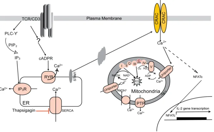
Figure 1-3. Ca2+ signaling in T cells.
Antigen recognition through the TCR results in the production of the second messengers IP3 and cADPR. IP3 and cADPR bind to IP3R and RYR and induce Ca2+ release from ER. A decrease in the ER Ca2+ induces a conformational change in the ER-localized STIM1 protein, which in turn activates CRAC channels in the plasma membrane. Ca2+ influx though CRAC channels and elevated intracellular Ca2+
concentration activate transcription factors and drive T cell activation. Sustained Ca2+ entry through the CRAC channel is maintained by mitochondria: following Ca2+ influx via the CRAC channel, the raising [Ca2+]c activates the Ca2+ uniporter on the mitochondrial membrane. This induces a slow, ΔΨm-driven uptake of Ca2+, which is released later via the Na+/ Ca2+ exchanger after the cessation of Ca2+ entry. Mitochondria is also a important site of ROS production and ATP generation.
1.4.2 Role of GIMAP5 in cellular Ca
2+homeostasis
Despite the uncertainty over the mechanisms underlying spontaneous apoptosis of
Gimap5lyp/lyp T cells, our work and that of many other labs have indicated that freshly isolated T cells from BB-DP rats showed a gradual loss of mitochondrial membrane potential over time upon culture in vitro (Keita et al., 2007; Pandarpurkar et al., 2003). Our group investigated whether GIMAP5 regulates Ca2+ signals generated following the cross-linking of the TCR in rat
T cells. T cells isolated from Gimap5lyp/lyp rats display reduced Ca2+ flux in response to stimulation via the TCR. This defect is manifested in single positive mature thymocytes, where the survival defect is first observed (Ilangumaran et al., 2009). Our results suggested that Ca2+ entry from extracellular milieu is defective in Gimap5lyp/lyp T cells since the mobilization of Ca2+ from the intracellular stores induced by TG, in the absence of extracellular Ca2+ appears to be normal in these cells. In human T cells from severe combined immunodeficiency (SCID) patients, Feske has shown that even when the Ca2+ release from ER stores is normal, the Ca2+ influx and T cell activation are impaired, which is due to the complete lack of CRAC activity (Feske et al., 2005). Similarly, Schwarz has shown that a large reduction of CRAC activity by specific CRAC channel blockers leads to the reduction of the T cell-based immune responses including T cell proliferation and IL-2 secretion (Schwarz et al., 2007). The defective Ca2+ response observed in Gimap5lyp/lyp T cells could arise entirely from faulty activity of the plasma membrane Ca2+ channels. Therefore, we first examined whether Ca2+ entry across the plasma membrane via the CRAC channels is compromised in Gimap5lyp/lyp T cells. Since the magnitude of Ca2+ influx through CRAC channel varies with changes in extracellular Ca2+, I first
investigated the correlation between the opening of the CRAC channel and different external Ca2+ concentration dependent T cell proliferation. In the presence of different amounts of ethylene glycol tetraacetic acid (EGTA), a Ca2+ chelator, the sensitization of CRAC channels to extracellular Ca2+ change is comparable between Gimap5lyp/lyp and control CD4+ lymphocytes (Figure 1-4A). Meanwhile, to directly confirm whether GIMAP5 regulates the CRAC channel, we measured the electro-physiological nature of the CRAC current (ICRAC) by the patch-clamp technique (Feske et al., 2005) in purified control and Gimap5lyp/lyp T cells. No significant difference was observed between Gimap5lyp/lyp and control T cells in the ICRAC current generated (Figure 1-4B). These observations ruled out defects in the CRAC channels in T lymphocytes from Gimap5-deficient rats and suggested that reduced Ca2+ flux in Gimap5lyp/lyp T cells may arise from defects in other mechanisms required for sustained Ca2+ entry through the CRAC channel. However, we do not know how this is related to the known T cell survival signals.
Figure 1-4. Reduced Ca2+ entry in Gimap5lyp/lyp T cells does not result from impaired opening of the CRAC channel.
A. Isolated CD4+ lymphocytes from Gimap5lyp/lyp and control rats cultured with irradiated syngeneic spleen cells were stimulated with A) ConA or B) anti-rat TCR monoclonal antibody R73 (10μg/ml) in the presence of different concentration of EGTA, followed by mesurement of 3H thymidine incorporation at day 3.
Results are expressed as percentage of 3H-thymidine uptake over no EGTA treatment.
B. ICRAC current was measured in T cells from Gimaplyp/lyp and control rats at room temperature. The
external solution contained 145mM NaCl, 2.8mM KCl, 10mM CaCl2, 2mM MgCl2, 10mM CsCl, 10mM
Glucose and 10mM Hepes pH7.4. The Internal Solution (in the microelectrode): 145mM Cesium glutamate, 1mM MgCl2, 10mM Hepes, 2 mM thapsigargin and 100uM EGTA, pH 7.2. The currents were
1.5 Survival signals for T lymphocytes
1.5.1 TCR tuning and signaling pathways involved in the maintenance of
naïve T cell survival
Following positive selection, T cells undergo a process of TCR tuning before entering the secondary lymphoid tissue (Grossman and Singer, 1996; Modiano et al., 2008). For these T cells, TCR tuning reduces TCR signaling to restrain cell cycle entry, thereby keeping the cells in a quiescent and self-tolerant state. T cell quiescence is enforced by upregulation of negative regulators, notably the adaptor Cbl-b, which blocks the activation of Vav (Bachmaier et al., 2000; Chiang et al., 2000; Teh et al., 2010), SHP-1 phosphatase (Stephen et al., 2009) and CD5 (Perez-Villar et al., 1999; Tarakhovsky et al., 1995). T cell quiescence also requires negative transcriptional regulators such as the transducer of ErbB2-1 (Tob1) (Tzachanis et al., 2001) and Krupple-like factor (KLF4) (Yamada et al., 2009), which downregulate the expression of genes essential for cell cycle progression. T cell quiescence also depends on repression of transcription factors NF-kB and NFAT, which are critically involved in cytokine production (Schorle et al., 1991). TCR tuning also alters expression of various cell-surface molecules with costimulatory function such as CD8 and CD2 (Teh et al., 1997).
After T cells mature in the thymus and enter the periphery as antigen-inexperienced naïve cells, they possess both proliferative and differentiative potential that is repressed by different homeostatic mechanisms to maintain T cell homeostasis. As far as we know T cell homeostasis requires signaling by both IL-7 and low affinity TCR engagements of self-ligands (Maraskovsky et al., 1997; Maraskovsky et al., 1996; Schluns and Lefrancois, 2003; Tan et al., 2001). Studies have shown that depriving naïve CD8+ cells of contact with MHC I ligands in either lymphopenic (Murali-Krishna et al., 1999; Tanchot et al., 1997) or nonlymphopenic hosts (Takada and Jameson, 2009) leads to a shortened lifespan of CD8+ T cells. Half-life of naïve CD8+ T cells is also reduced following TCR expression ablation (Labrecque et al., 2001; Polic et al., 2001). However, CD4+ T cells are less dependent on MHC contact for their survival because the half-life of CD4+ T cells ranged from several days to 46 days after transferring CD4+ T cells into MHC II-/- hosts (Rooke et al., 1997; Surh and Sprent, 2005; Takeda et al., 1996) or ablating CD4+ TCR expression (Polic et al., 2001).
The signals delivered by the interaction with self MHC that helps naïve T cell survival have not been well characterized (Figure 1-5). Partial tyrosine phosphorylation of the TCR -chain has
been observed in naïve T cells ex vivo, and it disappears rapidly after both physiological and experimental interruption of T cell contact with self-peptide MHC ligands (Seddon and Zamoyska, 2002; Stefanova et al., 2002). Furthermore, the compromised survival of naïve T cells in the absence of Fyn and Lck was associated with this loss of TCR -chain phosphorylation, indicating that the Src family kinases are critical for TCR-mediated survival signals (Zamoyska et al., 2003). The TCR hyporesponsiveness associated with the impaired lifespan of naïve T cells are observed in mice with deletions or mutations in Vav1 (Fujikawa et al., 2003), Wiskott-Aldrich syndrome protein (WASP) (Cotta-de-Almeida et al., 2007), the adaptor protein GrpL (Yankee et al., 2004), the adaptor Nck (Roy et al., 2010), the RNA-binding protein hnRNPLL (Wu et al., 2008), a Rho-Rac GTP exchange factor, Dock 8 (Randall et al., 2009), and the β3 regulatory subunit of voltage-gated calcium channels (Cavβ3) (Jha et al., 2009). However, the exact signaling pathways mediated by TCR-self-peptide-MHC encounters in naive T cell maintenance remain controversial.
Left: Several molecules including Lck, Fyn, Vav1, WASP and Nck are essential for T cell survival. Right: IL-7 binding to its receptor triggers a cascade of signaling events that induce cell survival. The activation of JAK/STAT5 pathway leads to STAT5 nuclear translocation and subsequent transcription factors induced Bcl-2 gene expression. The phosphorylation and activation of PI3K/Akt results in increased anti-apoptotic activities.
1.5.2 IL-7 acts as a survival factor at various stage of T cell development
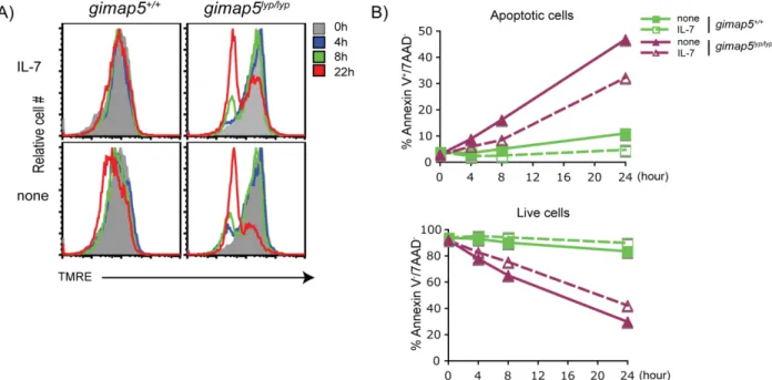
IL-7, a member of the common γ-chain Rc (CD132), family of cytokines, is essential for maintaining lymphocyte viability, lymphocyte differentiation and lymphocyte homeostasis (Surh and Sprent, 2008). Genetic ablation of IL-7 expression in mice has shown that normal development of T cells in the thymus is dependent on IL-7 (von Freeden-Jeffry et al., 1995). Adoptive transfer of naïve T cells into syngeneic IL-7-deficient mice or injection of IL-7 mAb into normal mice result in a marked impairment of T cell survival which is accompanied by decreased T cell numbers (Kondrack et al., 2003; Vivien et al., 2001). Conversely, overexpression of IL-7 in IL-7 transgenic mice or exogenous IL-7 treatment increased the size of the naïve T cell pool (Geiselhart et al., 2001; Kieper et al., 2002; Mertsching et al., 1995). Studies have shown that in humans, CD4+ lymphopenia is associated with supranormal levels of serum IL-7 (Bolotin et al., 1999; Fry et al., 2001; Napolitano et al., 2001). In line with this theory, I observed that the addition of IL-7 does not rescue Gimap5lyp/lyp CD4+ T lymphocytes from apoptosis. In addition, IL-7 did not prevent the loss of ΔΨm in Gimap5lyp/lyp T cells in vitro (Figure 1-6), which suggests that IL-7 is present at sufficient concentrations in Gimap5 deficient rats. Furthermore, reconstitution of lymphopenic rats with T cells from control rats restores the lymphopenic defect (Ramanathan and Poussier, 1999; Rossini et al., 1984; Rossini et al., 1986). Thus the spontaneous apoptosis of Gimap5 deficient T cells is not due to defects in the level of endogenous IL-7.
In the thymus and the periphery, IL-7 is produced by non-hematopoietic stromal cells rather than leukocytes. IL-7 receptor consists of the IL-7 receptor alpha chain (IL-7Rα/CD127) and the CD132, which is a shared component of several receptors of cytokines including IL-2, IL-4, IL-9, IL-15 and IL-21 (Kovanen and Leonard, 2004; Ma et al., 2006). IL-7Rα is expressed at all developmental stages in the thymus except in DP thymocytes. After re-expression at the SP stage, Il-7Rα is maintained by resting T cells and is downregulated following IL-7 mediated signaling and /or T cell activation (Mackall et al., 2011). The members of Forkhead box family, Foxo1 transcription factor, were found to be critical for the expression of IL-7R in peripheral T
cells (Kerdiles et al., 2010). T cells of Foxo1 deficient mice display a severe defect in IL-7R expression. In addition, the absence of Foxo3 exacerbated the effects of the loss of Foxo1 (Kerdiles et al., 2010). On the other hand, the deletion of a different Fox factor, Foxp1, in mature T cells resulted in increased IL-7R expression (Feng et al., 2010; Feng et al., 2011).
In all T cells, IL-7-mediated signaling triggers a cascade of downstream signaling events that inhibit apoptosis to maintain T cell survival (Figure 1-5). IL-7 binding to its receptor activates Janus kinase 1 and 3 (JAK1/3) and results in the phosphorylation and activation of signal transducer and activator of transcription 5 (STAT5), resulting in the migration of activated STAT5/ dimmers to the nucleus to regulate gene transcription including NF-κB and CREB to induce anti-apoptotic Bcl-2 and Mcl-1 gene expression (Mazzucchelli and Durum, 2007). Stat5 knockout mice have decreased T cell numbers (Lin et al., 1995; Onishi et al., 1998; Teglund et al., 1998), while T cells from Stat5 transgenic mice have an enhanced peripheral expansion (Kelly et al., 2003), suggesting a critical role for STAT5 in peripheral T cell homeostasis. IL-7 binding further triggers the phosphatidylinositol 3-kinase (PI3K) pathway, which results in translocation of PI3K to the membrane and catalyzes the phosphorylation of PIP2 to phosphatidylinositol (3,4,5)-trisphosphate (PIP3). PIP3 localizes the downstream target of AKT to the plasma membrane, where AKT is fully activated by sequential phosphorylation at threonine 308 (Thr308) and serine 473 (Ser473) (Vivanco and Sawyers, 2002). AKT can promote cell survival by preventing the intrinsic mitochondrial pathway of apoptosis. In this respect, the increased expression of anti-apoptotic molecules Bcl-2 and myeloid cell leukaemia sequence 1 (Mcl-1) either directly regulate the activity of the death effector Bcl-2-associated X protein (Bax) and Bak or interact with the pro-apoptotic molecules Bcl-2-interacting mediator of cell death (Bim) and Bcl-2 antagonist of cell death (Bad) (Chipuk and Green, 2008), which terminally prevent apoptosis by inhibiting the activation of caspases (Khaled and Durum, 2002).
Figure 1-6. IL-7 does not prevent the loss of ΔΨm and apoptosis in Gimap5lyp/lyp CD4+ T cells.
CD4+ T lymphocytes from Gimap5lyp/lyp and control rats, freshly isolated or after incubation with or without IL-7 for different time points, were loaded with A) TMRE for 20min and B) Annexin V/7AAD for 5min. The dye-loaded cells were analyzed by flow cytometry. A) TMRE signal in gated live cells was evaluated to determine the ΔΨm. Apoptotic cells lose the ΔΨm and thus fail to retain TMRE. B) Annexin V+/7AAD-
cells were defined as apoptotic cells.
1.5.3 Metabolic pathways regulate T cell survival in the quiescent state
There is a growing appreciation for the importance of cell metabolism as a key regulator of T cell number and functions. The immune quiescence of naïve T cells is characterized by catabolic metabolism. Naïve T cells normally undergo OXPHOS and have low metabolic requirements. They consume glucose, amino acids, and lipids to catabolically fuel the TCA cycle for generating ATP in the mitochondria (Guppy et al., 1993). Both TCR and IL-7 are important in this process. Weak TCR signaling is required to maintain basal expression of the glucose transporter Glut-1 in resting T cells, thus glucose uptake and subsequent baseline ATP production and biosynthetic processes are induced (Jacobs et al., 2008; Rathmell et al., 2000). Similarly, IL-7 promotes cell survival, glucose uptake and surface expression of Glut-1, which is mediated by rapid STAT5 activation and a low and sustained activation of AKT (Wofford et al., 2008). Moreover, IL-7 is involved in amino acid metabolism in T cells. IL-7 induced growth of CD8+ T cells and maintenance of cell size is dependent on amino acids and that amino acid transporter are specific transcriptional targets of IL-7 signaling (Pearson et al., 2012).
Collectively, without these essential extracellular signals, T cells fail to maintain sufficient glucose uptake and metabolism, resulting in decreased mitochondrial membrane potential and a fall of cellular ATP, and leading to accelerated T cell death (Rathmell et al., 2000). During an immune response, T cells switch from catabolic to anabolic state and then again transition into catabolic state in memory T cells. The activation of T cells requires increased cellular metabolism by aerobic glycolysis, in which glucose is converted to lactate in the cytosol that are required to sustain clonal cell expansion and subsequent differentiation into effector cells (Bauer et al., 2004; Frauwirth et al., 2002).
Recent studies suggest that metabolic pathways linking immune signals and metabolic cues for the development, function and maintenance of T cells involves AMP activated protein kinase (AMPK) (Tamas et al., 2006), mammalian target of rapamycin (mTOR) (Delgoffe et al., 2009; Delgoffe et al., 2011; Lee et al., 2010; Yang et al., 2013), MYC (Wang et al., 2011) and hypoxia-inducibel factor 1 (HIF1).
AMPK is an evolutionarily conserved enzyme composed of a catalytic subunit and and regulatory subunits, which functions as an energy sensor in energy starved states. AMPK maintains bioenergetic homeostasis by promoting catabolic pathways including mitochondrial biogenesis, transcription, lipid oxidation and antagonizes anabolic pathways like mRNA translation, lipid biosynthesis, proliferation (Hardie, 2007; Misra and Chakrabarti, 2007). AMPK deficient T cells display increased activity in response to metabolic stress. However, AMPK is dispensable for activation and expression of effector function in response to Ag stimulation (Mayer et al., 2008). In agreement with previous work, AMPKα1-deficient mice display normal T cell development and TCR-mediated proliferation. But the T cell viability is reduced and the basal metabolism of resting T cells is increased ex vivo. Moreover, AMPKα-deficient CD8+ T cells have elevated glycolytic activity and inflammatory cytokine production in vitro, which suggests AMPK as a negative regulator of T cell activation (MacIver et al., 2011). Recently, the group of Dr. Jones showed that AMPK regulates mitochondrial metabolic homeostasis to maintain T cell bioenergetics and viability upon T cell activation (Blagih et al., 2015).
In T cells, the activation of AMPK is mediated by three upstream kinases: Liver kinase B1 (LKB1) in response to TCR and CD28 signal or bioenergetic stress like elevated AMP:ATP ratio (Hawley et al., 2003); calmodulin-dependent protein kinase kinase (CamKK) in response to TCR-mediated Ca2+ flux (Mayer et al., 2008; Tamas et al., 2006); TGF-activated kinase-1