Sustained, targeted, high-level transgene expression in primary B lymphocytes may be useful for gene ther-apy in B cell disorders. We developed several candidate B-lineage predominant self-inactivating lentiviral vectors (LV) containing alternative enhancer/promoter elements including: the immunoglobulin β (Igβ) (B29) promoter combined with the immunoglobulin μ enhancer (EμB29); and the endogenous BTK promoter with or without Eμ (EμBtkp or Btkp). LV-driven enhanced green fluorescent protein (eGFP) reporter expression was evaluated in cell lines and primary cells derived from human or murine hematopoietic stem cells (HSC). In murine primary cells, EμB29 and EμBtkp LV-mediated high-level expression in immature and mature B cells compared with all other lineages. Expression increased with B cell maturation and was maintained in peripheral subsets. Expression in T and myeloid cells was much lower in percentage and intensity. Similarly, both EμB29 and EμBtkp LV exhibited high-level activity in human primary B cells. In contrast to EμB29, Btkp and EμBtkp LV also exhibited modest activ-ity in myeloid cells, consistent with the expression profile of endogenous Bruton’s tyrosine kinase (Btk). Notably, EμB29 and EμBtkp activity was superior in all expression models to an alternative, B-lineage targeted vector con-taining the EμS.CD19 enhancer/promoter. In summary, EμB29 and EμBtkp LV comprise efficient delivery plat-forms for gene expression in B-lineage cells.
Received 10 June 2010; accepted 28 October 2010; published online 7 December 2010. doi:10.1038/mt.2010.259
IntroductIon
B cells represent an important target for gene transfer because single gene defects impacting B-lineage function have significant roles in the pathogenesis of immunodeficiency and autoimmu-nity.1 One B cell disorder predicted to benefit from safe strategies
for gene delivery is X-linked agammaglobulinemia (XLA). XLA is an immunodeficiency caused by a recessive gene defect in Bruton’s tyrosine kinase (Btk),2 that results in a block in B cell development
at the pro-B cell stage, reduced numbers of circulating B cells, and a near absence of antibody responses in affected males.3 XLA is a
good candidate for gene therapy for several reasons: a relatively high disease frequency, the ability to treat without interruption of clinical therapy, and a strong selective advantage for corrected cells.4,5 In order to facilitate such therapies, we focused
on designing a lentiviral vector (LV) optimized to drive gene expression in primary B cells, with a special emphasis on vectors that mimic the expression profile of endogenous Btk.
Self-inactivating LVs (LV) comprise a promising gene delivery platform for treatment of genetic disorders, autoimmune diseases, and malignancies. In contrast to γ-retroviruses, LVs proficiently target nondividing cells such as multipotent hematopoietic stem cells (HSC) at low viral copy number.6–8 Self-inactivating-LV also
limit the risk of viral long-terminal repeat enhancer mutagenesis and concurrently permit the use of lineage-specific promoters.9
Furthermore, there is evidence of less transcriptional silencing of internal promoters within integrated LV, and a reduced bias for integration near transcription start sites.10 These combined
fea-tures likely reduce the overall risk of viral enhancer-mutagene-sis responsible for adverse events in several γ-retroviral clinical trials.11,12 Previous work has demonstrated efficient transgene
expression with LV utilizing a range of internal promoter and other regulatory elements.8,13,14
To decrease potential side effects of nonspecific transgene expression in HSC-derived lineages, various vectors have been designed to restrict transgene expression to one or several lin-eages. For example, specific regulatory elements have been used for targeted expression within erythroid, T, antigen-presenting and myeloid cells, respectively.15–18 Notably, Moreau et al. showed that
LV incorporating the regulatory sequences from the human CD19 promoter allowed preferential transgene expression in B-lineage cells.19 Importantly, addition of the immunoglobulin heavy chain
μ intronic enhancer (Eμ) and its associated matrix attachment regions both significantly increased gene expression and promoted uniformity of expression compared with either the PGK20 or CD19
minimal promoters alone.21,22 However, the levels of expression
obtained with the B-restricted LV reported to date are low in com-parison with that mediated by promiscuous γ-retroviral-derived Correspondence: David J Rawlings, Center for Immunity and Immunotherapies, Seattle Children’s Research Institute, 1900 Ninth Avenue, Seattle, Washington 98101, USA. E-mail address: drawling@u.washington.edu
Development of B-lineage Predominant
Lentiviral Vectors for Use in Genetic
Therapies for B Cell Disorders
Blythe D Sather
1, Byoung Y Ryu
1, Brigid V Stirling
1, Mikhail Garibov
1, Hannah M Kerns
1,
Stéphanie Humblet-Baron
2, Alexander Astrakhan
3and David J Rawlings
1,3,41Center for Immunity and Immunotherapies, Seattle Children’s Research Institute, Seattle, Washington, USA; 2Center for Cellular and Molecular
Therapy, GIGA-R, University of Liège, FNRS, Liège, Belgium; 3Department of Immunology, University of Washington School of Medicine, Seattle,
enhancer/promoter elements. Thus, it has remained unclear as to whether candidate B cell-specific LV will mediate transgene expres-sion at levels that will restore function in B-lineage disorders, an important consideration as low levels of B-lineage Btk expression is insufficient to rescue murine models of XLA.23,24
In the studies described here, we sought to design LVs that will mimic the expression pattern of endogenous Btk, with the ultimate goal of utilizing such constructs for gene therapy in XLA patients. We generated LV containing three alternative B cell-specific internal enhancer/promoters and evaluated their ability to drive enhanced green fluorescent protein (eGFP) reporter gene expression. As a reference point, we compared the specificity of expression driven by these elements to a previously described B cell-specific LV, EμS. CD19.21 In parallel, each was compared to a LV containing a strong
murine leukemia virus-derived, enhancer/promoter, myeloprolif-erative sarcoma virus enhancer, negative control region deleted, dl587rev primer-binding site substituted (MND), capable of driving high-level transgene expression in all hematopoietic lineages,25 and
used in a recent successful LV clinical trial.14 LVs were compared with
respect to relative expression in cell lines and primary murine and human cells at low viral copy number. We found that both EμB29 and EμBtkp LV preferentially direct high-level, sustained, transgene expression in immature and mature B lymphoid cells, and support future testing of these constructs in XLA gene therapy models. results
design of B-lineage targeted lV
As shown schematically in Figure 1, we generated a number of LV-containing alternative enhancer/promoter elements within
the parental pRRL backbone.9 The first, MND-eGFP, incorporated
a ubiquitous, γ-retroviral enhancer/promoter. In comparison to other mammalian-derived ubiquitous elements such as elonga-tion factor 1α or PGK, MND drives much higher expression lev-els in primary murine and human cells providing a benchmark for very high-level gene expression (data not shown). Second, we generated a construct equivalent to the previously reported, B cell-specific vector, EμS.CD19,19 comprised of the minimal CD19
promoter (230 bp) in conjunction with the Eμ enhancer. While it has been reported that inclusion of a larger portion of the CD19 promoter element (1,274 bp—designated EμL.CD19) generates higher expression levels in B cells, we found little difference in expression levels between these promoters when in association with Eμ (Supplementary Figure S1a). Next, we generated EμB29-eGFP LV, which contains the B29 (Igβ, CD79b) minimal promoter element26 in association with the murine immunoglobulin heavy
chain, intronic enhancer, Eμ.27 B29, a key signaling component of
the B cell antigen receptor, is initially expressed in pro-B cells and its expression increases in immature/mature B cells.26,28,29 Because
the B29 promoter alone exhibited only low-level basal expression (data not shown), the Eμ enhancer was added to increase activ-ity in maturing B cells. The total size of the enhancer/promoter elements for EμB29 and EμS.CD19 were nearly identical. Finally, we created two unique LV constructs designated Btkp-eGFP and EμBtkp-eGFP, that contain regulatory elements derived from the human BTK locus either alone or in combination with the Eμ enhancer, respectively. The 37 kb BTK locus is comprised of 19 exons at Xq22.30,31 Transcriptional start sites of human Btk were
previously identified within −5 and −30 bp of exon 1 with a mini-mal promoter critical for lineage-restricted expression between −200 and −1 bp from the transcriptional start site.32–34 Additional
reporter studies implicated putative regulatory elements between −450 and −200, as well as two conserved NF-κB binding sites within −800 to −600, as positive regulators of Btk expression.35
We therefore used the first 788 bp of the Btk promoter including these elements, hypothesizing that Btkp, in conjunction with the Eμ enhancer, might provide strong B cell-predominant expression and also retain endogenous control elements essential for lineage-appropriate Btk expression.
eµB29 lV drives B-lineage predominant expression that increases with cell maturation
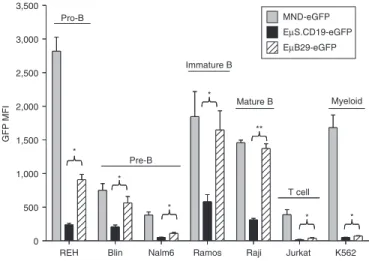
We first compared the expression of eGFP driven by EμB29, EμS. CD19, and MND LV in human T (Jurkat), myeloid (K562), and B cell lines including a panel of different B developmental stages: pro-B (REH), pre-B (Blin and Nalm6), immature B (Ramos), and mature B (Raji) cells. Transduction rates were adjusted via matching eGFP marking to ~10–15% in order to evaluate expression medi-ated by mostly single viral-integration. In this setting, eGFP mean fluorescent intensity (MFI) provides a direct measure of relative promoter strength. Compared to MND, both EμB29 and EμS.CD19 LV exhibited B cell-predominant eGFP expression with the highest expression levels in more mature B cell lines (Figure 2). Importantly, eGFP MFI was significantly higher in all B cell lines transduced with EμB29 compared to EμS.CD19 LV; with the greatest expres-sion in lines representative of mature stages, e.g., Ramos and Raji B cells. Substitution of the extended CD19 promoter (to generate
eGFP wPRE ∆U3 R U5 R U5
∆U3 gag RRE MND
348 bp MND-eGFP EµS.CD19-eGFP EµL.CD19-eGFP hS.CD19 Eµ 204 bp 986 bp 1224 bp 986 bp Eµ hL.CD19 Btkp-eGFP EµBtkp-eGFP EµB29-eGFP Eµ 180 bp 986 bp B29 Eµ 788 bp 986 bp Btkp 788 bp Btkp
Figure 1 schematic of lentiviral constructs. LV were constructed using a pRRL backbone containing an enhancer-deleted U3 region to generate a self-inactivating (SIN) LTR and differ only in their internal promoter/ enhancer region. MND is a retroviral LTR-derived ubiquitous promoter that drives transgene expression in most hematopoietic lineages. Both EμS.CD19-eGFP and EμL.CD19-eGFP employ regulatory sequences from human CD19 and EμB29-eGFP utilizes a murine B29 minimal promoter. Btkp-eGFP LV and EμBtkp-eGFP LV contain a788 bp element derived from endogenous human BTK locus (described in Materials and Methods section). EμB29, EμS.CD19, EμL.CD19, and EμBtkp each also include the murine immunoglobulin intronic enhancer, Eμ. Btk, Bruton’s tyrosine kinase; eGFP, enhanced green fluorescent protein; LV, lentiviral vec-tors; LTR, long-terminal repeats; MND, myeloproliferative sarcoma virus enhancer, negative control region deleted, dl587rev primer-binding site substituted; wPRE, woodchuck hepatitis virus posttranscriptional regula-tory element.
EμL.CD19 LV) resulted in slightly higher levels of eGFP compared with EμS.CD19 LV (Supplementary Figure S1a). However, the expression level for both constructs remained significantly lower than EμB29 LV (Supplementary Figure S1b).
eµB29 drives high-level eGFP expression in primary murine B-lymphocytes
To assess expression and lineage specificity in vivo, congenically marked, B6.Ly5.1 recipient mice were lethally irradiated and trans-planted with B6.Ly5.2, Lin− HSC transduced with either: MND-eGFP; EμS.CD19-MND-eGFP; or EμB29-eGFP LV. Mice with donor chimerism of >90% were sacrificed 25 weeks post-transplant and spleen and bone marrow (BM) cells analyzed for relative eGFP expression in myeloid and lymphocyte subsets (Figures 3 and 4).
Figure 3a,b shows representative data from a single experiment (3–5 recipients/group). Of note, the MFI of eGFP expression across independent experiments was influenced by differences in the instrument used for analysis and/or the voltage settings of the laser used to detect eGFP (488 nm argon). While this limited our ability to present a combined dataset, we observed consistent relative differences among our panel of LV constructs in multiple experiments. For comparison, the results from a second indepen-dent experiment are displayed in Figure 3c and representative flu-orescence-activated cell sorting (FACS) plots comparing B-lineage promoter activity for EμB29 versus EμS.CD19 LV from both experiments are shown in Supplementary Figure S2. Analysis of B cells, T cells, neutrophils and macrophages revealed expression by in all lineages. eGFP MFI, however, was markedly lower in non-B
versus B cells. EμB29 LV exhibited up to fivefold higher eGFP MFI in B cells compared to all other lineages (Figure 3d). Furthermore, although both LV lead to B cell-predominant expression, nearly all EμB29 recipients exhibited approximately fourfold higher MFI in B cells compared with EμS.CD19 (Figure 3b,c). One EμB29 recip-ient animal (in experiment #1) exhibited eGFP expression levels similar to that present in EμS.CD19 recipients; and this result cor-related with the lowest viral copy number among the eight EμB29 animals shown. By comparison, MND LV exhibited similar, albeit variable, eGFP expression in all lineages. Average viral copy num-bers were similar in all LV cohorts (Figure 3e) indicating that dif-ferential LV promoter activity did not reflect altered transduction efficiency.
We next determined whether LV-mediated expression was modulated during BM and/or peripheral B cell develop-ment and selection (Figure 4). Figure 4a shows representative FACS plots of eGFP expression in developing BM B cells. The subsets of developing B cells were gated-based CD43, IgM and IgD expression as described in the figure legend. eGFP MFI was significantly higher in all BM B cells derived from EμB29-eGFP compared to EμS.CD19-EμB29-eGFP recipients (Figure 4a) and increased as cells matured from the pro- to mature B cell stage in EμB29 LV recipients (Figure 4b). In contrast, there was little or no change in eGFP MFI in EμS.CD19-eGFP recipients and MND-eGFP recipients exhibited greater variability with a trend for reduced expression in mature cells. Splenic transitional 1 and 2 (T1 and T2), follicular mature, and marginal zone/marginal zone precursor B cells were identified based on of CD21 and CD24 expression within B220+ B cells36 and eGFP expression in
each subset determined (Figure 4c). Most MND-eGFP LV recip-ients exhibited a decline, and EμS.CD19 LV reciprecip-ients little or no change, in relative eGFP MFI in immature versus more mature B cells, respectively. In contrast, the majority of EμB29-LV recipients exhibited a consistent increase in eGFP MFI in more mature, follicular mature, and marginal zone/marginal zone precursor, compared to less mature, T1 B cells. Thus, EμB29-LV drives high-level B predominant expression that increases with B cell maturation.
eµB29 mediates high-level B-lineage expression in primary human B-lymphocytes
We also examined the activity of candidate LV in primary human cells derived from transduced HSC. We utilized a NOD/SCID/ γc−/− (NSG) xenograft system in which MND-eGFP,
EμS.CD19-eGFP or EμB29-EμS.CD19-eGFP LV transduced human cord blood CD34+ cells were transplanted into recipient immune-deficient NSG mice.37,38 Engrafted mice were sacrificed at 20–25 weeks
transplant and human-derived, BM and splenic hematopoietic subsets were analyzed for relative eGFP expression (Figure 5). The level of human cell engraftment in each mouse is depicted as the percentage of DAPI-live cells that express the human leukocyte common antigen marker CD45 (Figure 5b). A large percentage of splenic CD45+ cells were CD19+ B cells similar to other reports using the NSG model. Moderate populations of CD4+ and CD8+ T cells, as well as CD33+ myeloid cells, were also identified in all recipients. MND LV drove high-level eGFP expression in all lineages. In contrast, EμS.CD19 and EμB29 lead
REH Blin Nalm6 Ramos Raji Jurkat K562
0 500 1,000 1,500 2,000 2,500 3,000 3,500 Pro-B Pre-B Immature B Mature B T cell Myeloid GFP MFI * * * * ** * * MND-eGFP EµS.CD19-eGFP EµB29-eGFP
Figure 2 eµB29 exhibits increased levels of transgene expression compared to eµs.cd19 in B cell lines. A panel of human cell lines was transduced with MND-eGFP (gray bars), EμS.CD19-eGFP (black bars) or EμB29-eGFP (striped bars) LV to achieve 10–15% eGFP marking. Cells were evaluated on day 4 for eGFP expression by FACS. B cell lines selected were derived from a representative of different developmental stages: REH (pro-B), Blin or Nalm6 (pre-B), Ramos (immature B), and Raji (mature B). Expression was also compared using Jurkat (T) and K562 (myeloid) cell lines. Data shows mean fluorescence intensity (MFI) of eGFP in each line based upon three experiments. *indicate statistically significant results in this and all subsequent figures, with *<0.05, **<0.01 and ***<0.001. Only the statistically significant data is highlighted in this manner. eGFP, enhanced green fluorescent protein; FACS, fluorescence-activated cell sorting; MND, myeloproliferative sarcoma virus enhancer, negative con-trol region deleted, dl587rev primer-binding site substituted.
GFP
FCS
B cells T cells Monocytes Neutrophils
MND EµS.CD19 EµB29 a 1,000 800 600 400 200 0 1,000 800 600 400 200 0 1,000 800 600 400 200 0 100 101 102 103 104100 101 102 103 104100 101 102 103 104 100 101 102 103 104 804 993 959 1,679 37.1 64.5 34 251 1,141 79.4 205 71.7 N = 2 b 0 300 600 900 1,200 1,500 1,800 0 300 600 900 1,200 1,500 1,800 0 300 600 900 1,200 1,500 1,800 0 300 600 900 1,200 1,500 1,800 GFP MFI
B cells T cells Monocytes Neutrophils
N = 3 N = 5 N = 5
Mock MND EµS.CD19 EµB29 Mock MND EµS.CD19 EµB29 Mock MND EµS.CD19 EµB29 Mock MND EµS.CD19 EµB29
* * ** Exp#1 0.0 0.5 1.0 1.5 Viral co py nu m be r e
Mock MND EµS.CD19 EµB29
N = 2 N = 7 N = 8 N = 8 1.3 1.2 1.1 1.0 0.9 0.8 0.7 0.6 0.5 0.4 0.3 0.2 0.1 0.0 Ratio to B cell e xpression
T cells Monocytes Neutrophils
N = 7 N = 8 N = 8 ** ** ** ** d
MND EµS.CD19 EµB29 MND EµS.CD19 EµB29 MND EµS.CD19 EµB29
c 0 30,000 20,000 10,000 0 30,000 20,000 10,000 0 30,000 20,000 10,000 0 30,000 20,000 10,000 Exp#2 N = 4 N = 3 N = 3
MND EµS.CD19 EµB29 MND EµS.CD19 EµB29 MND EµS.CD19 EµB29 MND EµS.CD19 EµB29
**
*
GFP MFI
Figure 3 eµB29 drives high-level, B cell-predominant, transgene expression in primary murine B cells. Lin− BM cells were isolated from congeni-cally marked Ly5.1 B6 mice, transduced with MND-eGFP, EμS.CD19-eGFP, or EμB29-GFP LV at an MOI of 10 for 6 hours and 1 × 10⁶ transduced cells
transplanted into lethally irradiated Ly5.2 B6 recipients. Twenty-five weeks post-transplantation, mice were sacrificed and splenic cells were stained for B220, CD4, CD8, CD11b, GR1, Ly5.1, and Ly5.2 to identify donor B cells (B220+), T cells (CD4+ or CD8+), neutrophils (B220−CD4−CD8−CD11b+GR1+)
and monocytes (B220−CD4−CD8−CD11b+GR1−). (a) Representative FACS plots showing eGFP expression in splenic-derived lineages in one mouse per condition. Numbers indicate MFI for eGFP+ cells. (b) Graphs display eGFP MFI for each lineage in all mice from the experiment shown in (a) (2–5 mice/group). (c) Graphs show eGFP MFI for each lineage in all mice from a second independent experiment (3–4 mice/group). (d) Graphical repre-sentation of the ratio of eGFP expression in T cells, myeloid cells or neutrophils verses B cells. (e) Viral copy number in spleen cells. Combined data in d–e are derived from experiments shown in b and c. BM, bone marrow; Btk, Bruton’s tyrosine kinase; eGFP, enhanced green fluorescent protein; FACS, fluorescence-activated cell sorting; LV, lentiviral vectors; LTR, long-terminal repeats; MFI, mean fluorescent intensity; MND, myeloproliferative sarcoma virus enhancer, negative control region deleted, dl587rev primer-binding site substituted; MOI, multiplicity of infection.
to B cell-predominant eGFP expression with lower expression in T and myeloid cells (Figure 5b–d). Although substantial varia-tion was observed among individual recipients, the average level of eGFP expression in B cells was fourfold higher for EμB29 com-pared to EμS.CD19 LV. Viral copy numbers were similar in all experimental groups (Figure 5e).
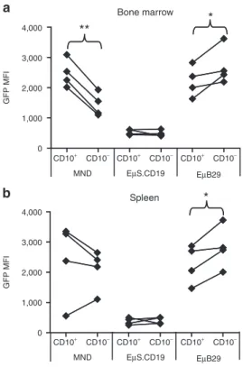
The cell surface marker, CD10, is expressed specifically on immature human B cells.39 We therefore evaluated relative eGFP
expression in immature CD10+ versus more mature CD10− CD19+
BM B cells in recipient NSG mice. EμB29-eGFP LV recipients exhibited a progressive increase in eGFP MFI in CD10− com-pared to CD10+ B cells (Figure 6a). In contrast, little difference was observed in EμS.CD19 recipients, and MND LV recipients exhibited a decline in eGFP MFI in CD10− cells. Immature, splenic CD24hiCD38hi B cells in NSG recipients also express CD10,40
con-sistent with the presence of recent BM emigrant B cells. Splenic B cells in EμB29-eGFP LV recipients exhibited a similar increase in eGFP MFI in mature CD10− compared to immature CD10+ B
Pro Pre Immature Transitional Mature
GFP FSC MND EµS.CD19 EµB29 a 1,000 800 600 400 200 0 1,000 800 600 400 200 0 1,000 800 600 400 200 0 1,371 946 800 741 486 153 93.5 111 174 165 319 386 495 571 589 100 101 102 103 104100 101 102 103 104100 101 102 103 104100 101 102 103 104100 101 102 103 104 b
Pro Pre Immature Transitional Mature Pro Pre Immature Transitional Mature Pro Pre Immature Transitional Mature
MND 0 250 500 750 1,000 1,250 1,500 0 250 500 750 1,000 1,250 1,500 0 250 500 750 1,000 1,250 1,500 GFP MFI EµS.CD19 EµB29 MND 0 100 200 300 400 500 F
old change in MFI
T1 T2 FM MZ-MZP T1 T2 FM MZ-MZP EµS.CD19 0 100 200 300 400 500 600 700 T1 T2 FM MZ-MZP 0 100 200 300 400 500 600 EµB29 MZ-MZP T2 FM T1 CD24 C D 21 c 100 100 104 104 103 103 102 102 101 101
Figure 4 eµB29 facilitates higher transgene expression during B cell maturation. BM from chimeras described in Figure 3 were stained with antibodies against B220, CD43, IgM, IgD, Ly5.1 and Ly5.2 and B cell subsets were identified as: CD43hiIgM−IgD− (pro-B); CD43loIgM−IgD−
(pre-B); CD43−IgM+IgDlo (immature); CD43−IgM+IgDint (transitional); and CD43−IgM+IgDhi (mature) B cells. (a) FACS plots show representative MFI of
eGFP in BM B cell subsets from one mouse per condition. Numbers in each plot show MFI for eGFP+ cells. (b) Graphs showing relative MFI of eGFP in B cell subsets in all mice from one experiment (3–6 mice per group). Data shown are representative of two independent experiments. (c) Splenic B220+ B cells were subdivided into developmental subsets based upon CD21 and CD24 expression (as in left panel) identify transitional 1 (T1), transitional 2 (T2), follicular mature (FM), and marginal zone/marginal zone precursors (MZ-MZP) B cells. MFI of eGFP expression in each subset is shown for individual mice transduced with each LV. BM, bone marrow; Btk, Bruton’s tyrosine kinase; eGFP, enhanced green fluorescent protein; FACS, fluorescence-activated cell sorting; Ig, immunoglobulin; LV, lentiviral vectors; MFI, mean fluorescent intensity; MND, myeloproliferative sarcoma virus enhancer, negative control region deleted, dl587rev primer-binding site substituted.
cells (Figure 6b) whereas no increase in transgene expression was observed in EμS.CD19 or MND LV recipients. Together, these data demonstrate that EμB29 LV mediate high-level B predominant expression in primary human B cells and that expression increases with B-lineage maturation.
eµBtkp lV mediates high-level expression primary B cells as well as modest activity in myeloid subsets
LV containing key endogenous regulatory elements may provide improved safety with regard to nonspecific transgene expression.41
Thus, based upon the potential goal of treating the B-lineage
c B cells Myeloid cells
5,000 4,000 3,000 2,000 1,000 0 5,000 4,000 3,000 2,000 1,000 0 5,000 4,000 3,000 2,000 1,000 0 T cells GFP MFI N = 7 N = 8 N = 7 ** ** ** ** **
MND EµS.CD19 EµB29 MND EµS.CD19 EµB29 MND EµS.CD19 EµB29
d
N = 9 N = 11 N = 11
MND EµS.CD19 EµB29 MND EµS.CD19 EµB29 MND EµS.CD19 EµB29 0 500 1,000 1,500 2,000 2,500 0 500 1,000 1,500 2,000 2,500 0 500 1,000 1,500 2,000 2,500 F
old change in MFI
*** * * *** ** N = 9 N = 11 N = 11
MND EµS.CD19 EµB29 MND EµS.CD19 EµB29 MND EµS.CD19 EµB29 0.0 0.5 1.0 1.5 2.0 Ratio to B cell e xpression T cells Myeloid cells * * *** *** e 0.0 0.3 0.6 0.9 1.2 1.5 Viral co py nu m be r N = 6 N = 6 N = 7 f a GFP C D 45
B cells T cells Myeloid cells
MND EµS.CD19 EµB29 100 101 102 103 104 100 101 102 103104 100 101 102 103104 100 101 102 103 104 100 101 102 103 104 100 101 102 103 104 b CD45+ engraftment MND EµB29 EµCD19 0 25 50 75 100 % of liv e cells N = 9 N = 11 N = 11 2,693 3,181 4,657 470 131 80.2 195 537 1,879
Figure 5 eµB29 drives high-level, lineage predominant eGFP expression in primary human B cells. Human cord blood CD34+ cells were trans-duced with MND-eGFP, EμS.CD19-eGFP, or EμB29-eGFP LV and transplanted into irradiated NSG recipient mice. Engrafted recipients were sacrificed 20 weeks after transplantation for analysis of human cells. Spleen cells were stained with CD45 to identify human cells with hematopoietic subsets defined as B cells (B220+CD4−CD8− CD33−), T cells (B220−CD4+ or CD8+, CD33−), and myeloid cells (B220−CD4−CD8−CD33+), respectively. (a) Representative FACS plots showing eGFP expression in splenic populations from one mouse per condition. Numbers show MFI for eGFP+ cells. (b) Level of human cell engraftment in the spleen depicted as the percentage of live (DAPI−) cells that express the human leukocyte antigen, CD45. (c) Graph showing MFI of eGFP in human hematopoietic subsets with data from three experiments (7–8 mice per vector). (d) Graphs showing rela-tive fold change in MFI levels between eGFP− and eGFP+ cells with data pooled from four experiments. (e) Graphical representation of the ratio of eGFP expression in T versus B cells or myeloid verse B cells. (f) Viral copy number in spleen cells. BM, bone marrow; Btk, Bruton’s tyrosine kinase; eGFP, enhanced green fluorescent protein; FACS, fluorescence-activated cell sorting; Ig, immunoglobulin; LV, lentiviral vectors; MFI, mean fluorescent intensity; MND, myeloproliferative sarcoma virus enhancer, negative control region deleted, dl587rev primer-binding site substituted.
immune disorder, XLA, we generated LV containing regulatory elements derived from the human BTK locus. As described above (Figure 1), we generated Btkp and EμBtkp LV and compared these constructs with our best-performing, B-lineage expression vector, EμB29 LV, in murine and human-based in vivo models. First, we established mouse BM chimeras using HSC transduced with EμB29-eGFP, Btkp-eGFP, or EμBtkp-eGFP LV using the same methods as Figure 3. Recipient mice were sacrificed at 25 weeks post-transplant and the relative percentage and intensity of eGFP expression assessed within various BM (data not shown) and splenic populations (Figure 7). As in Figure 3, due to dif-ferences in the 488-nm argon laser voltage settings, the results from two independent experiments are shown (Figure 7b,c). Again, while overall eGFP MFI differed between experiments, we observed consistent results with respect to the panel of LV vectors. Representative FACS plots for both experiments are also shown in
Supplementary Figure S2 (as experiment #3 and 4, respectively).
EμBtkp LV-mediated high-level eGFP expression in splenic B cells, although by comparison EμB29 LV showed a slightly higher MFI in most animals (Figure 7a–c). In contrast, expression in B cells using Btkp LV was tenfold lower level than Eμ enhancer containing LV. As anticipated, only low-level eGFP expression
was present in T cells. Consistent with its lower activity in B cells, the relative T:B cell expression ratio was ~0.35 (approximately threefold less) for Btkp versus ~0.05 (~20-fold less) for EμBtkp and EμB29 (Figure 7d). Consistent with the expression pattern of endogenous Btk, Btkp LV mediated similar levels of eGFP in B and myeloid cells. This feature was most evident when the MFI of eGFP in monocytes or neutrophils was analyzed as a ratio rela-tive to the MFI in B cells (Figure 7d). The monocyte:B and the neutrophil:B expression ratios for Btkp LV were close to 1.0. An increase in myeloid expression ratio was also evident, albeit to a lesser degree, with EμBtkp when compared to EμB29 LV. In con-trast, EμB29 expressed eGFP at significantly lower levels in mono-cytes or neutrophils versus B cells. All recipient animals exhibited similar viral copy numbers (Figure 7e).
To assess LV function in human primary cells, cord blood CD34+ HSC were transduced with EμB29-eGFP, Btkp-eGFP, or EμBtkp-eGFP LV and transplanted into NSG recipient mice. Overall, the expression pattern in splenic-derived human cells mirrored those from the murine chimeric setting (Figure 8). EμBtkp LV drove high-level eGFP expression in human B cells whereas in Btkp LV eGFP expression was at least fivefold lower. Combined data from several experiments showed high-level, albeit variable, B cell expression for both EμB29 and EμBtkp LV (Figure 8b). T cell expression was very low in all recipients with a B:T cell expression ratio as high as 60 in some EμBtkp recipients. In contrast to EμB29, EμBtkp LV also led to intermediate levels of transgene expression in monocytes. However, because expression was variable this data did not reach statistical significance for the number of animals studied. As in murine studies, Btkp LV exhib-ited consistent, low-level eGFP expression in both B cells and monocytes. All recipient animals exhibited similar average viral copy numbers (Figure 8c).
Collectively, these data demonstrate that EμBtkp LV mediates high-level transgene expression in primary murine and human B cells, presumably facilitated by the Eμ element; and also drives intermediate levels of transgene expression in myeloid cells in a manner analogous to the endogenous Btk promoter.
dIscussIon
Successful LV-based HSC gene therapy requires efficient gene trans-fer into multipotent HSC followed by sustained, therapeutic levels of transgene expression within key target cell populations. This study focused on developing LV that facilitate gene expression in developing and mature B-lineage cells for use in candidate B-lineage disorders. Our data demonstrate efficient, B cell-predominant gene expression using LV vectors containing two distinct promoter/ enhancer combinations. Our initial studies focused on the com-posite vector, EμB29. This construct mediated high-level reporter transgene expression in multiple human B cell lines and in both human and mouse primary B cells derived from transduced HSC. In contrast to previous observations showing highly variable B-lineage expression following HSC transduction using γ retroviral vectors,42
cell differentiation did not lead to appreciable evidence for silenc-ing or variegation of EμB29 LV. Consistent with this interpretation, sustained eGFP expression was observed in both immature and mature B cells for at least 20 weeks despite low-level viral marking (<1.5 copies/cell). Notably, EμB29 LV exhibited significantly higher
GFP MFI Bone marrow CD10+ CD10− CD10+ CD10− CD10+ CD10− 4,000 3,000 2,000 1,000 0 4,000 3,000 2,000 1,000 0 MND EµS.CD19 EµB29 CD10+ CD10− CD10+ CD10− CD10+ CD10− MND EµS.CD19 EµB29 GFP MFI Spleen a b ** * *
Figure 6 eµB29 drives higher eGFP expression in mature versus immature human B cells. Splenic and BM cells were isolated from NSG recipients as shown in Figure 5. Cells were stained with CD45 and CD19 to identify engrafted human B cells and with CD10 to distinguish immature (CD10+) versus more mature (CD10−) B cells. (a–b) Graphs showing change in MFI of eGFP in immature versus mature B cells derived from (a) BM or (b) spleen in mice from one experiment (3–4 mice per group). Data shown are representative of one of three indepen-dent experiments. BM, bone marrow; eGFP, enhanced green fluorescent protein; MFI, mean fluorescent intensity; MND, myeloproliferative sar-coma virus enhancer, negative control region deleted, dl587rev primer-binding site substituted.
levels of reporter gene expression than EμS.CD19 LV, even though both LV contained minimal promoters activated at nearly identical points following B-lineage commitment. EμB29 LV expression also increased progressively in parallel with BM B cell differentiation in
both murine and human models and was maintained in the periph-eral B lymphoid compartment. Expression levels were further increased during splenic transitional development, leading to high-est expression within mature murine marginal zone and follicular
EµB29 EµBtkp Btkp EµB29 EµBtkp Btkp EµB29 EµBtkp Btkp 0.0 0.5 1.0 1.5 Ratio to B cell e xpression
T cells Monocytes Neutrophils
N = 6 N = 6 N = 6 ** ** ** *** *** ** d EµB29 EµBtkp Btkp N = 6 N = 6 N = 6 0.0 0.2 0.4 0.6 0.8 1.0 1.2 1.4 1.6 1.8 2.0 2.2 Cop y nu m be r e
b Exp#1 B cells T cells Monocytes Neutrophils
N = 4 N = 3 N = 3
EµB29 EµBtkp Btkp EµB29 EµBtkp Btkp EµB29 EµBtkp Btkp EµB29 EµBtkp Btkp 30,000 20,000 10,000 0 MFI of GFP 3,000 2,000 1,000 3,000 2,000 1,000 3,000 2,000 1,000 0 *** ** * * N = 2 N = 3 N = 3 Exp#2 c
EµB29 EµBtkp Btkp EµB29 EµBtkp Btkp EµB29 EµBtkp Btkp EµB29 EµBtkp Btkp
MFI of GFP 0 20,000 40,000 60,000 0 2,000 4,000 6,000 8,000 10,000 0 10,000 20,000 30,000 40,000 0 2,000 4,000 6,000 8,000 10,000 ** * *** ** * ** ** a GFP FCS
B cells T cells Monocytes Neutrophils
EµBtkp Btkp EµB29 0 102 103 104 105 0 102 103 104 105 0 102 103 104 105 0 102 103 104 105 0 50K 100K 150K 200K 250K 0 50K 100K 150K 200K 250K 0 50K 100K 150K 200K 250K 21,815 1,117 911 856 2,063 2,473 769 14,744 1,613 488 1,589 1,808
Figure 7 eµBtkp drives high-level eGFP expression in primary murine B cells and also exhibits modest activity in myeloid cells. Mouse BM chi-meras were generated, as in Figure 3, using Lin− cells transduced with EμB29-eGFP, EμBtkp-eGFP, or Btkp-eGFP LV. Recipient animals were sacrificed 25 weeks after transplantation. (a) Representative FACS plots showing eGFP expression in splenic populations. Numbers indicate MFI for eGFP+ cells. (b) Graphs display eGFP MFI for splenic subsets in all animals from the experiment shown in a (3–4 mice per group). (c) Graphs show eGFP MFI all animals from a second independent experiment (2–3 mice per group) (d) Graphical representation of the ratio of eGFP MFI in T cells, monocytes, or neutrophils versus B cells. (e) Viral copy number in spleen cells. Combined data shown in d–e are derived from experiments shown in b and c. BM, bone marrow; Btk, Bruton’s tyrosine kinase; eGFP, enhanced green fluorescent protein; FACS, fluorescence-activated cell sorting; LV, lentiviral vectors; MND, myeloproliferative sarcoma virus enhancer, negative control region deleted, dl587rev primer-binding site substituted.
mature B cells, and CD10− human B cells, respectively. By con-trast, EμS.CD19 LV exhibited lower level, stable expression levels throughout BM and peripheral B cell development.
Together these data suggest that EμB29 LV might be useful for clinical disorders where progressively higher levels of transgene expression are required in developing and mature B cells. Although we observed some variability in expression levels of eGFP driven by EμB29 LV in both the murine and human stem cell models, the overall trend for higher-level transgene expression (compared
to EμS.CD19) suggests that this promoter would be a superior choice for B cell-specific expression. Notably, previous work has shown that subendogenous Btk expression levels are insufficient to mediate functional rescue of mature B cells Btk-deficient mice.23,24 Based on these ideas, we recently evaluated ex-vivo gene
therapy in a murine model of XLA using the EμB29 LV to deliver human Btk. This approach lead to near-complete developmental and functional B cell rescue.43 Importantly, our findings paralleled
earlier work from our group using a highly active, ubiquitously
c B cells T cells Myeloid cells
0 200 400 600 800 1,000 0 250 500 750 1,000 0 500 1,000 1,500 2,000 2,500 F
old change in MFI
*
*
EµB29 EµBtkp Btkp EµB29 EµBtkp Btkp EµB29 EµBtkp Btkp
N = 10 N = 5 N = 5 d 0.0 0.3 0.6 0.9 1.2 1.5 1.8 Viral co py nu m be r EµB29 EµBtkp Btkp N = 8 N = 5 N = 5 a GFP C D4 5
B cells T cells Myeloid cells
EµB29 EµBtkp Btkp 0 102 103 104 105 0 102 103 104 105 0 102 103 104 105 0 103 104 105 0 103 104 105 0 103 104 105 CD45+ engraftment EµB29 EµBtkp Btkp 0 25 50 75 100 P ercentage of liv e cells N = 10 N = 5 N = 5 b 1.57 × 10⁵ 1.47 × 10⁵ 7,688 1,095 2,472 4,723 16,092 26,709 5,511
Figure 8 eµBtkp drives high-level transgene expression in primary human B cells and also exhibits activity in myeloid cells. Human cord blood CD34+ cells were transduced with EμB29-eGFP, EμBtkp-eGFP, or Btkp-eGFP LV and transplanted into irradiated NSG recipient mice (as in Figure 6). Engrafted recipients were sacrificed 20 weeks after transplantation for analysis. (a) Representative FACS plots showing eGFP expression in splenic cells with numbers indicating MFI in eGFP+ cells. (b) Level of human cell engraftment in the spleen. (c) Graphs showing relative fold change in MFI levels between eGFP− and eGFP+ cells in CD45+ human hematopoietic subsets (data pooled from four experiments) (d) Relative viral copy number in spleen cells in three experimental cohorts. BM, bone marrow; Btk, Bruton’s tyrosine kinase; eGFP, enhanced green fluorescent protein; FACS, fluorescence-activated cell sorting; LV, lentiviral vectors; MFI, mean fluorescent intensity; MND, myeloproliferative sarcoma virus enhancer, negative control region deleted, dl587rev primer-binding site substituted.
expressed, murine stem cell virus-based γ retroviral vector.5 Thus,
equivalent clinical efficacy was achieved via this unique B-lineage targeted, potentially safer, LV delivery platform.
Despite its efficacy in the murine XLA model, the EμB29 enhancer/promoter may not be ideal for cell-specific Btk expres-sion. Notably, while its functional role is most evident in B cells, Btk is also expressed in all myeloid subsets and megakaryocytes; and Btk also impacts signaling via a range of cell surface receptors.3,44
Recent work has demonstrated a requirement for Btk kinases in Toll-like receptor signals45–47 implying that Btk deficiency may
impact innate, as well as adaptive, immune responses in vivo. Based on these considerations, we generated LV constructs containing the endogenous BTK promoter. As anticipated, Btkp LV exhibited sustained, albeit low-level activity in primary murine and human B and myeloid cells. Importantly, addition of the Eμ enhancer lead to a marked increase in relative B-lineage expression, with activ-ity levels closely mimicking EμB29 LV in both murine and human primary B cells. In contrast to EμB29 LV, however, EμBtkp LV also retained modest transgene expression in myeloid populations sug-gesting this LV may more faithfully mimic endogenous Btk expres-sion and potentially rescue Btk-dependent myeloid activity.
Taken together, our data indicate that EμB29 LV provides an efficient platform for high-level mammalian B cell gene expres-sion. Furthermore, these data also suggest that EμBtkp LV may represent an optimal system for regulated Btk gene delivery in XLA. This latter possibility is currently being investigated using murine models of XLA. Finally, it is important to note that we observed no evidence for Eμ enhancer-dependent mutagenesis in this study or in >200 primary/secondary/or tertiary recipient mice treated to date using these LV for Btk delivery. Formal studies, however, using genome targeted integration48 and BM
immortal-ization assays49 will be required to more fully address the potential
risk(s) of these new LV constructs. MaterIals and Methods
LV constructs and viral production. The parental pRRL-SIN-cppt-PGK-eGFP-WPRE vector9 was obtained from Didier Trono (Addgene plasmid
#12252) and the internal PGK promoter was replaced to generate pRRL-EµB29-eGFP, pRRL-EμBtkp-eGFP, pRRL-MND-eGFP, or pRRL-EµCD19-eGFP. MND is a retroviral long-terminal repeats-derived promoter.50 The
immunoglobulin μ heavy chain enhancer Eµ is comprised of the 1-kb murine Ig heavy chain intronic core enhancer and the 5′ and 3′ matrix attachment region elements27 and was added to all other promoters except
MND and Btkp alone. B29 contained the minimal promoter element derived from the B29 gene comprising 184 bp of the murine promoter including −166 to +18 from the major transcriptional start site.26 S.CD19
and L.CD19 contained the minimal (204 bp) versus extended (1,224 bp), human CD19 promoter element and adjacent upstream enhancer region, respectively.19 Btkp contains a 788 bp sequence located directly upstream
of exon 1 within the human BTK locus. LV were produced by transient transfection of 293T cells using PEI method, concentrated by low-speed centrifugation, and titered as described previously.43
Cell culture and transduction. The following cell lines were utilized for lentiviral transduction experiments; Blin and Nalm6 (gift from Dr Tucker LeBien, University of Minnesota, Minneapolis, MN), REH, Ramos, Raji, Jurkat, and K562 (ATCC, Manassas, VA). All cells were maintained in complete RPMI (RPMI plus l- glutamine, 10% fetal bovine serum, 0.1% β-mercaptoethanol and 10 μmol/l HEPES) media and transduced with the indicated lentiviral construct overnight with 4 μg/ml polybrene at variable
multiplicity of infection’s to obtain similar transduction rate (from 10 to 15%). Cells were incubated for 4 days and the MFI of eGFP was deter-mined via flow cytometer.
Murine BM transplantation. C57BL/6 mice were bred and maintained in the specific pathogen-free barrier facilities at Seattle Children’s Research Institute. Animal studies were carried out according to the guidelines of Seattle Children’s Research Institute Institutional Animal Care and Use Committee. BM from 6 to 8 weeks mice was harvested and Lin− cells were obtained using EasySep Mouse Hematopoietic Progenitor Cell Enrichment kit according to manufacture’s instruction (StemCell Technologies, Vancouver, BC), transduced overnight in media (StemSpan SFEM, StemCell Technologies) containing mSCF and mTPO (10 μg/ml; PeproTech, Rocky Hills, NJ) with the indicated LV overnight with 4 μg/ ml polybrene. Cells were washed, counted, and 1 × 10⁶ transduced cells
were transplanted into mice receiving 2 × 500 Gy total body irradiation conditioning. Recipient mice were evaluated by serial peripheral blood analyses beginning at 5 weeks, and cohorts were sacrificed at 24–26 weeks for detailed analyses.
Cord blood CD34+ cell isolation, transduction, and transplantation of NSG recipients. Human umbilical cord blood was obtained from the Puget Sound Blood Center cord blood donor program and CD34+ cells were isolated using Miltenyi CD34+ beads (Miltenyi, Biotec, Germany) according to the manufacturer’s instructions. Purity of CD34+Lineage− cells was >90% in all experiments. CD34+ cells were incubated for 8–12 hours in IMDM with human cytokines (100 ng/ml SCF, TPO, and FLT3-L; PeproTech) and 5% fetal bovine serum. After incubation, cells were trans-duced with the indicated LV at an multiplicity of infection of 10 for 8 hours in media with cytokines and 4 μg/ml polybrene. Transduced CD34+ cells were washed and transplanted into NOD/SCID/cγ−/− (NSG) mice. For all
experiments, 6–12 weeks recipient mice were conditioned with 250 cGy total body irradiation and transplanted with 2–3 × 10⁵ transduced, or mock
transduced, CD34+ cells via retro-orbital injection. Peripheral blood sam-ples were obtained from recipients 8 weeks after transplantation and the level of HSC engraftment was accessed via CD45 expression. Cohorts were sacrificed at 20–30 weeks for detailed FACS analysis of BM and spleen. Flow cytometric analysis. Single-cell suspensions prepared from BM or spleen were incubated in phosphate-buffered saline with 3% fetal calf serum and fluorochrome-conjugated antibodies for 20 minutes at 4 °C, and washed twice before resuspension in phosphate-buffered saline with 3% fetal calf serum for acquisition. The following antibodies were used: antihuman CD19, CD33, CD45, IgM, IgD, 38, CD24, CD10, CD34, anti-mouse CD3, CD4, CD8, B220, CD21, CD24, CD43, IgM, IgD, Gr-1, CD11b, and CD45.1 (BD Biosciences, San Jose, CA and eBioscience, San Diego, CA). Cell analysis were performed using FACSCalibur, Aria II, and LSRII flow cytometers (BD Biosciences) and FlowJo software (Tree Star, Ashland, OR).
Viral copy number determination. Quantification of viral integration was adapted from a previously described protocol.5 Briefly, genomic DNA was
obtained from spleen or total BM from sacrificed animals. Gag-specific primer/probe sets were used to amplify viral integrations in both mouse and human cells. To normalize DNA content in mouse cells, a murine β-actin primer/probe set was used; in human cells, a human RNaseP primer probe set was used (Applied Biosytems, Carlsbad, CA). Viral-integration number was determined via a standard curve established using genomic DNA extracted from A20 murine B cell line or Nalm6 human B cell line, each containing a single LV integration.
Statistical analysis. Statistical analyses of relative eGFP marking and viral copy numbers were carried out using a two-tailed paired Student’s t test comparing the indicated cell populations from the groups of experimental mice or cell lines.
suPPleMentarY MaterIal
Figure S1. Comparison of EμS.CD19, EμL.CD19, and EμB29 LV activ-ity in human B cell lines.
Figure S2. Representative FACS plots from independent experiments shown in Figures 3 and 7.
acKnoWledGMents
We thank Jit Khim for excellent technical assistance with murine trans-plantation studies, Dr Maria Garcia-Lloret (UCLA) for initial work with first-generation EμB29-GFP vectors, and Angel Hui for administra-tive support. This work was supported by a CRI fellowship award (to B.D.S.), CMB Training grant, GM07270 (to A.A.), and NIH R01 awards CA081140 and HL075453 (to D.J.R.). The authors declared no conflict of interest.
reFerences
1. Gaspar, HB, Howe, S and Thrasher, AJ (2003). Gene therapy progress and prospects: gene therapy for severe combined immunodeficiency. Gene Ther 10: 1999–2004. 2. Tsukada, S, Saffran, DC, Rawlings, DJ, Parolini, O, Allen, RC, Klisak, I et al. (1993).
Deficient expression of a B cell cytoplasmic tyrosine kinase in human X-linked agammaglobulinemia. Cell 72: 279–290.
3. Smith, C and Witte, ON (1999). Functional aspects of Btk signaling in Primary
Immunodeficiency Diseases, a Molecular and Genetic Approach. Oxford University Press.
4. Moreau, T, Calmels, B, Barlogis, V, Michel, G, Tonnelle, C and Chabannon, C (2007). Potential application of gene therapy to X-linked agammaglobulinemia. Curr Gene
Ther 7: 284–294.
5. Yu, PW, Tabuchi, RS, Kato, RM, Astrakhan, A, Humblet-Baron, S, Kipp, K et al. (2004). Sustained correction of B-cell development and function in a murine model of X-linked agammaglobulinemia (XLA) using retroviral-mediated gene transfer. Blood
104: 1281–1290.
6. Uchida, N, Sutton, RE, Friera, AM, He, D, Reitsma, MJ, Chang, WC et al. (1998). HIV, but not murine leukemia virus, vectors mediate high efficiency gene transfer into freshly isolated G0/G1 human hematopoietic stem cells. Proc Natl Acad Sci USA 95: 11939–11944.
7. Case, SS, Price, MA, Jordan, CT, Yu, XJ, Wang, L, Bauer, G et al. (1999). Stable transduction of quiescent CD34(+)CD38(-) human hematopoietic cells by HIV-1-based
lentiviral vectors. Proc Natl Acad Sci USA 96: 2988–2993.
8. Naldini, L, Blömer, U, Gage, FH, Trono, D and Verma, IM (1996). Efficient transfer, integration, and sustained long-term expression of the transgene in adult rat brains injected with a lentiviral vector. Proc Natl Acad Sci USA 93: 11382–11388. 9. Zufferey, R, Dull, T, Mandel, RJ, Bukovsky, A, Quiroz, D, Naldini, L et al. (1998).
Self-inactivating lentivirus vector for safe and efficient in vivo gene delivery. J Virol 72: 9873–9880.
10. Schröder, AR, Shinn, P, Chen, H, Berry, C, Ecker, JR and Bushman, F (2002). HIV-1 integration in the human genome favors active genes and local hotspots. Cell 110: 521–529.
11. Hacein-Bey-Abina, S, von Kalle, C, Schmidt, M, Le Deist, F, Wulffraat, N, McIntyre, E
et al. (2003). A serious adverse event after successful gene therapy for X-linked severe
combined immunodeficiency. N Engl J Med 348: 255–256.
12. Howe, SJ, Mansour, MR, Schwarzwaelder, K, Bartholomae, C, Hubank, M, Kempski, H
et al. (2008). Insertional mutagenesis combined with acquired somatic mutations
causes leukemogenesis following gene therapy of SCID-X1 patients. J Clin Invest 118: 3143–3150.
13. Rivella, S, May, C, Chadburn, A, Rivière, I and Sadelain, M (2003). A novel murine model of Cooley anemia and its rescue by lentiviral-mediated human β-globin gene transfer. Blood 101: 2932–2939.
14. Cartier, N, Hacein-Bey-Abina, S, Bartholomae, CC, Veres, G, Schmidt, M, Kutschera, I
et al. (2009). Hematopoietic stem cell gene therapy with a lentiviral vector in X-linked
adrenoleukodystrophy. Science 326: 818–823.
15. Pawliuk, R, Westerman, KA, Fabry, ME, Payen, E, Tighe, R, Bouhassira, EE et al. (2001). Correction of sickle cell disease in transgenic mouse models by gene therapy. Science
294: 2368–2371.
16. Indraccolo, S, Minuzzo, S, Roccaforte, F, Zamarchi, R, Habeler, W, Stievano, L et al. (2001). Effects of CD2 locus control region sequences on gene expression by retroviral and lentiviral vectors. Blood 98: 3607–3617.
17. Cui, Y, Golob, J, Kelleher, E, Ye, Z, Pardoll, D and Cheng, L (2002). Targeting transgene expression to antigen-presenting cells derived from lentivirus-transduced engrafting human hematopoietic stem/progenitor cells. Blood 99: 399–408. 18. Gough, PJ and Raines, EW (2003). Gene therapy of apolipoprotein E-deficient mice
using a novel macrophage-specific retroviral vector. Blood 101: 485–491. 19. Moreau, T, Bardin, F, Imbert, J, Chabannon, C and Tonnelle, C (2004). Restriction of
transgene expression to the B-lymphoid progeny of human lentivirally transduced CD34+ cells. Mol Ther 10: 45–56.
20. Lutzko, C, Senadheera, D, Skelton, D, Petersen, D and Kohn, DB (2003). Lentivirus vectors incorporating the immunoglobulin heavy chain enhancer and matrix attachment regions provide position-independent expression in B lymphocytes. J Virol
77: 7341–7351.
21. Moreau, T, Barlogis, V, Bardin, F, Nunes, JA, Calmels, B, Chabannon, C et al. (2008). Development of an enhanced B-specific lentiviral vector expressing BTK: a tool for gene therapy of XLA. Gene Ther 15: 942–952.
22. Taher, TE, Tulone, C, Fatah, R, D’Acquisto, F, Gould, DJ and Mageed, RA (2008). Repopulation of B-lymphocytes with restricted gene expression using haematopoietic stem cells engineered with lentiviral vectors. Gene Ther 15: 998–1006.
23. Faust, EA, Rawlings, DJ, Saffran, DC and Witte, ON (1995). Development of btk transgenic mice. Curr Top Microbiol Immunol 194: 363–370.
24. Satterthwaite, AB, Cheroutre, H, Khan, WN, Sideras, P and Witte, ON (1997). Btk dosage determines sensitivity to B cell antigen receptor cross-linking. Proc Natl Acad
Sci USA 94: 13152–13157.
25. Robbins, PB, Yu, XJ, Skelton, DM, Pepper, KA, Wasserman, RM, Zhu, L et al. (1997). Increased probability of expression from modified retroviral vectors in embryonal stem cells and embryonal carcinoma cells. J Virol 71: 9466–9474.
26. Thompson, AA, Wood, WJ Jr, Gilly, MJ, Damore, MA, Omori, SA and Wall, R (1996). The promoter and 5’ flanking sequences controlling human B29 gene expression.
Blood 87: 666–673.
27. Banerji, J, Olson, L and Schaffner, W (1983). A lymphocyte-specific cellular enhancer is located downstream of the joining region in immunoglobulin heavy chain genes. Cell
33: 729–740.
28. Hagman, J and Grosschedl, R (1994). Regulation of gene expression at early stages of B-cell differentiation. Curr Opin Immunol 6: 222–230.
29. Hermanson, GG, Eisenberg, D, Kincade, PW and Wall, R (1988). B29: a member of the immunoglobulin gene superfamily exclusively expressed on β-lineage cells. Proc
Natl Acad Sci USA 85: 6890–6894.
30. Rohrer, J, Parolini, O, Belmont, JW, Conley, ME and Parolino O [corrected to Parolini, O (1994). The genomic structure of human BTK, the defective gene in X-linked agammaglobulinemia. Immunogenetics 40: 319–324.
31. Oeltjen, JC, Malley, TM, Muzny, DM, Miller, W, Gibbs, RA and Belmont, JW (1997). Large-scale comparative sequence analysis of the human and murine Bruton’s tyrosine kinase loci reveals conserved regulatory domains. Genome Res 7: 315–329.
32. Müller, S, Sideras, P, Smith, CI and Xanthopoulos, KG (1996). Cell specific expression of human Bruton’s agammaglobulinemia tyrosine kinase gene (Btk) is regulated by Sp1- and Spi-1/PU.1-family members. Oncogene 13: 1955–1964.
33. Himmelmann, A, Thevenin, C, Harrison, K and Kehrl, JH (1996). Analysis of the Bruton’s tyrosine kinase gene promoter reveals critical PU.1 and SP1 sites. Blood 87: 1036–1044.
34. Rohrer, J and Conley, ME (1998). Transcriptional regulatory elements within the first intron of Bruton’s tyrosine kinase. Blood 91: 214–221.
35. Yu, L, Mohamed, AJ, Simonson, OE, Vargas, L, Blomberg, KE, Björkstrand, B et
al. (2008). Proteasome-dependent autoregulation of Bruton tyrosine kinase (Btk)
promoter via NF-kappaB. Blood 111: 4617–4626.
36. Pillai, S, Cariappa, A and Moran, ST (2005). Marginal zone B cells. Annu Rev Immunol
23: 161–196.
37. Ito, M, Hiramatsu, H, Kobayashi, K, Suzue, K, Kawahata, M, Hioki, K et al. (2002). NOD/SCID/γ©(null) mouse: an excellent recipient mouse model for engraftment of human cells. Blood 100: 3175–3182.
38. Hiramatsu, H, Nishikomori, R, Heike, T, Ito, M, Kobayashi, K, Katamura, K et al. (2003). Complete reconstitution of human lymphocytes from cord blood CD34+ cells
using the NOD/SCID/γcnull mice model. Blood 102: 873–880.
39. Blom, B and Spits, H (2006). Development of human lymphoid cells. Annu Rev
Immunol 24: 287–320.
40. Watanabe, Y, Takahashi, T, Okajima, A, Shiokawa, M, Ishii, N, Katano, I et al. (2009). The analysis of the functions of human B and T cells in humanized NOD/shi-scid/ γc(null) (NOG) mice (hu-HSC NOG mice). Int Immunol 21: 843–858. 41. Nienhuis, AW, Dunbar, CE and Sorrentino, BP (2006). Genotoxicity of retroviral
integration in hematopoietic cells. Mol Ther 13: 1031–1049.
42. Klug, CA, Cheshier, S and Weissman, IL (2000). Inactivation of a GFP retrovirus occurs at multiple levels in long-term repopulating stem cells and their differentiated progeny. Blood 96: 894–901.
43. Kerns, HM, Ryu, BY, Stirling, BV, Sather, BD, Astrakhan, A, Humblet-Baron, S et al. (2010). B cell-specific lentiviral gene therapy leads to sustained B-cell functional recovery in a murine model of X-linked agammaglobulinemia. Blood 115: 2146–2155.
44. Plebani, A, Soresina, A, Rondelli, R, Amato, GM, Azzari, C, Cardinale, F et al.; Italian Pediatric Group for XLA-AIEOP. (2002). Clinical, immunological, and molecular analysis in a large cohort of patients with X-linked agammaglobulinemia: an Italian multicenter study. Clin Immunol 104: 221–230.
45. O’Neill, LA (2008). When signaling pathways collide: positive and negative regulation of toll-like receptor signal transduction. Immunity 29: 12–20.
46. Jefferies, CA and O’Neill, LA (2004). Bruton’s tyrosine kinase (Btk)-the critical tyrosine kinase in LPS signalling? Immunol Lett 92: 15–22.
47. Jefferies, CA, Doyle, S, Brunner, C, Dunne, A, Brint, E, Wietek, C et al. (2003). Bruton’s tyrosine kinase is a Toll/interleukin-1 receptor domain-binding protein that participates in nuclear factor kappaB activation by Toll-like receptor 4. J Biol Chem
278: 26258–26264.
48. Ryu, BY, Evans-Galea, MV, Gray, JT, Bodine, DM, Persons, DA and Nienhuis, AW (2008). An experimental system for the evaluation of retroviral vector design to diminish the risk for proto-oncogene activation. Blood 111: 1866–1875. 49. Modlich, U, Navarro, S, Zychlinski, D, Maetzig, T, Knoess, S, Brugman, MH et al.
(2009). Insertional transformation of hematopoietic cells by self-inactivating lentiviral and γ retroviral vectors. Mol Ther 17: 1919–1928.
50. Halene, S, Wang, L, Cooper, RM, Bockstoce, DC, Robbins, PB and Kohn, DB (1999). Improved expression in hematopoietic and lymphoid cells in mice after transplantation of bone marrow transduced with a modified retroviral vector. Blood