Science Arts & Métiers (SAM)
is an open access repository that collects the work of Arts et Métiers Institute of
Technology researchers and makes it freely available over the web where possible.
This is an author-deposited version published in:
https://sam.ensam.eu
Handle ID: .
http://hdl.handle.net/10985/17457
To cite this version :
Daniel GEORGE, Rachele ALLENA, Yves REMOND - Cell nutriments and motility for
mechanobiological bone remodeling in the context of orthodontic periodontal ligament
deformation - Journal of Cellular Immunotherapy - Vol. 4, p.26-29 - 2018
Any correspondence concerning this service should be sent to the repository
Administrator :
archiveouverte@ensam.eu
Cell nutriments and motility for mechanobiological bone remodeling in the
context of orthodontic periodontal ligament deformation
Daniel George
a,∗, Rachele Allena
b, Yves Rémond
aaICube Laboratory, Université de Strasbourg, CNRS, 2 rue Boussingault, 67000, Strasbourg, France
bArts et Métiers ParisTech, LBM/Institut de Biomécanique Humaine Georges Charpak, 151 bd de l’Hôpital, 75013, Paris, France
Keywords: Stimulus Bone remodeling Oxygen Glucose Cell motility Periodontal ligament A B S T R A C T
Bone remodeling is a complex phenomenon during which old and new bone is continuously removed and re-placed. This phenomenon involves several processes at different length scales such as mechanical, biological, molecular, and chemicals. In the current work, we study the influence of the biological (cells) and molecular (oxygen and glucose) factors coupled with mechanical loads in order to predict bone remodeling for orthodontic treatments. A coupled theoretical mechanobiological model is proposed to extract the oxygen variation due to the deformation of the periodontal ligament leading to cell differentiation and activation. The mechan-obiological stimulus is then calculated. The model is applied on a simplified two dimensional geometry to highlight the density variations and migrations of cells and molecular factors influencing the bone remodeling process.
1. Introduction
Bone is a continually renewed living material [1]. It undergoes continual adaptation under externally applied mechanical loads as in-itially phenomenologically modeled by Wolff under the well-known Wolff's Law [2]. Many multiscale and/or multiphysics theoretical and numerical models have followed since predicting of the global kinetics of bone remodeling was tried [3–14]. However, there are still many difficulties to obtain a precise understanding of the mechanotransduc-tion processes driving this bone remodeling [15]. For example, bone density evolution is highly dependent on vascularization and nutrient supply [16–18], is difficult to comprehend due to its highly hetero-geneous structure [19–23], and depends strongly on the biology dis-tribution and activation processes inside its porous matrix [24–27].
We present here this influence for an application of the mechan-obiological couplings in orthodontic bone remodeling due the applied orthodontic forces [28,29]. The cell proliferation is activated through oxygen variation in the periodontal ligament [30–33] being partially occluded due to the applied mechanical forces. We study the variations in the supply chain of nutrients and oxygen to predict cell recruitment, proliferation and migration assuming that bone remodeling occurs by the osteoblasts proliferating with oxygen increase [30] and bone re-sorption occurs by the osteoclasts proliferating in hypoxia [31,32].
2. Model development
Bone remodeling comes via the application of a mechanobiological stimulus ΔS, defined from a variation of the mechanobiological equi-librium [8,12,14] and newly expressed [18] as:
= = S S exp( D ( )X ( ) )X dX i n i i 1 0 0 (1)
where n is the total number of external sources Si (mechanical,
biolo-gical, electrical, neurolobiolo-gical, …) involved in the remodeling process and αi are their weighting coefficients, triggered by genetic and/or
epigenetic factors, allowing to simultaneously control their impact on the overall response of the system as well as their interactions. ( )X
and X( )0 are the kinematical fields that associate to any material point
its current (X) and reference (X0) position respectively, and Di is a
characteristic distance accounting for each independent effect. The external sources Siconsidered in this work are: (i) the mechanical
en-ergy accounting for the mechanical loads sustained by the bone cells and triggering bone density evolution, (ii) the concentration of cell nutriments (here being oxygen and glucose) expressed as function of the hydrostatic pressure in specific regions of the system, and (iii) the cells activity triggered by specific levels of oxygen and glucose con-centration together with the intensity of the mechanical force applied. The cells recruiting and migration are described via two diffusion
https://doi.org/10.1016/j.jocit.2018.09.006
∗ Corresponding author.
equations [25,33] reading: = D + c t div c (1 c c) c j j j j j j j (2) = + = D jI j i 1 3 i i i (3)
wherecjis the cell density (with j being the osteoblasts or osteoclasts), t
is the time, j and j are two coefficients of proliferation and
differ-entiation respectively. The diffusion tensor D depends on the principal
strains ( )i and strain directions( )i and with jand jtwo coefficients
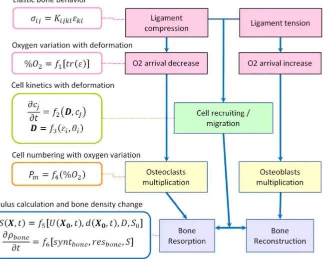
and I the identity matrix. The bone density variation in time is calcu-lated by the rates of bone synthesis and resorption respectively, de-pending on the positiveness of the defined mechanobiological stimulus. The chosen application proposes to solve the mechanobiological effects through a stepped analysis of coupled partial differential equa-tions as presented inFig. 1.
The presented schematic shows that the applied mechanical force leads to a partial compression or tension of the periodontal ligament. Through elastic mechanical behavior, a variation of oxygen con-centration is observed due to blood flow variation inside the period-ontal ligament vascularization, which has a direct impact on the os-teoblasts [30] or osteoclasts [31,32] concentration. In parallel, compression (resp. tension) of the periodontal ligament influences cells recruiting and migration [33]. The mechanical effect, together with the cellular combined effects, will then impact the calculated mechan-obiological stimulus driving the bone density variation.
The proposed schematic ofFig. 1was implemented in a simplified 2D finite element (FE) numerical model of the periodontal ligament to predict cell density variation and, sugar and glucose concentration variations. As the periodontal ligament is very thin, a simply strained 2D rectangular geometry can highlight the corresponding kinetics (see
Fig. 2).
The geometry is anchored on the left side and distributed force is applied on the right side. Biology that is initially distributed on the left side only (vivid zone) will migrate towards the right side (initially non-vivid zone). The challenge to obtain a satisfactory prediction in the bone remodeling process lies in the adequate identification and im-portance of each of the external sources and parameters used together
Fig. 1. Schematic of the stepped mechanobiological couplings leading to bone remodeling.
Fig. 2. Schematic of the 2D model used to obtain the cells and molecular
mi-gration kinetics.
Table 1
Initial cells and molecular distributions inside the geometry.
Osteoclasts concentration (%) Osteoblasts concentration (%) Oxygen concentration (%) Glucose concentration (%) Vivid zone 5 10 20 10 Non-vivid zone 0
with their mutual interactions and quantification of each of the applied individual kinetics involved in the process.
3. Results and discussion
The defined model being a strained simplified geometry under simple tension mechanical load, the expected results are the kinetics and extract cells and molecular migration between the two sides of the geometry. The initial parameters distributions are defined inTable 1.
Preliminary results are presented inFig. 3for the cells density and molecular evolutions as a function of time (defined arbitrary between 0 and 1).
For osteoclasts, only apoptosis and migration is taken into account, no proliferation. For osteoblasts, proliferation comes from osteoclasts differentiation in addition to migration. Finally, molecular (oxygen and glucose) absolute quantities are not supposed changing, only migrating geographically as a function of time and depending on the applied strain.
Overall, the results show migrations between the small strained (left) and large strained (right) area for each parameter. For osteoclasts, as no proliferation is defined, an initial migration (mid-length) is ob-served at the beginning of the analysis. But differentiation between osteoclasts to osteoblasts becomes then predominant and osteoclasts density degrades quickly to reach almost zero at the end of analysis. For osteoclasts, both migration and proliferation are observed since the start of the analysis. The maximum density reaches a value of 13.4% (left) then migration become predominant as no more osteoclasts are present to be differentiated and osteoblasts density increases more on the large strained region (right) than on the small strained one (left). Finally, both kinetics of oxygen and glucose being defined identical, the migration between the two regions is completely symmetrical and reaches equilibrium at the end of the analysis since it is supposed not being influenced by other parameters in this model.
Evolutions of these four densities (osteoblasts, osteoclasts, oxygen and glucose) impact directly the mechanobiological stimulus. More specifically, the bone remodeling process will be concentrated where these densities will be the highest at any given time of evolution. It is
therefore crucial to know their distributions as it will help to predict the bone density evolution and remodeling process.
4. Conclusion
We presented a coupled multiphysic theoretical numerical analysis integrating the mechanical and biological phenomena within a single mechanobiological stimulus influencing the bone density evolution and remodeling process. This coupled model could help predict the bone remodeling for patient specific orthodontic applications and the or-thodontist understanding and optimization of the procedure to follow for each patient's case.
References
[1] Frost HM. Bone “mass” and the “mechanostat”: a proposal. J Anat Rec
1987;219:1–9.
[2] Cowin SC. Wolff's law of trabecular architecture at remodeling equilibrium. J
Biomed Eng 1986;108(1):83–8.
[3] Carter DR, Orr TE, Fyhrie DP. Relationship between loading history and femoral
cancellous bone architecture. J Biomech 1989;22:231–44.
[4] Weinans H, Huiskes R, Grootenboer HJ. The behavior of adaptative bone
re-modeling simulation models. J Biomech 1992;25:1425–41.
[5] Ruimerman R, Hilbers P, van Rietbergen B, Huiskes R. A theoretical framework for
strain-related trabecular bone maintenance and adaptation. J Biomech
2005;38:931–41.
[6] Lekszycki T. Modeling of bone adaptation based on an optimal response hypothesis.
Meccanica 2002;37:343–54.
[7] Madeo A, Lekszycki T, Dell'Isola F. A continuum model for the bio-mechanical
in-teractions between living tissue and bio-resorbable graft after bone reconstructive
surgery. C R Mécanique 2011;339:625–40.
[8] Madeo A, George D, Lekszycki T, Nierenberger M, Rémond Y. A second gradient
continuum model accounting for some effects of micro-structure on reconstructed
bone remodeling. C R Mécanique 2012;340:575–89.
[9] Lekszycki T, Dell'Isola F. A mixture model with evolving mass densities for
de-scribing synthesis and resorption phenomena in bones reconstructed with
bio-re-sorbable materials. ZAMM 2012;92:426–44.
[10] Madeo A, George D, Rémond Y. Second gradient models for some effects of
micro-structure on reconstructed bone remodeling. Comput Meth Biomech Biomed Eng
2013;16:S260–1.
[11] Andreaus U, Giorgio I, Lekszycki T. A 2-D continuum model of a mixture of bone
tissue and bio-esorbable material for simulating mass density redistribution under
load slowly variable in time. ZAMM 2014;94:978–1000.
[12] Scala I, Spingarn C, Rémond Y, Madeo A, George D. Mechanically-driven bone remodeling simulation: application to LIPUS treated rat calvarial defects. Math
Mech Solid 2016;22(10):1976–88.
[13] Giorgio I, Andreaus U, Scerrato D, Dell'Isola F. A visco-poroelastic model of
func-tional adaptation in bones reconstructed with bio-resorbable materials.
Biomechanics Model Mechanobiol 2016;15(5):1325–43.
[14] George D, Spingarn S, Dissaux C, Nierenberger M, Abdel Rahman R, Rémond Y.
Examples of multiscale and multiphysics numerical modeling of biological tissues.
Bio Med Mater Eng 2017;28:S15–27.
[15] Lemaire T, Capiez-Lernout E, Kaiser J, Naili S, Sansalone V. What is the importance
of multiphysical phenomena in bone remodelling signals expression? A multiscale
perspective. J Mech Behav Biom Mat 2011;4(6):909–20.
[16] Bednarczyk E, Lekszycki E. A novel mathematical model for growth of capillaries
and nutrient supply with application to prediction of osteophyte onset. ZAMP (Z
Angew Math Phys) 2016;67:94.
[17] Lu Y, Lekszycki T. A novel coupled system of non-local integro-differential
equa-tions modelling Young's modulus evolution, nutrients' supply and consumption
during bone fracture healing. ZAMP (Z Angew Math Phys) 2016;67(5):111.
[18] George D, Allena R, Rémond Y. Mechanobiological stimuli for bone remodeling:
mechanical energy, cell nutriments and mobility. Comput Meth Biomech Biomed
Eng 2017;20:S91–2.
[19] Goda I, Ganghoffer JF, Czarnecki S, Wawruch P, Lewinski T. Optimal internal
ar-chitecture of femoral bone based on relaxation by homogenization and isotropic
material design. Mech Res Commun 2016;76:64–71.
[20] Rémond Y, Ahzi S, Baniassadi M, Garmestani M. Applied RVE reconstruction and
homogenization of heterogeneous materials. Wiley-ISTE978-1-84821-901-4; 2016.
[21] Bala Y, Lefevre E, Roux JP, Baron C, Lesague P, Pithioux M, et al. Pore network
microarchitecture influences human cortical bone elasticity during growth and
aging. J Mech B Bio Mat 2016;63:164–73.
[22] Sansalone V, Gagliardi D, Descelier C, Haiat G, Naili S. On the uncertainty
propa-gation in multiscale modeling of cortical bone elasticity. Comput Meth Biomech
Biomed Eng 2015;18:2054–5.
[23] Martin M, Lemaire T, Haiat G, Pivonka P, Sansalone V. A thermodynamically
consistent model of bone rotary remodeling: a 2D study. Comput Meth Biomech
Biomed Eng 2017;20(S1):127–8.
[24] Bonewald LF. The amazing osteocyte. J Bone Miner Res 2011;26:229–38.
[25] Allena R, Maini PK. Reaction-diffusion finite element model of lateral line
pri-mordium migration to explore cell leadership. Bull Math Biol
2014;76(12):3028–50.
[26] Yi W, Wang C, Liu X. A microscale bone remodeling simulation method considering
the influence of medicine and the impact of strain on osteoblast cells. FE An Des
2015;104:16–25.
[27] Lemaire T, Kaiser J, Sansalone V. Three-scale multiphysics modeling of transport
phenomena within cortical bone. Math Probl Eng 2015;2015:398970.
[28] Wagner D, Bolender Y, Rémond Y, George D. Mechanical equilibrium of forces and
moments applied on orthodontic brackets of a dental arch : correlation with lit-terature data on two and three adjacent teeth. Bio Med Mater Eng
2017;28:S169–77.
[29] Zargham A, Geramy A, Rouhi G. Evaluation of long-term orthodontic tooth
move-ment considering bone remodeling process and in the presence of alveolar bone loss
using finite element method. Orthod Waves 2016;75:85–96.
[30] Tuncay OC, Daphne Ho BS, Melissa K, Barker BS. Oxygen tension regulates
osteo-blast function. Am J Orthod Dentofacial Orthop 1994;105(5):457–63.
[31] Arnett TR, Gibbons DC, Utting JC, Orris IR, Hoebertz A, Rosendaal M, et al. Hypoxia
is a major stimulator of osteoclast formation and bone resorption. J Cell Physiol
2003;196(1):2–8.
[32] Utting JC, Robins SP, Brandao-Burch A, Orris IR, Behar J, Arnett TR. Hypoxia
in-hibits the growth, differenciation and bone-forming capacity of rat osteoblasts. Exp
Cell Res 2006;312(10):1693–702.
[33] Schmitt M, Allena R, Schouman T, Frasca S, Collombet JM, Holy X, et al. Diffusion
model to describe osteogenesis within a porous titanium scaffold. Comput Meth