HAL Id: tel-00771435
https://tel.archives-ouvertes.fr/tel-00771435
Submitted on 8 Jan 2013
HAL is a multi-disciplinary open access
archive for the deposit and dissemination of sci-entific research documents, whether they are pub-lished or not. The documents may come from teaching and research institutions in France or abroad, or from public or private research centers.
L’archive ouverte pluridisciplinaire HAL, est destinée au dépôt et à la diffusion de documents scientifiques de niveau recherche, publiés ou non, émanant des établissements d’enseignement et de recherche français ou étrangers, des laboratoires publics ou privés.
development
Filippo Maria Massa
To cite this version:
Filippo Maria Massa. The crucial roles played by HNF1β during kidney development. Human health and pathology. Université René Descartes - Paris V, 2012. English. �NNT : 2012PA05T034�. �tel-00771435�
THESE DE DOCTORAT DE L’UNIVERSITE PARIS DESCARTES
Ecole doctorale « Génétique, Cellules, Immunologie, Infectiologie, Développement », ED 157 Spécialité : Développement
Présentée pour obtenir le titre de DOCTEUR de l’Université Paris Descartes
“
The crucial roles played by HNF1β during kidney development”
Par
M. Filippo Maria Massa
Soutenance le 14 novembre 2012 Composition du jury:
M. Marco Pontoglio Directeur de thèse Mme Evelyne Fischer Codirectrice de thèse M. Michel Cohen-Tannoudji Rapporteur
Mme Brigitte Lelongt Rapportrice M. Bertrand Knebelmann Examinateur
M. Gerard Waltz Examinateur
M. Rémi Salomon Examinateur
M. Weitzman Jonathan Examinateur
Equipe "Expression Génique, Développement et Maladies" (EGDM) INSERM U1016/ CNRS UMR 8104 / Université Paris-Descartes Institut Cochin, Dpt. Génétique et Développement 24, Rue du Faubourg Saint Jacques, 75014 Paris, France
« Considerate la vostra semenza: fatti non foste a viver come bruti ma per seguir virtute e canoscenza»
Ulisse
Aknowledgments - Remerciements – Ringraziamenti
Four years of thesis are a long journey; fortunately I have be surrounded and supported by a lot of people that I will try to thank in this page, too short to be exhaustive for all the gratitude I have to them.
In first place, I would like to thank the members of the jury that have kindly accepted to evaluate my work: Pr. Knebelmann, Pr Waltz, Pr Knebelman, Pr Salomon. A special thank goes to Mme Lelongt and M Cohen-Tannoudji to have reviewed my thesis and to give very useful suggestions in order to ameliorate it.
A long journey needs good leading. For the precious and irreplaceable help and support I would like to thank Marco and Evelyne that overviewed all my work (and corrected the not-so-few mistakes I did on the way). They avoid me to get lost in failing experiments and frustrating periods, as well as to encourage my enthusiasm. I’m still in love with the research! Once the journey has started, I found a very important help from the entire group of colleagues: the “Pontoglio’s team” crew always supported me and offered me the pratical/technical/moral support to achieve my work. I would like to thank the actual members of the lab (Serge, Michel, Magali, Jon, Cecile and Claire) and the people I have the pleasure to meet in the lab and that are now in the different corner of the world (the spanish Miguel and Kiko, Celine and Tristan).
A big thank to all the friends that I meet during my thesis: too many to number them without forgiving someone… Thanks for the support you gave me, for the antibodies protocols and suggestion you gave me, without forgetting the long nights spent partying around!
In conclusion, useless to check the list of the names, I could not double the size of my thesis!! But I will remember all of you wherever I will go
Of course, a very special thank goes to my parents Ezio and Grazia that supported me (even with excellent food supplies) all along my thesis and to my brothers Corrado and Emanuele! Thanks also to Giulia and Veronica to support and stand my bros, and without forgetting the newcomer Edoardo.
Finally, THE very special thanks to Eva that stand me all along the way and gave me the strength to keep going on, even if sometimes the things seem not to work and I was tempted to give up. Thank youuuu!!
3
Table of Contents
Introduction I
I. Kidney morphology and functions ... 3
Introduction II
II. Principal signaling pathways during renal morphogenesis ... 12The Notch Pathway ... 13
The Wnt signaling pathways ... 17
Bone Morphogenic Proteins (BMPs) signaling pathways ... 22
Fibroblast Growth Factor (FGF) signaling pathway ... 25
Introduction III
III. Kidney development ... 281. Pronephros and mesonephros ... 30
Morphology ... 30
Molecular mechanisms ... 31
2. Metanephros Development ... 34
Metanephric mesenchyme identity. ... 34
The emergence of the ureteric bud. ... 40
Branching induction and regulation. ... 46
Metanephric mesenchyme condensation. ... 50
Mesenchyme derived structures formation... 51
Pre-Aggregates and Renal Vesicle formation ... 51
Comma-shaped body development ... 56
S-shaped body development ... 56
The mature nephron morphogenesis ... 57
1. Formation of the glomerulus ... 57
4
Introduction IV
IV. Transcription factors and gene expression ... 71
1. Structure of transcription factors and their specificity ... 75
2. The Hepatocyte Nuclear Factor 1 family ... 77
3. The Hepatocyte Nuclear Factor 1 in the kidney ... 82
Introduction V
V. Hnf1 transcription factors and human diseases (MODY syndrome) ... 861. Pancreatic defects correlated to MODY syndrome ... 88
2. Renal defects correlated to MODY syndrome ... 89
Project Design ... 93
Material and Methods ... 96
Results Part I
Role of Hnf1b in early step of kidney development and ureteric bud branching... 103Results Part II
Role of Hnf1b in tubulogenesis ... 133
Discussion
The role of Hnf1b role during kidney morphogenesis... 166Perspectives
Analysis of gene expression in Hnf1b deficient nephron precursors ... 175Role of Hnf1b during tubular specification and elongation.. ... 176
3D reconstruction of nephron precursors ... 178
Introduction I
3
I. Kidney morphology and functions
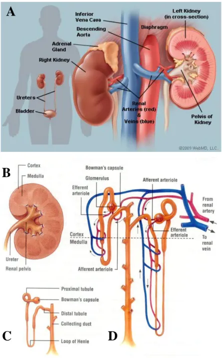
The kidney is a complex organ, located at the rear of the abdominal cavity in
the retroperitoneum
(
figure 1A).
The kidneys play crucial roles in the maintenance of the homeostasis including the regulation of blood pressure (salts and water balance), themodulation of acid-base balance and the elimination of wastes derived from the metabolism. The tight controlled balance between glomerular filtration, tubular excretion and reabsorption is at the basis of kidney functions and importance. Around 40 highly specialized cell types are organized in peculiar compartments of the kidney to exert all these functions. The kidney
has also endocrine functions: it produces erythropoietin, responsible for inducing red blood cells maturation, active Vitamin D that participates to the homeostasis of calcium and phosphorus, important for bone morphogenesis and maintenance, and renin, an active actor in the blood pressure regulation.
The functional unit of the kidney is the nephron. It is composed by a blood filtration unit, called glomerulus, and a tubular part in charge of processing the primary urine filtered by the glomerulus. The epithelial tubular portion of the nephron is divided in successive segments, composed by highly specialized cells: starting from the Bowman’s capsule of the glomerulus, the nephron is composed by the Proximal Tubule (subdivided in S1, S2 and S3 segments), the Henle’s Loop, the Distal Convoluted Tubule and the Collecting Duct system (figure 1C-D). Morphologically, the kidney is formed by the cortex, characterized by the presence of S1 and S2 convoluted segments of proximal tubules, the globular structures of the glomeruli and the convoluted distal tubules that are connected to collecting tubules via a short connecting segment. The inner part of the kidney, the medulla, contains the straight S3 segment of proximal tubule, the Henle’s loop branches and the collecting duct system. The collecting system will progressively form pyramids (that is a single structure in mouse and rat), renal calyces, pelvis and finally the ureter that will flow the urine into the bladder (figure 1B).
4
Figure 1. Anatomical structure of the kidney. (A) Macroscopical representation of kidneys and their
connections with the vascular system: the renal artery coming from the aorta in red and the renal vein going to the vena cava in blue (adapted from http://www.webmd.com). (B) The kidney is composed by a cortex that contains glomeruli, proximal and distal convoluted tubular portions of the nephron, and a medulla that contains the straight part of the proximal tubule, Henle’s loop and part of the collecting system. (C) Representation of the different segments of the nephron and (D) their interaction with the peritubular capillaries network (adapted from: http://classconnection.s3.amazonaws.com).
A
B
5 The glomerulus is formed by a complex net of anastomosed capillaries that arises from a single afferent artery and that finally converges into a single efferent artery: the glomerular capillaries are fenestrated and surrounded by a specific cell type, the podocytes. The structural support to the vascular flocculus is provided by the mesangial cells. The blood is filtered through a filtration barrier that allows only small molecules (low molecular weight proteins or ions) to pass. This barrier is composed by a fenestrated endothelial layer, a basal membrane and a podocyte zip-shaped slit diaphragm. Cells and high molecular weight proteins (for example, albumin) are normally retained in the circulation and their presence in the urine is a symptom of alteration of this filtration barrier. The vascular tuft is surrounded by the Bowman’s capsule that collects the primary urine derived from filtration in the urinary space. The Bowman space is in direct connection with the proximal tubule at the urinary pole (figure 2).
Figure 2. Structure of the glomerulus. The glomerulus (on the left panel) is composed by a net of capillaries,
arising from the afferent artery and flowing into the efferent artery. These capillaries are wrapped by the podocytes. After the filtration process, the primary urine flows in the urinary space of Bowman’s capsule and is drained away by the proximal tubule (image adapted from http://www.sharinginhealth.ca). In the right panel, a scanning electron microscopy picture of the podocytes and their foot processes. These foot processes are linked by a zip-shaped slight diaphragm, which is part of the filtration barrier (this image has been gently provided by Michel Leibovici).
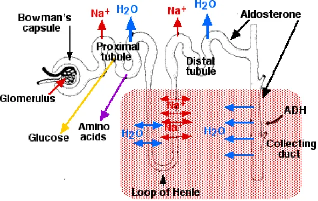
6 The Proximal Tubule (PT) is responsible for the massive reabsorption of most of the primitive urine components. Around 60% of the water and salt and up to 95% of phosphate, glucose and amino acids filtered by the glomerulus are reabsorbed in this segment (figure 3). To perform this massive reabsorption, the surface of tubular cells is increased by the presence of brush borders on the apical membrane and by interdigitations of the basal membrane. In the proximal tubule, the passive reabsorption of ions occurs via several Na+-coupled cotransporters that are located on the luminal surface. The driving force of this reabsorption is generated by an electrochemical gradient of Na+ that is performed and maintained by the Na+ -K+-ATPase, an active transporter located baso-laterally and intimately associated with large prominent mitochondria (Brenner, 1996). The transport of water through the epithelial layer is a passive mechanism, while the reabsorption of small molecular weight proteins physiologically filtered by the glomerulus requires a lysosomal system, known as apical vacuolar endocytotic apparatus. A more detailed observation shows a sub-compartmentalization of the PT in three different segments called S1, S2 (convoluted segments) and S3 (straight segment). These segments are characterized by the expression of a different set of genes that confer to each subdomain specific functions. As an example, among the small molecules that pass the glomerular filter, the glucose is an important metabolite that is reabsorbed entirely in the proximal tubule via the glucose transporters Slc5a1 and Slc5a2. These glucose transporters present different affinity and stochiometries for the glucose and they are specifically expressed in sub-segments according to the concentration of the glucose along the tubule. The low affinity Na+/glucose transporter Slc5a2 is present mostly in the first convoluted segment S1 where the concentration of glucose is high in the primary urine, whereas the Na+/glucose high affinity transporter Slc5a1 is located in the straight segment S3 where only few molecules of glucose have still to be reabsorbed (Kamiyama et al., 2012; Kanai et al., 1994).
Henle’s Loop segment is composed by the thin descending limb (TDL), which is in direct continuation with the S3 segment, and the thick ascending limb (TAL). The Henle’s loop has a characteristic U conformation that enters in the medulla of the kidney and that goes up again in the cortex where it is in contact with its own glomerulus. The major role of Henle’s loop is to generate the cortico-medullary gradient that plays a crucial role in the ability of the kidney to concentrate or dilute urine (figure 3). The TDL is extremely permeable to water and less permeable to ions. In this portion of Henle’s loop, the reabsorption is limited to water, whereas the solutes are retained in the urine (Kokko, 1970). This process increases
7 the concentration of the urine that reaches its maximum around the tip of the loop in the medulla. In the TAL the situation is reversed: the tubule becomes impermeable to water and permeable to ions principally via active transports. The ions are reabsorbed mainly via the Na-K-2Cl cotransporter (Slc12a1), localized at the apical pole of tubular cells. The intracellular ion gradient is maintained by a Na+/K ATPase that actively pumps Na+ ions in the peritubular compartment, that will ultimately be reabsorbed by the vasa recta (Greger, 1985), (Imai and Kokko, 1976).
A specific set of cells, composing the Macula Densa, is localized at the end of the Henle’s loop segment. This peculiar tubular portion, with the two glomerular arteries and the extraglomerular mesangium, forms the juxta-glomerular apparatus (Barajas, 1970) (Barajas, 1979). This specific domain, through its peculiar anatomical conformation has been demonstrated to be important for the regulation of blood pressure, via the renin angiotensin system. It is also acting as chemo-mechanical sensor of the urine and it participates to the tubulo-glomerular feedback via mechanisms that are not yet completely elucidated (Brenner, 1996).
The Distal Convoluted Tubule (DCT) is the last portion of the nephron before the connection to the collecting system. The DCT plays a crucial role in refining the urine composition and concentration, via the action of circulating hormones on the distal tubule cells (Greger and Velazquez, 1987) (figure 3). In particular, the DCT participates to the homeostasis of Na+, mediated by aldosterone, and to the reabsorption of Ca+(Borke et al., 1987).
The DCT is then connected via a short connecting duct to the Collecting Duct (CD) system. This set of tubules collects the urine produced by the nephrons and brings it via a series of structures (the pyramids, the calices, the pelvis and the ureter) into the bladder. The CD has a different embryological origin than the other tubules from the nephron as we will discuss it later. It is separated in cortical and medullary collecting ducts, according to the kidney compartments it crosses. It is composed mainly by “principal cells” (around 60%), rich in transporters and channels located at the apical membrane, that are mainly sensible to the action of hormones (figure 3). For example, the aldosterone increases the activity and, later, the number of Na+/K+ transporters to increase the sodium reabsorption and the potassium secretion, whereas the vasopressin modulates the localization of aquaporin channels on the cell surface. Together, aldosterone and vasopressin are key players in the final regulation of
8 urine volume and concentration in this last portion of the nephron (Naray-Fejes-Toth and Fejes-Toth, 1990) (Verrey et al., 1987). The other 20% of cells composing the CD are “intercalated cells” type α and β: they participate to the acid-base homeostasis by regulating the absorption/excretion of acid and bicarbonates (Malnic et al., 1994), (Brenner, 1996).
In conclusion, the kidney fulfills several important functions in order to maintain the body homeostasis through the filtration, reabsorption or secretion of water and small molecules present in the blood. These functions are accomplished thanks to the cellular and anatomical complexity of the kidney: the adult kidney is composed by around 40 different cell types. Its functional unit, the nephron, is composed by highly differentiated segments that have different characteristic and perform specific functions (figure 3). Such high level of complexity is achieved by a tightly controlled and regulated developmental process during embryogenesis: several signaling pathways and crosstalk take place since the early steps of nephrogenesis and lead an aggregate of multipotent cells to become a complete and functional organ. In the next chapter I will introduce the state of the art in the knowledge of these processes during the embryonic development and their implication in renal morphogenesis.
9
Figure 3. Schematic representation of tubular absorption and secretion functions. After glomerular
filtration, the primary urine is processed all along the tubule in order to reach its final concentration and composition. The proximal convoluted part is responsible of massive reabsorption of water, solutes and glucose, whereas the successive segments refine the urine composition.
10
II. Principal signaling pathways during renal morphogenesis
In higher organisms, the interactions and communications between different cells and with environment are crucial. Autocrine or paracrine signals deliver information to cells via short range signaling whereas hormones and soluble molecules can act via long range signalization. These external signals can modify cell behavior through multiple aspects, from structural cell modification to gene expression modulation. The ability of cells to perceive and correctly respond to their environment is at the basis of normal tissue homeostasis, tissue repair, as well as development. During embryonic development, several signaling pathways are involved in organ morphogenesis: despite the huge difference in shape and function between different organs and different species, most of the signaling pathways are evolutionary conserved and are able to fulfill different roles according to the environment in which they act.
In order to have a general overview of the pathways I will discuss during my thesis, I will briefly illustrate the principal signaling pathways that are implicated in kidney morphogenesis. In particular, I will focus on:
- Notch pathway
- Wnt canonical (β-catenin dependent) and Wnt non-canonical (β-catenin independent) signaling
- Bone Morphogenic Proteins (BMPs) signaling pathways - Fibroblast Growth Factors (FGFs) signaling
11 The Notch Pathway
The Notch signaling pathway is a highly conserved cell signaling system present in most of multicellular organisms. The canonical Notch pathway is considered as a short range signaling, involving cell-to-cell contact. Indeed, both ligand and receptor are membrane proteins. In order to initiate the pathway, the signal-sending cell has to express the ligand at the cell membrane, whereas the adjacent signal-receiving cell must expose the receptor on its surface. In mammals, the Notch receptors family is composed by four members (Notch1 to 4) and they can interact with five ligands, two Jagged proteins (Jag1 and Jag2, homologous of Serrate in Drosophila) and three Delta-like ligands (Dll1, Dll3 and Dll4). Notch signaling can elicit opposite responses depending on the cell nature, such as proliferation or apoptosis program, can drive the acquisition of a specific cell fate or the maintenance of self-renewing status. The peculiar characteristic of the canonical Notch pathway depends on the mechanism of signal transduction. Once a ligand is bound to the receptor, a complex set of events leads to the release of the extracellular portion of the receptor after cleavage. The intracellular domain of the receptor is then able to translocate into the nucleus, where it plays the role of a transcriptional co-activator.
After their synthesis in the endoplasmic reticulum, Notch receptors undergo a first cleavage (S1) by a furin-like convertase in the trans-Golgi. This cleavage converts the original molecule in a heterodimeric molecule joined by non-covalent interactions (Logeat et al., 1998). The mature Notch receptor is composed by an extra cellular domain, a transmembrane part and an intracellular domain.
The extracellular portion is composed by a conserved array of 36 Epidermal Growth Factor (EGF) like repeats that are involved in the binding of the ligand (Wharton et al., 1985). In addition, three juxtamembrane repeats of cysteine rich regions, known as Lin-12, modulate the interaction with the juxtamembrane portion of the intracellular domain. These repeats compose the negative regulatory region (NRR). In the absence of ligand, the receptor is folded in a structure that makes the S2 cleavage site inaccessible and the receptor inactive (Gordon et al., 2007). The large modular Notch intracellular domain (NICD) is composed, starting from the juxtamembrane domain, by a region called RAM (RBPjκ associated molecule) that is followed be a repeated structural motif, flanked by nuclear localization signals. This repeated structural domain is composed by seven Ankirin, in charge of modulating the interaction with CBF1/Su(H). The N-terminal part of NCID domain is
12 composed by a transactivation domain (TAD, only in Notch1 and 2) and a proline-glutamine-serine-threonine rich domain (PEST) that is involved in the degradation/recycling of Notch (Oberg et al., 2001) (figure 4).
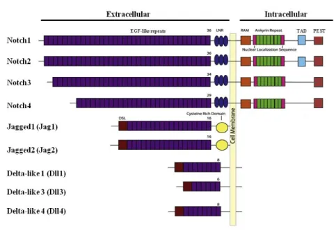
All the Notch ligands are, as well, membrane proteins, composed by a short N-terminal intracellular domain and a large extracellular domain. The major difference between Jag and Dll ligands is represented by the length of EGF-like domains, composed by 16 repeats for Jag and 6-8 repeats for the three Dll homologous. In addition to the number of EGF-like-tandem repeats, Jag differs from Dll by the presence of a cysteine rich domain close to the transmembrane region. The most important part for the signaling role of the ligand is located in the C-terminal portion of the extracellular part of the protein and it is common to both Jag and Dll. This domain is known as DSL (Delta-Serrate-Lag2) and is responsible for interaction with Notch receptor at the level of EGF-like repeat 11 and 12 (reviewed in (D'Souza et al., 2010)) (figure 4).
Figure 4. Schematic representation of the four Notch receptors (Notch1 to 4) and the five ligands present in mammals (Jag1 and 2, Dll1-2 and 4). The receptors differ mainly by the number of EGF-like repeats and the
presence of a TAD domain in the intracellular portion of Notch1 and 2. All the ligands are characterized by a DSL sequence at the C-terminal part, whereas the cysteine rich domain is specific for the two Jagged ligands (adapted from (Wang, 2011)).
13 The activation of the pathway via ligand-receptor binding is known as canonical Notch signaling pathway (figure 5, left panel). In order to be active, the Notch receptor needs to undergo a first cleavage of the extracellular portion. Constitutively, the cleavage site is not accessible due to the 3D protein conformation that maintains the receptor in an inactive configuration. The binding of the ligand is able to modify this conformation, resulting in the exposition of a metalloproteinase site (S2) to the caspase activity and the shedding of the ectodomain. The enzyme in charge of this cleavage is a membrane bound complex, composed by ADAM (a desintegrin and metallopeptidase) and TACE (tumor necrosis factor α converting enzyme) (Brou et al., 2000).
The cleaved extracellular domain of the receptor is transferred inside the cell expressing the ligand by trans-cytosis. This complex is then disassembled inside the endosome and the ligand is again able to be expressed on the cell surface. The remnant cleaved part of the receptor, still inserted in the membrane, is a transient intermediate called Notch extracellular truncation (NEXT). NEXT becomes now accessible to another caspase, the γ-secretase, a multicomponent member of the intramembrane cleaving proteases (I-CLiPs). The γ-secretase is able to progressively cut NEXT at S3 and S4 sites, probably after endocytosis of the complex NEXT/γ-secretase. This process leads to the release of the transcriptionally active Notch intracellular domain (NICD). Its translocation in the nucleus leads to its interaction with different DNA-binding CSL proteins through the RAM domain. These proteins, also known as CBF1, Su(H), Lag1 contain RBPjκ binding interfaces. Subsequently, the Ankirin domain of NICD interacts with CSL to recruit another co-activator, Mastermind (Mam) (reviewed in (Kovall, 2008)). In order to elicit a transcriptional response, this complex has to bind RBPjk. In fact, in absence of NICD, RBPjk is bound to a repressor complex and silences gene expression. The intervention of NICD displaces the co-repressors complex from RBPjk, that switches from repressor to activator via the recruitment of the described transcriptional co-activators. The best known target genes of Notch canonical pathway are the members of hairy/enhancer of split (HES/HEY) family: they are helix-loop-helix transcription factors that mainly act as repressor during the embryonic development and the modulation of cell fate (Iso et al., 2003)
14 Recent experimental evidences disclosed the importance of a second Notch signaling pathway, known as non-canonical Notch pathway (figure 5, right panel). Preliminary experiments suggest that activation of Notch could be either independent from ligand-receptor interaction or uncoupled from the canonical intracellular action of the NICD. In this context, the extracellular domain of Notch could be bound by non canonical ligands or soluble proteins. This binding could modulate Notch pathway in several ways, including competitive inhibition, dissociation of the receptor or enhanced endocytosis (review in (Wang, 2011)). As an alternative, non canonical signaling can also act via the intracellular domain of Notch, which can behave separately from the canonical CSL-dependent way. Some genes have been shown to be affected, in different models and cell types, by non-canonical Notch signaling, but the precise mechanisms are still elusive. The most studied and well conserved non-canonical Notch pathway is the regulation of Wnt/β-catenin signaling: NICD domain can bind and titrate the active β-catenin, the major component of the Wnt/β-catenin pathway, as we will discuss it later (reviewed in (Andersen et al., 2012)).
Figure 5. Canonical and non-canonical activation of Notch intracellular signaling. (A) The canonical Notch
pathway involves the translocation of the NICD (once released by the secreatase) in the nucleus, necessary to the expression of target genes. (B) In the non-canonical pathway, NICD does not migrate in the nucleus, and more and more evidences suggest an interaction with other partners to elicit the modulation of several intracellular cascades (from (Andersen et al., 2012))
15 The Wnt signaling pathways
The Wingless-related MMTV integration site (Wnt) genes were first identified in the Drosophila. They were called Wingless (Wg) due to the absence of wings in the mutant flies. (Nusslein-Volhard et al., 1980). In mammals, the Wnt family is composed by 19 different soluble ligands that act, in association with lipoproteins, as middle-long range morphogens in a classical concentration-dependent manner (Panakova et al., 2005). Many of the 19 members are conserved in several multicellular organisms: the fact that monocellular organisms lack Wnt proteins suggests its importance for the development of more complex organims (reviewed in (Petersen and Reddien, 2009)). Wnt members are 40kDa proteins that contain many conserved cysteines, but their biochemical characterization still remains challenging. During the transit in the endoplasmatic reticulum, Wnt undergoes a lipid modification by the Porcupine (Porc) enzyme before being shuttled in Golgi vesicles and released outside the cell. The interaction of these ligands with the signaling receiving cell can give rise to two different cellular responses, according to the intracellular mechanisms involved: similar to Notch pathway, we can distinguish between a canonical and non-canonical Wnt signaling pathway.
The canonical Wnt signaling pathway, also known as Wnt/β-catenin signaling, involves a complex network of proteins that is activated once the ligand/receptor interaction occurs on the surface of the cell (figure 6). This signaling cascade ends up with the transcription of specific target genes in the nucleus. The canonical Wnt/β-catenin signaling is involved in several aspects of embryo and adult biology: its main role is the maintenance of self-renewal properties of stem cells and altered Wnt/β-catenin signaling is correlated with cancer and developmental abnormalities. The β-catenin is an important component of the cytoskeleton interacting complexes at the adherent junctions: these proteins have multiple copies of the so-called armadillo repeat domain, which is specialized for protein-protein interaction and allows the catenin to interact with cadherins and alpha-catenin. When β-catenin is not involved in membrane complexes, it can interact with other proteins and, in particular, it plays a role in Wnt intracellular signaling pathway. In absence of Wnt signalling, the cytoplasmic β-catenin is constantly degraded by the action of a complex composed by the scaffold protein Axin, the modular protein Dishevelled (Dsh, composed by three domains DIX, PDZ and DEP), the tumor suppressor adenomatous polyposis coli gene product (APC), and two different kinases, the casein kinase 1 (CK1) and the glycogen synthase kinase 3 (GSK3). The two kinases act sequentially by phosphorylating the amino
16 terminal region of β-catenin. This modification results in β-catenin recognition by β-Trcp, an E3 ubiquitin ligase subunit, and subsequent β-catenin ubiquitination and targeting to proteasome for degradation (Ha et al., 2004). The continuous elimination of β-catenin prevents its cytoplasmic accumulation and its migration in the nucleus: the β-catenin target genes are maintained repressed by the DNA-bound T cell factor/lymphoid enhancer factor (TCF/LEF) family members. In fact, in absence of β-catenin, these factors act as transcription repressors via the binding of a co-repressor Groucho that can both modify chromatin conformation and recruiting other co-repressors (Jennings and Ish-Horowicz, 2008).
The signaling becomes active when Wnt proteins interact with a surface heterodimeric receptor complex, composed by the transmembrane receptor Frizzled (Fz) and the single-pass transmembrane receptor LRP5/6 protein. The N-terminal cysteine-rich domain of Fz (CRD) contains a hydrophobic groove that gives a docking platform for the lipid component of Wnt (Janda et al., 2012). Once bound by Wnt, Fz undergoes a conformational modification that leads to its heterodimerization with LRP5/6 and the recruitment of Dsh. The activation of Dsh induces the successive phosphorylation of the LRP5/6 receptor by CK1 and GSK3 that allows the binding of the Axin complex and its sequestration at the membrane (Tamai et al., 2004). The new conformation leads to the inhibition of β-catenin ubiquitination. Two different models have been proposed. The current model suggests that Axin segregation at the membrane disassembles partially the complex leading to the absence of β-catenin phosphorylation and its subsequent β-Trcp-mediated ubiquitination (figure 6A). Recent studies have proposed a new model, suggesting that the relocalisation of the whole Axin complex leads, instead of disassociating the kinases, to the recruitment and phosphorylation of β-catenin that rapidly saturates the complex. In this context, the new produced non-phosphorylated β-catenin accumulates in the cytoplasm and finally translocates in the nucleus (Li et al., 2012) (figure 6B). In the nucleus, β-catenin interacts with TCF/LEF transcription factors. The β-catenin is able to displace Groucho and recruit several elements (as CBP, Cdc47, Bcl9 and Pygous), switching the TCF/LEF transcription factors from a repressor status to an activator of gene expression (figure 7).
17
Figure 6. Schematic representation of Canonical Wnt/β-catenin signaling. (A) According to the current
hypothesis, in absence of Wnt signaling, the β-catenin is rapidly phosphorylated and degraded by the proteasome via βTrCP transport (left panel). In the presence of Wnt, the phosphorylation complex is sequestered and dismantled by the receptor, leading to the β-catenin accumulation in the cytoplasm and finally to its migration into the nucleus (right panel). (B) An alternative hypothesis suggests that in absence of signaling, β-catenin is directly transported by the phosphorylating complex to the proteasome (left panel). In the presence of Wnt, the complex is sequestrated at the membrane by the activated receptor, rapidly saturated by phosphorylated β-catenin and not more able to interact with βTrCP and the proteasome: newly synthesized β-β-catenin is able to accumulate in the cytoplasm and migrate then in the nucleus (right panel) (Clevers and Nusse, 2012).
Figure 7. β-catenin mediated transcription. In absence of β-catenin, the transcription factors TCF/LEF are
bound to the target genes, but in a repressive status, via the binding to the inhibitor Groucho (left panel). When the β-catenin enters the nucleus, Groucho is displaced and the newly recruited co-activators can assemble with TCF/LEF transcription factors and elicit gene transcription (right panel) (Clevers and Nusse, 2012).
18 The second pathway involving Wnt is the non-canonical Wnt signaling: this pathway acts in a β-catenin independent way and involves a central role for Dishevelled (Dsh). Until today, two main non-canonical pathways involving Wnt signaling have been indentified: the planar cell polarity pathway (PCP) and the non-canonical Wnt/Ca+ pathway. The planar cell polarity is an intrinsic property that confers to the cell a specific orientation in the plan. As an example, it has been shown that planar cell polarity is crucial during cell division to maintain a correct orientation of the mitosis axis during tubular elongation (Fischer et al., 2006) .
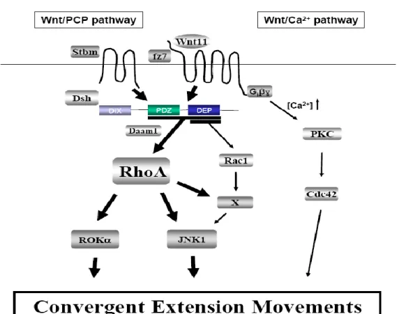
The non-canonical Wnt PCP pathway emerged from the study of the Drosophila in which Wnt mutations affected the orientation of several structures such as hair, sensory bristles and ommatidia in the eye (Mlodzik, 2002) (figure 8). In this pathway, Wnt signaling is mediated by Fz alone, without interaction with LRP receptor. Once activated, Fz is then able to recruit and activate Dsh. Dsh is composed by three different domains, DIX, PDZ and DEP. These domains are involved in the interaction with specific partners that can activate different signals, such as Rho or Rac signaling cascades, involved in different cell responses (Wallingford and Habas, 2005). To activate the GTPase Rho pathway, Dsh interacts, via its PDZ and DEP domains, with Daam1 (Dishevelled associated activator morphogenesis-1), a Formin homologous protein able to bind Dsh and RhoA. This interaction activates, via Rho, the downstream Rho-associated kinase (ROCK) and JNK1, which are responsible for cytoskeletal rearrangement and actin modification. The parallel activation of Rac GTPase is Daam1 independent and is mediated by the DEP domain of Dsh and leads to JNK activation, even if the downstream effectors are not clear. Rho and Rac are known to have opposite functions: it is then considered that both pathways are responsible for cytoskeletal rearrangments during morphological convergent extension movements (Veeman et al., 2003).
The second non-canonical pathway is Wnt/Ca+ pathway: it has been noticed that the
interaction Wnt/Fz can stimulate intracellular Ca+ release from the endothelial reticulum in G-protein dependent way without affecting β-catenin stabilization (Harada et al., 2007). Ca+
waves have been demonstrated to be crucial for early embryo patterning in Xenopus and Zebrafish (Wallingford et al., 2001), (Gilland et al., 1999) (figure 8). Intracellular release of calcium activates Ca+ sensitive proteins such as protein kinase C (PKC) and calcium/calmodulin-dependent kinase II (CamKII): the downstream pathways result in the regulation of important morphological events during embryonic development. These processes include the regulation of cell fate in the blastula stage and the modulation of morphogenetic convergent extension movements during gastrulation via phosphorylation of
19 Dsh and Lef. This phosphorilation leads to their inactivation and has as a consequence the downregulation of the convergent extension movements (reviewed in (De, 2011)).
Figure 8. Schematic representation of Non-canonical Wnt signaling. The two non-canonical Wnt pathways
are mediated by the interaction of Wnt with Fz alone, without any interaction with LRP receptors. In the
Wnt-PCP pathway, Fz recruits the protein Dsh which in turn interacts with Daam1 in order to elicit the signalization
cascade of RhoA and the activation of ROKα and JNK1. Fz activation can also act in a Daam1independent way and activate Rac1: this cascade leads to the activation of JNK1and its downstream effectors.. The second non canonical pathway is known as Wnt-Ca+ pathway: the interaction of Wnt with Fz is able to elicit intracellular release of Ca+ that activates PKC cascade. These pathways (PCP signaling and Wnt-Ca+) are known to modulate the convergent extension movements during embryogenesis.
20 Bone Morphogenic Proteins (BMPs) signaling pathways
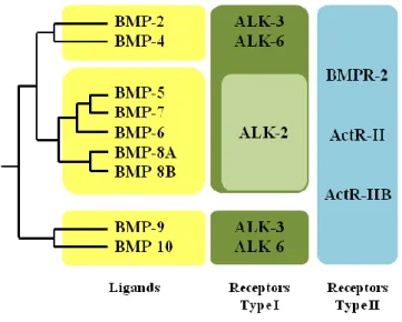
BMPs (Bone morphogenic proteins) are a subgroup of growth factors belonging to the TGF-β superfamily, except for BMP1 that is a metalloprotease. BMPs were originally discovered by their ability to induce the formation of bone and cartilage (Urist, 1965). Nowadays, a more precise knowledge of BMPs functions pointed out their important morphogenetic functions during embryonic development. The BMP signalling is mediated by the interaction of BMP proteins with BMP receptors type I and type II. These two groups of receptors are composed by serine/threonine kinase receptors. The BMPR class II is composed by three types of proteins that share similar structures: BMP type II receptor (BMPR-II), Activin type II receptor (ActR-II) and Activin type IIb receptor (ActR-IIb). All the members of these receptor subgroups can interact with all the different BMPs. The BMPR class I is composed by seven members of Activin receptor-like kinase (Alk1 to Alk7). These receprtors show different affinity for the TGFβ-BMPs ligands: in particular, BMPs bind to the receptors Alk1 Alk2, Alk3 and Alk6 with different specificity, whereas the other receptors of the group are involved specifically in the TGFβ signaling pathway (reviewed in (Miyazono et al., 2010)) (figure 9).
The binding of BMPs to the BMPR type II allows its heterodimerisation with the type I receptor. This heterodimerisation promotes the phosphorylation of Gly-Ser (GS) domain of the intracellular part of the type I receptor, via the constitutively active serine/threonine kinase present on type II receptor. This type I receptor phosphorylation leads to the activation of its own kinase. Therefore, intracellular specificity of the signal is dictated by the type I intracellular domain: the activated type I receptor kinase propagate the signal by phosphorylating proteins of the Smad family transcription factors. In mammals, eight different Smad proteins are identified: Smad1, Smad5 and Smad8 are receptor-regulated Smads (R-Smad) involved in the transmission of BMPR signaling whereas Smad2 and Smad3 are involved in TGFβ specific pathways. Smad4 is a Co-Smad, common to both pathways, whereas Smad6 and Smad7 fulfill inhibitory functions (I-Smad). Structurally, all the Smad family members share a highly conserved Mad homology domain in N-terminal region (MH2), whereas a second Mad homology domain (MH1) is conserved only in R-Smad and Co-Smad. MH2 domain is responsible for interaction with type I BMPR, dimerization with other Smads and activation of transcription, whereas MH1 is responsible for DNA binding (reviewed in (Miyazono et al., 2005)). In the absence of phosphorylation, MH1 and MH2 are
21 folded together by a linker of variable amino acids length: the inactive conformation of R-Smad cannot dimerize with other R-Smads and it is anchored to the membrane via transmembrane proteins as SARA (Qin et al., 2002).
R-Smads have a characteristic Ser-Ser-Val/Met-Ser (SSXS) sequence at their C-terminal part, through which they can transiently and directly interact with activated type I receptors: the activated kinase in the intracellular N-terminal of BMPR-I phosphorylates the SSXS sequence, disrupting the MH1-MH2 bridge. This disruption allows R-Smads to form hetero-oligo dimers that are translocated into the nucleus via interaction via Co-Smad (Qin et al., 2001). In the nucleus, Smad complex can bind gene promoter regions and interact with other DNA binding proteins, Co-activator/repressor, in order to modulate downstream target gene expression (reviewed in (Miyazono et al., 2010)).
The I-Smads negatively control the Smad intracellular signaling: Smad6 preferentially inhibits the BMP signaling, whereas Smad7 is involved in both TGFβ-BMP signaling pathways inhibition via different mechanisms (figure 10). The first mechanism of regulation involves the capacity of I-Smad to interact directly with R-Smads, inhibiting their heterodimerization with Co-Smad and the subsequent translocation in the nucleus. On the other hand, I-Smad can directly degrade BMPR by interacting with the Smurf proteins. Smurf1 and Smurf2 proteins are members of HECT type E3 ligase, known to directly regulate the R-Smad cellular pool (Zhang et al., 2001). In the nucleus, Smurf proteins can also bind to I-Smad, and translocate outside the nucleus. The complex Smurf/I-Smad directly targets the active BMPR type I and promote its ubiquitination and proteasome-dependent degradation (Horiki et al., 2004). In addition to Smurf driven degradation, the I-Smad can negatively regulate the BMPR signaling via protein phosphatase-1 (PP1): I-Smad, in fact, interacts with a regulatory subunit of PP1, recruiting the catalytic domain (PP1c) to dephosphorylate and silence the active BMPR type I (Shi et al., 2004). The I-Smads are modulated by Arkadia-mediated dowregulation. The expression of this RING type ligase is downregulated by TGFβ, suggesting a negative feedback control of TGFβ on Smad signaling. Contrary to Smurf complex, Arkadia does not interact with the BMPR type I when engaged by Smad7, but it elicit the ubiquitin-depend degradation of Smad7 that can not anymore play is role in the control of TGFβ-BMP signaling (Koinuma et al., 2003).
22
Figure 9. Schematic representation of BMPs family. The bone morphogenic proteins can be subdivided in
three major groups according to their homology degree: BMP2 and 4 belongs to the first group, BMP5 to BMP-8B to the second group, and BMP-9 and 10 form the third group. All the BMPs members are able to bind to type II receptors (BMPRII, ActR-II and ActR-IIB). In the type I group, ALK2 can be bound only by the second group of BMP members, i.e. BMP5 to BMP8B. (Modified from (Miyazono et al., 2010))
Figure 10. Schematic representation of Smad signaling modulation by I-Smad. The inhibitory Smads can
regulate the intracellular signaling in several ways: they can either dephosphorylate directly the receptor or degrade it via Smurf ubiquitination. The I-Smad can also interfere in the interaction R-Smad/Co-Smad and block the nuclear translocation of this complex. In addition, I-Smad directly interferes with R-Smad phosphorylation at the membrane. In the nucleus, the I-Smad are modulated by Arkadia (Modified from (Miyazono et al., 2010)).
23 Fibroblast Growth Factor (FGF) signaling pathway
Fibroblast Growth Factors (FGFs) and their specific cell surface receptors (FGFR) make up a large and complex family of signaling molecules that have been shown to play an important role in several crucial processes ranging from embryonic development to tissue homeostasis: the importance of this pathway is underlined by its role in a wide range of organisms, from the nematode to the human. In vertebrates, FGFs family counts 22 members, whose molecular weight varies from 17 to 34 kDa. These members share a high affinity for heparin or heparan sulfate proteoglycan and are composed by a central domain of 28 highly conserved amino acids: among them, six amino acids residues are identical in all the FGF members and are in charge of the interaction with FGFRs (Plotnikov et al., 2000). These proteins are almost all readily secreted molecules even if some of them (FGF9, FGF16 and FGF20) lack the amino-terminal sequence peptide, common to the other members. On the contrary FGF1 and FGF2 are not secreted, but released from damaged cells in exocytotic mechanism independent from endoplasmic/Golgi reticulum pathways (Mignatti et al., 1992). The signaling of the FGFs family members passes through their interaction with four high related members of tyrosine/kinase (RTKs) receptors.
The fibroblast growth factor receptors (FGFR1 to FGFR4) are composed by a single transmembrane domain that links the extracellular globular portion, in charge of ligand binding, with the intracellular domain that contains the tyrosine kinase activity domain. The extracellular domain is composed by three immunoglobulin-like domains (D1-3), a seven to eight acidic residues domain in the linker connecting D1 and D2, designated as “acid box”, and finally a conserved positively charged region in D2 that serves as a binding site for heparin (reviewed in (Schlessinger et al., 2000)).
It is hypothesized that the acid box can participate to the inhibition of the FGFR auto induction. The multiple acidic sites carried by the acid box can mimic the negative potential surface of heparin-like compounds. Binding of the acid box to the heparin-binding sites on the D2 region of the FGF receptor leads to a 3D compacted conformation of D1 on D2, without initiating a biological response (figure 11). A peculiar characteristic of the FGFR family is that alternative splicing of FGFR transcripts generates a variety of FGFR isoforms: the different FGFR isoforms include receptors with extracellular domain composed of either two or three immunoglobulin-like domains or soluble secreted FGFR forms. In addition, alternative splicing in the D3 domain can profoundly modify the ligand-binding specificity
24 (reviewed in (Mohammadi et al., 2005)). Ligand-induced receptor dimerization is a prerequisite for PTK activation. Receptor dimerization brings the cytoplasmic domains of the receptors in close vicinity one to the other. This provides the opportunity for receptor trans-autophosphorylation, subsequent tyrosine kinase activation and initiation of downstream signaling pathways. The heparan sulfate (HS) proteoglycans have a very important role in the ligand-receptor interaction: in the inactive form of the receptor, the heparin is bound to a specific part of the D2 domain, preventing the juxtaposition of intracellular tyrosine kinase domain of adjacent receptors. The asymmetric binding of FGFs with D2-D3 receptor domains leads to their dimerization and the modification of heparin interaction: the heparin is reoriented by the binding of the ligand and stabilized by the receptor new conformation (Uematsu et al., 2000).
Once the receptor is activated, multiple tyrosines on its intracellular domain are phosphorylated by the intrinsic tyrosine kinase. These phosphorylated tyrosines allow the docking of regulatory proteins implicated in different intracellular signaling cascades (reviewed in (Eswarakumar et al., 2005)) (figure 12). One of the most studied and characterized pathway activated by FGF involves the docking of the protein FRS2α that serves as core for the assembling of Shp2-Grb2-GAB1 activating complex. On one hand, via the recruitment of the transmembrane guanine nucleotide exchange factor SOS, this complex is able to activate Ras and the downstream effectors of MAP kinase: these latter proteins regulate the activity of downstream kinases (as ERKs, P38 and JNKs families) or modulate the action of transcription factors. On the other hand, the FRS2α complex is also able to promote the anti-apoptotic pathways via AKT activation: the P-I-3 kinase signaling cascade activation leads to PDK dependent activation of AKT that is able to block or silence apoptotic effectors like Caspase9, FKHR and BAD (reviewed in (Schlessinger et al., 2000)). Another pathway mediated by the FGFR activation is the modulation of the cytoskeletal organization via PLCγ: the phospho tyrosine kinase part of the FGFRc can dock and activate PLCγ, that will hydrolize Pt-Ins-(4-5)P2 to form diacylglycerol (DAG) and Inositol triphosphate. Ins-(1-4-5)P3 release will stimulate intracellular calcium release and calcium-calmodulin dependent protein kinase activation. Finally, in some cell types, specific proteins (i.e. Src or Shc) are directly recruited to the active phosphorylation sites where they are, in turn, activated by addition of phosphate groups.
25
Figure 11. Structure of FGF receptors. FGFR are transmembrane receptors that are composed by an
intracellular kinase domain and three conserved globular extracellular domains (D1-D3): the D1 and D2 domains are involved in the ligand binding, whereas the D1 and the acid box AB between D1 and D2 are responsible for the auto-inhibitory function. In the absence of the ligand, the D1 domain is folded on the HBS site, maintaining the receptor in a closed conformation. The binding of the ligand opens this conformation, which is stabilized by heparan sulfate interaction, via the Heparan binding site (HBS) in the domain D2. Different isoforms of the receptors have been described: as an example, different isoforms of the D3 domain of FGFR2 give rise to FGFR2-IIIb and FGFR2-IIIC (Mohammadi et al., 2005).
Figure 12. Schematic representation of FGF intracellular signal transduction. Activated FGFRs (red
rectangles) can activate PLCγ cascade pathway (blue highlight) modulating the cytoskeletal organization, the P-I-3 Kinase-AKT/PKB pathway (yellow highlight) to elicit the cell surviving program and the FRS2-Ras-MAP kinase pathway (grey highlight). The activated MAP kinases (ERKs, p38, or JNKs) translocate into the nucleus where they phosphorylate specific transcription factors, regulating target genes expression (Dailey et al., 2005).
26
III. Kidney development
In human, renal morphogenesis is a tightly controlled process that starts with the specification of a condensed cluster of cells in the intermediate mesoderm, the metanephric mesenchyme. This structure will lead to the formation of a definitive kidney, composed by more than 40 different cell types. These cell types acquire during organogenesis a complex spatial organization and activate the expression of a peculiar subset of genes which is crucial for kidney functions. Kidney development is a morphogenetic process controlled mostly at the transcriptional level. A complex network of transcription factors regulate the expression of effectors of several signaling pathways that interact or act successively in a complex spatio-temporal process.
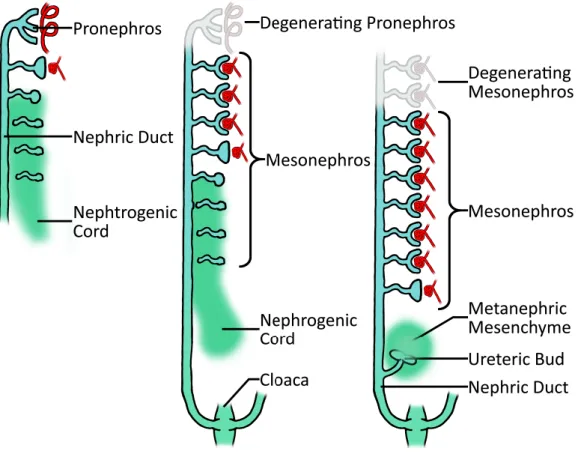
In mammals, kidney development is based on the formation of three successive structures: the pro-, meso- and metanephros (figure 13). The first two are transient structures, whereas the third one will give rise to the definitive kidney (figure 13). All these structures derive from a common nephrogenic territory in the intermediate mesoderm. They develop successively following a rostro-caudal pattern, the pronephros being the most rostral. During evolution, all these successive embryonic renal structures have been adopted to play a functional role. The pronephros is the functional kidney in the amphibious tadpole and in the fish larvae. The study of pronephros development in these animals has been very useful to identify some crucial genes involved in kidney development in mammals. In fact, the molecular mechanisms at the basis of the formation of this rudimentary kidney are conserved through evolution in different species (Drummond and Davidson, 2010). The mesonephros is the functional kidney for adult fishes and frogs, whereas in mammals this structure is able to perform some blood filtration, but it is functional only during a short period of time and rapidly degenerates. Only few vestigial tubular structures derived from the mesonephros are involved in the development of the male reproductive system. The metanephros is a much more complex organ, based on branching morphogenesis and it develops only in amniotes (mammals birds and reptiles), (reviewed in (Joseph et al., 2009)).
27
Figure 13. Schematic representation of kidney development during embryogenesis in mammals. Temporal
succession of three structures during kidney morphogenesis: the pronephros, on the left, and the mesonephros, in the center, are transient organs that disappear when the definitive metanephros on the right starts to develop (adapted from (Sawle A, 2009)).
28 III.1 Pronephros and mesonephros
Morphology
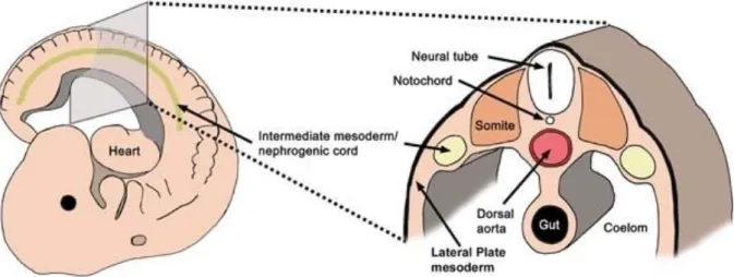
During gastrulation, multipotent cells migrate from the primitive streak to form the endodermal and mesodermal layers (figure 14). The mesoderm is composed by paraxial mesoderm, intermediate mesoderm at the origin of the nephric field and lateral plate mesoderm. Intermediate mesoderm is further specified by dorsal-ventral gradient expression of morphogen proteins, such as the Bone Morphogenic Proteins (BMPs), and by signals from the paraxial mesoderm (De Robertis and Kuroda, 2004). In the mouse, at the 8th day of embryonic development (around 3 weeks of human embryo development), a small cluster of cells, located around the 5th and 6th somites, generates the nephrogenic cord. Part of these cells undergo an epithelialisation process and form the primary nephric duct (ND), also known as Wolffian Duct (WD), that begins to elongate caudally. At the same time, some clusters of cells in the intermediate mesoderm facing the nephric duct start to condense to form tubules. The few pronephric tubules connect to the nephric duct to form the pronephros. In mouse embryos, in a matter of days, this first renal structure undergoes rapidly a massive apoptosis and degenerates around 12 days of gestation, reviewed in (Dressler, 2006).
After the formation of the pronephros, the more caudal part of the nephric duct continues to elongate in parallel with the migration of the nephric cord that will give rise to the mesonephric mesenchyme. This event takes place at 9 days of gestation in the mouse and 3.5 weeks in the human.
The mesonephros, located more caudally in respect to the pronephros (between somites 10-17) (Smith and Mackay, 1991) is composed by two different populations of tubules derived from the condensation of the mesonephric mesenchyme: they can be divided in cranial and caudal tubules (Sainio et al., 1997a). The cranial tubules are in number of 4-6 and they are formed by rudimentary glomeruli and short tubular structures. These tubules are directly connected to the Wolffian Duct (WD) by a short connecting portion derived from the nephric duct (Mugford et al., 2008a), (Brenner-Anantharam et al., 2007). In contrast, the caudal tubules (in number of 10-12) derive only from the nephrogenic mesenchyme and do not contact the WD. They are formed by condensation of cells that undergo mesenchymal-to-epithelial transition, forming the typical nephron precursors. The aggregates initially condense in “renal vesicles” and they rearrange until they assume the typical shape of “sigma-shaped bodies (S-shaped body)” (Smith and Mackay, 1991). However, the mesonephros is not able to
29 develop further and begins to degenerate by E14.5. The tubules disappear starting from the caudal to the cranial ones in 24 hours (Sainio et al., 1997a). In the male, some cranial tubules are maintained in the adult and they are part of the epididymis (Joseph et al., 2009).
Molecular mechanisms
A few genes have been identified to play a role in pronephros formation in vertebrates. Among them, in mouse, Osr1 (Odd skipped related 1) and Lhx1, two transcription factors are expressed in the lateral mesoderm just after gastrulation. This territory is broader than the intermediate mesoderm, and progressively, the expression pattern of Lhx1, and to a lesser extent, of Osr1 become restricted to the intermediate mesoderm around E8.5.
At this stage, the two paired-box homeotic transcription factors Pax2 and Pax8 start to be expressed in few cells of the intermediate mesoderm. These two factors represent the first markers of the nephric field (Bouchard et al., 2002). They are expressed both in the nephrogenic cord and in the nephric duct, an epithelial structure that form in the intermediate mesoderm. In mouse, the inactivation of Pax8 doesn’t affect the kidney development (Mansouri et al., 1998), whereas the absence of Pax2 leads to a degeneration of the nephric duct that is not able to reach the urogenital sinus (Torres et al., 1995). Interestingly, in double Pax2/Pax8 mutants, the nephric duct fails to form and the intermediate mesoderm undergoes a massive apoptosis (Bouchard et al., 2002). Gata3 is a transcription factor whose expression has been shown to be controlled by Pax2 and Pax8. Its expression pattern is restricted to the nephric duct. Inactivation of Gata3 leads to morphogenic abnormalities in the mesonephros formation and to the absence of metanephros development due to a defective elongation of the nephric duct (Grote et al., 2006),(Pedersen et al., 2005). In the same way, inactivation of Lhx1 leads to kidney agenesis, linked to a defective elongation and survival of the nephric duct. {Shawlot, 1995 #3131}(table1). In addition, Lhx1 inactivation in early stage of gastrulation leads to severe defect in intermediate mesoderm differentiation (Tsang et al., 2000). Inactivation of Osr1, which is expressed only in the nephrogenic cord and is excluded from the nephric duct, leads to defective mesonephric tubule formation and absence of metanephric differentiation (James et al., 2006).
During pronephros formation, the epithelial derivatives emanating from the nephrogenic cord and the nephric duct form independently. On the contrary, the development of mesonephric tubules is due to the induction of the mesonephric blastema by inductive signals send by the nephric duct. Wnt9b encodes a member of the Wnt family signaling
30 molecules secreted by the nephric duct. Interestingly, the mutants lacking Wnt9b show also defective mesonephric tubules and metanephric nephrons formation (Carroll et al., 2005), suggesting a common mechanism of formation of epithelial derivatives from the mesonephric and metanephric mesenchymes. Several other genes involved in the mesonephros development play also a role in metanephric development, including the transcription factors Pax2, Wt1, Osr1 and Six1 and they will be discussed in detail later. Inactivation of these transcription factors leads to variable defects, ranging from total mesonephros agenesis (Pax2 inactivation) or defective mesonephric tubules formation (Wt1, Osr1 and Six1 mutants) (Torres et al., 1995),(Sainio et al., 1997a),(James et al., 2006),(Mugford et al., 2008b).
Figure 14. Schematic representation of intermediate mesoderm position. Lateral and transversal section of
the mouse embryo showing the position of the nephrogenic cord (intermediate mesoderm) in respect of body axis (on the left) and of the other embryonic layers (on the right) (Davidson, 2008).
31 Table 1. Summary of the principle actors implicated in the nephric duct development in the mouse
Function Gene involved Malformation Ref.
Nephric duct formation and elongation
Pax2
Pax2/Pax8
- Abortive ND elongation and degeneration - Absence of ND formation - Defect in epithelialization of ND (Torres et al., 1995) (Bouchard et al., 2002) Gata3 - Defective elongation of ND - Formation of ectopic NDs (Grote et al., 2006) Lhx1 - Impaired ND extension - Absence of IM differentiation (Pedersen et al., 2005) {Shawlot, 1995 #3131} (Tsang et al., 2000)
32 III.2 Metanephros Development
In mammals, the metanephros is the last and definitive kidney: it is a much more complex organ compared to the previous pro- and mesonephros. Evolutionary, the metanephros appeared when animals had to adapt to terrestrial life. The complex organization of the metanephros provides the capacity of efficient water reabsorption. In addition, metanephros structural modifications have been selected depending on the external environment. For example, small rodents that live in desertic areas have developed very long Henle’s loops. This anatomical particularity confers to these rodents the capacity to concentrate very efficiently their urine.
The metanephros is formed mainly by the interaction between two different compartments: the ureteric bud, derived from the epithelial nephric duct, and the metanephric mesenchyme, a structure that differentiates from the intermediate mesoderm (Shah et al., 2009).
Metanephric mesenchyme identity.
The metanephric mesenchyme is a sub territory of the intermediate mesoderm that is characterized by the expression of a specific set of genes, whose expression can be partially activated even in the absence of the nephric duct elongation (Grote et al., 2006). The correct differentiation of this territory is crucial for the invasion of the ureteric bud, the first step of metanephros morphogenesis. This event occurs around E10.5 in the mouse and at five weeks of gestation in the human. Several genes has been demonstrated to have a crucial role in the metanephric mesenchyme specification and survival including Alk3, COUP-TFII, Osr1, Eya1, Sall1, Wt1, Six1-4, and the Hox11 paralogous family (Hoxa11, Hoxc11 and Hoxd11) (table 2). All these genes are important for the expression of Gdnf, a crucial molecule for the outgrowth of the ureteric bud (figure 15). I will discuss briefly the role of these different genes in the following paragraphs.
33 A very important role of the BMP signaling in the metanephric mesenchyme has been recently demonstrated by the selective inactivation in this territory of the receptor Activin like Kinase 3 (Alk3). This receptor starts to be expressed at E9.5 in the nephric field in the intermediate mesoderm (Mishina et al., 1995), (Hartwig et al., 2008). The Rarb2-Cre inactivation of Alk3 in the nephrogenic field leads to hypoplasia, characterized by a reduced number of normally developed nephrons. This defect is due to the reduction of the nephrogenic and mesenchymal precursor cells in the metanephric mesenchyme that lacks Alk3. This defect is correlated to the decreased expression of Osr1, one of the earliest marker of nephrogenic commitment in the intermediate mesenchyme (Di Giovanni et al., 2011).
Odd-skipped related1 (Osr1) encodes a zinc-finger transcription factor that is already expressed in some mesodermal cells just after their migration from the primitive streak (Wang et al., 2005a). Later on, it is restricted to the commited metanephric mesenchyme and is not expressed in the nephric duct (So and Danielian, 1999). Embryos lacking Osr1 suffer from an absence of metanephric mesenchyme differentiation leading to renal agenesis. The metanephric mesenchymal cells lacking Osr1 do not express Eya1 and Pax2 and show a drastic downregulation of Wilm’s tumor suppressor1 (Wt1) expression (James et al., 2006), (Wang et al., 2005b). Since Wt1 expression is necessary for metanephric mesenchyme cell survival, its downregulation in absence of Osr1 causes a massive increase of apoptosis in the metanephric blastema (Donovan et al., 1999), (Davies et al., 2004).
Recent studies have defined the role of the orphan nuclear receptor Chicken Ovalbumin Upstream transcription factor II (COUP-TFII) in metanephric mesenchyme specification. During kidney development, it is expressed specifically in the nephrogenic field since E9.5 and later on in the metanephric mesenchyme and in the developing nephrons. The inactivation of COUP-TFII does not affect the mesonephros development, but it leads to the absence of all markers of metanephric mesenchyme specification (Eya1, Pax2, Six2 and Gdnf). COUP-TFII has been shown to directly regulate the expression of Eya1 and Six2 in an Osr1-independent way, suggesting an interaction between these two factors in the first steps of metanephric mesenchyme specification. Similarly, COUP-TFII is also able to control the expression of Wt1 in order to maintain the metanephric mesenchyme survival (Yu et al., 2012a).