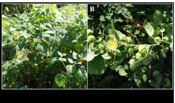
Figure 1: Physalis angulata (A) asymptomatics and (B) exhibiting typical begomovirus symptoms (apical chlorosis).
245x143mm (150 x 150 DPI)
1 Identification of Physalis angulata (Solanaceae) as a Natural Alternative Weed
2 Host of Tomato Severe Rugose Virus in Brazil
3
4 M. F. Duarte, Plant Pathology Department, University of Brasilia, Brasilia–DF, Brazil;
5 M. E. N. Fonseca and L. S. Boiteux, Embrapa Vegetable Crops (CNPH), Brasilia–DF,
6 Brazil; H. Costa, INCAPER, Venda Nova do Imigrante–ES, Brazil; B. M. Ribeiro, Cell
7 Biology Department, University of Brasilia, Brasilia–DF; F. L. Melo and R. C.
Pereira-8 Carvalho, Plant Pathology Department, University of Brasilia, Brasilia–DF, Brazil.
9
10 A diverse array of Begomovirus species (family Geminiviridae) can induce economically
11 important diseases in all major tomato-producing areas in Brazil (Fernandes et al., 2008).
12 Weeds of the Solanaceae family have been identified as important alternative hosts of the
13 viral species reported infecting tomato crops in the country (Barreto et al., 2013). In 2016,
14 about 10% of the plants of the weed Physalis angulata L. were observed with
15 begomovirus-like symptoms (apical chlorosis and stunting; Supplementary Fig. 1) around
16 and within tomato fields in Venda Nova do Imigrante–ES (Southeast Brazil). Leaf tissue
17 was collected from ten symptomatic and five healthy-looking plants, respectively. Total
18 DNA was extracted using the CTAB method in combination with organic solvents
19 (Boiteux et al., 1999). PCR assays with the universal begomovirus primers for DNA-A
20 and DNA-B detection (Rojas et al., 1993) were carried out using total genomic DNA
21 samples (20 ng/μL) as templates. Amplicons of ≈ 1,100 bp and ≈ 550 bp were obtained
22 only in the ten symptomatic samples, which are in agreement with the expected fragment
23 sizes of DNA-A and DNA-B from begomoviruses (Rojas et al. 1993). These amplicons
24 were directly Sanger sequenced at the Embrapa Vegetables Genomic Analysis
25 Laboratory. BLASTn analyses of the amplicon sequences indicated high nucleotide
26 identities (99%, 98%) with DNA-A and DNA-B of tomato severe rugose virus (ToSRV)
27 isolates (accession numbers JX415196, MG837739) from Brazil, suggesting the infection
28 of ToSRV in these plants. To fully prove the infection and to characterize the ToSRV
29 isolate associated with P. angulata, the DNA samples were submitted to rolling circle
30 amplification (RCA) method followed by Nanopore sequencing (Oxford Nanopore
31 Technologies), as previously described (Naito et al., 2019). The full-length DNA-A
32 component of one isolate obtained from P. angulata (MN059848) was 98% identical to
33 the DNA-A genome of a Brazilian ToSRV isolate from tomato (JX415196). The
34 complete DNA-B sequence from the P. angulata isolate (MN059849) was 96% identical
35 to the DNA-B genome of a tomato-infecting ToSRV isolate obtained in Southeast Brazil
36 (MG837739). The complete DNA-A component (2,592 nts) displayed all genomic
37 features of the New World species, with one virion-sense ORF (AV1) and four
38 complementary sense ORFs (AC1, AC2, AC3, and AC4). The complete DNA-B
39 component (2,571 nts) displayed two ORFs, virion-sense (BV1) and complementary
40 sense (BC1). The common regions of DNA-A and DNA-B components were identified
41 (183 nts in length) and the presence of iterons was confirmed. ToSRV is so far the most
42 prevalent tomato-infecting begomovirus in Brazil (Fernandes et al., 2008). To our
43 knowledge, this is the first report of natural infection of P. angulata by ToSRV,
44 expanding the viral host range to this widespread tropical and subtropical weed.
45 Considering that P. angulata may function as a relevant natural source of ToSRV
46 inoculum to tomato crops across tropical and subtropical regions, control of the weed
47 should be a part of the integrated management of ToSRV in tomato crops.
48
49 References
50 Barreto, S. S. et al. 2013. Phytopathology 103: 436
51 Boiteux, L. S. et al. 1999. J. Am. Soc. Hort. Sci. 124: 32.
52 Fernandes, F. R. et al. 2008. Virus Genes 36: 251.
53 Naito, F. et al. 2019. Arch. Virol. 164: 1907.
54 Rojas, M. R. et al. 1993. Plant Dis. 77: 340. Page 3 of 3