Université de Montréal
Study of the Diffusion in Polymer Solutions and Hydrogels
by NMR Spectroscopy and NMR Imaging
par WANG Yu Juan Département de chimie Faculté des arts et des sciences
Mémoire de Maîtrise présenté à la Faculté des études supérieures en vue de l’obtention du grade de maîtrise en chimie
Novembre 2010 © WANG Yu Juan, 2010
Université de Montréal Faculté des études supérieures
Ce mémoire intitulé
Study of the Diffusion in Polymer Solutions and Hydrogels
by NMR Spectroscopy and NMR Imaging
présenté par WANG Yu Juan
a été évalué par un jury composé des personnes suivantes :
Prof. Michel Lafleur président-rapporteur
Prof. Julian X. Zhu directeur de recherche
Prof. Françoise Winnik membre du jury
i
Résumé
Afin d'étudier la diffusion et la libération de molécules de tailles inférieures dans un gel polymère, les coefficients d'auto-diffusion d'une série de polymères en étoile avec un noyau d'acide cholique et quatre branches de poly(éthylène glycol) (PEG) ont été déterminés par spectroscopie RMN à gradient de champ pulsé dans des solutions aqueuses et des gels de poly(alcool vinylique). Les coefficients de diffusion obtenus ont été comparés avec ceux des PEGs linéaires et dendritiques pour étudier l'effet de l'architecture des polymères. Les polymères en étoile amphiphiles ont des profils de diffusion en fonction de la concentration similaires à leurs homologues linéaires dans le régime dilué. Ils diffusent plus lentement dans le régime semi-dilué en raison de leur noyau hydrophobe. Leurs conformations en solution ont été étudiées par des mesures de temps de relaxation spin-réseau T1 du noyau et des
branches.
L'imagerie RMN a été utilisée pour étudier le gonflement des comprimés polymères et la diffusion dans la matrice polymère. Les comprimés étaient constitués d'amidon à haute teneur en amylose et chargés avec de l'acétaminophène (de 10 à 40% en poids). Le gonflement des comprimés, ainsi que l'absorption et la diffusion de l'eau, augmentent avec la teneur en médicament, tandis que le pourcentage de libération du médicament est similaire pour tous les comprimés.
Le gonflement in vitro des comprimés d'un complexe polyélectrolyte à base d'amidon carboxyméthylé et de chitosane a également été étudié par imagerie RMN. Ces comprimés sont sensibles au pH : ils gonflent beaucoup plus dans les milieux acides que dans les milieux neutres en raison de la dissociation des deux composants et de la protonation des chaînes du chitosane. La comparaison des résultats avec ceux d'amidon à haute teneur en amylose indique que les deux matrices ont des gonflements et des profils de libération du médicament semblables dans les milieux neutres, alors que les comprimés complexes gonflent plus dans les milieux acides en raison de la dissociation du chitosane et de l'amidon.
Mots-clés: RMN à gradient de champ pulsé, Imagerie RMN, Coefficients d'auto-diffusion, Polymère en étoile, Amidon à haute teneur en amylose, Chitosane
ii
Abstract
In an effort to study the diffusion and release of small molecules in a polymeric system, the self-diffusion coefficients of a series of star polymers with a cholic acid core bearing four poly(ethylene glycol) (PEG) arms in aqueous solutions and gels of poly(vinyl alcohol) were determined by pulsed gradient spin-echo NMR techniques. The results have been compared with those of linear and dendritic PEGs to elucidate the effect of the architecture of the polymers. The amphiphilic star polymers show similar concentration-dependent diffusion behaviors in the dilute regime to their linear homologues. They diffuse more slowly in the semi-dilute regime than the linear PEGs due to the presence of the hydrophobic core. The conformation of the star polymers in the solutions was studied by measuring the T1 values of
the core and the arms of the diffusants.
NMR imaging was used to study the swelling of polymeric tablets and diffusion in the polymer matrix. The tablets investigated were made of cross-linked high amylose starch (CHAS) and loaded with acetaminophen (10, 20 and 40 wt%). The swelling, water uptake and diffusion in the CHAS network are faster at higher drug loading levels, while the drug release rates are similar among all the tablets.
The in vitro swelling of the tablets made of a polyelectrolyte complex based on chitosan and carboxymethylated starch has also been studied by NMR imaging. These tablets showed pH-sensitive behavior. They swelled much more in acidic media than in neutral media due to dissociation of the two components and the protonation of the amino groups in the chitosan residues. The comparison of the results with those obtained with the CHAS tablets indicates that the two matrices have similar swelling and drug release profile in neutral media, while the complex tablets showed a greater extent of swelling in acidic media due the dissociation of the chitosan from the complex.
Keywords: Pulsed gradient NMR spectroscopy, NMR imaging, Self-diffusion coefficients,
iii
Table of Contents
Résumé ... i
Abstract ... ii
Table of Contents ... iii
List of Figures ... vi
List of Tables ... viii
List of Symbols and Abbreviations ... ix
Acknowledgements ... xi
1. Introduction ... 1
1.1. Polysaccharides as Drug Delivery Excipients ... 2
1.1.1. Starch ... 3
1.1.2. Cellulose ... 4
1.1.3. Chitosan ... 5
1.2. Drug Release Kinetics ... 6
1.3. The Characterization of Polymer Matrices ... 8
1.3.1. XRD and FT-IR Studies ... 8
1.3.2. SEM and CMT Studies ... 9
1.3.3. Dissolution Studies ... 9
1.3.4. Optical Imaging ... 10
1.3.5. NMR Imaging Studies ... 11
1.3.6. Relaxation Time Studies ... 11
1.4. Diffusion in Polymer-Water-Diffusant Systems ... 12
1.4.1. Molecular Size of the Diffusant ... 12
1.4.2. Concentration of the Polymer ... 13
1.4.3. Molecular Weight and Hydrolysis Degree of the Polymer ... 13
1.4.4. Geometry of the Diffusant ... 13
1.4.5. Effect of Temperature ... 13
1.4.6. Interaction Between the Diffusants and the Polymers ... 14
1.4.7. Various Polymer Matrices ... 14
1.5. Water Diffusion in CHAS Tablets ... 14
iv
1.5.2. Effect of Temperature ... 16
1.5.3. Effect of the Tablet Size ... 16
1.5.4. Effect of Drug Loading ... 16
1.6. Objectives of This Study ... 17
1.7. Scope of This Study... 17
1.8. References ... 18
2. Effect of Molecular Architecture on the Self-Diffusion of Polymers in Aqueous Systems: A Comparison of Linear, Star, and Dendritic Poly(ethylene glycol)s ... 23 2.1. Abstract... 23 2.2. Introduction ... 23 2.3. Experimental... 25 2.3.1. Materials ... 25 2.3.2. Sample Preparation ... 26
2.3.3. NMR Measurements of Self-Diffusion Coefficients ... 27
2.3.4. T1 Measurements ... 28
2.4. Results and Discussion ... 28
2.4.1. Diffusion Behaviors in PVA-Water-Diffusant Tenary Systems ... 28
2.4.2. Scaling Relation between the Hydrodynamic Radius and the Molecular Weight of the Diffusant in Water-Diffusant Binary Systems ... 32
2.4.3. Effect of Diffusant Concentration on the Self-Diffusion Coefficient in Water-Diffusant Binary Systems ... 33
2.4.4. The Longitudinal Relaxation Times of the Diffusants (T1) ... 36
2.5. Conclusion ... 37
2.6. Acknowledgements ... 38
2.7. References ... 38
3. NMR Imaging Study of Cross-linked High Amylose Starch Tablets: Effect of Drug Loading ... 41 3.1. Abstract... 41 3.2. Introduction ... 41 3.3. Experimental... 42 3.3.1. Preparation of Tablets ... 42 3.3.2. NMR Imaging ... 42
v
3.3.4. Water Uptake Experiments ... 43
3.3.5. In Vitro Drug Release Tests ... 43
3.4. Results and Discussion ... 44
3.5. Conclusion ... 51
3.6. Acknowledgements ... 51
3.7. References ... 52
4. Swelling Behavior of Chitosan, Carboxymethyl Starch and Chitosan-Carboxymethyl Starch Mixture As Studied by NMR Imaging ... 54
4.1. Abstract... 54
4.2. Introduction ... 54
4.3. Experimental Section ... 56
4.3.1. Preparation of Matrices and Tablets. ... 56
4.3.2. Preparation of Media. ... 57
4.3.3. NMR Imaging. ... 57
4.4. Results and Discussion ... 57
4.4.1 NMR Imaging and Proton Density Profiles. ... 57
4.4.2. Comparison of Polymer Matrices. ... 59
4.4.3. Swelling Characteristics of the Tablets ... 61
4.5. Conclusion ... 64
4.6. Acknowledgements ... 65
4.7. Supporting Information ... 66
4.8. References ... 70
5. Conclusion ... 72
5.1. The diffusion of Star Polymers in PVA Solutions and Gels ... 72
5.2. The Study of the Effect of Drug Loading ... 73
5.3. The Study of Various Polymer Matrices ... 74
5.4. Future Work... 75
5.4.1. Research Techniques ... 75
5.4.2. Choice of Diffusing Probes ... 75
5.4.3. Choice of Drugs ... 76
vi
List of Figures
Figure 1.1. The chemical structures of (A) amylose and (B) amylopectin. ... 3 Figure 1.2. The chemical structures of (A) cellulose and (B) hydroxypropyl methyl
cellulose (HPMC). ... 4
Figure 1.3. The chemical structures of (A) chitin and (B) chitosan. ... 5 Figure 2.1. The chemical structure of the star polymers used in this study. They are
prepared by anionic polymerization of ethylene oxide on a core of cholic acid.. ... 24
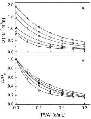
Figure 2.2. Self-diffusion coefficients (A) and reduced self-diffusion coefficients
(B) of the star polymers as a function of PVA concentration at 25oC. ... 28
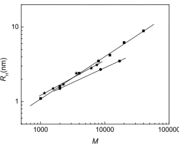
Figure 2.3. Logarithmic plot of the hydrodynamic radius RH as a function of
molecular weight for linear PEOs, dendrimers, and the star polymers in aqueous solutions at 23oC.. ... 31
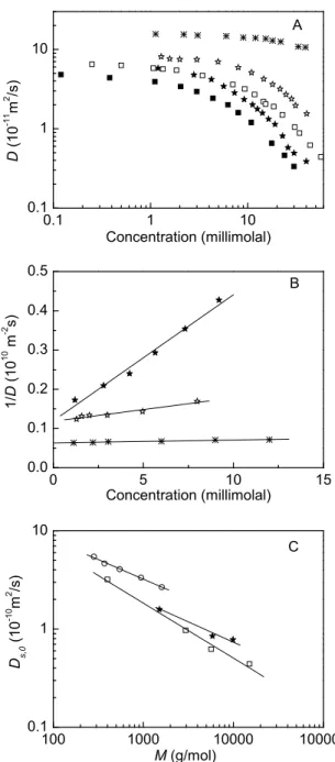
Figure 2.4. (A) The dependence of self-diffusion coefficient D on the concentration
of polymer diffusants in water: CA(EG6)4, CA(EG31)4, CA(EG54)4,
linear PEO-6k, linear PEO-10k. (B) Variation of 1/D as a function of concentration of the star polymers. (C) Variation of Ds,0 as a function of
molecular weight of the star polymers in D2O at 23oC, linear PEGs in
D2O at 25oC, and linear PEGs in D2O at 30oC.. ... 34
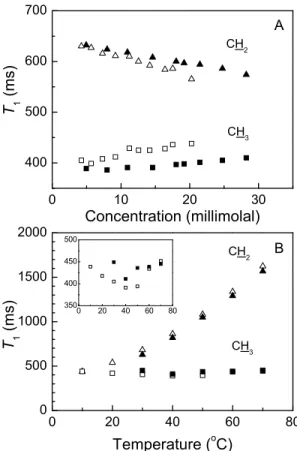
Figure 2.5. The 1H T1 values for CH2 of PEG chains and CH3 of the cholane core
measured for the star polymers. (A) Effect of concentration for two polymers CA(EG31)4, CA(EG54)4 at 25oC; (B) Effect of temperature for
CA(EG54)4 at 1.2 and 19.1 millimolal.. ... 37
Figure 3.1. NMR images of CHAS tablets immersed in water at 37oC for 1, 2, 5, 10, 15, and 20 h. ... 45
Figure 3.2. Mass uptake (A), radial swelling (B), and axial swelling (C) of the
CHAS tablets loaded with 10%, 20%, and 40% acetaminophen. ... 47
Figure 3.3. The water proton spin density profile and the fit to Eq. 3.2 of a CHAS
tablet with 20% acetaminophen swelled in water at 37oC for 30 min. ... 48
Figure 3.4. Diffusion coefficients of water in the inner core of the tablets loaded
with 10%, 20%, and 40% acetaminophen obtained by
vii
Figure 3.5. Release of drugs of CHAS tablets loaded with 10%, 20%, and 40%
acetaminophen.. ... 50
Figure 4.1. The NMR images of the CMS-chitosan complex tablets immersed in
various media at 37oC for 1, 2, 3, 5, 7 and 10 h.. ... 58
Figure 4.2. The change of the proton density profiles of the tablets made of the
CMS-chitosan complex immersed in (A) H2O, (B) SGF, (C) SIF, (D)
SGF-SIF at 37oC at different immersion times. ... 60
Figure 4.3. The radial and axial swelling of the CHAS tablets (A and B), of the
CMS tablets (C and D), and of the complex tablets (E and F) in H2O,
SGF, SIF, and SGF-SIF. ... 62
Figure 4.4. The NMR images of the CHAS tablets immersed in various media at
37oC for 1, 2, 5, 7, 10 and 15 h. ... 66
Figure 4.5. The NMR images of the CMS tablets immersed in various media at
37oC for 1, 2, 3 and 4 h. ... 66
Figure 4.6. The NMR images of the chitosan tablets immersed in various media at
37oC for 1, 2, 5, 10, 15 and 20 h. ... 67
Figure 4.7. The change of the proton density profile of the CHAS tablets immersed
in (A) H2O, (B) SGF, (C) SIF and (D) SGF-SIF at 37oC. ... 67
Figure 4.8. The change of the proton density profile of the CMS tablets immersed in
(A) H2O, (B) SGF, (C) SIF and (D) SGF-SIF at 37oC. ... 68
Figure 4.9. The change of the proton density profile of the chitosan tablets
immersed in (A) SGF and (B) SGF-SIF at 37oC. ... 68
Figure 4.10. The radial and axial swelling of the CHAS, CMS and CMS-chitosan
complex tablets in H2O, SGF, SIF and SGF-SIF. ... 69
Figure A1. The chemical structure of pectin. ... 77 Figure A2. The chemical structures of G and M blocks of alginate. ... 78 Figure A3. The chemical structures of (A) κ-carrageenan, (B) ι-carrageenan, and
viii
List of Tables
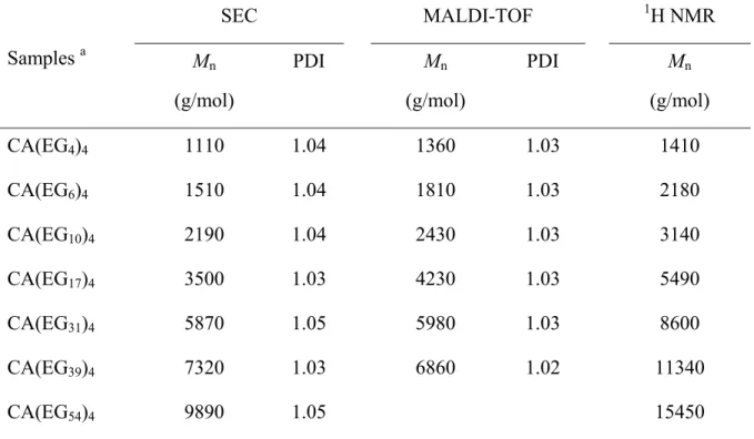
Table 2.1. The molecular weights of the star polymers CA(EGn)4 determined by
SEC, MALDI-TOF mass spectrometry, and 1H NMR spectroscopy ... 26
Table 2.2. Self-diffusion coefficients (D0), hydrodynamic radii (RH), and fitting
parameters kβ2 and ν obtained for the star polymers CA(EGn)4 in
PVA-water-diffusant ternary systems ... 30
Table 2.3. Molecular weights and hydrodynamic radii of the polymers shown in Figure 2.4A. ... 33
Table 3.1. The initial diffusion coefficients of water fitted to Eq. 3.2 and the average diffusion coefficients fitted to Eq. 3.5 in the CHAS tablets. ... 45
Table 4.1. Swelling of the tablets made of CHAS, CMS, CMS-chitosan complex. Parameters obtained by fitting to Eq. 4.2. ... 63
ix
List of Symbols and Abbreviations
APAP Acetaminophen
CAC Critical aggregation concentration CHAS Cross-linked high amylose starch
CMS Carboxymethyl starch
CMT X-ray computed microtomography CP-MAS Cross-polarization magic angle spinning δ Length of a gradient pulse
Δ Time interval between the two gradient pulses (diffusion time)
D Self-diffusion coefficient
D0 Initial diffusion coefficient of water
Self-diffusion coefficient in pure water DMF N,N-dimethylformamide
FT-IR Fourier transform infrared spectroscopy γ Gyromagnetic ratio of 1H
G Gradient strength
HPMC Hydroxypropyl methylcellulose
Mn Number average molecular weight
NMR Nuclear magnetic resonance
NMRI NMR imaging
PDI Polydispersity index
PEG Poly(ethylene glycol)
PEO Poly(ethylene oxide)
PGSE Pulsed-gradient spin-echo
pKa Logarithmic acid dissociation constant
x methyl ether
PVA Poly(vinyl alcohol)
R2 Coefficients of determination RH Hydrodynamic radius
SEM Scanning electron microscopy SEC Size exclusion chromatography
SEC-LS SEC coupled with a light scattering detector SEC-RI SEC coupled with a refractive index detector SGF Simulated gastric fluid
SIF Simulated intestinal fluid T1 Longitudinal relaxation time
T2 Transverse relaxation time
Tg Glass-transition temperature
TE Echo time
TR Repetition time
xi
Acknowledgements
I would like to thank all the people who supported, helped and inspired me during my study at the Université de Montréal.
First of all, I would like to thank my supervisor, Prof. Julian. X. Zhu, for his guidance through my study and research, for his enthusiasm, inspiration, and immense knowledge. He has also provided good teaching on how to make presentations and write papers, which will greatly benefit my future career.
I would like to thank Dr. Cédric Malveau for his training in NMR spectroscopy and valuable discussions. It is a great pleasure to work with him. He has made the NMR experiments very interesting and inspiring.
I am grateful to Prof. Mircea A. Mateescu and Mr. Elias Assaad at the Univesity of Québec at Montréal for their generous help with tablet preparation, dissolution tests and constructive discussions.
Mr. Sylvain Essiembre and Mr. Pierre Ménard-tremblay provided trainings on the instruments in the group and the maintenance of the instruments used for the characterization of polymers.
I am indebted to all members in our research group for providing a stimulating and fun environment in which to learn and grow.
Last but not least, I would like to thank my parents for their unflagging love and support throughout my life.
1
1. Introduction
Polymers have been widely used as excipients in the field of drug delivery. The pharmaceutical polymers provide the researchers with a wide choice of physical and chemical characteristics such as different molecular weight and possibility of copolymerisation, modification or blending with other polymers.1 They include both natural polymers (such as starch, cellulose, chitosan, and pectin) and synthetic polymers (such as polyanhydrides, polyesters, polyacrylic acids, poly(methyl methacrylates), and polyurethanes). The wide range of physicochemical properties may be utilised to improve the clinical use, manufacturing, and stability of dosage forms.1 More specifically, polymers have been used as binders, flow-controlling agents, and coatings in the conventional solid and liquid dosage forms. They are also essential parts to modify release characteristics in sustained drug release formulations.2-4
Drug dosage forms can be oral, inhalational, parenteral, topical, or suppository. Each of the forms has advantages and disadvantages. Different medical conditions or different drugs warrant different routes of administration. Around 84% of the 50 most-sold drug products in the United States and European markets are administered orally.5 Once swallowed, with an adequate volume of water, a tablet will leave the oral cavity and rapidly pass along the esophagus into the stomach, where disintegration of the tablet might occur within minutes in the case of immediate-release dosage forms. The released drug will then dissolve in the gastrointestinal fluids, and the drug solution will pass directly into the small intestine, the optimal site for the absorption of most drugs into the systemic circulation. Drug absorption is normally completed in the small intestine, although on some occasions drug enters the large intestine where absorption of certain drugs is possible.2
However, some drugs may irritate the stomach or degrade at a pH between 2 or 3. It is advantageous to protect such drugs during passage into the small intestine. Colon delivery becomes increasingly important due to the increasing demand of oral administration of proteins and other macromolecules, such as insulin and heparin.6 In order to minimize the exposure to proteolytic enzymes, enteric coating and controlled release are normally used to keep the drugs intact before they reach the target site of absorption.
2 In addition to providing the protection to the drug before reaching the absorption site, a controlled delivery can also maintain a prolonged therapeutic effect at a reduced dosing frequency.
A dosage form generally consists of one or more active ingredients together with a varying number of excipients added to facilitate the preparation and administration, promote the consistent release and bioavailability of the drug, and protect it from degradation.7 According to their functions, excipients could be glidants, binders, diluents, and disintegrants. For example, an aminophylline table comprises aminophylline, corn starch, polyvinylpyrrolidone, magnesium stearate, hydrated magnesium silicate, and water.
Hydrogels are hydrophilic polymers commonly used as sustained release agents in pharmaceutical formulations because of their ability to form a gel network upon swelling, which entraps the drug and acts as a barrier to its release to the surrounding medium.
1.1. Polysaccharides as Drug Delivery Excipients
Polysaccharides are polymeric carbohydrate structures with repeating units joined together by glycosidic bonds. These structures are often linear, but may contain various degrees of branching. Polysaccharides are the most abundant natural polymers that exist in algae (e.g., alginate, carrageenan), plants (e.g., starch, cellulose, pectin, guar gum), microbes (e.g., dextran, xanthan gum), and animals (e.g., chitosan, chondroitin). Polysaccharides are highly stable, non-toxic, hydrophilic, degradable, and bioadhesive (adhesion to mucus gel). All of these properties are highly desirable for drug excipients. The pharmaceutical applications of polysaccharides have drawn much interest due to their proved ability to control drug release. The number of available polymers has substantially increased, leading to a wider range of applications.
Polysaccharides can be divided into three categories according to the functional groups attached to the glucose rings: anionic, cationic, and non-ionic polysaccharides. Starch and cellulose are non-ionic; chitosan is cationic due to the presence of –NH2 groups; pectin,
alginate, carrageenan are anionic due to the presence of –COOH or SO3- groups. Non-ionic
pharmaceutical polymer matrices exhibit pH-independent drug release profiles while ionic matrices show interesting pH-sensitive swelling behaviors, which is of great significance for
3 designing controlled release systems and cancer drug delivery due to the pH gradients in gastrointestinal tract from stomach to colon and across tumor cell compartments.8, 9
1.1.1. Starch
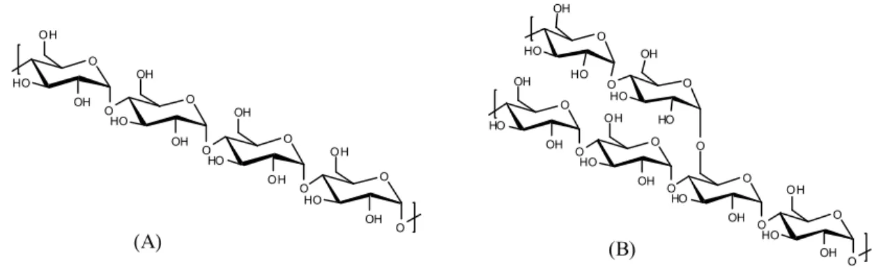
Starch is the safest carbohydrate. It consists of two types of molecules: the linear and helical amylose (20 – 25 wt%) and the highly branched amylopectin (75 – 80 wt%) (Figure 1.1). Amylose has a molecular weight approximately between 40,000 and 340,000, while amylopectin has a much higher molecular weight which may reach 80,000,000.
O HO OH O O HO OH OH OH O O HO OH OH O O HO O OH OH O HO OH O O HO OH OH OH O O HO O OH O O HO O OH OH O HO OH O O HO OH HO HO (Α) (B)
Figure 1.1. The chemical structures of (A) amylose and (B) amylopectin.
Starch is widely used as glidant, diluent, disintegrant and binder for tablets. It is generally recognized that drug release is strongly influenced by the starch origin and its degree of cross-linking in the tablet.7, 10-12 The degree of cross-linking is directly related to the capacity of the starch to undergo a transition from V- to B-type double helix arrangement upon hydration, which is very important in controlling water transport and drug release rate.13, 14
The presence of –OH group at positions C2, C3, C6 of glucose allows for various modifications of amylose and amylopectin. Carboxymethyl starch has been studied for its gastroprotection capacity as it has a pKa around 4.2. In acidic medium, carboxylic groups are
protonated, giving a compact shape of the tablets.15, 16 In the absence of cross-linking, drug release from monolithic tablets made of carboxymethyl starch is controlled by a combination of tablet erosion and diffusion of the drug from the swollen matrix.15, 17, 18
4 Administration (USFDA) for use as inactive ingredients in oral tablets and capsules.19
1.1.2. Cellulose
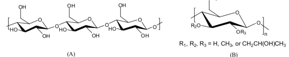
Cellulose is the skeletal substance of all vegetable tissues and the most abundant polymer on earth. It consists of a linear chain of several hundred to over ten thousand D-glucose units which are β-1,4 linked (Figure 1.2 A). Every other glucose in cellulose is flipped over due to the β-1,4 linkages, which promotes intrachain and interchain hydrogen bonds, as well as van der Waals interactions. These interactions make cellulose linear and highly crystalline.7 Cellulose is insoluble in water and in most of the common solvents; the poor solubility is attributed primarily to the strong hydrogen bonding. However, the solubility may be enhanced by substitutions and is highly dependent on the degree of substitution. The cellulose derivatives are mainly obtained by esterifications and etherifications at the hydroxyl groups of cellulose, yielding products such as sodium carboxymethyl cellulose and methyl cellulose, etc. Hydroxypropyl methylcellulose (HPMC) (Figure 1.2 B) is one of the most commonly used polymers to retard the release of water-soluble drugs.20-23 Since the hydroxypropyl group is hydrophilic and methoxyl group is hydrophobic, the ratio of hydroxypropyl to methoxyl content affects the extent of polymer interaction with water. This property will in turn influence water mobility in a hydrated gel layer and drug release.20, 24, 25 The viscosity of HPMC plays an important role regulating tablet swelling and drug release. Higher viscosity grades of HPMC normally lead to slower swelling and drug release due to the greater effect of chain entanglement associated with higher molecular weights.26
Many cellulose derivatives have been approved by USFDA for use as inactive ingredients in oral tablets and capsules. They include carboxymethyl cellulose, cellulose acetate, ethylcellulose, methyl hydroxyethyl cellulose, and HPMC, etc.19
O HO OH OH O O HO OH OH O O HO OH OH O O R2O OR3 O OR1 R1, R2, R3= H, CH3, or CH2CH(OH)CH3 n (A) (B)
Figure 1.2. The chemical structures of (A) cellulose and (B) hydroxypropyl methyl cellulose
5
1.1.3. Chitosan
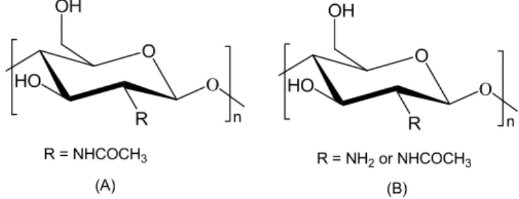
Chitosan has attracted research attention for decades.27, 28 Chitosan is produced commercially by deacetylation of chitin, which is the structural element in the exoskeleton of crustaceans (crabs, shrimps, etc.). Chitosan is a linear cationic polysaccharide composed of randomly distributed β-1,4 linked D-glucosamine (deacetylated unit) and N-acetyl-D-glucosamine (Figure 1.3). Very interestingly, chitosan can prolong residence time in gastrointestinal tract through mucoadhesion, and enhance absorption by increasing permeability, which are major factors contributing to its widespread evaluation as a component of oral dosage forms. The pKa value of chitosan is around 6.3. Chitosan is thus
insoluble in water at neutral pH but soluble under slightly acidic conditions due to the protonation of the amine groups.27 The pH sensitivity makes chitosan a unique polymer for oral drug delivery applications. However, the physicochemical properties of chitosan vary by its molecular weight, degree of deacetylation, the salt form, and charge density.
O HO R Ο OH n R = NH2 or NHCOCH3 O HO R Ο OH n R = NHCOCH3 (A) (B)
Figure 1.3. The chemical structures of (A) chitin and (B) chitosan.
Chitosan's amine groups and hydroxyl groups allow chemical derivatization by which the properties of this polymer can be modulated and adjusted to the intended application. This leads to a large variety of chitosan derivatives with different physical and biological properties, such as improved solubility and permeation enhancement.
To date, neither chitosan nor its derivatives has been approved by USFDA for use as inactive ingredients in oral drug dosage forms due to safety and tolerability concerns. Chitosan might cause mineral and vitamin depletion in the GIT.
To summarize, polysaccharides are biocompatible, biodegradable, mucoadhesive, and chemically versatile. The physicochemical properties and the drug delivery performances of
6 polysaccharides are influenced by factors such as ratio of components (amylose/amylopection ratio for starch, deacetylation degree of chitosan), molecular weight and viscosity, substitution degree, interaction between drug and matrix, cross-linking degree, and charge density, etc. The desirable pharmacokinetics may be tailored by functionalizations of the suitable polysaccharides. In the expanding field of controlled drug delivery, pharmaceutical polymers based on polysaccharides are playing an increasingly important role.
Other polysaccharides of pharmaceutical interest are presented in Appendix I for reference.
1.2. Drug Release Kinetics
Nearly all of the oral extended-release dosage forms fall into one of the following two technologies: matrix (monolithic) systems which consist of a rate-controlling polymer matrix through which the drug is dissolved or dispersed, and reservoir (coated) systems where drug-containing core is enclosed within a polymer coating.20 A matrix tablet is the simplest and the most cost-effective method to fabricate an extended-release dosage form.
The mechanism of drug release from hydrophilic matrix tablets is based on diffusion of the drug and erosion of the outer hydrated polymer on the surface of the matrix.20 Typically, when the matrix tablet is exposed to gastrointestinal fluids, the surface of the tablet is wetted and a gel layer formed around the matrix, which acts as a diffusing barrier to drug release. During the process, the polymer matrix undergoes a transition from the glassy to rubbery state. This leads to the relaxation of the matrix which also contributes to the mechanism of drug release. In the case of a highly soluble drug, an initial burst release occurs due to the presence of the drug on the surface of the matrix tablet. The gel layer (rubbery state) grows with time as more water permeates into the matrix, along with the shrinkage of the glassy core.
If the matrix is not cross-linked, as the outer layer becomes fully hydrated, the polymer chains become completely relaxed and can no longer maintain the integrity of the gel layer, thereby leading to disentanglement and erosion of the surface of the matrix. Water continues to penetrate towards the core of the tablet through the gel layer until it has been completely eroded.20
7 swelling after immersion in the media for a few hours, well before the whole tablet becomes fully hydrated. But the diffusing rates of the water towards the core and the drug out of the gel still increase until the processes reach equilibrium. The duration varies remarkably with the polymer matrix from 2-3 hours to days.
Drug solubility is an important factor determining the mechanism of drug release from hydrophilic matrix. Soluble drugs can be released by a combination of diffusion and erosion mechanisms whereas erosion is the predominant mechanism for insoluble drugs.20, 29 The insoluble drug may dissolve slowly and have slow diffusion through the gel layer of the hydrophilic matrix. Therefore, the drug is released through the erosion of the surface of the hydrated matrix. For drugs with very high solubility, the drug dissolves within the gel layer and diffuses out into the media. Take HPMC tablets as an example, the drug release rate is controlled mainly by diffusion and erosion (or polymer dissolution) for soluble and insoluble drug, respectively.26
The diffusion in polymers can be divided into three categories depending on the rates of diffusion and relaxation. If the rate of diffusion is much lower than that of relaxation, the diffusion is called Case I or Fickian diffusion. In Case II, diffusion is much more rapid than the relaxation processes. Non-Fickian or anomalous diffusion occurs when the diffusion and relaxation rates are comparable.30 Glassy polymers generally exhibit non-Fickian behavior while the diffusion in rubber polymers is Fickian because rubber polymers respond very rapidly to changes.30
A large number of mathematical models have been developed to describe drug release profiles from matrix systems.20, 22, 31-33 The simplest yet most widely used model is the one
derived by Korsmeyer et al.34:
/ n
t
M M∞ = ⋅ (1.1) k t where Mt / M∞ is the fraction of drug release, k is the diffusion rate constant, t is the release
time and n is the exponent indicative of the mechanism of drug release. If a tablet is analogous to a slab, the exponent n is 1.0 or 0.5, the drug release mechanism follows zero-order release kinetics (also termed as Case II transport) or Fickian diffusion (Case I transport), respectively.35 Values of n between 0.5 and 1 indicate the contribution of both the diffusion process as well as polymer relaxation in controlling the release kinetics (non-Fickian, anomalous or first-order release). For cylindrical tablets, these values are 0.45, 0.45
8 < n < 0.89 and 0.89 for Fickian, anomalous or Case II transport, respectively.22 Drug release profiles from HPMC hydrophilic matrices are generally first-order for highly soluble drugs or zero-order for practically insoluble drugs, with the release exponent n ranging from 0.5 to 0.8.20 The anomalous release is owing to the contribution of a mechanism other than diffusion of drug transport.36
More complex mechanistic theories that consider diffusion, swelling and dissolution processes simultaneously have been developed.22 The choice of the appropriate mathematical model strongly depends on the desired predictive ability.
1.3. The Characterization of Polymer Matrices
The polymer matrix and the resultant tablets have been studied by various techniques, such as optical imaging,12, 26, 37-39 mechanical method,40 Fourier transform infrared spectroscopy (FT-IR),11, 41 X-ray diffraction,11, 41 X-ray computed microtomography (CMT),42 scanning electron microscopy (SEM),40, 42 cross-polarization magic angle spinning (CP-MAS) nuclear magnetic resonance (NMR) spectroscopy,14, 43, 44 and NMR imaging.45-50 Gravimetric experiments and dissolution tests are also widely used in the aim to study the drug release mechanism.
1.3.1. XRD and FT-IR Studies
The drug release rate of the tablets based on cross-linked high amylose starch (CHAS) was studied as a function of cross-linking degree, which showed that sustained release up to 20 h was achieved at a low cross-linking degree of 6% (the weight ratio of epichlorohydrin to high amylose starch). The CHAS tablets of higher cross-linking degree up to 20% showed a sharp decrease in the release time. Typically, a tetrafunctional polymer network would be created by increasing the degree of cross-linking, leading to a reduced swelling capacity. However, CHAS yielded a nonlinear response for swelling capacity and drug release.51 X-ray diffraction studies suggested that the increase in cross-linking degree induces a transition of amylose from B-type (double helix) to a predominant V-type (single helix) and a loss in crystallinity. A high cross-linking degree also limited chain flexibility. Another consequence was that fewer chains were stabilized by interchain hydrogen-bonding and thus more
9 hydroxyl groups were available for dynamic exchange with free water, which facilitates hydration of the CHAS power, as reflected by the intensity of FT-IR water deformation vibration mode.41 The conformation studies suggested that an optimized crystalline/amorphous ratio is responsible for tablet integrity during swelling, water penetration, and drug release of the CHAS-based monolithic dosage forms.11
1.3.2. SEM and CMT Studies
The SEM and CMT experiments demonstrated the skin-core structure of the CHAS tablets as a result of interaction with water and reorganization.42 The core is surrounded by the skin consisting of two concentric layers, an inner porous membrane and an outer very dense membrane, which acts as pseudo-crosslinks and retain the integrity of the tablets.
The gravimetric method can be used to record water uptake of drug tablets. At appropriate time intervals, each tablet is removed from water and weighed. The water uptake data can be used to determine water diffusion mechanism. For example, the dynamic swelling and equilibrium swelling of poly(2-hydroxyethyl methacrylate-co-methacrylic acid) (poly(HEMA-co-MAA)) in buffer of different pH values were studied gravimetrically.52 The mechanism of water diffusion in the gel became more anomalous as the pH of the swelling medium increased and as the ionic strength decreased at a constant pH ≥ pKa. The mechanism
of water diffusion was Fickian and independent of ionic strength at a pH lower than the pKa.
The anionic polymeric networks ionized as the pH of the medium rose above the pKa of that
ionizable moiety. Increasing ionization at higher pH might affect the relative magnitude of diffusion and relaxation times. The reason is that the electrostatic repulsion between adjacent ionized carboxylate groups led to chain expansion, which in turn affected polymer chain relaxation. Despite the easy access, the gravimetric method has a disadvantage that the tablet transfer process and the ex-situ method would inevitably affect the swelling process and alter the fragile outer region of the gel, to the detriment of accuracy.
1.3.3. Dissolution Studies
Dissolution test is the only way to measure the rate of in vitro drug release as a function of time. Dissolution tests are commonly conducted according to the procedure now rigorously
10 and comprehensively defined in US Pharmacopeia. The objective of the dissolution test is to establish dissolution mechanism. In general, analytical methods used for quantifying drug release in dissolution tests can be classified into four categories: spectrophotometric, chromatographic, mass spectrometric, and potentiometric. Direct UV-vis spectrophotometric determination is most widely used for drug containing chromophores. The drug release profile is then used to determine whether the drug release is controlled by diffusion or by polymer relaxation mechanism.
A dissolution study was done to follow the behavior of CHAS derivatized with cationic (carboxymethyl), anionic (aminoalkyl) groups and less polar group (acetate) to study their release control properties and interactions with charged drugs (acetylsalicylic acid and metformin) and an uncharged drug (acetaminophen).53 Ionic interactions retarded drug release effectively, the effect being more significant at high drug loading in the case of drugs of high solubility (such as metformin).
1.3.4. Optical Imaging
Optical imaging has been used to track the penetration of water into the tablets.12, 26, 37-39 This method offers ease of manipulation with relatively low instrument cost. The gel layer normally appears as a bright ring and the dry core as a dark part due to the scattering of light by the hydrated polymer.37 In digital image processing, the image is converted into a discrete number of points called pixels, which are assigned a numeric location and grey level value. The intensity of the signal was related to the water content in the tablets, the method is therefore not only used to monitor the tablet swelling, but also to characterize the moving front and the solvent concentration gradient. Optical imaging also demonstrated that the equilibrium swelling state was attained much after the solvent penetration fronts had met.12 On the basis of the empirical relationship between scattered light intensity and HPMC concentration, the apparent gel region of a swelling HPMC tablet can be defined with respect to polymer concentration. The polymer concentration within the gel layer of HPMC tablets is around 5-50%.37 As a matter of fact, the solvent penetration front goes slightly deeper than the apparent gel front. A very small portion of the gel region of higher polymer concentration is beyond the detection limit of optical imaging due to the opacity.
11
1.3.5. NMR Imaging Studies
NMR imaging (NMRI) has been used to study the matrix systems for more than a decade as NMRI can provide cross-sectional images from inside solid materials.24, 39, 46-50, 54-63 Contrary to this, optical imaging can only observe the surface and the averaged diffraction signal of a tablet. NMRI uses magnetic field gradients to encode the NMR signal with spatial information. The amplitude of the signal indicates the water concentration at a particular position within the sample. The noninvasive and nondestructive nature of NMRI is especially advantageous for tracking the water penetration and the swelling of the matrix of the tablets in terms of both the development of the gel layer and the dimensional change of the core. Quantitative information can be obtained regarding the kinetics of the diffusion process and the polymer or drug concentration gradient upon hydration exclusively by NMRI rather than optical imaging.12 A number of investigations47, 49, 55, 56, 64, 65 aimed at characterizing the formation of the gel layer and determining how its properties influence drug release. Normally the images of the dimensional changes in the gel layer and the core are monitored and solvent concentration profiles (proton density profiles) are extracted from the images. NMRI can also be used to monitor the distribution of drug inside the tablets. For example, 19F NMRI was chosen to measure triflupromazine–HCl and 5-fluorouracil concentration in HPMC-based tablets.29
Diffusion-weighted NMR imaging experiments are essential to studying the mobility of water inside the gel layer and determine the spatial distribution of self-diffusion coefficient of water. Such kind of studies showed that there was a water mobility gradient across the gel layer of HPMC and poly(vinyl alcohol) (PVA) matrices and the limited mobility of polymer chains near the glassy region hindered solvent transport.24 From the outer gel to the glassy core, the self-diffusion coefficient of water decreases from that of free water (2×10⁻⁹ m2/s at 25oC) to zero.24
1.3.6. Relaxation Time Studies
T2 (transverse relaxation time) measurement of tablets is an interesting method to deduce
the polymer concentration of hydrated layer of the swollen tablets.45 Firstly, the dependence of the 1H T2 values on the HPMC weight fraction was obtained by measuring the relaxation
12 percentages were then calculated from T2 values with the determined calibration. At the same
time, the percentages of the water bound to polymer and the free water were obtained.
1.4. Diffusion in Polymer-Water-Diffusant Systems
The diffusion of small molecules to macromolecules in polymer solutions and gels has attracted increasing research interest in the past decade. A particular important application involves the hydrogels as controlled release carriers of drugs. The effects of polymer concentration, size, and shape of the diffusant, temperature, and specific interactions within the polymer matrix are important in determining the diffusion properties in a polymer system. Our group has extensively studied the self-diffusion of small and large molecules in polymer-water-diffusant systems by pulsed-gradient spin-echo (PGSE) NMR spectroscopy.66-73
The diffusants having been studied included water, methanol, tert-butanol, formamide, acetic acid, trimethylamine, tetramethylammonium cation, oligo- and poly(ethylene glycol) of increasing molecular weight from 62 to 10,000,66-69 a series of end-capped ethylene glycol and oligo(ethylene glycol),70 poly(propyleneimine) dendrimers (generations 2, 4, and 5),71 hyperbranched polyglycidols,72 and carboxylated dendrimers.73
The matrices are mainly poly(vinyl alcohol) (PVA).66-73 Besides, hydroxypropyl methyl cellulose (HPMC),69 poly(N,N-diethylacrylamide) (PNNDEA),69 poly(N-isopropylacrylamide) (PNIPA),69 and poly(allyl amine) (PAAm)73 were also used to study the effects of different matrices and ionic interactions between the diffusants and the polymer matrices.
1.4.1. Molecular Size of the Diffusant
It was found that at a given polymer concentration the size of the diffusant has the most significant effect on its diffusion in the absence of strong interactions between the diffusants and the polymer network.66-72
13
1.4.2. Concentration of the Polymer
The self-diffusion coefficient of diffusants ranging from small molecules to macromolecules in PVA solutions and gels decreased with increasing PVA concentration (from 0 to 0.38 g/mL).66-73
1.4.3. Molecular Weight and Hydrolysis Degree of the Polymer
The diffusion of both small molecules and linear poly(ethylene glycol)s (PEGs) did not vary significantly with the molecular weight (50,000 and 115,000) of the PVA matrix, and only a small variation was observed with the degree of hydrolysis of the PVA (87% and 99%).66
1.4.4. Geometry of the Diffusant
The molecular geometry of the diffusant plays an important role in the diffusion process, as reflected by a study of a series of end-capped ethylene glycol and oligo(ethylene glycol)s in PVA aqueous solutions and gels.70 The end groups included both flexible groups (methyl, ethyl, hexyl) and rigid groups (tert-butyl and aromatic groups). The results showed that the methyl groups reduced or prevented the binding of ethylene glycol to PVA.70 Diffusants with a bulkier end group (such as tert-butyl group) diffused less rapidly than those with a smaller linear end group even though the molecular weights of the molecular are comparable.70
The dendritic polymers, poly(propyleneimine) dendrimers with hydrophilic triethylenoxy methyl ether terminal groups diffused faster in PVA aqueous solutions and gels than the linear PEGs when the molecular weights were similar.71 Hyperbranched polyglycidols of molecular weights from 300 to 2000 were also studied.72 For diffusants of similar molecular weight and without specific interactions, the activation energy increased from the dendrimers to hyperbranched polymers and then to linear polymers.72
1.4.5. Effect of Temperature
The studies on the effect of temperature on the diffusion of oligo- and poly(ethylene glycol)s showed that the self-diffusion coefficients increased with increasing temperature.67,
14
1.4.6. Interaction Between the Diffusants and the Polymers
The study of the diffusion of small molecules with different functional groups (alcohol, amine, ammonium salt, amide, and acid) in PVA solutions and gels showed that the interactions had little impact on the diffusion behaviour because the diffusion was primarily influenced by the size of the diffusant.66
The ionic interaction between the carboxylated dendritic diffusants and the cationic poly(allyl amine) network significantly reduced the diffusion rate of the diffusant. The self-diffusion coefficients were an order of magnitude lower than those in PVA and the values were more widely distributed.73
1.4.7. Various Polymer Matrices
The effect of the polymer matrices on the self-diffusion of PEG with a molecular weight 600 was studied for different ternary polymer-water-PEG systems (PVA, HPMC, PNNDEA, and PNIPA).69 The diffusion in hydrophilic polymers was mostly affected by formation of the hydrogen bonds between the solute and the polymer matrix.
Several pertinent physical models of diffusion were evaluated and the physical significance of the parameters were analyzed as well.67 These models were based on the obstruction effect, the free volume effect, or hydrodynamic interactions. The difficulties in their applications include failure to describe large probes, lack of knowledge of some parameters that are not readily available, and lack of physical significance of the parameters.67-69 Our group proposed a model suitable for both small and large diffusants in dilute and concentrated polymer systems.74 This model has been validated by a number of diffusants in the polymer-water-diffusant ternary systems, including oligo- and poly(ethylene glycol)s,67, 68, 70 dendrimers,71, 73 hyperbranched polyglycidols72 in PVA, water and PEG69 in PVA, HPMC, PNNDEA, and PNIPA. In all the cases, the model successfully described the effects of polymer concentration, temperature, and molecular size of the diffusants.
1.5. Water Diffusion in CHAS Tablets
15 content of amylose was chosen because it yielded a stronger gel.51 Firstly, high-amylose starch was activated by heating in sodium hydroxide solution. Phosphorous oxychloride was used as the cross-linking agent along with propylene oxide added to further functionalize amylose and amylopectin molecules. Covalent cross-links and hydroxypropyl side chains of the final product allow greater stability by hindering retrogradation over time.51 Retrogradation of starch is used to define the changes from amorphous state to a more ordered or crystalline state.
CHAS attracted attention due to its sustained drug release property as an excipient of oral drug dosage form. A membrane at the outer layer of CHAS tablets quickly formed upon hydration and the tablets could maintain the integrity in water for days. The absence of erosion and limited swelling allow for further explorations of the applications in drug delivery.13
CHAS tablets have been extensively studied by NMRI and a few other polymer characterization techniques by our group.46-50, 55 The result obtained were summarized hereafter.
1.5.1. Swelling of CHAS Tablets
Similar to other tablets, when a CHAS tablet is immersed into water, water penetrates into the hydrophilic polymer matrix easily to form a hydrogel, leading to a steep water gradient. The formation of the membrane which acts as a barrier opposing water and drug transport was well characterized by NMRI.42, 49 The glassy-rubbery transition is based on the lowering of the glass-transition temperature (Tg) of the polymer, which is controlled by the
water concentration and depends on temperature and thermodynamic interactions of the polymer-water system.36 The swelling of the CHAS tablets approaches its maximum at around 10 h at 37 oC but 60% of swelling can be achieved within the first 4 h.48
The swelling is anisotropic with the axial swelling being much more significant than the radial swelling for all the tablets of different sizes and within the temperature range from 25 to 60oC.46-48 The more pronounced axial swelling was related to the relief of the stresses induced during compaction of the matrix tablets.77 However, the gel layer along both directions are similar in thickness.39
16
1.5.2. Effect of Temperature
Four temperatures (25, 37, 45, and 60 oC) were employed to study the water diffusion and the swelling of the CHAS tablets.46, 48 The tablets have a faster swelling with increasing temperature up to 60 oC. The diffusion process is Fickian between 25 and 45 oC and Case II at 60 oC. The difference is caused by the different degrees of the transformation from V-type single helix polymorph to B-type double helices during water uptake.47, 48 The double helical conformation acts as physical cross-links which in turn limits the swelling and thus is essential for sustained drug release, which is verified by CP-MAS 13C NMR spectroscopy. The details of the transformation include the following steps with water penetrating into a tablet to act as a plasticizing agent. This plasticizing effect increases intermolecular space and thus allows greater mobility to starch molecules which rapidly start reorganizing into the thermodynamically most stable conformations: B-type double helices.51
1.5.3. Effect of the Tablet Size
The water uptake and the tablet swelling strongly depend on the size of the tablets.47 The swelling rate is faster for the small tablet along both swelling directions due to its larger surface/volume ratio. In both cases, the decrease of the dry thickness is more rapid than that of the dry diameter with immersion time.
1.5.4. Effect of Drug Loading
The drug loading effect has been studied by comparing tablets loaded with 10 wt% soluble drugs (ciprofloxacin and acetaminophen) and tablets containing exclusively polymer matrix.49 The extent of the swelling of the tablets was not noticeably influenced by a 10 wt% drug-loading, since the presence of drugs at this content did not interfere with the formation of double helices which limited the overall swelling of the CHAS tablets.49 However, the presence of drug molecules caused a faster water uptake, as reflected by the higher diffusion coefficients of water.
17
1.6. Objectives of This Study
The objective of the study is to elucidate the effect of architecture of polymers, the level of drug loading, and the pH value and ionic strength of the environment on the diffusion of the diffusant and the swelling of the polymer matrices. A justification for these choices follows.
Every natural or synthetic polymer falls into one of the following categorized architectures: linear, graft, branched, cross-linked, star-shaped, and dendritic. The effect of shape of the diffusants has been preliminarily investigated by comparing the diffusion of linear, dendritic, and hyperbranched polymers in polymer aqueous solutions and gels.71, 72 As a series of star-shaped polymers with four arms have been successfully synthesized in our group, a comparison among the polymers of different architectures becomes highly desirable.
Hydrophilic drugs have been believed to trigger water uptake and water diffusion into the polymer matrix, and thus it is challenging to yield a sustained release when the level of drug loading is high. The CHAS tablets loaded with only 10 wt% of drug have been studied up to now. It is important to measure the rate of water diffusion in the tablets with different drug loading levels and to correlate it with the swelling of the tablets and the rate of drug dissolution.
In the past, all the NMR Imaging studies of the CHAS tablets used only water as the medium. To study the swelling of the tablets and the solvent uptake in the physiological environment (for example, the media simulating the stomach and the small intestine fluids) is of significant importance. To understand the effect of pH on the drug release from a certain matrix is also highly desirable due to the different pH values in human’s gastrointestinal tract. The pH value and ionic strength are expected to significantly affect the swelling of the polymer matrices containing ionic groups. A chitosan-based polyelectrolyte complex is an interesting material which shows pH-dependent association and dissociation among the ionic groups attached to the polymer chains. It should be compared with CHAS.
1.7. Scope of This Study
This thesis comprises of a series of studies leading to the development of a relationship between drug release kinetics and physicochemical properties of polymers such as the
18 diffusion coefficients of the polymer and water, liquid uptake, and kinetics of swelling of the matrices.
Chapter 2 focuses on the diffusion of a star polymer in aqueous solution and PVA gel studied by PGSE NMR spectroscopy. An amphiphilic star polymer is compared with linear polymers and dendrimers.
Chapter 3 presents the effect of drug loading on water uptake kinetics, swelling, drug release of CHAS tablets. The effect is studied by NMR imaging, drug dissolution experiments complementary to a quantitative measurement of diffusion coefficients in the hydrated tablets with acetaminophen loading up to 40 wt%.
Chapter 4 describes the effect of pH and ionic strength of the external media on the swelling of tablets made of a polyelectrolyte complex. The complex is formed between chitosan and sodium carboxymethyl starch (CMS).
The thesis concludes with a summary and suggestions for future work. Studies on the effect of polymer architecture, drug loading, and various media would be valuable for the development and optimization of biopolymer systems for drug delivery.
1.8. References
1. Jones, D., Pharmaceutical Applications of Polymers for Drug Delivery. Rapra Technology Limited: Shropshire, UK, 2004; Vol. 15.
2. Kendall, R. A.; Basit, A. W., The Role of Polymers in Solid Oral Dosage Forms. In Polymer in Drug Delivery, Uchegbu, I.; Schatzlein, A., Eds. CRC Press: Boca Raton, 2006. 3. Alexander, C. Expert Opin. Emerging Drugs 2001, 6, 345-363.
4. Chaubal, M. V., Polymeric Excipients for Controlled Release Applications. In Excipient Development for Pharmaceutical, Biotechnology, and Drug Delivery Systems, Katdare, A.; Chaubal, M. V., Eds. Informa Healthcare USA, Inc. : New York, 2006.
5. Abrahamsson, B.; Lennernäs, H., Application of the Biopharmaceutic Classification System Now and in the Future. In Drug Bioavailability: Estimation of Solubility, Permeability, Absorption and Bioavailability, Waterbeemd, H. v. d.; Lennernäs, H.; Artursson, P., Eds. Wiley-VCH: Weinheim, 2003.
19 7. Klein, S., Polysaccharides in Oral Drug Delivery –Recent Applications and Future Perspectives In Polysaccharide Materials: Performance by Design, Edgar, K. J.; Heinze, T.; Buchanan, C. M., Eds. ACS: 2009.
8. Na, K.; Bae, Y. H., pH-Sensitive Polymers for Drug Delivery In Polymeric Drug Delivery Systems, Kwon, G. S., Ed. Taylor & Francis Group: Boca Raton, FL, 2005.
9. Shen, Y.; Tang, H.; Radosz, M.; Kirk, E. V.; Murdoch, W. J., pH-Responsive Nanoparticles for Cancer Drug Delivery. In Drug Delivery Systems, Jain, K. K., Ed. Humana Press: Totowa, NJ, 2008.
10. Onofre, F.; Wang, Y.-J.; Mauromoustakos, A. Carbohydr. Polym. 2009, 76, 541-547 11. Ispas-Szabo, P.; Ravenelle, F.; Hassan, I.; Preda, M.; Mateescu, M. A. Carbohydr. Res.
2000, 323, 163-175.
12. Moussa, I. S.; Cartilier, L. H. J. Control. Release 1996, 42, 47-55.
13. Lenaerts, V.; Moussa, I.; Dumoulin, Y.; Mebsout, F.; Chouinard, F.; Szabo, P.; Mateescu, M. A.; Cartilier, L.; Marchessault, R. J. Control. Release 1998, 53, 225-234.
14. Thérien-Aubin, H.; Janvier, F.; Baille, W. E.; Zhu, X. X.; Marchessault, R. H. Carbohydr. Res. 2007, 342, 1525-1529.
15. Massicotte, L. P.; Baille, W. E.; Mateescu, M. A. Int. J. Pharm. 2008, 356, 212-223. 16. Calinescu, C.; Mateescu, M. A. Eur. J. Pharm. Biopharm. 2008, 70, 582-589. 17. Sen, G.; Pal, S. J. Appl. Polym. Sci. 2009, 114, 2798-2805.
18. Brouillet, F.; Bataille, B.; Cartilier, L. Int. J. Pharm. 2008, 356, 52-60.
19. Inactive ingredients database. http://www.accessdata.fda.gov/scripts/cder/iig/index.cfm (April 3, 2010),
20. Tiwari, S. B.; Rajabi-Siahboomi, A. R., Extended-Release Oral Drug Delivery Technologies: Monolithic Matrix Systems. In Drug Delivery Systems, Jain, K. K., Ed. Humana Press: Totowa, NJ 2008.
21. Cao, Q. R.; Choi, Y. W.; Cui, J. H.; Lee, B. J. J. Control. Release 2005, 108, 351-361. 22. Siepmann, J.; Peppas, N. A. Adv. Drug Deliver. Rev. 2001, 48, 139–157.
23. Rowe, R. C.; Sheskey, P. J.; Quinn, M. E., Handbook of Pharmaceutical Excipients. 6th ed.; Pharmaceutical Press: London, 2009.
24. Rajabi-Siahboomi, A. R.; Bowtell, R. W.; Mansfield, P.; Davies, M. C.; Melia, C. D. Pharm. Res. 1996, 13, 376-380.
20 25. McCrystal, C. B.; Ford, J. L.; Rajabi-Siahboomi, A. R. J. Pharm. Sci. 1999, 88, 797-801. 26. Pham, A. T.; Lee, P. I. Pharm. Res. 1994, 11, 1379-1384.
27. Kumar, M. N. V. R.; Muzzarelli, R. A. A.; Muzzarelli, C.; Sashiwa, H.; Domb, A. J. Chem. Rev. 2004, 104, 6017-6084.
28. Park, J. H.; Saravanakumar, G.; Kim, K.; Kwon, I. C. Adv. Drug Deliver. Rev. 2010, 62, 28-41.
29. Fyfe, C. A.; Blazek-Welsh, A. I. J. Control. Release 2000, 68, 313-333.
30. Crank, J., The Mathematics of Diffusion. 2nd ed.; Oxford University Press: Oxford, 1975.
31. Narasimhan, B. Adv. Drug Deliver. Rev. 2001, 48, 195-210.
32. Siepmann, J.; Göpferich, A. Adv. Drug Deliver. Rev. 2001, 48, 229-247. 33. Parker, R. S.; Doyle III, F. J. Adv. Drug Deliver. Rev. 2001, 48, 211-228.
34. Korsmeyer, R. W.; Gurny, R.; Doelker, E.; Buri, P.; Peppas, N. A. J. Pharm. Sci. 1983, 72, 1189–1191.
35. Bajwa, G. S.; Hoebler, K.; Sammon, C.; Timmins, P.; Melia, C. D. J. Pharm. Sci. 2006, 95, 2145–2157.
36. Colombo, P.; Bettini, R.; Santi, P.; Peppas, N. A. Pharm. Sci. Technol. To. 2000, 3, 198-204.
37. Gao, P.; Meury, R. H. J. Pharm. Sci. 1996, 85, 725-731.
38. Bussemer, T.; Peppas, N. A.; Bodmeier, R. Eur. J. Pharm. Biopharm. 2003, 56, 261-270. 39. Moussa, I. S.; Lenaerts, V.; Cartilier, L. H. J. Control. Release 1998, 52, 63–70.
40. Ravenelle, F.; Marchessault, R. H.; Legare, A.; Buschmann, M. D. Carbohydr. Polym.
2002, 47, 259-266.
41. Dumoulin, Y.; Alex, S.; Szabo, P.; Cartilier, L.; Mateescu, M. A. Carbohydr. Polym.
1998, 37, 361-370.
42. Chauve, G.; Ravenelle, F.; Marchessault, R. H. Carbohydrate Polymers 2007, 70, 61-67. 43. LeBail, P.; Morin, F. G.; Marchessault, R. H. Int. J. Biol. Macromol. 1999, 26, 193-200. 44. Shiftan, D.; Ravenelle, F.; Mateescu, M. A.; Marchessault, R. H. Starch-Starke 2000, 52, 186-195.
21 46. Baille, W. E.; Malveau, C.; Zhu, X. X.; Marchessault, R. H. Biomacromolecules 2002, 3, 214-218.
47. Malveau, C.; Baille, W. E.; Zhu, X. X.; Marchessault, R. H. Biomacromolecules 2002, 3, 1249-1254.
48. Thérien-Aubin, H.; Baille, W. E.; Zhu, X. X.; Marchessault, R. H. Biomacromolecules
2005, 6, 3367-3372.
49. Thérien-Aubin, H.; Zhu, X. X.; Ravenelle, F.; Marchessault, R. H. Biomacromolecules
2008, 9, 1248-1254.
50. Thérien-Aubin, H.; Zhu, X. X. Carbohydr. Polym. 2009, 75, 369-379.
51. Ravenelle, F.; Rahmouni, M., Contramid: High-Amylose Starch for Controlled Drug Release. In Polysaccharides for Drug Delivery and Pharmaceutical Applications, Marchessault, R. H.; Ravenelle, F.; Zhu, X. X., Eds. American Chemical Society: Washington, DC, 2006; pp 79-104.
52. Khare, A. R.; Peppas, N. A. Biomaterials 1995, 16, 559-567.
53. Mulhbacher, J.; Ispas-Szabo, P.; Lenaerts, V.; Mateescu, M. A. J. Control. Release 2001, 76, 51-58.
54. Dahlberg, C.; Fureby, A.; Schuleit, M.; Dvinskikh, S. V.; Furo, I. J. Control. Release
2007, 122, 199-205.
55. Thérien-Aubin, H.; Zhu, X. X., Water diffusion in drug delivery systems made of high-amylose starch as studied by NMR imaging. In Polysaccharides For Drug Delivery And Pharmaceutical Applications, Marchessault, R. H.; Ravenelle, F.; Zhu, X. X., Eds. American Chemical Society: Washington, D. C., 2006; pp 105-120.
56. Richardson, J. C.; Bowtell, R. W.; Mader, K.; Melia, C. D. Adv. Drug Deliv. Rev. 2005, 57, 1191-1209.
57. Kowalczuk, J.; Tritt-Goc, J.; Pislewski, N. Solid State Nucl. Magn. Reson. 2004, 25, 35-41.
58. Chowdhury, M. A.; Hill, D. J. T.; Whittaker, A. K.; Braden, M.; Patel, M. P. Biomacromolecules 2004, 5, 1405-1411.
59. Chowdhury, M. A.; Hill, D. J. T.; Whittaker, A. K. Biomacromolecules 2004, 5, 971-976.
22 61. Fahie, B. J.; Nangia, A.; Chopra, S. K.; Fyfe, C. A.; Grondey, H.; Blazek, A. J. Control. Release 1998, 51, 179-184.
62. Hopkinson, I.; Jones, R. A. L.; Black, S.; Lane, D. M.; McDonald, P. J. Carbohydr. Polym. 1997, 34, 39-47.
63. Rajabi-Siahboomi, A. R.; Bowtell, R. W.; Mansfield, P.; Henderson, A.; Davies, M. C.; Melia, C. D. J. Control. Release 1994, 31, 121-128.
64. Baumgartner, S.; Lahajnar, G.; Sepe, A.; Kristl, J. Eur. J. Pharm. Biopharm. 2005, 59, 299-306.
65. Kulinowski, P.; Dorozynski, P.; Jachowicz, R.; Weglarz, W. P. J. Pharm. Biomed. 2008, 48, 685-693.
66. Petit, J.-M.; Zhu, X. X.; Macdonald, P. M. Macromolecules 1996, 29, 70-76. 67. Masaro, L.; Zhu, X. X.; Macdonald, P. M. Macromolecules 1998, 31, 3880-3885.
68. Masaro, L.; Zhu, X. X.; Macdonald, P. M. J. Polym. Sci., Part B: Polym. Phys. 1999, 37, 2396-2403.
69. Masaro, L.; Ousalem, M.; Baille, W. E.; Lessard, D.; Zhu, X. X. Macromolecules 1999, 32, 4375-4382.
70. Masaro, L.; Zhu, X. X. Macromolecules 1999, 32, 5383-5390.
71. Baille, W. E.; Malveau, C.; Zhu, X. X.; Kim, Y. H.; Ford, W. T. Macromolecules 2003, 36, 839-847.
72. Baille, W. E.; Zhu, X. X.; Fomine, S. Macromolecules 2004, 37, 8569-8576.
73. Thérien-Aubin, H.; Zhu, X. X.; Moorefield, C. N.; Kotta, K.; Newkome, G. R. Macromolecules 2007, 40, 3644-3649.
74. Petit, J.-M.; Roux, B.; Zhu, X. X.; Macdonald, P. M. Macromolecules 1996, 29, 6031-6036.
75. Lenaerts, V.; Beck, R. H. F.; Van Bogaert, E.; Chouinard, F.; Hopcke, R.; Desevaux, C. Cross-linked high amylose starch for use in controlled-release pharmaceutical formulations and processes for its manufacture. U.S. Patent 6,607,748, August 19, 2003.
76. Mateescu, M. A.; Lenaerts, V.; Dumoulin, Y. Use of cross-linked amylose as a matrix for the slow release of biologically active compounds U. S. Patent 5,456,921, Oct.10, 1995. 77. Papadimitriou, E.; Buckton, G.; Efentakis, M. Int. J. Pharm. 1993, 98, 57-62.
* Modified from a research article published: Y. J. Wang, H. Therien-Aubin, W. E. Baille, J. T. Luo, X. X. Zhu, Polymer, 2010, 51, 2345-2350.
2. Effect of Molecular Architecture on the Self-Diffusion of
Polymers in Aqueous Systems: A Comparison of Linear, Star,
and Dendritic Poly(ethylene glycol)s
*2.1. Abstract
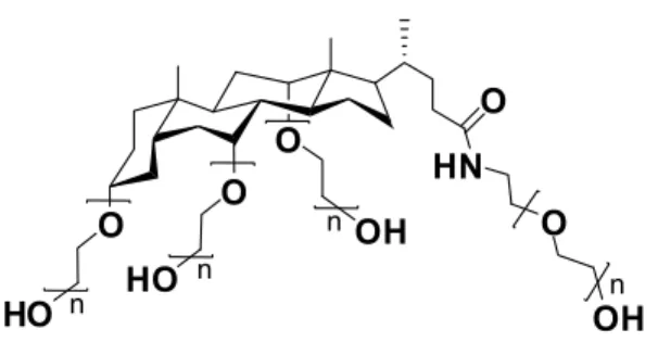
Star polymers with a hydrophobic cholane core and four poly(ethylene glycol) (PEG) arms, CA(EGn)4, have been synthesized by anionic polymerization. Pulsed-gradient spin-echo
NMR spectroscopy was used to study the diffusion behavior of the star polymers, ranging from 1000 to 10,000 g/mol, in aqueous solutions and gels of poly(vinyl alcohol) (PVA) at 23oC. The star polymers have a lower self-diffusion coefficient than linear PEGs at equivalent hydrodynamic radius. In water alone, the star polymers and their linear homologues have a similar diffusion behavior in the dilute regime, as demonstrated by the similar concentration dependence of the self-diffusion coefficients. In the semidilute regime, the star polymers tend to aggregate due to their amphiphilic properties, resulting in lower self-diffusion coefficients than those of linear PEGs. 1H NMR T
1 measurements at 10-70 oC revealed that the PEG arms
of the star polymers are more mobile than the core, suggesting the star polymers in solution have a conformation similar to that of poly(propylene imine) dendrimers.
2.2. Introduction
The study of diffusion is of fundamental importance in describing macromolecular solution dynamics. The determination of diffusion coefficients of macromolecules in solutions or gels of polymer matrices is also important for applications such as controlled delivery of drugs, gel electrophoresis, permeation through membranes, plasticizers in plastic materials, and encapsulation of drugs and fragrances.1-4 The diffusion behavior of a variety of oligomers and polymers, including linear poly(ethylene glycol) (PEG),5-8 dendrimer,9, 10 hyperbranched,11-13 and star polymers14-16 have been studied. The understanding of the dependence of the transport behaviors of the diffusants on their size and shape may help in the design of polymer systems with predictable properties.17, 18 The shape of a macromolecule
24 may have a pronounced effect on its diffusion coefficient. For example, the rodlike protein tropomyosin (aspect ratio R = 26) and globular protein myoglobin (aspect ratio R = 1.6) exhibited similar behavior in agarose gels but markedly different diffusion in carrageenan gel,19 since agarose gel has a mesh size about 6 times of that of carrageenan gel, in which the diffusion of stiff tropomyosin was hindered more significantly. In aqueous solutions of poly(vinyl alcohol) (PVA), a cyclic poly(ethylene oxide) (PEO) of a lower molecular weight (Mn = 6 000) was found to have almost the same self-diffusion coefficients as linear PEO of a
higher molecular weight (Mn = 10 000).20
O O O HN O HO n O HO n OH n OHn
Figure 2.1. The chemical structure of the star polymers used in this study. They are prepared by anionic polymerization of ethylene oxide on a core of cholic acid.21 Four PEG chains are attached and the chain length n = 4, 6, 10, 17, 31, 39, and 54.
Star polymers have attracted significant research interests due to their compact structures and unique physical properties.22-24 Pulsed-gradient spin-echo (PGSE) NMR experiments have revealed that the molecular mobility of star-branched polyisoprenes in C6F5Cl and CCl4
solutions depends largely on the weight fraction of the polymer, and only weakly on the number of the arms.14 Similarly, no marked difference was observed between linear and three-armed polystyrenes and polybutadienes (Mn = 3,000 – 1,000,000) in CCl4 solution from
dilute to semidilute regime.15 Although considerable research concerning the shape effect on both the static and dynamic parameters of polymer solutions has been conducted, very few general conclusions can be drawn. The factor of molecular shape is more difficult to address than the molecular size and the accumulation of results helps in the elucidation of such effects. We have previously compared the self-diffusion of linear PEGs and poly(propylene imine) dendrimers bearing triethylenoxy methyl ether as end groups (PPI(TEG)n).9 In this
25 cholane core (Figure 2.1) by anionic polymerization21 and studied their diffusion behaviors using the PGSE NMR technique. The star polymers with bile acid cores were characterized in a previous study.21 All three series of polymers, star polymers CA(EGn)4, linear PEGs, and
poly(propylene imine) dendrimers, share the same repeat unit, ethylene glycol, while the cores of the star polymers (cholic acid) and dendrimers (poly(propylene imine)) add structural variants for the comparative studies. The self-diffusion coefficient measurements were performed in either binary solutions of the diffusants or ternary systems of PVA-water-diffusant.
2.3. Experimental
2.3.1. Materials
PVA (MW = 89,000 – 98,000, 99% hydrolyzed) and deuterium oxide (D2O) were
purchased from Sigma-Aldrich (Milwaukee, WI). All chemicals were used as received. The star polymers (Figure 2.1) were synthesized as reported previously.21 The molecular weights of the star polymers measured by size exclusion chromatography (SEC), MALDI-TOF mass spectrometry, and NMR spectroscopy21 are listed in Table 2.1 and all results show very low polydispersity indices (PDI = 1.02 ~ 1.05). The molecular weight were obtained both by SEC coupled with a refractive index detector (SEC-RI) calibrated with linear homologues and by SEC with a light scattering detector (SEC-LS). Both detection methods provided similar results, while the absolute molecular weights measured by SEC-LS are systematically 1.1 times of the values obtained by SEC-RI. It should be noted, however, that linear PEGs were used as the standards for SEC-RI. The molecular weights listed in the report are those obtained by SEC-RI unless otherwise specified. The refractive index increment, dn/dc, was measured with a series of 8 solution samples in the concentration range of 0.2 – 3.0 mg/mL for each polymer, using a refractive index detector from Wyatt. The molecular weights were determined by SEC equipped with a differential refractometer (Optilab) and a multiangle light scattering detector (DAWN EOS, wavelength 690 nm) in N,N-dimethylformamide (DMF) at a flow rate of 0.5 mL/min at 25 °C with a set of styragel columns (a TSK-gel α-M, particle size 13 μm, exclusion limit 1×10⁷ Da for polystyrene in DMF, and a TSK-gel α-3000, particle size 7 μm, exclusion limit 1×10⁵ Da for polystyrene in DMF) (Tosoh Biosep).