DOI: 10.1051/vetres:2004012
Original article
Pathophysiological changes occurring during
Escherichia coli endotoxin and Pasteurella multocida
challenge in piglets: relationship with cough
and temperature and predicitive value
for intensity of lesions
David J. HALLOYa, Sandrine BOUHETb, Isabelle P. OSWALDb,
Michèle GORET-NICAISEc, Marylène KOBISCHd, Jacques MAINILe,
Pascal G. GUSTINa*
a Department of Functional Sciences, Unit of Pharmacology, Pharmacotherapy and Toxicology, Faculty of Veterinary Medicine, University of Liège, Boulevard de Colonster B-41 4000 Liège, Belgium
b INRA, Unit of Pharmacology and Toxicology, BP 3, 31931 Toulouse Cedex, France c Human Anatomy Research Unit, Faculty of Medicine, Catholic University of Louvain (UCL),
Brussels, Belgium
d AFSSA, Laboratory of avian and swine research, Unit of mycoplasmology and Bacteriology, BP 53, 22440 Ploufragan, France
e Department of Infectious and Parasitic Disease, Unit of Bacteriology, Faculty of Veterinary Medicine, University of Liège, Boulevard de Colonster B-43 4000 Liège, Belgium
(Received 2 May 2003; accepted 30 January 2004)
Abstract – The aims of this study were (1) to correlate cough and body temperature (BT) with the severity of bronchopneumonia in pigs, (2) to determine whether these clinical signs can be used to early diagnose bronchopneumonia and (3) to assess the predictive values of cough and BT regarding lung lesions. Bronchopneumonia was induced by administering E. coli endotoxin (LPS) combined with Pasteurella multocida type A (PmA) in the trachea of 13 piglets. Saline-instilled negative controls (n = 8), PmA inoculated (n = 6) and LPS instilled (n = 5) groups were also constituted. Cough and BT were recorded daily while the bronchopneumonia severity was assessed using bronchoalveolar lavage fluid (BALF) cytology, cytokines and measurement of lung lesion volume. Changes in expiratory breathing pattern were also measured (Penh). The combination of LPS and PmA induced a subacute bronchopneumonia characterised by macrophage, neutrophil, and lymphocyte infiltration, changes in Penh and an increase in the mRNA level of IFN-γ whileIL8, IL-18 and TNF-α mRNA levels remained unchanged. The daily body weight gain of infected animals was significantly reduced. Cough and BT changes were proportional to the intensity of the lung inflammatory process, functional respiratory changes and to the extent of macroscopic lesions. When comparing the individual values of cough and BT to thresholds defined for both parameters, an early diagnosis of pneumonia was possible. Considering the pooled data of each group, it was possible to define thresholds allowing an early segregation between the groups of diseased and healthy piglets. The daily values of cough and BT were predictive for the volume of lung lesions recorded at the end of the trial. In conclusion, cough and BT appear as potential indicators for the intensity and the evolution of the respiratory disease. They also seem to be good predictors for the magnitude of lung lesions and weight gain recorded at the study endpoint.
lipopolysaccharides / bronchopneumonia / breathing pattern / whole body barometric plethysmography / cytokines
1. INTRODUCTION
In pigs, Pasteurella multocida type A (PmA) is the most frequent secondary path-ogen, which can generate a respiratory dis-order called swine pneumonic pasteurello-sis [31]. This disease represents the most common final stage of pig respiratory myc-oplasmosis which causes important eco-nomic losses in swine herds [9, 22]. Only a few moderate clinical signs like coughing and hyperthermia are expressed during sub-acute to chronic respiratory infections with PmA explaining why infected animals are often only detected in slaughterhouses with extended lung lesions [25]. These clinical signs can also be detected with a variable intensity during other respiratory diseases induced by Mycoplasma hyopneumoniae and Bordetella bronchiseptica possibly com-bined with PmA and also in response to sev-eral viral infections often complicated with bacteria [1, 5, 9, 23, 26]. In order to prevent the development of these pneumonic lung lesions and the associated weight gain losses by appropriate curative treatments, an early detection of these diseases is needed.
The quantitative assessment of some clinical signs could be very useful to deter-mine the respiratory status in swine herds, to detect diseased animals and also to record the response of infected animals to treat-ments. Recently, cough detection measure-ment using an on-line cough recogniser sys-tem has been described in pigs [24, 28]. If such techniques become commonly availa-ble in the future, the cough and body tem-perature could be used to improve the man-agement of respiratory diseases in swine. The relationship between the cough, body temperature and lung lesions is poorly doc-umented. The relationship between cough and lung damage has been investigated in pigs with enzootic pneumonia by scoring the cough and assessing the extent of lung lesions at the slaughterhouse [25] but no significant correlation has been obtained. It was suggested that a daily monitoring rather than a weekly recording procedure would
have strengthened this study. Hyperthermia has also been monitored during swine res-piratory infections involving Pasteurella multocida but the relationship between the body temperature with lung lesions was not investigated [6, 9, 15].Finally, the relation-ships between the cough, body temperature and other pathological parameters, scarcely measured in clinical conditions, as the mag-nitude of the pulmonary inflammatory proc-ess and the pulmonary functional changes, have never been investigated so far.
In this study, E. coli endotoxin, which is known to be a common airborne pollutant in swine housings [12] has been intratrache-ally administered to predispose the animals to a Pasteurella multocida type A infection. Cough frequency and body temperature were monitored during the course of this experimental subacute bronchopneumonia with the aim to correlate these clinical events with cytokine production, broncho-alveolar lavage fluid composition, respira-tory function, weight gain and macroscopic lung lesions. The relationship between these clinical signs and the severity of the disease was especially investigated to determine whether an early diagnosis of pneumonia was possible using these parameters. More-over, the relationships between the cough, body temperature and the major clinical end-points recorded 14 days after the inoc-ulation were also assessed to check the pre-dictive values of the cough and body tem-perature regarding the final issue of the disease.
2. MATERIALS AND METHODS
2.1. Animals
A total of 32 conventional piglets (9.05± 2.82 kg) free from Mycoplasma hyopneu-moniae infection according to polymerase chain reaction (PCR) and the bacteriologi-cal culture of bronchoalveolar lavage fluid and lung tissue samples were used in this
study. The pigs originated from the Univer-sity of Liège, Belgium, swine herd. The ani-mal experimental protocol was approved by the local animal care committee.
2.2. Experimental design
The study was conducted to evaluate the relationship between the cough, body tem-perature, pulmonary inflammatory process, functional changes and lung lesions during the course of a pneumonic pasteurellosis model combining an intra-tracheal E. coli endotoxin administration followed by a subsequent intra-tracheal inoculation with Pasteurella multocida. Pigs were randomly allocated into four groups, which were treated according to the following sched-ule: group 1: negative controls correspond-ing to two successive inoculations with E. coli endotoxin free saline and sterile growth medium for Pasteurella multocida (n = 8); group 2: inoculation with E. coli endotoxin free saline plus Pasteurella mul-tocida (n = 6); group 3: inoculation with E. coli endotoxin enriched saline plus ster-ile growth medium for Pasteurella multoc-ida (n = 5); group 4: combined inoculations with E. coli endotoxin plus Pasteurella mul-tocida (n = 13). This experimental design was selected to investigate the individual role of endotoxins and PmA in our model. One day before inoculation with Pas-teurella multocida, pulmonary function was investigated using whole body baro-metric plethysmography and pigs were then anaesthetised. A bronchoalveolar lavage (BAL) was performed as described below and saline or E. coli enriched saline was intra-tracheally instilled. A second anaes-thesia was performed at day 0 to inoculate the animals with sterile growth medium or growth medium with Pasteurella multoc-ida. Then, pulmonary function measure-ments were repeated at days 1, 6, 8 and 14 post-inoculation (dpi). The animals were anaesthetised again at 14 dpi to perform a last BAL, which was followed by eutha-nasia. Cough and body temperature were recorded each day from –2 to 14 dpi.
2.3. Experimental procedure
2.3.1. Anaesthesia
The piglets were anaesthetised with xylazine 2 mg/kg IM (Rompun®, Bayer, Brussels, Belgium), ketamin 10 mg/kg IM (Imalgene®1000, Mérial, Brussels, Bel-gium), and thiopental 10 mg/kg IV (Pen-tothal®, Abott, Louvain-La-Neuve, Bel-gium).
2.3.2. Administration of Escherichia coli endotoxins and challenge with
Pasteurella multocida
A 0.2 mg/mL endotoxin solution was prepared by diluting purified endotoxins (E. coli lipopolysaccharides, serotype O127:B8, Sigma Chemical CO, St Louis, USA) in sterile saline (9‰) and it was instilled in the trachea by bronchoscopy (70 cm × 6.0 mm × 2 mm, ETM GMBH, Germany) in anaesthetised animals at a rate of 1 mL/kg of the solution so that each ani-mal received 100 µg/kg of endotoxins.
A field isolate of non-toxigenic Pas-teurella multocida type A (PmA) from a pig with bronchopneumonia was grown over-night in a tryptose sterile broth (tryptose 10 g, glucose 0.5 g, NaCl 2.5 g, yeast extract 1.25 g, 500 mL water, pH = 7.4) with 10% sterile horse serum at 37 °C. The piglets were anaesthetised and 5 mL of the broth culture (> 2 × 10⁹ Colony Forming Unit/mL) was intra-tracheally injected through a nee-dle (No. 21G) introduced in the midnee-dle extra-thoracic segment of the trachea.
2.3.3. Bronchoalveolar lavage (BAL) and lung cell isolation
Bronchoalveolar lavages (BAL) were performed in anaesthetised animals by bronchoscopy. Twenty milliliters of sterile phosphate-buffered saline were injected in the right principal bronchi and at least 5 mL of BAL fluid (BALF) were recovered. One-hundred microliters of BALF were mixed
with 900 µL Turk solution. Fifty microliters of this solution were placed on a Thoma cell (Superior, Germany) for a total cell count. Cytospin (Cytospin 2, Shandon (1400 tours/ min, 6 min)) slides were stained with Hema-color® (Merck, Diagnostica, Darmstadt, Germany) and cytology was performed by light microscopy (at least 100 cells were counted).
2.3.4. Clinical observations: cough, temperature and daily weight gain
Every day, the cough frequency was individually counted between 8.30 am and 9.00 am by one trained person able to simul-taneously observe a maximum of 10 ani-mals clearly identified by a number on their back. The rectal temperature was measured between 9.30 am and 10.00 am with a dig-ital thermometer. The cumulative cough count (CCC) was calculated daily by add-ing the daily cough counts from the begin-ning of the experiment (–2 dpi) with the aim to obtain a clinical parameter integrating the evolution of the pathological process. All the animals were weighed at –2 and 14 dpi in order to calculate the daily weight gain of each animal.
2.3.5. Whole body barometric plethysmography: the Penh measurement
A whole body barometric plethysmo-graph (WBBP) adapted to the size of piglets was built to assess the pulmonary function as previously described by Halloy et al. [16]. Briefly, the pig was introduced in the main chamber of the plethysmograph and a differential pressure signal (pressure trans-ducer, Emka Technologies, Paris, France) was measured between this WBBP box and a reference box maintained at atmospheric pressure. The pressure waveform signal gen-erated by the respiratory cycles was ampli-fied (AC264, Emka Technologies, Paris, France) and analysed by the IOX software (Emka Technologies, Paris, France) before
the Penh was calculated on a breath-by-breath basis using the following formula [18]:
Penh = (PEP/PIP) × Pause where Pause = ((Te-RT)/RT)); Te = expi-ration time; RT = relaxation time i.e. the time corresponding to pressure decay to 36% of the total box pressure during expi-ration; PEP = peak of expiratory pressure; PIP = peak of inspiratory pressure.
Penh is an index validated in mice and currently used to assess airway responsive-ness in laboratory species [7, 11, 17, 19]. In pigs, Penh has been demonstrated to be a screening index allowing to quantify respi-ratory pattern disorders and is currently used to investigate airway reactivity to pharma-cological agents in freely moving pigs but also to predict the amplitude of the pulmo-nary functional changes occurring during pulmonary diseases especially those of air-way resistance [16].
To minimise the influence of stress due to handling and the new environment, the piglets were previously trained by placing them for 15 min during three consecutive days into the WBBP. Each measurement was performed during 10 min when the ani-mal was breathing regularly and quietly.
2.3.6. Pathology and histology
Euthanasia was performed by an IV overdose of barbiturate at 14 dpi. The extent of macroscopic lesions was assessed by estimating the lung volume showing abnor-malities such as congestion and red or grey hepatisation. The volume of macroscopic pneumonic lesions was determined by adapt-ing a previously described method [15]. The lung volume (VL) was determined by water displacement before the lungs were transversely cut into 2 cm slices. A 0.2 cm × 0.2 cm grid was applied to the flat side of the cranial transverse section to assess the proportion of injured lung tissue. The counts of squares corresponding to injured
tissue and covering the total lung section were measured for each section and added. The relative proportion of injured paren-chyma (Vp) was then calculated by dividing total lesion grid points by the total grid points counted. An approximate volume of pneumonic lung was determined by multi-plying VL by Vp.
After examining the macroscopic lung lesions, specimens for light microscopy were sampled and fixed in 4% formaldehyde. The fixed specimens were embedded in paraffin, sectioned and stained with hema-toxylin and eosin.
2.3.7. RNA extraction, RT-PCR detection of cytokine mRNA and
quantification of PCR products
Pneumonic lung samples collected after euthanasia in the right cardiac lobe were maintained in Trizol (Life Technologies, Eragny, France) at –80 °C before they were homogenised using a Cat homogeniser. Total RNA was extracted as recommended by the manufacturer. The RNA was resus-pended in 50 to 200 µL of ultrapure water containing 0.02% (wt/vol) diethyl pyrocar-bonate (Sigma, St. Quentin Fallavier, France) and 1 mM EDTA. Total RNA was quanti-fied using a spectrophotometer at an optical density of 260 nm (OD260) and the purity was assessed by determining the OD260/ OD280 ratio. All of the samples had an OD260/OD280 ratio above 1.8.
An RT-PCR procedure was performed as previously described [13] with minor modifications. Briefly, 1.5 µg of RNA were reverse transcribed using murine moloney leukaemia virus reverse transcriptase (Point Mutant; Promega, Charbonnières, France) and suspended to a final volume of 100 µL. Then, 5 µL of cDNA were amplified for cyclophilin and tested for cytokines using 2.5 U of DNA Taq Polymerase enzyme (Invitrogene, Cergy Pontoise, France). The primer sequences, annealing temperatures and the number of PCR cycles for IFN-γ, TNF-α, IL-18 and the house-keeping gene
cyclophilin have already been described [10, 13, 14]. For IL-8, the primers used were 5’-TCTCTGGCAACCCTATGTC-3’ (sense) and 5’-AGGACCAGAGCCAGGAAGA-3’ (antisense), the annealing temperatures was 55 °C and 30 PCR cycles were realized. Amplified DNA were analysed following electrophoresis on 1% TBE (Tris-Borate-EDTA) agarose gels, which were stained with ethidium bromide. The level of each PCR product was quantified by densitom-etry using a Quantity One programme (Bio-Rad, Hercules, USA). To compare the rel-ative cytokine mRNA expression levels among samples, the values were presented as the ratio of the band intensity of the cyclo-philin-specific RT-PCR product over that of the corresponding constitutively expressed “house-keeping” gene, cyclophilin.
2.3.8. Pasteurella multocida isolation and identification
At necropsy, samples of pneumonic lungs were collected from caudal and cranial lobes and homogenised within a sterile mixer (Servall, Norwalk, USA) in 4 mL of sterile saline. One-hundred microliters of the homogenised solution were placed on Columbia agar with 5% sheep blood (BD, Erembodegem, Belgium), Muller-Hinton chocolate agar (BD, Erembodegem, Bel-gium) and Gassner agar (Oxoïd, Drongen, Belgium). After overnight growth at 37 °C in aerobic conditions, the colonies were examined using standard laboratory proce-dures and identified using the API 20 NE kit (Bio-Mérieux, Marcy-l’Étoile, France).
2.4. Data analysis and statistics
The normal and variance homogeneity hypotheses were tested by using Kol-mogorov-Smirnov and Levene tests, respec-tively. When both hypotheses were signifi-cantly (p < 0.05) confirmed, data within and between groups were subjected to one-way or two-way analysis of variance (ANOVA). When the F test was significant (p < 0.05), further differences between means were
determined by the least square difference (LSD) Fisher procedure. The Pearson linear correlation coefficient was calculated to establish statistical relationships between parameters. When non-normal data distri-bution was obtained, unpaired data were subjected to Kruskal-Wallis analysis of var-iance and when the H was significant (p < 0.05), further differences between the raw sums were established using Mann-Whit-ney U test. The non-parametric paired data were analysed with Friedman analysis of variance before they were compared with the Wilcoxon test. The Spearman linear correlation coefficient was used to calculate statistical relationships between non-para-metric data and other parameters. All data were expressed as means ± standard errors on the mean (SEM) while the p value defin-ing significance was set at 0.05.
To determine whether CCC and body temperature measured during the course of pneumonia could be used to predict the magnitude of lung lesions, the severity of the pulmonary inflammation or the impact of the respiratory disease on weight gain at
the end of the experimental protocol, the individual daily data of CCC and body tem-perature obtained from 0 up to 14 dpi were correlated with end-point values of the parameters measured at 14 dpi.
3. RESULTS
3.1. Changes induced in inoculated piglets
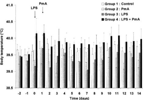
Cumulative cough count (CCC) was increased significantly (p < 0.05) from 1 to 14 dpi in the animals inoculated with E. coli endotoxin and PmA (group 4) (Fig. 1). No significant changes were detected in the other groups. Similar results were obtained when data were expressed as the daily mean of cough counted one hour per day (2.1 ± 2.7 and 8.1 ± 9.4 on 1 and 14 dpi respec-tively versus 0.2 ± 0.4 at –2 dpi). A signif-icant (p < 0.05) hyperthermia was detected in group 4 between 0 and 14 dpi while the body temperature recorded in groups 1, 2 and 3 remained within a physiological Figure 1. Evolution of the mean cumulative cough count (CCC) during the course of the disease. Control: group 1 (n = 8); Pasteurella multocida type A (PmA): group 2 (n = 6); E. coli lipopolysaccharides (LPS): group 3 (n = 5); E. coli lipopolysaccharides plus Pasteurella multocida type A: group 4 (n = 13). CCC was significantly increased (p < 0.05) from 1 to 14 dpi in animals inoculated with E. coli endotoxin and PmA. No significant changes were detected in other groups.
range (Fig. 2). Except coughing and hyper-thermia observed in group 4, no other clin-ical signs appeared in any group.
Piglets receiving LPS alone (group 3) (0.24 ± 0.02 kg) or in combination with PmA (group 4) (0.19 ± 0.07 kg) presented a similar but significant (p < 0.05) reduction in daily weight gains compared to those of control (0.45 ± 0.12 kg) and PmA inocu-lated groups (0.31 ± 0.15 kg).
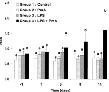
The whole body barometric plethysmog-raphy measurements revealed a significant Penh increase from 8 to 14 dpi in group 4 as presented in Figure 3. No significant changes were observed in other groups.
Cell counts measured at –1 and 14 dpi are presented in Figure 4. Significant increases in total lung cells, macrophages, neutrophils and lymphocytes were measured in group 4 while no significant changes occurred in
other groups except in group 1 exhibiting a slight but significant increase in total lung cells, and macrophages.
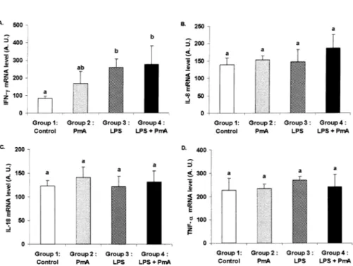
Fourteen days post-inoculation, the cytokine/cyclophilin mRNA absorbance ratio of IFNγ was significantly (p < 0.05) increased in group 4 compared to the con-trol group (Fig. 5). In group 3 inoculated with LPS alone, a slight but significant (p < 0.05) increase was also observed. No sig-nificant changes in IL-8, IL-18 and TNFα levels were observed in any groups.
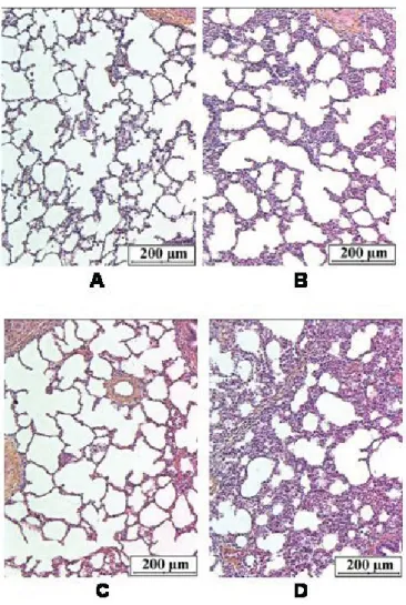
Low volume macroscopic lung lesions preferentially located in caudal lung lobes were detected in group 1 (controls) (34.4 ± 23.3 cm3), group 2 (11.4 ± 6.2 cm3) and group 3 (7.0 ± 3.6 cm3). In group 1, the alve-olar walls likely consisted of flattened epi-thelial cells in close contact with capillaries and of a delicate connective tissue frame-work. It also contained scanty macrophages. Figure 2. Evolution of the mean rectal temperature in the four groups during the course of the dis-ease. Control: group 1 (n = 8); Pasteurella multocida type A (PmA): group 2 (n = 6); E. coli lipopol-ysaccharides (LPS): group 3 (n = 5); E. coli lipopollipopol-ysaccharides plus Pasteurella multocida type A: group 4 (n = 13). A significant (p < 0.05) hyperthermia was detected in group 4 between 0 and 14 dpi while the body temperature recorded in groups 1, 2 and 3 remained within a physiological range.
The alveolar spaces were well expanded and, as the bronchiolar lumina, were free of inflammatory cells (Fig. 6A). Sometimes, the alveolar walls were enlarged by the presence of a slight lymphocytic and a mod-erate macrophage infiltrate. However, no inflammatory infiltrate was found in the bronchi. In group 2, the alveolar framework was preserved but the walls were enlarged by the presence of a slight lymphocytic and a moderate macrophage infiltrate. A subtle lymphocytic infiltrate was also seen in the bronchiolar and bronchial walls (Fig. 6B). In group 3, there was a minimal enlarge-ment of the alveolar septa due to the increase of macrophages and lymphocyte number. Scanty macrophages were free in the alveoli. There was no inflammation of the bronchial wall (Fig. 6C). In group 4 where LPS and PmA were combined, sig-nificantly more extended lung damage
(119.7 ± 39.2 cm3) was measured. These lesions were located in cranial lobes as well as in caudal lobes. The lung parenchyma was characterised by an accumulation of intra-alveolar fluid, extravased red cells and some fibrin together with polymorphs and macrophages (Fig. 6D). A moderate bronchial inflammatory infiltrate and a pleuritis characterised by a lympho-plas-mocytic infiltrate and a florid reactive mes-othelial proliferation were observed. The lesions of pleurisy were recorded in 4 of the 13 animals of group 4.
No Pasteurella multocida or other swine respiratory tract pathogens were isolated from the control, LPS and PmA groups. In group 4, Pasteurella multocida was iso-lated from one pig (cranial lung lobe) while no other swine respiratory tract pathogen was founded.
Figure 3. Evolution of the mean Penh value recorded in the different groups (unit: –). Control: group 1 (n = 8); Pasteurella multocida type A (PmA): group 2 (n = 6); E. coli lipopolysaccharides (LPS): group 3 (n = 5); E. coli lipopolysaccharides plus Pasteurella multocida type A: group 4 (n = 13). Letters differing between groups indicate a significant difference (p < 0.05). When at least one letter is common between the two groups, the difference is not significant. Within a group, the values were compared to those obtained on –2 dpi. Comparisons between groups were performed by testing the values measured the same day.
3.2. Relationship between pathophysiological changes and clinical signs
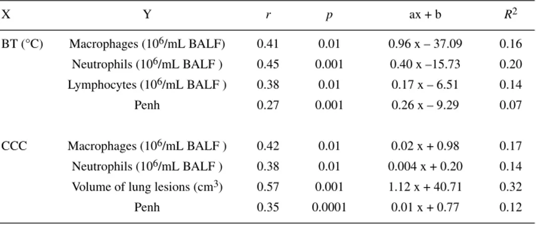
Significant linear correlation coefficients were obtained when individual values of the volume of lung lesions, Penh, number of BALF macrophages and neutrophils were correlated with the corresponding individ-ual values of CCC measured simultaneously during the protocol. Penh, BALF macro-phage, neutrophil and lymphocyte numbers were also significantly (p < 0.01) correlated with body temperature (Tab. I). Individual values of Penh were correlated with the BALF neutrophil number (r = 0.64; p > 0.0001) and the volume of lung lesions (r = 0.35; p < 0.05) was measured simultaneously.
3.3. Use of cough and body temperature for the detection of diseased animals
When selecting a threshold for CCC of five coughs per animal, it appeared that among the animals from group 4 (n = 13), CCC exceeded this threshold at least one day during the course of the disease in 12 piglets (92.3%), all of them exhibiting severe lung lesions at the end of the protocol (lung lesions volume: 119.6 ± 42.4 cm3). The mean detection time corresponding to the moment when the threshold was exceeded was 4.5 ± 3.1 dpi. In this group, only one pig with extended lung lesions (120 cm3) showed CCC values remaining under the selected threshold. In animals Figure 4. Inflammatory lung cells counted in bronchoalveolar lavage fluids. Control: group 1 (n = 8); Pasteurella multocida type A (PmA): group 2 (n = 6); E. coli lipopolysaccharides (LPS): group 3 (n = 5); E. coli lipopolysaccharides plus Pasteurella multocida type A: group 4 (n = 13). Letters differing between groups indicate a significant difference (p < 0.05). When at least one letter is common between two groups, the difference is not significant. Within a group, the values were compared to those obtained on –2 dpi. Comparisons between groups were performed by testing the values measured the same day.
from other groups (1, 2, 3) (n = 19), only three (16%) developed coughs while their mean lung lesion volume was very low (11.7 ± 5.1 cm3). The others (84%) exhib-ited low values of CCC and lung lesion vol-umes (21.5 ± 16.5 cm3). When a threshold for body temperature was considered (> 40.0 °C), it appeared that all animals from group 4 (100%) developed hyperther-mia at least one day during the protocol (mean detection time: 2.6 ± 2.3 dpi), all of them having severe lung lesions (119.7 ± 39.2 cm3). However, in the other groups, 11 animals exceeded the threshold (57%) while their lung lesion volume was not sig-nificantly increased (21.5 ± 15.7 cm3). When both criteria were combined (CCC > 5 and body temperature > 40.0 °C), only one pig
from groups 1, 2 and 3 (5%) was considered as a diseased animal while its lung lesion volume was very low (4.1 cm3). In group 4 (n = 13), 11 animals were detected on the basis of this combined criteria.
To check whether the total cumulative cough counted daily in a group could be used to differentiate groups composed of diseased or healthy animals, a threshold of five coughs per animal multiplied by the number of animals included in the group was selected. The thresholds of CCC calcu-lated for group 1 (n = 8), 2 (n = 6), 3 (n = 5) and 4 (n = 13) were 40, 30, 25 and 75, respectively. These thresholds were never reached for groups 1, 2 and 3 while pneu-monia was diagnosed at 2 dpi in group 4. Figure 5. Cytokine production in lung tissue sampled at 14 dpi in: control: group 1 (n = 6);
Pasteurella multocida type A (PmA): group 2 (n = 5); E. coli lipopolysaccharides (LPS): group 3
(n = 5); E. coli lipopolysaccharides plus Pasteurella multocida type A: group 4 (n = 13). Total RNA was isolated and assayed for expression of (A) IFN-γ, (B) IL-8, (C) IL-18, (D) TNF-α and the cyclophilin housekeeping genes by RT-PCR. Quantification of these cytokine mRNA is shown. Letters differing between groups indicate a significant difference (p < 0.05). When at least one letter is common between two groups, the difference is not significant.
3.4. Predictive value of CCC and body temperature
CCC and body temperature measured from 1 to 14 dpi were significantly (p < 0.05)
correlated with the lung lesion volume and BALF cells recorded at the end of the pro-tocol while only CCC was significantly (p < 0.05) correlated with the daily body weight gain from 12 to 14 dpi (Tab. II). Figure 6. Lung parenchyma of a control animal (group 1, A), an animal inoculated with Pasteurella
multocida only (group 2, B), an animal instilled with E. coli lipopolysaccharides only (group 3, C)
and of an animal instilled with E. coli lipopolysaccharides and inoculated with Pasteurella multocida (group 4, D). (A) Free of inflammatory cells in alveolar wall and lumina. Respiratory bronchiole with smooth muscle lined by flattened epithelial cells and a normal arteriole. (B) Thickening of the alveolar septa by a moderate macrophage infiltrate and a slight lymphocytic inflammatory infiltrate. (C) Minimal inflammatory infiltrate consisting of macrophages and lymphocytes observed in the alveolar wall; presence of scanty alveolar macrophages. (D) Alveolar framework is modified by the presence of a massive confluent exsudation with red cells, neutrophils, macrophages and some fibrin filling the alveolar spaces. Alveolar wall congestion and inflammatory infiltrate are also seen (× 248).
4. DISCUSSION
In this model of subacute bronchopneu-monia, daily weight gain reduction and only a few clinical signs such as cough and hyperthermia were detected. A pulmonary inflammatory process involving macro-phages, neutrophils, lymphocytes and IFNγ was responsible for the lung lesions observed at the end of this study. Functional disorders consisting of the alteration of the expiratory airflow pattern also occurred. While cough and body temperature were moderately expressed, their magnitude was propor-tional to the intensity of the lung inflamma-tory process, the pulmonary functional changes and the extent of gross lesions. Moreover, cough and body temperature measured during the course of the disease were also related to the severity of the inflammation and lung injuries recorded at the end of the pathological process. Only cough was related to the reduction of the daily weight gain.
Airway clearance of non-attached organ-isms is very effective in pigs so that the epithelium integrity needs to be impaired to successfully induce pneumonic pas-teurellosis. Mycoplasma hyopneumoniae, Aujeszky disease virus, porcine reproduc-tive and respiratory syndrome virus with Bordetella bronchiseptica and crude Actin-obacillus pleuropneumoniae cytotoxin are very effective agents to promote PmA-induced pneumonia [5, 8, 9, 22]. In our study, E. coli endotoxins, which have already been shown to inhibit mucociliary clear-ance in rats and to induce a transient acute lung inflammation in pigs which remains in the lung for a maximum of 48 h, were intrat-racheally injected to promote PmA infec-tion [20, 27]. The lung injuries observed in the present study, especially subacute exu-dative bronchopneumonia involving migra-tion of neutrophils in alveoli and bronchi, marked alveolar macrophage prolifera-tion, and peribronchiolar and alveolar lym-phocytic infiltration were in full agreement with studies devoted to Pasteurella
multo-cida infection [9, 15]. However, PmA was scarcely isolated from lung lesions suggest-ing that a prolonged colonisation of the res-piratory tract was not necessary to strongly worsen the pulmonary inflammatory proc-ess initiated by E. coli endotoxins. The fact that PmA was cleared from the lungs during the experiment might explain the baseline levels of IL-8, IL-18 and TNF-α (Fig. 5) observed at 14 dpi which illustrated that the inflammatory process was decreasing in intensity. Indeed, inflammatory cytokines have been shown to be expressed during acute Pasteurella multocida infection [3]. However in the latter study, the animals were infected with a higher dose of bacteria (5 mL of 4.8 × 10⁹ CFU mL–1 versus 5 mL of 2 × 10⁹ CFU mL–1 in the present study) and were killed at an earlier time point of the respiratory infection (5 versus 14 dpi). A very transient production of cytokines within the first eight hours post infection has also been described during Actinobacil-lus pleuropneumoniae infection [2]. The mRNA synthesis of IFN-γ detected sug-gests that lymphocytes observed within the lung tissue and the BALF participate in the regulation of both innate and acquired anti-microbial immunity [4, 30].
The Penh measured by whole body plethysmography is a functional parameter which has previously shown changes in the expiratory airflow pattern during choliner-gic or endotoxin challenges [16]. In this experiment, the increase of Penh recorded on 8 and 14 dpi in group 4, was likely due to airway obstruction and/or to a decrease in the lung elastic recoil induced by the pul-monary inflammatory process as reported in anaesthetised piglets with other infec-tious pulmonary diseases [21]. To the best of our knowledge, it is the first time that such pulmonary functional alterations are reported in non-sedated, freely moving piglets with bronchopneumonia. Since the raise in Penh was proportional to the inten-sity of the lung inflammatory reaction and to the extent of lung lesions, measurement of this functional parameter might be an
interesting tool for a non-invasive follow-up of a lung disease accompanied by air-flow limitation.
Pigs from group 4 exhibited moderate cough and hyperthermia. These clinical findings are in agreement with previous
data and field observations collected in pigs with PmA-induced pneumonia [9]. With-out any accurate quantitative recording pro-cedure, these symptoms are not so easy to detect explaining why an early diagnosis of such pulmonary diseases remains difficult Table I.
R
elationship between individual values of cumulative cough count (CCC) or body temperature (BT) and the corresponding individual values of bronchoalveolar lavage fluid cell numbers, Penh and volume of lung damage measured simultaneously. Regression equations with significant linear correlation coefficients are only indicated in this figure. The corresponding determination coefficients are also presented.X Y r p ax + b R2 BT (°C) Macrophages (106/mL BALF) 0.41 0.01 0.96 x – 37.09 0.16 Neutrophils (106/mL BALF ) 0.45 0.001 0.40 x –15.73 0.20 Lymphocytes (106/mL BALF ) 0.38 0.01 0.17 x – 6.51 0.14 Penh 0.27 0.001 0.26 x – 9.29 0.07 CCC Macrophages (106/mL BALF ) 0.42 0.01 0.02 x + 0.98 0.17 Neutrophils (106/mL BALF ) 0.38 0.01 0.004 x + 0.20 0.14 Volume of lung lesions (cm3) 0.57 0.001 1.12 x + 40.71 0.32
Penh 0.35 0.0001 0.01 x + 0.77 0.12
Table II. Linear correlation coefficients (r) between clinical signs and bronchoalveolar lavage fluid cells, volume of lung lesions and daily weight gain. Values of clinical signs were day by day correlated with the clinical end-points recorded at d14. Days corresponding to significant (p < 0.05) r values are indicated in this table with the range of daily r values obtained during the indicated period of time.
Cumulative cough count Body temperature
Total lung cells days p.i. 3 to 14 1 to 14
r range 0.35 to 0.58 0.37 to 0.64
Macrophages days p.i. 1 to 14 1 to 14
r range 0.41 to 0.53 0.43 to 0.63
Neutrophils days p.i. 4 to 14 1 to 14
r range 0.38 to 0.50 0.51 to 0.69
Lymphocytes days p.i. 6 to 14 1 to 14
r range 0.42 to 0.48 0.48 to 0.68
Volume of lung lesions days p.i. 1 to 14 1 to 14
r range 0.31 to 0.59 0.34 to 0.55
Daily weight gain days p.i. 12 to 14 –
in field conditions. Since other clinical signs are often missing or cannot be detected, the use of quantitative measurement of cough-ing and body temperature might improve the diagnosis of respiratory disorders in pigs. The relationship between coughing and lung damage has been investigated in pigs with enzootic pneumonia by scoring the cough on a weekly basis and measuring lung lesions at slaughterhouses but no sig-nificant correlation was obtained probably due to a non appropriate assessment of cough and lung lesions [25]. Cough was counted by mixing pigs in the pen for 30 s and observing the response (cough or no cough). Moreover, the extent of lung lesions was semi-quantitatively determined by scor-ing them as a percentage of the damaged lung surface. As concluded by the authors of this trial, a daily monitoring of cough and a better record of the lung lesion volume could improve the predictive value of this symptom regarding the intensity of the pul-monary disease. Hyperthermia has also been monitored during swine respiratory infec-tions involving Pasteurella multocida but the relationship between the body temper-ature and lung lesions was not investigated [6, 9, 15].
The significant linear correlation coeffi-cients obtained in our study between cough, body temperature and the corresponding values of BALF cells, lung lesions and Penh (Tab. I) suggest that these clinical signs could be used to predict the severity and evolution of the inflammatory process and lung lesions during the course of an infec-tious respiratory disease. To assess the potential use of CCC and body temperature to early diagnose a respiratory disease in a pig, thresholds for both parameters have been defined. Hence, all piglets reaching five coughs or 40 °C at least one time during the protocol were considered as diseased animals and the accuracy of the diagnosis was confirmed by the presence of lung lesions on 14 dpi. Applying this method with CCC as the criteria, 92.3% of the ani-mals with lung lesions were detected early
while only 16% of the pigs with no signif-icant lesions compared to controls were erroneously declared as diseased (false pos-itive response). Considering body temper-ature as the discriminative criteria, the cor-responding percentages were 100 and 57% respectively. A combination of both criteria allowed a marked reduction of the number of false positive cases (85% of positive responses versus 5% of false positives). When a threshold for CCC was defined for a group, the respiratory disease was detected at 2 dpi in group 4 while the others (groups 1, 2, 3) never overcame the thresh-old in our experimental conditions. Thus, if cough recognition systems are available in field conditions in the future, this might be a real advance to determine the respiratory status of animals, to early and non-inva-sively diagnose respiratory diseases in herds but also to perform the follow-up of bronchopneumonia [28, 29]. However, a prior validation of these systems is needed to confirm our observations in a large swine population which contains animals with several common respiratory diseases. Data from Table II also show that BT and CCC could be used to predict early-on the extent of major end-points regarding the intensity of respiratory disease in piglets.
We conclude that the E. coli lipopolysac-charide-induced lung inflammatory process might be worsened by Pasteurella multoc-ida while it does not allow lung colonisa-tion. The resulting inflammatory process is characterised by an inflammatory cell influx into the airways which limits the air-flow and by extended lesions of subacute bronchopneumonia explaining the weight gain reduction. Cough and body tempera-ture, while moderately exhibited, could be used as indicators to assess the intensity and the evolution of the lung inflammatory reaction and functional changes during the course of the disease. They are potential predictors for the magnitude of lung lesions and weight gain reduction assessed at the end of the process.
ACKNOWLEDGEMENTS
We thank N. Kirschvink for her interest and advice during the preparation of the manuscript. The technical assistance of F. Delvaux, D. Beerens and S. Martinez is gratefully appreciated. This work was supported by the Ministry of Agricul-ture (DGVI), Brussels, Belgium and by special funds for research (Ulg).
REFERENCES
[1] Amass S.F., Clark L.K., van Alstine W.G., Bowersock T.L., Murphy D.A., Knox K.E., Albregts S.R., Interaction of Mycoplasma hyopneumoniae and Pasteurella multocida infections in swine, J. Am. Vet. Med. Assoc. 204 (1994) 102–107.
[2] Baarsch M.J., Scamurra R.W., Burger K., Foss D.L., Maheswaran S.K., Murtaugh M.P., Inflammatory cytokine expression in swine experimentally infected with Actinobacillus pleuropneumoniae, Infect. Immun. 63 (1995) 3587–3594.
[3] Berndt A., Heller M., Kosmehl H., Cytokine mRNA expression in experimental porcine pneumonia, Dtsch. Tierarztl. Wochenschr. 109 (2002) 205–209.
[4] Boehm U., Klamp T., Groot M., Howard J.C., Cellular responses to interferon-gamma, Annu. Rev. Immunol. 15 (1997) 749–795. [5] Brockmeier S.L., Palmer M.V., Bolin S.R.,
Rimler R.B., Effects of intranasal inoculation with Bordetella bronchiseptica, porcine repro-ductive and respiratory syndrome virus, or a combination of both organisms on subsequent infection with Pasteurella multocida in pigs, Am. J. Vet. Res. 62 (2001) 521–525. [6] Carvalho L.F., Segales J., Pijoan C., Effect of
porcine reproductive and respiratory syn-drome virus on subsequent Pasteurella mul-tocida challenge in pigs, Vet. Microbiol. 55 (1997) 241–246.
[7] Chong B.T., Agrawal D.K., Romero F.A., Townley R.G., Measurement of bronchocon-striction using whole-body plethysmograph: comparison of freely moving versus restrained guinea pigs, J. Pharmacol. Toxicol. Methods 39 (1998) 163–168.
[8] Chung W.B., Backstrom L.R., Collins M.T., Experimental model of swine pneumonic pas-teurellosis using crude Actinobacillus pleu-ropneumoniae cytotoxin and Pasteurella mul-tocida given endobronchially, Can. J. Vet. Res. 58 (1994) 25–30.
[9] Ciprian A., Pijoan C., Cruz T., Camacho J., Tortora J., Colmenares G., Lopez-Revilla R., de la Garza M., Mycoplasma hyopneumoniae increases the susceptibility of pigs to experi-mental Pasteurella multocida pneumonia, Can. J. Vet. Res. 52 (1988) 434–438. [10] Darwich L., Pié S., Rovira A., Segalés J.,
Domingo M., Oswald I.P., Mateu E., Cytokine mRNA expression profiles in lymphoid tis-sues of pigs naturally affected by postweaning multisystemic wasting syndrome, J. Gen. Virol 84 (2003) 2117–2125.
[11] Djuric V.J., Cox G., Overstreet D.H., Smith L., Dragomir A., Steiner M., Genetically transmit-ted cholinergic hyperresponsiveness predis-poses to experimental asthma, Brain Behav. Immun. 12 (1998) 272–284.
[12] Donham K.J., Association of environmental air contaminants with disease and productiv-ity in swine, Am. J. Vet. Res. 52 (1991) 1723– 1730.
[13] Dozois C.M., Oswald E., Gautier N., Serthelon J.P., Fairbrother J.M., Oswald I.P., A reverse transcription-polymerase chain reaction method to analyze porcine cytokine gene expression, Vet. Immunol. Immunopathol. 58 (1997) 287– 300.
[14] Fournout S., Dozois C.M., Yerle M., Pinton P., Fairbrother J.M., Oswald E., Oswald I.P., Cloning, chromosomal location, and tissue expression of the gene for pig interleukin-18, Immunogenetics 51 (2000) 358–365. [15] Hall W.F., Bane D.P., Kilroy C.R.,
Essex-Sorlie D.L., A model for the induction of Pas-teurella multocida type-A pneumonia in pigs, Can. J. Vet. Res. 54 (1990) 238–243. [16] Halloy D., Kirschvinck N., Vincke G., Hamoir
J., Delvaux F., Gustin P., Whole body baro-metric plethysmography: a screening method to investigate airway reactivity and acute lung injuries in freely moving pigs, Vet. J. (in press).
[17] Hamelmann E., Schwarze J., Takeda K., Oshiba A., Larsen G.L., Irvin C.G., Gelfand E.W., Noninvasive measurement of airway responsiveness in allergic mice using baro-metric plethysmography, Am. J. Respir. Crit. Care Med. 156 (1997) 766–775.
[18] Hamelmann E., Schwarze J., Takeda K., Oshiba A., Larsen G.L., Irvin C.G., Gelfand E.W., Noninvasive measurement of airway responsiveness in allergic mice using baro-metric plethysmography [see comments], Am. J. Respir. Crit. Care Med. 156 (1997) 766–775.
To access this journal online: www.edpsciences.org
[19] Hoffman A.M., Dhupa N., Cimetti L., Airway reactivity measured by barometric whole-body plethysmography in healthy cats, Am. J. Vet. Res. 60 (1999) 1487–1492.
[20] Hosoe H., Kaise T., Ohmori K., Erdosteine enhances mucociliary clearance in rats with and without airway inflammation, J. Pharma-col. ToxiPharma-col. Methods 40 (1998) 165–171. [21] Intraraksa Y., Engen R.L., Switzer W.P.,
Pul-monary and hematologic changes in swine with Mycoplasma hyopneumoniae pneumo-nia, Am. J. Vet. Res. 45 (1984) 474–477. [22] Kobisch M., Friis N.F., Swine
mycoplas-moses, Rev. Sci. Tech. 15 (1996) 1569–1605. [23] Kobisch M., Blanchard B., Le Potier M.F., Mycoplasma hyopneumoniae infection in pigs: duration of the disease and resistance to rein-fection, Vet. Res. 24 (1993) 67–77. [24] Loughmiller J.A., Spire M.F., Dritz S.S.,
Fenwick B.W., Hosni M.H., Hogge S.B., Relationship between mean body surface tem-perature measured by use of infrared thermog-raphy and ambient temperature in clinically normal pigs and pigs inoculated with Actino-bacillus pleuropneumoniae, Am. J. Vet. Res. 62 (2001) 676–681.
[25] Morris C.R., Gardner I.A., Hietala S.K., Carpenter T.E., Enzootic pneumonia:
com-parison of cough and lung lesions as predic-tors of weight gain in swine, Can. J. Vet. Res. 59 (1995) 197–204.
[26] Thacker E.L., Thacker B.J., Janke B.H., Inter-action between Mycoplasma hyopneumoniae and swine influenza virus, J. Clin. Microbiol. 39 (2001) 2525–2530.
[27] Urbain B., Prouvost J.F., Beerens D., Ansay M., Gustin P., Acute effects of endotoxin inhalation on the respiratory tract in pigs: interaction with ammonia, Inhal. Toxicol. 8 (1996) 947–968.
[28] Van Hirtum A., Berckmans D., Assessing the sound of cough towards vocality, Med. Eng. Phys. 24 (2002) 535–540.
[29] Van Hirtum A., Berckmans D., Automated recognition of spontaneous versus voluntary cough, Med. Eng. Phys. 24 (2002) 541–545. [30] Varma T.K., Lin C.Y., Toliver-Kinsky T.E.,
Sherwood E.R., Endotoxin-induced gamma interferon production: contributing cell types and key regulatory factors, Clin. Diagn. Lab. Immunol. 9 (2002) 530–543.
[31] Von Altrock A., Occurrence of bacterial infectious agents in pathologically/anatomi-cally altered lungs of pigs and compilation of resistance spectra, Berl. Muench. Tieraerztl. 111 (1998) 164–172.