CLINICAL STUDY
The changing spectrum of TSH-secreting pituitary adenomas:
diagnosis and management in 43 patients
H Valdes Socin, P Chanson1, B Delemer2, A Tabarin3, V Rohmer4, J Mockel5, A Stevenaert and A Beckers
Service d’Endocrinologie et Neurochirurgie, CHU, Lie`ge, Belgium,1Service d’Endocrinologie et des Maladies de la Reproduction, CHU, Biceˆtre-Paris, France,2Service d’Endocrinologie, CHU, Reims, France,3Service d’Endocrinologie, CHU, Bordeaux, France,4Service d’Endocrinologie, CHU, Angers, France and5Service d’Endocrinologie, CHU, Erasme, Brussels, Belgium
(Correspondence should be addressed to A Beckers, Service d’Endocrinologie, CHU de Lie`ge, Domaine Universitaire du Sart-Tilman, 4000 Lie`ge, Belgium; Email: Albert.Beckers@chu.ulg.ac.be)
Abstract
Objective: Our aim was to report the recent changes in diagnosis and management of TSH-secreting pituitary adenomas.
Methods: We retrieved 43 consecutive patients with TSH-secreting pituitary tumors (23 male and 20 female) among 4400 pituitary adenomas followed between 1976 and 2001 in six Belgian and French centers.
Results: TSH was elevated in 18/43 and a subunit in 13/32 patients. In patients with intact thyroid ðn ¼ 30Þ; mean free tri-iodothyronine was 13.1 pmol/l (range 3.5 – 23) and mean free thyroxine was 38.4 pmol/l (range 10.2 – 62.7). Hyperprolactinemia and acromegaly were associated in 9/43 and 8/43 cases. The number of associated hypersecretions was higher in macroadenomas than in micro-adenomas (Chi square = 11.2, P , 0:01). Two women had sporadic multiple endocrine neoplasia type 1-associated syndrome. The proportion of microadenomas versus macroadenomas was 1/11 (period 1974 – 1986) and 8/32 (period 1987 – 2001). Bilateral petrosal sinus sampling, 111 In-pen-treotide scintigraphy and (11C)-L-methionine positron emission tomography scan confirmed diagnosis in four questionable microadenomas. Macroadenomas with extrasellar extension (31 cases) had a tendency to be medially located. Medical treatment with somatostatin analogs was initiated as first-line treatment in 26 patients. TSH levels were reduced by more than 50% in 23/26 cases. A tumoral shrinkage of more than 20% was observed in 5/13 cases. Surgery was performed in 36 patients. After 1 year, 21 of them (58.3%) met the criteria of surgical favorable outcome. Pituitary radiotherapy ðn ¼ 8Þ and somatostatin analogs allowed normalization in cases not cured by surgery. Conclusion: Ultrasensitive methods for TSH measurement led to an earlier recognition of TSH-secreting pituitary tumors. In this series, we observed that TSH-secreting pituitary tumors are today more fre-quently found at the stage of microadenomas, medially located, without associated hypersecretions and needing new exploration methods as compared with older series. This changing spectrum in the presentation of TSH-secreting pituitary tumors and the excellent response to somatostatin analogs has been accompanied by an improvement in the prognosis of the disease.
European Journal of Endocrinology 148 433–442
Introduction
Jailer & Holub postulated in 1960 (1) that a pituitary tumor could be responsible for excessive quantities of thyrotropin (TSH) and thyrotoxicosis. During the 20 years following this publication, reports of TSH-secret-ing pituitary tumors were scarce. Since 1980, a few limited series have been published dealing with particu-lar aspects of diagnosis and treatment (2 – 9). In fact, the diagnosis and management of these rare tumors have evolved considerably in the last two decades, due to the spread of the concept of inappropriate secretion of TSH, ultrasensitive methods for TSH measurement, the improvement in pituitary imaging leading to a better recognition of pituitary microadenomas and
lastly, to the availability of somatostatin analogs. We report herein the clinical, radiological and follow-up features of TSH-secreting pituitary adenomas in a large number of patients recruited from a limited number of centers. Analysis of this series provides an accurate description of the changing spectrum over the last two decades in the presentation and manage-ment of these rare tumors.
Subjects and methods
Patients
Forty-three patients, aged 19 – 84 years (23 men, 20 women) with TSH-secreting pituitary adenomas have
been followed from 1976 to 2001. These patients were collected from six Belgian and French centers. They represent less than 1% of 4400 pituitary adenoma patients seen during that period. Partial data of patients nos 1, 2, 4, 5, 6, 7 and 36 have already been published (3, 10). All patients underwent a com-plete physical examination including palpation of the thyroid and evaluation of visual fields using Goldman’s perimetry.
Diagnosis of TSH-secreting pituitary tumors was based on clinical findings (tumoral syndrome and/or thyrotoxicosis), biochemical evidence of unsuppressed TSH levels contrasting with elevated serum thyroid hormone concentrations and the identification of an adenoma by pituitary computerized tomography (CT) scan or magnetic resonance imaging (MRI). Additional diagnostic tools were dynamic response of TSH and a subunit (aSU) to thyrotropin-releasing hormone (TRH), tri-iodothyronine (T3) suppression test in questionable cases, and measurement of prolac-tin (PRL), growth hormone (GH), insulin-like growth factor-I (IGF-I), luteinizing hormone (LH), follicle-stimu-lating hormone (FSH) and adrenocorticotropin (ACTH) levels.
Endocrine studies
Endocrine investigations were carried out before administration of anti-thyroid drugs or somatostatin analog treatment. Hormone measurements were per-formed in the laboratories of each institution using commercially available immunometric assays. TSH ultrasensitive assays have been available in participat-ing centers since 1987. In the 11 patients in whom diagnosis was made before 1987, TSH levels were above 1 mU/l.
For the different assays used, the normal range for hormonal determination was set as follows: TSH (0.3 – 4.3 mU/l), free T3(fT3; 2.3 – 4.6 pmol/l), free thyr-oxine (fT4; 9 – 21.8 pmol/l), aSU (0 – 1.2 mg/l for men and less than 2 mg/l for premenopausal women), PRL (1 – 20 mg/l), mean GH (0 – 2.5 mg/l), IGF-I (90 – 300 mg/l) and ACTH (10 – 90 ng/l).
Thyroid autoimmunity was tested in 33 cases, includ-ing thyroperoxydase antibodies (TPO Ab) levels and TSH-binding inhibiting immunoglobulin (TBII). The stimulatory effect of TRH (200 mg i.v) on TSH, GH and aSU was also assessed. The TSH response was considered as positive when hormonal levels increased more than 50% of basal values and when the net increase was at least 4 mU/l. Insulin tolerance test at a dose of 0.1 U/kg i.v. was performed to assess GH deficiency (GH peak , 3 mg/l) and ACTH deficiency (cortisol , 190 ng/l). Patients undergoing a TSH suppression test were treated with 100 mg T3orally per day for 10 days as previously described (11).
Imaging studies
Contrast-enhanced CT scan of the pituitary was per-formed in 11 patients between 1974 and 1986. In 32 patients, MRI of the pituitary gland was performed from 1987 until the present at 1.5 Tesla. Three milli-meter-thick coronal T1- and T2-weighted images were performed before and after gadolinium i.v. The maximal diameter of each adenoma was assessed in a frontal view. Tumors were considered as macroadenomas when the maximal diameter was more than 10 mm and giant adenomas when the diameter exceeded 40 mm. Invasion was suspected on the basis of an exten-sion of the adenomatous tissue in the cavernous sinus in both frontal and horizontal view when available. In 13 patients presurgically treated with octreotide the per-centage change in tumor volume was calculated according to the Di Chiro and Nelson formula: volume = height £ length £ width £ 0.5233 (12).
Less usual procedures were performed in particular patients (patients without thyroid or pituitary medical treatment): a bilateral petrosal sinus sampling (BPSS) was performed in patient no. 1 (10). Single-photon emission tomography (SPECT) was performed by injec-tion (i.v.) of 110 Mbq 111In-pentreotide in seven patients (nos 3, 7, 10, 32, 34, 36 and 40) who under-went a tomographic imaging of the head and whole body. Pituitary accumulation was expressed as the ratio between the uptake of radioactivity by the ade-noma and the normal brain tissue. We expressed the index threshold of positivity as twice the normal ratio of a control group. In seven untreated patients, results of pentreotide uptake were analyzed with regard to TSH inhibition by chronic octreotide treatment and surgery outcome. (11C)-L-methionine positon emission tomog-raphy (PET) scan was carried out in three patients (nos 10 and 36 with a microadenoma and no. 35 with a macroadenoma). The highest resolution of this device is 6 mm. (11C)-L-methionine PET index of
posi-tivity was expressed as a ratio (. 1), calculated as the methionine uptake of the tumor divided by the whole brain slice (13).
Immunocytochemistry
Tumor specimens were available for microscopy studies in 36 operated patients (both the original tumor and the recurrence were available for analysis in two patients) and for immunohistochemical characteri-zation of cellular hormonal contents in 32 patients. The peroxidase – antiperoxidase immunocytochemical method was applied immediately to pituitary slices obtained at surgery, using antisera specific to bLH, bFSH, bTSH, GH, PRL, ACTH, aSU and b-chorionic gonadotropin (bCG) (in 12 cases). Hormonal positivity was established if more than 5% of the cells showed immunopositivity.
Genetic studies
All patients gave their informed consent for genetic studies. Tumor specimen and leukocyte DNA were investigated for multiple endocrine neoplasia type 1 (MEN1) gene inactivation. Loss of heterozygosity and MEN1 gene mutation were assessed as previously described (14).
Treatment
Prior to the diagnosis of pituitary adenoma some patients ðn ¼ 13Þ had received a treatment targeted to the thyroid: antithyroid drugs in five patients (nos 1, 5, 28, 29 and 35) for a mean duration of 6 months (range 2 – 18), near total thyroidectomy in eight (nos 6, 7, 16, 19, 33, 34, 36 and 37) or131iodine in one (no. 6) on average 5 years (range: 2 months – 12 years) before pituitary surgery.
Medical treatment with somatostatin analogs was initiated as a first-line treatment and pursued for a mean time of 6 months (7 days – 4 years) in 26 patients. Nineteen of them were thereafter operated (presurgical treatment group, s.c. octreotide (300 mg/day) in 15 cases and lanreotide Slow Release (SR) in four cases) while somatostin analogs (octreotide s.c., lanreotide SR i.m. or octreotide Long-Acting Release depot i.m.) were the sole treatment for a mean time of 24 months (range 7 – 48 months) in seven patients. Bromocriptine (5 mg/day) and quinagolide (75 mg/day) were given to five and two patients respectively. Transsphenoidal surgery was performed in 36 patients in their respective center. Among these, seven required a second operation, three of them by a subfrontal approach. Tumoral consistency, invasion and the com-pleteness of adenomectomy were assessed according to the surgeon’s protocol: invasion was defined during surgery as the bone or dura invasion by the tumor; tumoral consistency was evaluated as soft or hard. Eight patients considered to be uncontrolled by surgery underwent external beam pituitary (one gamma knife irradiation). Mean follow-up after radiotherapy was 6 years (range 5 – 10). Total dose ranged from 42 to 45 Gy in 28 treatments via at least three ports.
Follow-up and criteria of remission after
surgery
Clinical and biochemical evaluations were repeated during the 7 days following surgery. Thereafter, clinical and biochemical assessments and neuroradiological evaluations were repeated 3 months and 1 year later, then at least yearly. All but two patients were followed for at least 1 year after surgery: 41 out of 43 patients were contacted by their respective center for the last follow-up in 2001. Two patients were lost to follow-up after 60 and 88 months. Surgical remission was con-sidered as complete if normalization of all clinical
signs and hypersecretions (including basal and dynamic responses of TSH, GH, PRL, aSU and thyroid hormones in patients with intact thyroid) was still attained 1 year after surgery alone; moreover, neuroradiological controls must also rule out the presence of tumoral resi-due. The patient was considered as having tumoral recurrence if clinical, biochemical and/or neuroradiolo-gical signs of tumor activity were detected after 1 year of apparent remission. Thyroid-ablated patients under-went a suppression test. Only five patients with intact thyroids and questionable hormonal levels (nos 6, 8, 11, 13 and 19) had the T3test.
Statistical analysis
Results are expressed as means^S.D.Fisher’s exact test
was used for comparisons between parametric data and Wilcoxon’s test for continuous variables. Chi square and Student’s t-test were employed when appropriate. The level of significance was set at P , 0:05: All data were analysed by a Statistica package 1999 (Stat Soft, Tulsa, OK, USA).
Results
Clinical features
Diagnosis of a TSH-secreting adenoma was made at the age of 44^13 years, with a latency between onset of hyperthyroidism and diagnosis of pituitary adenoma of 4^6 years. Symptoms of severe thyrotoxicosis were found in only two patients (no. 39, who presented with cardiac failure and no. 40, who had an atrial fibrillation). Thyrotoxicosis was moderate in 20 patients and 21 patients had only mild hyperthyroidism or no symptoms (13 patients had had previous treat-ments targeted to the thyroid).
In one patient hyperthyroidism did not contribute to the diagnosis. This patient was treated for hypothyroid-ism and Hashimoto’s thyroiditis. Although he received substitutive thyroid hormone treatment with L-T4 (150 mg) TSH values were inappropriately elevated with regard to T3and T4serum levels. A T3test failed to suppress TSH whereas TRH did not stimulate TSH. There was an associated aSU hypersecretion. Pituitary MRI identified a 41 mm macroadenoma that was suc-cessfully operated on and histologically confirmed.
Concomitant hyper- or hyposecretory syndromes were observed in 18 patients: eight patients presented with acromegalic features, six women had amenor-rhea – galactoramenor-rhea, four men complained of impotence and reduced libido.
Biochemistry
At diagnosis, biochemical hyperthyroidism was found in only 32/43 patients. In patients with intact thyroid ðn ¼ 30Þ; mean fT3 was 13.1 pmol/l (range 3.5 – 23)
and fT4 was 38.4 pmol/l (range 10.2 – 62.7). Serum TSH levels ranged between 1 and 12 mU/l in 29 cases, but in one patient the level was 36.3 mU/l. In the whole series, TSH levels were above the upper limit of normal range in 18/43 patients (42%). Follow-ing TRH administration there was no TSH response in 31/38 patients (81%). TPO Ab and antithyroglobulin antibodies (TG Ab) were negative except in one of the 33 patients evaluated. TBII was negative in all cases.
Serum PRL and IGF-I were elevated in 9/43 and 8/43. In the nine patients with hyperprolactinemia, the mean PRL level was 47 mg/l (range 25 – 85). In the eight acromegalics, mean IGF-I was 733 mg/l (range 310 – 1600). In the 13 patients with high aSU level, the mean level of aSU was 16.8 mg/l (range 2 – 140). TRH stimulated aSU more than 100% in 14/32 cases. A paradoxical rise of GH following TRH administration was observed in six out of the nine patients tested. Two out of ten patients studied with the insulin tolerance test had GH deficiency. Corticotro-pic function was normal in all patients.
Neuroradiological studies
Pituitary CT scanning and MRI revealed the presence of lesions in all but two patients: 34 had a macroadenoma (three giants) and an extrasellar extension for 31 of them; seven had a microadenoma and in two patients no pituitary adenoma was seen (nos 10 and 40). Twenty of 31 macroadenomas with an extrasellar extension were medially located. Additional data con-cerning microadenomas and macroadenomas are given in Tables 1 and 2.
A BPSS was performed in patient no. 1 in order to con-firm a questionable 3 mm pituitary adenoma. Results are shown in Table 3. A central to peripheral TSH gradi-ent was evidgradi-ent. Additionally, there was a right to left gradient contrasting with MRI data. MRI findings were later confirmed at surgery and immunohistochemistry identified a TSH-secreting microadenoma.
The hypothesis of an ectopic TSH-secreting tumor was raised for patients nos 10 and 40: total body 111In-pentreotide SPECT scan, with abdominal and
thoracic views, failed to identify pituitary or extrapitui-tary lesions. (11C)-L-methionine PET scan showed a
pituitary hyperfixation in patient no. 10. In both patients, despite the absence of a patent microadenoma on pituitary imaging, a pituitary tumoral origin of TSH hypersecretion was considered as the most likely alternative and the hypothesis of thyroid hormone resistance was ruled out on the following grounds: T3 administration did not suppress TSH and lanreotide chronic treatment persistently normalized thyroid hor-mone levels and TSH hypersecretion.
An (11C)-L-methionine PET pituitary scan was posi-tive in two patients with microadenomas (nos 10 and 36) not detected by MRI or111In-pentreotide scan. An (11C)-L-methionine PET pituitary scan was also positive
in another patient with a macroadenoma (no. 35). The number of associated hypersecretions was higher in macroadenomas than in microadenomas (Table 4).
Pituitary adenomas and MEN1
Two women developed a sporadic MEN1 syndrome. In both patients, hyperthyroidism and pituitary adenoma were diagnosed before the other tumors: one had a mixed TSH/GH/FSH-secreting adenoma and the other had a pure thyrotropic adenoma. This last patient also developed Zollinger Ellison syndrome. Primary hyperparathyroidism occurred later in both patients.
Treatments targeted to the thyroid
Diagnosis in patients previously treated either with sur-gery or 131I was significantly delayed compared with patients treated with antithyroid drugs ðP , 0:001Þ: Mean pituitary tumoral diameter at diagnosis was significantly smaller in this last group than in thyr-oid-operated patients ð10:2^6:1 ðn ¼ 5Þ vs 21:3^8:5 ðn ¼ 8Þ mm; P ¼ 0:03Þ: Outcome of pituitary surgery, mean tumor volume and TSH levels were not signifi-cantly different in patients who had received treatments
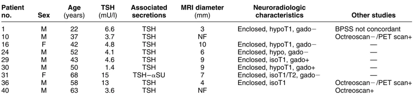
Table 1 Anthropometric, biochemical and imaging characteristics in a series of nine TSH-secreting microadenomas. Patient no. Sex Age (years) TSH (mU/l) Associated secretions MRI diameter (mm) Neuroradiologic
characteristics Other studies
1 M 22 6.6 TSH 3 Enclosed, hypoT1, gado2 BPSS not concordant
10 M 37 3.7 TSH NF Octreoscan2 /PET scan+
16 F 42 4.8 TSH 10 Enclosed, hypoT1, gado2 —
24 M 52 4.1 TSH 6 Enclosed, hypo, gado2 —
29 M 43 4.6 TSH 9 Enclosed, isoT1, gado+ —
30 M 50 1.4 TSH 9 Enclosed, hypoT1, gado+ —
31 F 68 15 TSH– aSU 7 Enclosed, isoT1/T2, gado2 —
36 M 58 13 TSH 4 Enclosed, isoT1 Octreoscan2 /PET scan+
40 M 63 3.6 TSH NF Octreoscan+
targeted at the thyroid ðn ¼ 13Þ and in the thyroid-untreated patients ðn ¼ 30Þ (data not shown).
Treatment of pituitary adenomas
Among the 26 patients who received somatostatin ana-logs as first-line treatment, 19 underwent surgery while the treatment was pursued chronically in seven. A reduction of more than 50% in TSH levels was observed in 23/26 (88%) cases. Normalization of free thyroid hormones was observed in 22/26 (85%) cases. Normal-ization of aSU was recorded in three out of four cases. A tumoral shrinkage of more than 20% was demon-strated in 5/13 (36%) cases.
Pituitary111In-pentreotide SPECT and response to octreotide 111In-pentreotide SPECT was performed in seven patients (nos 3, 7, 10, 32, 34, 36 and 40). While all of them were considered as responders to
octreotide treatment (suppression of TSH levels by more than 50%), only three had a positive pituitary scan ðIU . 2Þ: Two of these tumors were mixed macroadenomas secreting both TSH and GH, the third was a microadenoma secreting both TSH and aSU, not identified by pituitary MRI. Among the four patients with negative scans, one (no. 10) had no iden-tifiable pituitary lesion on MRI. In the three remaining patients with negative scans, a 4 mm microadenoma (no. 36) and two macroadenomas were evidenced (nos 3 and 34).
Patients who received medical treatment with somatostatin analogs only Thyrotoxicosis was present in all seven patients. Reasons for medical treat-ment as the main therapy were: presurgical treattreat-ment (two patients are waiting for surgery and two patients ultimately declined surgery (nos 3 and 39), poor gen-eral condition (no. 13) or absence of a visible adenoma on MRI (nos 10 and 40). As medical treatment gave satisfactory results, patients declined further investi-gations. Somatostatin analogs (lanreotide SR (30 mg i.m.) twice monthly, octreotide (300 mg/day s.c.)) were effective in normalizing thyroid secretion; TSH levels were reduced by more than 50% in all patients for a mean time of 24 months (range 7 – 48). fT3 and fT4 normalized in all cases. The aSU hypersecretion was also well controlled by somatostatin analogs in patients nos 13 and 40. Tumoral shrinkage was not observed in the three patients with evident tumors. Mild adverse effects such as nausea and diarrhea were reported, but treatment was not discontinued.
Patients treated with both medical and surgical treatment Nineteen patients were treated with presur-gical somatostatin analogs for a mean time of 6 months. After at least 2 weeks of treatment, a signifi-cant reduction in TSH levels and euthyroidism was obtained in 16/18 patients (90%). A significant tumoral shrinkage was observed in only three of the 11 cases evaluated after at least 3 months. Presurgical octreotide treatment did not modify surgical outcome (remission group: 11/19 vs failure group: 8/19; P . 0:05).
Seven patients were treated with dopamine agonists. A significant reduction in TSH and prolactin levels was noticed after bromocriptine (5 mg/day) in only one of five cases, without normalization of thyroid hormone levels. This patient (no. 9) had a mixed prolac-tin/TSH-secreting tumor. No suppression of TSH levels was seen in the two cases treated by quinagolide. Neither a tumoral shrinkage nor an improved surgical outcome was obtained in those patients.
Surgical outcome
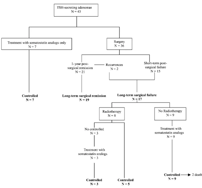
Transsphenoidal surgery was performed in 36 patients (Fig. 1). After 1 year, 21 of them (58.3%) met the
Table 2 Pattern of neuroradiological localization and invasion in a series of 34 TSH-secreting macroadenomas.
Localization Number Intrasellar 3/34 Extrasellar extension 31/34 Cavernous sinus 12*/31 Suprasellar 22/31 Sphenoidal sinus 14/31
* Of the 12 macroadenomas invading the cavernous sinus seven were mixed GH/TSH and had clinical signs of acromegaly and two others were mixed PRL/TSH with hyperprolactinemia.
Table 3 BPSS in a patient with a TSH-producing pituitary microadenoma with the TRH stimulation test.
TSH Time (min) (mU/l) 0 7.5 15 PS right 14.4 77.1 60.1 PS left 7 9.1 11.5 Periphery 5.6 9.9 11.2 PS, petrousal sinus.
Table 4 Tumoral diameter and hormonal hypersecretions in a series of 43 patients with TSH-secreting adenomas.
Tumoral maximal diameter (mm) Isolated TSH secretion (no.) Associated secretions (no.) 3 – 10 8 1 (aSU)
11 – 20 6 12 (aSU and/or GH and/or PRL)
21 – 30 7 5 (aSU and/or GH and/or PRL)
31 – 55 0 4 (aSU and/or GH and/or PRL)
The correlation between the number of patients with isolated TSH and the tumoral diameter is significant (Chi square = 11.2, P , 0:01).
criteria of successful surgical outcome (normalization of TSH and thyroid hormone levels, along with absence of tumoral remnant). The other 15 patients had evidence of residual adenoma after surgery (see also Fig. 1). Surgical remission rate in microadenomas (6/7) was better than in macroadenomas (15/29).
Macro- and microscopic findings
Invasion (defined as macroscopic dura or bone invasion) was found by the surgeon in 9/28 well-documented cases. Tumoral consistency was evaluated in 30 cases: 60% of the tumors had a soft consistency (the tumor could gently be removed or aspirated), the remaining had fibrotic characteristics and were difficult to excise.
All but one microadenoma displayed only TSH immunostaining, whereas macroadenomas predomi-nantly immunostained with TSH, aSU, GH and PRL antibodies. Thirty-one adenomas showed positivity for bTSH, 18 of them being also strongly positive for GH, 13 for PRL and only 13 for aSU. In addition, six of 12 tumors studied were positive for bCG, one for bLH and another for both bLH and bFSH. Unexpectedly, four ade-nomas were mildly positive for ACTH although none of them had biochemical evidence of hypersecretion or had the clinical features of Cushing’s syndrome. Finally, one tumor was found to have a changing immunohisto-chemical pattern (bTSH/bFSH/GH to bTSH/GH) at recurrence. Three of them did not immunostain for bTSH although patients had overt biochemical
hyperthyroidism with inappropriately unsuppressed TSH levels (nos 2, 7 and 25).
Date of diagnosis and tumoral findings
As routine MRI and ultrasensitive methods of detec-tion of TSH had been available in our centers since approximately 1987, we arbitrarily analyzed our data according to the date of diagnosis of the pituitary adenoma and separated the patients between those for whom the diagnosis has been made before 1987 and those for whom the diagnosis was made after 1987. In the series, the number of diagnosed cases in the period 1974 – 1986 ðn ¼ 11Þ was one-third that of the period 1987 – 2001 ðn ¼ 32Þ: Before and after 1987, the proportion of microadenomas versus macroadenomas was 1/11 and 8/32 respectively.
Genetic studies
Sporadic tumors of five patients (nos 1, 6, 7, 8 and 9) were screened for mutations of MEN1 gene sequence (11) but none was found. DNA of peripheral leucocytes of patients nos 3 and 16, with a clinically proven MEN1 syndrome, were also negative for MEN1 gene mutation. Tumor tissue from these last patients was not available for analysis.
Follow-up
All patients were followed-up for a mean period of 8^6 years (range 1 – 25 years). Two out of 43 patients died during follow-up at the ages of 75 and 86 respectively. Outcome was assessed in the 36 operated patients who were followed-up for at least 1 year. Patients nos 31 and 35 developed a recurrence 84 and 24 months after sur-gery respectively. Hypersecretion and residual tumors were well controlled by lanreotide SR.
At the last follow-up examination (Fig. 1), 19 patients fulfilled all the biochemical criteria for remis-sion after pituitary surgery alone and none had evi-dence of regrowth of the pituitary adenoma. The second group of 17 patients did not meet the success-ful criteria. In this last group, eight patients underwent pituitary radiotherapy and were followed-up for a mean time of 6.8 years (range 5 – 10) thereafter. Five of them had biochemical criteria of control after a mean time of 3 years (range 1 – 4). The remaining three patients are currently under long-acting somato-statin analogs with acceptable biochemical control of the disease. The other nine non-irradiated patients have a residual adenoma and are controlled by somatostatin analogs. In summary, at the last follow-up, the 41 patients with TSH-secreting adenomas of this series who survived have thyroid and thyrotropic function in the normal range, this being obtained either by surgery alone ðn ¼ 19Þ; by somatostatin analog treatment alone ðn ¼ 7Þ or by a combination
of surgery+somatostatin analogs ðn ¼ 7Þ; or surgery+ radiotherapy ðn ¼ 5Þ; or surgery+radiotherapy+ somatostatin analogs ðn ¼ 3Þ (Fig. 1).
Discussion
Thyrotroph cells represent less than 5% of all pituitary cells (15). This may partially explain the low rate of occurrence of TSH-secreting adenomas which account for about 1% of all pituitary adenomas in surgical series (11). The presentation and management of these rare tumors has evolved considerably over the two last decades. Our series of 43 patients, the largest reported so far, provides new insights into the charac-teristics of these tumors. It provides a more accurate idea of the current management and therapeutic conse-quences of TSH-secreting adenomas in routine practice. The first noticeable finding of our study was that many of our patients during the period 1987 to 2001 were diagnosed with relatively short delay. The avail-ability of TSH ultrasensitive methods has improved the diagnosis of this disorder and the use of MRI has facilitated the localization of the adenoma. Moreover, clinicians are probably more familiar with the concept of inappropriate secretion of TSH.
The second important finding in this series was the higher proportion of microadenomas diagnosed during the second period. This raises new diagnostic challenges. Indeed, in spite of the powerful resolution of MRI, three of our cases were questionable or not detected. As ectopic (pharyngeal) pituitary adenoma has been described (16), the hypothesis of an ectopic source of TSH was thus investigated. In one case, we used BPSS with a TRH test which demonstrated a cen-tral to peripheral gradient of TSH level, but failed to localize accurately the right side of the adenoma which was surgically discovered as contralateral to the gradient. In the other two CT and MRI scan nega-tive cases, an accurate diagnosis was obtained using an 111
In-pentreotide scan in one case and an (11C)-L -methionine PET scan in the other. As these two patients were ultimately treated successfully with lanreotide, no pathological proof of the presence of a microadenoma was obtained. Therefore, resistance to thyroid hormone could not be definitely ruled out in these two cases. Nevertheless, the absence of a familial history, the bio-chemical characteristics (increased basal aSU and TSH levels, absence of TRH-stimulated responses of TSH as well as a negative T3suppression test), the long-term response to somatostatin analogs and the above-men-tioned imaging findings (17 – 19) make this diagnosis very unlikely.
Before 1990, increased plasma aSU levels were observed in all patients with TSH-secreting adenomas, and were used as a diagnostic tool. In 1991, we described the first case of a patient with a TSH-secreting adenoma and a normal aSU level (3). In the present
series, a normal aSU level was observed in more than 60% of the cases; these figures increased only when we retained cases seen during the last decade. This is probably related to the fact that a higher number of microadenomas are diagnosed nowadays. Indeed, our data show a relationship between multiple hypersecre-tion and tumor volume: the bigger the tumor, the higher the number and the amount of hormones secreted in excess. For instance, aSU level was found to be increased more frequently in macroadenomas than in microadenomas. This is also true for high GH and PRL levels that were only found in macroadeno-mas. However, it must be pointed out that immunohis-tochemical data and hybridization studies frequently show that the three PIT-1 factor-dependent hormones are generally over-expressed in TSH-secreting adeno-mas, sometimes in different cell populations (20), regardless of the size of the adenoma.
This high percentage of patients with normal aSU level (and also frequently harboring a microadenoma) could make the differential diagnosis with thyroid hor-mone resistance difficult. One challenging situation is that of patients with an invisible adenoma and normal aSU level. The absence of TSH response to TRH (which, unfortunately, is not constant) may be suggestive of the presence of a TSH-secreting adenoma (6). In rare difficult cases (non-visible pituitary ade-noma on MRI, normal response of TSH to TRH), T3 suppression tests as well as genetic analysis looking for the presence of a mutation in the sequence of the triiodothyronine receptor b (TRb) gene may easily help to discriminate between the two disorders (21). Indices evaluating the peripheral effects of thyroid hor-mones may also be useful (11).
An examination of neuroradiological data revealed a great diversity of tumors in this series. No microade-noma was found to invade the cavernous sinus. Inter-estingly, invasive macroadenomas had a tendency to be medially localized, with suprasellar or sphenoidal
extension in 22 out of 31 cases. This is at variance with GH-secreting adenomas which usually invade the cavernous sinus (22). This difference may be due to the respective histological localization of GH-secret-ing cells and TSH-secretGH-secret-ing cells since somatotrophs are found mostly in the lateral wings while thyrotrophs are uniformly distributed and largely present in the median part of pituitary (15).
Surgical treatment was performed in 36 out of 43 patients and produced a 58.3% remission rate after 1 year and 52.7% in the very long term. These good results (as compared with previous series; Table 5) may be partially explained by the greater percentage of small adenomas in our series, different criteria of remis-sion and the small number of patients having received thyroid-targeted treatments. In such cases, TSH may be high for reasons not related to the biology of the pitu-itary tumor but to altered feedback mechanisms.
TSH-secreting adenomas are considered to be fibrotic more often than other pituitary tumors (15). Fibroblast growth factor has been implicated in the fibrotic characteristics of these tumors (23). Such fibro-tic characterisfibro-tics were confirmed in our series where they were found in 40% of cases, principally in macro-adenomas. Fibrosis may worsen surgery results in TSH-secreting adenomas: this should be considered for deciding between somatostatin analog treatment and surgery as the first-line treatment in patients with macroinvasive TSH-secreting adenomas.
Medical treatment of TSH-secreting adenomas has been widely improved by the use of somatostatin ana-logs. The efficacy of this treatment in the present series is similar to that which has been previously reported (24 – 26) and allows the control of thyroid function and tumoral mass in all patients with TSH-secreting adenomas when surgery and/or radiotherapy has failed. However, it must be noticed that only five out of 13 cases demonstrated a significant tumoral shrinkage with somatostatin analogs when used as primary
Table 5 Repartition of microadenomas versus macroadenomas, surgical cure rate and criteria of remission in TSH-secreting adenomas. Review of the literature.
Series n Micro/macro Surgery remission Criteria of remission
1986 Grisoli et al. (2) 6 2/4 (33%) 3/6 (50%) a, b, c 1991 Beckers et al. (3) 7 0/7 (0%) 3/7 (43%) a, b, c, e 1992 Wynne et al. (4) 6 1/5 (17%) 5/6 (83%) a, c, d, e 1993 Mindermann et al. (5) 19 ND ND ND 1996 Losa et al. (6) 17 3/15 (17%) 7/16 (44%) a, b, c, d, e 1999 Bertholon-Gre´goire et al. (7) 12 1/11 (8%) ND ND 1999 Brucker-Davis et al. (8) 25 2/23 (9%) 8/23 (35%) a, c, d, e 2000 Sanno et al. (9) 16 2/14 (13%) 10/16 (62.5%) a, c, d, e* Total 108 11/79 (12%) — 2001 Present series 43 9/34 (21%) 21/36 (58.3%) a, b, c, d, e*
a, euthyroidism; b, normalization of a, PRL or GH hypersecretion; c, series with long-term follow-up; d, T3suppression test (*in a few selected cases); e, absence of radiological residual tumor.
treatment, while such shrinkage is observed in more than 50% of GH-secreting adenomas in most series (27). This may be due to the fibrotic characteristics of these tumors, as mentioned above. Octreoscan which was performed in seven cases was unable to predict the response to somatostatin analog treatment. Indeed, no uptake was observed in four cases which afterwards were good responders to somatostatin ana-logs. Interestingly, chelator substitution or metal repla-cement considerably affects the in vitro binding affinity of somatostatin analogs (28). Therefore, In-radiolabeled pentreotide has better affinity to type 3 somatostatin receptor, whereas octreotide is essentially a preferential type 2 agonist. These previous studies may explain the dissociation between biological and radiological responses to octreotide that we have observed.
Dopamine agonists that were used in seven cases proved to be effective in only one mixed TSH/PRL-secreting adenoma, suggesting a lactotroph differen-tiation. Such a poor response confirms data previously published, where only four out of 24 cases of patients with TSH-secreting adenoma challenged with dopa-mine agonists showed a suppression of TSH and thyroid hormone levels (29).
Finally, only a few patients with TSH-secreting ade-nomas and MEN1 syndrome have been described; we add two new patients. Genetic analysis of the ten exons of the MEN1 gene by two independent labora-tories failed to identify germinal mutations in leukocyte DNA in these patients. Tumor tissue was unfortunately not available for study in those cases. Such negative findings have been described in nearly 10% of MEN1 reported cases (30). Conversely, we did not find MEN1 mutation in five sporadic TSH-secreting adenomas. Taken together, these findings suggest that although MEN1 syndrome can be associated with TSH-secreting adenomas (4.6% of this series), MEN1 gene mutations do not seem to contribute to tumorigenesis in sporadic TSH-secreting adenomas.
In conclusion, patients with TSH-producing adeno-mas may present with a wide range of clinical symp-toms depending on the amount and biological activity of secretion (from overt to mild hyperthyroidism) or the existence of associated co-secretions: aSU, GH (acro-megaly) or PRL (hypogonadism/ galactorrhea – amenor-rhea). Modern neuroradiological techniques and improvement in recognition of TSH-secreting adenomas by physicians facing biological hyperthyroidism associ-ated with unsuppressed TSH levels have undoubtedly changed the presentation and diagnosis spectrum of these rare pituitary adenomas. This changing spectrum, with a better recognition of microadenomas, has been accompanied by an improvement in surgical remission rate, comparable with that observed in other types of pituitary microadenomas. This and the excellent response to somatostatin analogs of the tumors which cannot be cured by surgery have profoundly improved the prognosis of TSH-secreting adenomas.
Acknowledgements
Part of this study has been published as an abstract in the proceedings of the Socie´te´ Francaise d’Endocrino-logie Congress, Brest 1999 and of the 5th European Federation of Endocrine Societies Meeting, Turin 2001. We are very grateful to Dr G Sassolas and Dr M McNamara for kindly reviewing the manuscript and for excellent discussions about its contents. We especially thank Dr T’Sjoe¨n and Dr Sassolas for data about their patient.
References
1 Jailer JW & Holub DA. Remission of Grave’s disease following radiotherapy of a pituitary neoplasm. American Journal of Medicine 1960 28 497 – 500.
2 Grisoli F, Leclercq T, Winteler JP, Jaquet P, Guibout M, Diaz-Vaquez P et al. Thyroid-stimulating hormone pituitary adenomas and hyperthyroidism. Surgical Neurology 1986 25 361 – 368. 3 Beckers A, Abs R, Mahler C, Vandalem JL, Pirens J, Hennen G et al.
Thyrotropin-secreting pituitary adenomas: report of seven cases. Journal of Clinical Endocrinology and Metabolism 1991 72 477 – 483.
4 Wynne A, Gharib H, Scheithauer BW, Davis DH, Freeman SL & Horvath E. Hyperthyroidism due to inappropriate secretion of thyr-otropin in 10 patients. American Journal of Medicine 1992 1 15 – 24. 5 Mindermann T & Wilson CB. Thyrotrophin-producing pituitary
adenomas. Journal of Neurosurgery 1993 79 521 – 527. 6 Losa M, Giovanelli M, Persani L, Mortini P, Faglia G &
Beck-Peccoz P. Criteria of cure and follow-up of central hyperthyroidism due to thyrotropin-secreting pituitary adenomas. Journal of Clini-cal Endocrinology and Metabolism 1996 81 3084 – 3090. 7 Bertholon-Gre´goire M, Trouillas J, Guigard MP, Loras B &
Tourniaire J. Mono- and plurihormonal thyrotropic pituitary adenomas: pathological, hormonal and clinical studies in 12 patients. European Journal of Endocrinology 1999 140 519 – 527. 8 Brucker Davis F, Oldfield E, Skarulis M, Doppman JL & Weintraub BD. Thyrotropin-secreting pituitary tumours: diagnostic criteria, thyroid hormone sensitivity, and treatment outcome in 25 patients followed at the National Institutes of Health. Journal of Clinical Endocrinology and Metabolism 1999 84 476 – 486. 9 Sanno N, Teramoto A & Osamura RY. Long-term surgical
out-come in 16 patients with thyrotropin pituitary adenoma. Journal of Neurosurgery 2000 93 194 – 200.
10 Stadnik T, Stevenaert A, Beckers A, Luypaert R & Osteaux M. Diagnosis of primary thyrotrophin-secreting microadenoma by 1.5 T MR. European Journal of Radiology 1992 14 124 – 127. 11 Beck-Peccoz P, Brucker-Davis F, Persani L, Smallridge RC &
Weintraub BD. Thyrotropin-secreting pituitary tumors. Endocrine Reviews 1996 17 610 – 638.
12 Lundin P & Pedersen F. Volume of pituitary macroadenomas: assessment by MRI. Journal of Computer Assisted Tomography 1992 16 518 – 528.
13 Bergstrom M, Muhr C, Lundberg PO & Langstrom B. PET as a tool in the clinical evaluation of pituitary adenomas. Journal of Nuclear Medicine 1991 32 610 – 615.
14 Poncin J, Stevenaert A & Beckers A. Somatic MEN1 gene mutation does not contribute significantly to sporadic pituitary tumorigenesis. European Journal of Endocrinology 1999 140 573 – 576.
15 Kovacs K & Horvath E. Tumours of the pituitary gland. In Atlas of Tumour Pathology, fascicle 21, series 2. Eds WH Harthmann & LH Sobin. Washington DC, USA: Armed Forces Institute of Path-ology, 1986.
16 Cooper DS & Wenig BM. Hyperthyroidism caused by an ectopic TSH-secreting pituitary tumor. Thyroid 1996 6 337 – 343.
17 Losa M, Magnani P, Mortini P, Persani L, Acerno S, Giugni E et al. Indium-111 pentetreotide single-photon emission tomography in patients with TSH-secreting pituitary adenomas: correlation with the effect of a single administration of octreotide on serum TSH levels. European Journal of Nuclear Medicine 1997 24 728 – 731. 18 Muhr C, Bergstro¨m M, Lundberg PO & Langstro¨m B. Positron
emission tomography in the evaluation effects in pituitary adeno-mas. Journal of Endocrinological Investigation 1990 13 (Suppl 2) S79 (Abstract).
19 Duet M, Ajzenberg C, Benelhadj S, Lajeunie E, Lormeau B, Guillausseau PJ et al. Somatostatin receptor scintigraphy in pitu-itary adenomas: a somatostatin receptor density index can predict hormonal and tumoral efficacy of octreotide in vivo. Journal of Nuclear Medicine 1999 40 1252 – 1255.
20 Asa SL, Puy LA, Lew AM, Sundmark VC & Elsholtz HP. Cell type-specific expression of the pituitary transcription activator pit-1 in the human pituitary and pituitary adenomas. Journal of Clinical Endocrinology and Metabolism 1993 77 1275 – 1280.
21 Safer JD, Colan SD, Fraser LM & Wondisford FE. A pituitary tumor in a patient with thyroid hormone resistance: a diagnostic dilemma. Thyroid 2001 11 281 – 291.
22 Saeki N, Iuchi T, Isono S, Eda M & Yamaura A. Neuroradiology. MRI of growth hormone-secreting pituitary adenomas: factors determining pretreatment hormone levels. Neuroradiology 1999 41 765 – 771.
23 Ezzat S, Horvath E, Kovacs K, Smyth HS, Sriger W & Asa SL. Basic fibroblast growth factor expression by two prolactin and thyrotro-pin producing pituitary adenomas. Endocrinology and Pathology 1995 6 125 – 134.
24 Chanson P, Weintraub BD & Harris AG. Octreotide therapy for thyroid-stimulating hormones-secreting pituitary adenomas.
A follow-up of 52 patients. Annals of Internal Medicine 1993 119 236 – 240.
25 Kuhn JM, Arlot S, Lefebvre H, Caron P, Cortet-Rudelli C, Archambaud F et al. Evaluation of the treatment of thyrotropin-secreting pituitary adenomas with a slow release formulation of the somatostatin analog lanreotide. Journal of Clinical Endocrin-ology and Metabolism 2000 85 1487 – 1491.
26 Caron P, Arlot S, Bauters C, Chanson P, Kuhn JM, Pugeat M et al. Efficacy of the long-acting octreotide formulation (octreotide-LAR) in patients with thyrotropin-secreting pituitary adenomas. Journal of Clinical Endocrinology and Metabolism 2001 86 2849 – 2853. 27 Stevenaert A & Beckers A. Presurgical octreotide: treatment in
acromegaly. Metabolism 1996 45 (Suppl 1) 72 – 74.
28 Reubi JC, Schar JC, Waser B, Wenger S, Heppeler A, Schmitt JS et al. Affinity profiles for human somatostatin receptor subtypes SST1-SST5 of somatostatin radiotracers selected for scintigraphic and radiotherapeutic use. European Journal of Nuclear Medicine 2000 27 273 – 282.
29 Bevan JS, Burke CW, Esiri MM, Adams CB, Ballabio M & Faglia G. Studies of two thyrotrophin-secreting pituitary adenomas: evi-dence for dopamine deficiency. Clinical Endocrinology 1989 31 59 – 70.
30 Be´te´a D, Valdes Socin H & Beckers A. Pituitary pathology and MEN1. Annales d’ Endocrinologie 2000 61 214 – 223.
Received 11 July 2002 Accepted 10 January 2003