Orthopaedics & Traumatology: Surgery & Research (2011)97, 94—97
CLINICAL REPORT
Spinal cord compression due to C4 vertebral arch
osteochondroma
C. Eap
∗, C.-F. Litré, R. Noudel, J. Duntze, E. Theret, P. Rousseaux
Department of Neurosurgery, Reims Teaching Hospital Medical Center, 51100 Reims, France Accepted: 29 June 2010 KEYWORDS Osteochondroma; Spinal cord compression; Hereditary multiple exostoses; Cervical surgical treatment
Summary Osteochondromas are usually benign bone tumors found on the metaphysis of long
bones. These tumors are rarely located on the spine especially at cervical level. This report presents the case of a 23-year-old man who had previously developed tetraparesis at the age of 13 after infectious myelitis. Recent severe clinical neurological deterioration revealed the diagnosis of osteochondroma arising in the C4 vertebral arch compressing the spinal cord and associated with syringomyelia. Of note in his past history was a treated hip localization. The patient underwent complete surgical excision of the osteochondroma. Postoperative outcome was good with slow clinical recovery from the spinal cord compression. We report this rare cause of spinal cord compression and other cases reported in the literature.
© 2010 Elsevier Masson SAS. All rights reserved.
The patient is a 23-year-old male of tropical African origin. We noted in his past history myeloradiculitis at the age of 13 with neurological sequelae with pyramidal signs and right hemiparesis not limiting walking. The imaging studies per-formed at that time (plain cervical film) were unremarkable; in particular, there were no images of additional bone. Right hemiparesis persisted, predominant distally in the hand and foot without any sphincter deficits. At the age of 15, he was operated for an osteochondroma of the left trochanter developing at the expense of the intertrochanteric line, followed-up with plain radiographs.
∗Corresponding author. Tel.: +33 3 26 78 76 63; fax: +33 3 26 78 40 97.
E-mail address:[email protected](C. Eap).
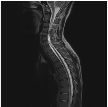
When seen in consultation, he had a 2-month history of progressive onset of fatiguability when walking associ-ated with worsening of the pre-existing right deficit and appearance of left hemiparesis. The physical examination found pyramidal signs involving all four limbs with bilateral Hoffmann and Babinski signs. He also presented bladder dys-function with urgency. The imaging studies, a cervical MRI and CT scan, showed bone condensation; the mass measured approximately 22 mm in greatest dimension and was devel-oping at the expense of the C4 neural arch (posterior arch) resulting in major spinal cord compression due to narrow-ing of the canal. The spinal canal was reduced to 3 mm in anteroposterior diameter in the narrowest zone. This was associated with a reactional syrinx measuring 70 mm in cran-iocaudal diameter involving levels C3 to C6 (Figs. 1—3). The rest of the craniospinal images were unremarkable. Consid-ering the imaging findings and the relatively urgent clinical
1877-0568/$ – see front matter © 2010 Elsevier Masson SAS. All rights reserved. doi:10.1016/j.otsr.2010.06.013
Spinal cord compression due to C4 vertebral arch osteochondroma 95
Figure 1 T2-weighted sagittal MRI showing posterior spinal cord compression by an osteochondroma in the posterior arch of C4 with intramedullary high signal intensity in C4 and a 70 mm syrinx.
presentation, immediate surgical excision was decided for decompression. A biopsy was not performed prior to surgery as this was considered to be dangerous and would not have had any effect on the indication, whatever the result.
Surgery enabled complete excision of the tumor by sim-ple laminectomy from C3 to C5. There were no adhesions with the dura mater. The procedure provided release of the dural sheath. The histology examination concluded in
osteo-Figure 2 Axial CT scan with bone window showing an osteo-chondroma measuring 22 mm in anteroposterior length and 17 mm in width, developing at the expense of the posterior arch of C4 and entering the spinal canal.
Figure 3 Sagittal CT scan with bone window showing the osteochondroma in C4 and its orientation within the spinal canal.
chondroma without histological abnormalities suggesting malignant transformation. The bony edges of the laminec-tomy margins were in healthy tissue.
Immediate postoperative recovery was marked by rapid progressive improvement in his neurological status. He was discharged from the unit on the 10th postoperative day to a physical medicine and rehabilitation unit. He was seen again in consultation 3 months later; he showed clinical recovery of his previous status with complete motor and sphincter autonomy. The check MRI performed 3 months after surgery showed complete release of the spinal cord with persistence of intramedullary signal at C4. The syrinx had reduced from 70 to 27 mm in greatest dimension. The subarachnoid spaces anterior and posterior to the spinal cord had been restored (Fig. 4).
Discussion
Osteochondromas or osteogenic oxostoses can be either soli-tary, or multiple in the case of hereditary familial exostoses. They represent 30 to 40% of benign bone tumors[1]. They usually involve the metaphysis of long bones. Prevalence of the multiple form is estimated at between 0.9 and 2 per 100,000 persons in the Caucasian population[2]. This figure is probably underestimated as many cases are not diagnosed because they are asymptomatic.
Spinal localizations are rare; their frequency varies from 3% for the solitary form to 7 to 9% for the multiple form
[3]. A review of the literature performed in 2005 found 165 cases of spinal osteochondroma [4]. Fifty percent of spinal osteochondromas involve the cervical spine[1,5]. This preferred localization could be contributed to by repeated microtrauma [1]. All levels can be involved, but the site the most affected by isolated osteochondromas is C1. In the multiple forms, it is C2[4].
96 C. Eap et al.
Figure 4 T2-weighted sagittal MRI 3 months after surgery showing reduction in the syrinx and the cord compression.
The physiopathology of osteochondroma remains unknown. The familial form has an autosomal dominant mode of transmission with full penetrance. The sex ratio is 2.5:1 with male preponderance[1]. Ionizing radiation has been implicated as this association is found in 12 to 15% of cases[6]. The time to onset is apparently 8 years after the initial irradiation, and mainly concerns patients who have been administered over 25 Gy before the age of 2 years[7]. The first clinical manifestations appear in the second and third decades, with an average age of 20 at the time of diag-nosis[8]. However, six cases in patients over the age of 60 have been reported[9—11]. The clinical manifestations of spinal involvement vary. The most frequent presentation is spinal cord compression[4]. Spinal cord compression is more frequent in the familial forms than the solitary forms[1]. Our literature analysis found some rarer clinical presentations. Dysphagia has been described[12] due to compression of the oesophagus, and Horner syndrome[13]due to compres-sion of the cervical sympathetic chain by osteochondromas arising in the vertebral body and developing anteriorly into the lateral spinal nerve groove. Cases of purely radicular involvement have also been published, with either sciatica pain[11,14], or cervicobrachial neuralgia[15,16], and also Arnold’s (occipital) neuralgia with C2 involvement [17]. A case of sudden death has also been reported[18]in odontoid osteochondroma.
Diagnosis is difficult on plain radiographs as the various spinal elements are visible in the area of the lesion. CT scan is the treatment of choice for diagnosis. MRI provides a precise study of the relationship between the osteochon-droma and the dural sheath, and the degree of spinal cord compression.
The clinical history and imaging studies are strongly sug-gestive of the diagnosis; a prior biopsy is not useful as it involves a risk of neurological worsening without modifying treatment management.
The rate of local recurrence after surgery is low, but does exist. A review of solitary spinal osteochondroma by the Bordeaux (France) University Hospital [19] only found six cases in the literature. Average time to recurrence is 5 years, varying from 6 months to 14 years[20,21]. For this reason, complete excision is recommended, even if it results in instability that could be protected with arthrodesis[19]. Apart from the risks of local compression, the main complication of osteochondromas is their malignant trans-formation. This risk has been calculated as 1 to 5% in the solitary forms and 10 to 25% in the multiple forms[7]. All patients with exostosis must have close clinical and radio-logical surveillance. The appearance of neuroradio-logical signs in a patient known to have familial osteochondrosis must initiate a search for spinal involvement.
Our patient’s case raised several questions. Firstly, it cast a doubt on the diagnosis of myeloradiculitis at the age of 13. This could have been spinal cord compression due to the early cervical osteochondroma. The plain radiographs performed at that time, and considered to be normal, do not exclude this diagnosis. If an osteochondroma was already present, the question of why it remained stable for 10 years and then worsened remains unanswered. There is also the question of possible factors contributing to the growth of osteochondromas.
Our experience, and current data in the literature, do not provide answers.
Conclusion
Osteochondromas are common benign bone tumors that rarely affect the spine. They can cause slowly develop-ing spinal cord compression. Treatment is complete surgical excision. The outcome is usually favorable.
Conflict of interest statement
None.
References
[1] Albrecht S, Crutchfield JS, SeGall GK. On spinal osteochondro-mas. J Neurosurgery 1992;77(2):247—52.
[2] Schmale GA, Conrad 3rd EU, Raskind WH. The natural his-tory of hereditary multiple exostoses. J Bone Joint Surg Am 1994;76(7):986—92.
[3] Solomon L. Hereditary multiple exostosis. Am J Hum Genet 1964;16:351—63.
[4] Bess RS, Robbin MR, Bohlman HH, Thompson GH. Spinal exos-toses: analysis of twelve cases and review of the literature. Spine 2005;30(7):774—80.
[5] Calhoun JM, Chadduck WM, Smith JL. Single cervical exosto-sis. Report of a case and review of the literature. Surgical neurology 1992;37(1):26—9.
[6] Cree AK, Hadlow AT, Taylor TK, Chapman GK. Radiation-induced osteochondroma in the lumbar spine. Spine 1994;19(3): 376—9.
[7] Sharma MC, Arora R, Deol PS, Mahapatra AK, Mehta VS, Sarkar C. Osteochondroma of the spine: an enigmatic tumor of the spinal cord. A series of 10 cases. J Neurosurg Sci 2002;46(2):66—70 [discussion].
Spinal cord compression due to C4 vertebral arch osteochondroma 97
[8] Labram EK, Mohan J. Diaphyseal aclasis with spinal cord compression. Report of two cases and review of the literature. J Neurosurg 1996;84(3):518—21.
[9] Kaneko K, Yasuma M, Yanase H. Cervical myelopathy due to an osteochondroma in a 73-year-old female. The oldest case in the literature. Bull NYU Hosp Jt Dis 2000;59(2):106—10. [10] Ratliff J, Voorhies R. Osteochondroma of the C5 lamina with
cord compression: case report and review of the literature. Spine 2000;25(10):1293—5.
[11] Sakai D, Mochida J, Toh E, Nomura T. Spinal osteochondromas in middle-aged to elderly patients. Spine 2002;27(23):E503—6. [12] Ilgenfritz HC. Vertebral osteochondroma. Am Surg
1951;17(10):917—22.
[13] Zhao CQ, Jiang SD, Jiang LS, Dai LY. Horner Syndrome due to a solitary osteochondroma of C7: a case report and review of the literature. Spine 2007;32(16):E471—4.
[14] Fiumara E, Scarabino T, Guglielmi G, Bisceglia M, D’Angelo V. Osteochondroma of the L-5 vertebra: a rare cause of sciatic pain. Case report. J Neurosurg 1999;91(Suppl. 2):219—22.
[15] Arasil E, Erdem A, Yuceer N. Osteochondroma of the upper cervical spine. A case report. Spine 1996;21(4):516—8. [16] Srikantha U, Bhagavatula ID, Satyanarayana S, Somanna S,
Chandramouli BA. Spinal osteochondroma: spectrum of a rare disease. J Neurosurg Spine 2008;8(6):561—6.
[17] Kouwenhoven JW, Wuisman PI, Ploegmakers JF. Headache due to an osteochondroma of the axis. Eur Spine J 2004;13(8):746—9.
[18] Rose EF, Fekete A. Odontoid osteochondroma causing sudden death. Report of a case and review of the literature. Am J Clin Pathol 1964;42:606—9.
[19] Gille O, Pointillart V, Vital JM. Course of spinal solitary osteo-chondromas. Spine 2005;30(1):E13—9.
[20] Pecker J, Vallee B, Desplat A, Guegan Y. Lateral interscalenic approach for tumors of the cervical intervertebral foramina (author’s transl). Neuro-Chirurgie 1980;26(2):165—70. [21] Roblot P, Alcalay M, Cazenave-Roblot F, Levy P, Bontoux D.
Osteochondroma of the thoracic spine. Report of a case and review of the literature. Spine 1990;15(3):240—3.