O
pen
A
rchive
T
OULOUSE
A
rchive
O
uverte (
OATAO
)
OATAO is an open access repository that collects the work of Toulouse researchers and
makes it freely available over the web where possible.
This is an author-deposited version published in :
http://oatao.univ-toulouse.fr/
Eprints ID : 18551
To link to this article : DOI:10.1148/radiol.2016151886
URL :
https://doi.org/10.1148/radiol.2016151886
To cite this version : Lapègue, Franck and André, Aymeric and
Meyrignac, Olivier and Pasquier-Bernachot, Etienne and Dupré, Pierre
and Brun, Caroline and Bakouche, Sarah and Chiavassa-Gandois,
Hélène and Sans, Nicolas and Faruch, Marie US-guided Percutaneous
Release of the Trigger Finger by Using a 21-gauge Needle: A
Prospective Study of 60 Cases. (2016) Radiology, vol. 280 (n° 2). pp.
493-499. ISSN 0033-8419
Any correspondence concerning this service should be sent to the repository
administrator:
[email protected]
1 From the Service d’Imagerie (F.L., O.M., E.P.B., P.D., C.B.,
S.B., H.C.G., N.S., M.F.) and Institut de l’Appareil Locomo-teur, Unité de Chirurgie de la Main et Chirurgie Réparatrice des Membres (A.A.), CHU de Toulouse-Purpan, Bâtiment Pierre Paul Riquet, TSA 40031-31059 Toulouse, France; Centres d’Imagerie du Languedoc, Narbonne, France (F.L.); and Laboratoire d’Anatomie, Faculté de Médecine de Toulouse, Toulouse, France (A.A.). Received August 26, 2015; revision requested October 14; final revision received November 19; accepted December 8; final version accepted December 16. Address correspondence to F.L.
(e-mail: [email protected]).
Purpose: To evaluate the efficacy of ultrasonographically (US)-guid-ed percutaneous treatment of the trigger finger by releas-ing the A1 pulley with a 21-gauge needle.
Materials and
Methods: This two-part study was approved by the ethics committee, and written consent was obtained from all patients. The first part consisted of 10 procedures on cadaver digits fol-lowed by dissection to analyze the effectiveness of the A1 pulley release and detect any collateral damage to the A2 pulley, interdigital nerves, or underlying flexor tendons. The second part was performed during an 18-month pe-riod starting in March 2013. It was a prospective clinical study of 60 procedures performed in 48 patients. Out-comes were evaluated through a clinical examination at day 0 and during a 6-month follow-up visit, where the trigger digit was evaluated clinically and the Quick Disabil-ities of the Arm, Shoulder and Hand outcome measure, or QuickDASH, and patient satisfaction questionnaires were administered.
Results: No complications were found during the cadaver study. However, the release was considered “partial” in all fin-gers. In the clinical study, the trigger finger was com-pletely resolved in 81.7% (49 of 60) of cases immediately after the procedure. Moderate trigger finger persisted in 10 cases, and one thumb pulley could not be released. A US-guided corticosteroid injection was subsequently per-formed in these 11 cases. At 6-month follow-up, only two cases still had moderate trigger finger and there were no late complications. The mean QuickDASH questionnaire score was 4; all patients said they were satisfied.
Conclusion: US-guided treatment of the trigger finger by using a 21-gauge needle is feasible in current practice, with mini-mal complications. Franck Lapègue, MD Aymeric André, MD Olivier Meyrignac, MD Etienne Pasquier-Bernachot, MD Pierre Dupré, MD Céline Brun, MD Sarah Bakouche, MD Hélène Chiavassa-Gandois, MD Nicolas Sans, MD, PhD Marie Faruch, MD
Us-guided Percutaneous
release of the Trigger Finger
by Using a 21-gauge needle:
A
underlying flexor tendons, nerves, and collateral vessels.
Clinical Study
Study population.—During an 18-month
period starting in March 2013, 53 adult patients were enrolled into our pro-spective study. The inclusion criterion was idiopathic trigger finger present for at least 4 months. The exclusion criteria were a previous history of open release for trigger finger, rheumatoid arthritis, a concomitant pathologic condition in the hand at the time of the first con-sultation with the surgeon (A.A.), ap-pearance of hand disease not related to the trigger finger during the 6 months following the procedure, absence of the 6-month follow-up visit.
During the first visit, the hand sur-geon (A.A.) proposed this procedure to 53 patients; written informed con-sent was obtained from all subjects. These 53 patients underwent US-guid-ed release of 65 fingers (12 patients had the release performed on two fingers in the same session). Five pa-tients were excluded during the course of our study: two were lost to follow-up at 6 months, one died, and two sub-sequently developed another hand disease that interfered with analysis of the results (carpal tunnel syndrome, finger wound). As a consequence, 60 fingers in 48 patients (27 women, 21 men) were available for analysis. The average patient age was 61 years. The described in 1958 (10). The
effective-ness was equal to that of an open pro-cedure (11–14); however, complications such as overly wide release that extends to the A2 pulley or damage to interdigi-tal nerves have been reported. Never-theless, the complication rate is low (0.02%) (13).
This type of procedure can also be US guided (15–17) and performed with a 2.5–2.6-mm hook (15,16) or a 19-gauge, 1.27-mm needle (17). This has the advantage of providing direct visualization of the vascular and nerve structures during the procedure.
The purpose of our study was to evaluate the efficacy of US-guided per-cutaneous treatment of trigger finger by releasing the A1 pulley with a 21-gauge needle.
Materials and Methods
Our two-part study was approved by our Research Ethics Committee. It con-sisted of cadaver and clinical studies conducted jointly by a hand surgeon (A.A., 10 years of experience) and an interventional radiologist specializing in musculoskeletal procedures (F.L., 15 years of experience).
Cadaver Study
A feasibility study was performed to confirm that the A1 pulley could be cut in a cadaver by using a 21-gauge needle; the bevel was oriented laterally to act as the cutting edge of a scalpel (Fig 1, Fig E2 [online]). On the basis of these findings, the radiologist performed a US-guided A1 pulley release in 10 digits of a fresh cadaver (80-year-old woman). Subsequently, the surgeon carefully dis-sected the treated fingers to analyze the condition of the A1 pulley, A2 pulley,
Published online before print
10.1148/radiol.2016151886 Content codes: Radiology 2016; 280:493–499
Author contributions:
Guarantors of integrity of entire study, F.L., A.A., E.P.B., P.D., C.B., S.B., H.C.G., N.S., M.F.; study concepts/study design or data acquisition or data analysis/interpretation, all authors; manuscript drafting or manuscript revision for important intellectual content, all authors; approval of final version of submitted manuscript, all authors; agrees to ensure any questions related to the work are appropriately resolved, all authors; literature research, F.L., P.D., C.B., S.B., H.C.G., M.F.; clinical studies, F.L., A.A., E.P.B., P.D., C.B., S.B., N.S., M.F.; experimental studies, F.L., E.P.B., C.B., N.S., M.F.; statistical analysis, F.L., C.B., N.S.; and manuscript editing, F.L., O.M., C.B., H.C.G.
Conflicts of interest are listed at the end of this article.
Advances in Knowledge
n US-guided treatment of the trigger finger by using a 21-gauge needle is feasible in current clin-ical practice.
n Complete resolution of trigger finger was achieved in 96.7% (58 of 60) of cases after 6 months. n The combined use of US-guided
procedure and a small needle (21 gauge) is very safe, making it possible to completely avoid iat-rogenic neurovascular or tendi-nous injuries (zero of 60) and minimizing the occurrence of minor adverse event (four of 60). n Our microinvasive procedure is
painless and requires less than 1 day off work for 100% of our subjects.
Implications for Patient Care
n An efficient, quick, safe, and low-cost alternative to surgery is pro-posed for trigger finger
treatment.
n Outpatient care of trigger finger is feasible even in very old patients and those with severe concurrent diseases.
S
napping and locking of the fingers are very common clinical findings, related mainly to an imbalance between the size of the flexor tendons and that of the tendon sheath (Fig E1 [online]). The likely cause is thickening of the A1 pulley secondary to repeated microtrauma (1). In the chronic stage, there is histologic evidence of deep fi-brocartilaginous metaplasia in this pul-ley (2–4).With modern ultrasonographic (US) equipment, the finger pulleys and tendons can be fully analyzed in their normal state (Fig E1 [online]) and the pathologic anatomic structures involved in trigger finger can be clearly seen. The signs of trigger finger have been well described (1): hypoechogenic or even Doppler hyperemic thickening of the A1 pulley with abnormal underlying flexor tendons (tenosynovitis, tendino-sis, dark tendon sign [5]).
Typically, trigger finger is first treated conservatively, with the patient wearing a splint and taking nonsteroidal anti-inflammatory drugs (6–8) or under-going cortisone injections (9). If conser-vative treatment fails, the A1 pulley can be released surgically; good results have been reported in 60%–97% of cases (6).
Blind percutaneous release by us-ing simple clinical landmarks was first
injected around the distal part of A1 pulley and in the tendon sheath when not contraindicated. This additional in-jection was required in 11 cases.
Performance of US-guided release in the thumb is technically more diffi-cult than in the long fingers. Since the thumb cannot be laid completely flat (Fig E4a [online]) on its dorsal side while keeping the flexor tendon pointed at 12 o’clock, the needle must be insert-ed while taking into account two bends (Fig E4b [online]). Anatomic variations in the position of the palmar interdigi-tal nerves can also make the procedure more challenging (Fig E4c [online]). Af-ter the procedure, we advised patients to avoid using their treated hand for 6 hours.
Assessment of clinical outcomes.—
On the day of the procedure (day 0), the radiologist also performed a clin-ical examination; a video of the fin-ger’s movement before and after the procedure was created with a camera (Movies 3 and 4 [online]).
The triger finger cases were classi-fied by using the following semiquanti-tative scale and McNemar test was used for statistical testing. Grade 0 indicated no triggering; grade 1, intermittent, moderate triggering; grade 2, contin-uous triggering that is eliminated with active extension; grade 3, triggering with flexion contracture that requires the patient to use the other hand to un-lock the involved finger; and grade 4, active flexion of finger is impossible.
Patients had a follow-up consultation with the surgeon after 6 months. The clinical outcome of the trigger finger was estimated based on the above scale, and a QuickDASH (Quick Disabilities of the Arm, Shoulder and Hand outcome measure) questionnaire was completed. The patient was also asked to state he or she was “very satisfied,” “satisfied,” “barely satisfied,” or “not satisfied” with the care of their trigger finger.
Results
Cadaver Study
The following observations were made after dissecting the 10 cadaver fingers and made it possible to determine the
bevel’s orientation even when it was hidden beneath the patient’s skin.
Once the 21-gauge needle was in the desired location, the radiologist slid it back and forth horizontally, parallel to the long axis of flexor tendons, four or five times along the trajectory of the A1 pulley (Fig 4a, Movie 2 [online]). While doing so, the hand of an experienced radiologist feels the typical slight resis-tance of the structure being cut. During the release, continuous US verification was performed in the longitudinal plane (Fig 4b), while making sure that the tip of the needle was visible. The position of the needle was also verified in the short axis of the tendon before the release. In general, a centrally located needle near the vertex of the pulley ensures there will be no complications in the long fin-gers; the interdigital pedicles remaining in this position are as far away as possi-ble from the needle (Fig 4c).
Once these four or five back and forth movements were completed, the needle was removed and the patient was asked to flex the treated finger. If the triggering was gone, the procedure was considered complete. If moderate triggering remained, the 21-gauge nee-dle was reintroduced for another four or five back and forth movements; this second set of needling was required in 42% of cases (25 of 60). If the trigger-ing still persisted after these two release attempts, a few drops of Cortivazol (Al-tim; Sanofi-Aventis, Paris, France) were procedures were distributed among
the following fingers: 18 thumbs, six index, 20 middle, 10 ring, and six small fingers; 28 fingers were in the right hand and 32 in the left hand.
Release procedure.—All the
proce-dures were performed by the radiologist (F.L.) using a US unit (model APLIO 500; Toshiba Medical Systems, Tokyo, Japan) with a high-frequency transducer (18 MHz). The patient was positioned supine on a stretcher, with the hand placed flat on a table. A sterile working area was prepared by disinfecting the hand, applying sterile drapes, and using a sterile probe cover and US gel.
First, a local anesthetic was injected with a 25-mm long, 25-gauge needle (orange hub). The needle’s entry point was in the proximal third of the proxi-mal phalanx directed toward the distal part of the A1 pulley. Our team places a gel pad (ie, extra gel heaped on the finger) between the transducer and skin, to make it easier to locate the needle and give it the correct trajectory before breaching the patient’s skin (Fig 2). Two cubic centimeters of 1% lidocaine HCl (Xylocaine; AstraZeneca, Rueil-Malmai-son, France) was injected along the nee-dle’s path and into the synovial sheath of the flexor tendons (Movie 1 [online]).
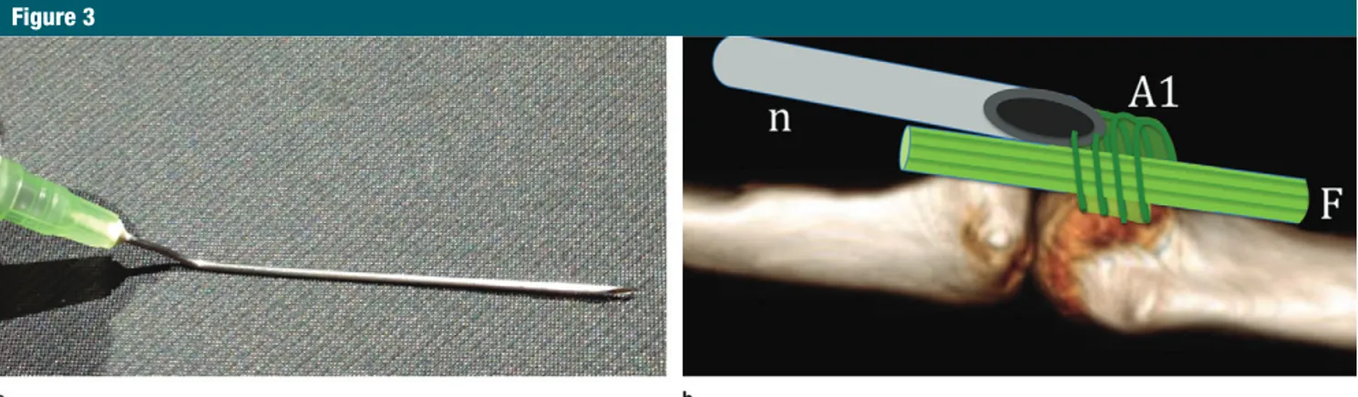
Next, the base of a 50-mm long, 21-gauge needle (green hub) was man-ually curved to a 140° angle so that its bevel faced laterally (Fig 3). This curva-ture had two effects: it placed the nee-dle in a completely horizontal position
Figure 1
Figure 1: Release of the A1 pulley by using a 21-gauge needle in a cadaver preparation. After the volar side of a fresh cadaver finger in an 80-year-old woman was dissected and the fibrous sheath exposed, the needle was slid longitudinally along the superficial aspect of the A1 pulley. The underlying flexor tendons are visible between the divided edges of the pulley (arrows). The pulley is completely cut after two back and forth movements of the needle.
resolved in 58 of the 60 cases (96.7%) (P , .001). No recurrence was ob-served in the treated digits.
The QuickDASH results at 6 months were as follows: (a) Thirty-two patients scored 0 (0 is the best possible score; it implies that there was no impact on activities of daily living), nine patients scored between 2 and 10, six patients scored between 10 and 21 (no signifi-cant impact), and one patient scored 38 (moderate aftereffects; this patient had concurrent shoulder problems that could modify the QuickDASH); note that 100 is the worse score possible; procedure, 81.7% (49 of 60) of the
pro-cedures (P , .001) resulted in complete mechanical release. One thumb (grade 4) could not be released, and 10 of 60 fingers (16.7%) still had minor inter-mittent catching that was not bother-some (grade 1). These 11 of 60 fingers (18.3%) were subsequently injected with cortisone.
At the 6-month follow-up, only two of 60 fingers still had a grade of 1 (3.3%). The failed thumb release procedure was eventually successful 3 weeks after the cortisone injection. The initial trigger finger was completely that underwent US-guided pulley
re-lease: (a) all 10 A1 pulleys were not fully released (Fig E5 [online]), superficial or deep grooves were visible or the pulley was partially divided, (b) none of the A2 pulleys were damaged, (c) the underly-ing flexor tendons were not damaged, and (d) the nerves and collateral blood vessels (palmar interdigital neurovascu-lar bundles) were not damaged.
Clinical Study
Detailed results by trigger finger grades are given in the Table and Table E1 (on-line). At day 0, immediately after the
Figure 2
Figure 2: Anesthesia performed over the release trajectory of the pulley by using the gel pad technique. Longitudinal US views in a 71-year-old woman. (a) A sterile gel pad (GP) made of extra gel heaped on the finger is placed between the transducer and its cover (PC = probe cover, superficial hyperechogenic line) and the finger. The needle (n) is precisely positioned before it enters the patient’s skin. The needle points at the distal part of the hypertrophied A1 pulley (dotted line) in a patient with trigger finger. (b) The needle (n) is advanced to the targeted site and the anesthetic is injected into the flexor tendon sheath (∗∗), around the A1 pulley (∗) and along the needle’s entire trajectory. The pulley is easier to discern as it is “molded” by the anechoic material. F = flexor tendons, MCP = metacarpal, PP = proximal phalanx.
Figure 3
Figure 3: Preparation of the needle used to release the pulley. (a) The base of a 50-mm-long, 21-gauge needle (green hub) is curved to about 140° so that its bevel points to the side. (b) Drawing of the volar side of the flexor mechanism at the metacarpophalangeal joint shows why the needle’s bevel (n) must face laterally. By it facing laterally, it can cut the fibers of the A1 pulley that are perpendicular to the long axis of the finger and will cause minimal damage to the longitudinally oriented fibers of the underlying flexor tendons (F).
following complaints were noted at the 6-month follow-up: seven of 60 fingers had slight, intermittent pain (1 of 10 in the numeric rating scale for pain) with-out triggering; two fingers had minimal residual triggering (grade 1); and trig-ger fintrig-ger developed in one fintrig-ger that had been initially asymptomatic.
Procedure Tolerance and Early Complications
The mean procedure time (including patient set-up) was 15 minutes (stan-dard deviation, 2.2 minutes); once lo-cal anesthetic had been injected into the finger, the patients no longer com-plained of pain during the procedure. Only one complication, a hematoma, was observed in four patients; it ap-peared a few hours after the procedure and became less noticeable 1 week later. Two of the patients were taking At the 6-month follow-up, 39 of 48
pa-tients were very satisfied (81.2%) and nine of 48 were satisfied (18.8%). The
(b) The mean QuickDASH score was 4,
with a median of 0 and standard devi-ation of 8.5.
Figure 4
Figure 4: US-guided release of the A1 pulley in a long finger. (a) Photo obtained during the procedure: The 140° curved needle is held between the radiologist’s thumb and index finger; the patient’s treated finger is extended. (b) Longitudinal US view in a 55-year-old woman. The 21-gauge needle (n) is inserted by using the gel pad (GP) method, with the transducer aligned in the finger’s longitudinal plane. The needle’s curved base allows it to be tilted to achieve a horizontal trajectory and to make four or five back and forth move-ments over the trajectory of the thickened A1 pulley (arrows). The radiologist must make sure that the tip of the needle does not damage the underlying flexor tendons (F). PP = proximal phalanx, MCP = head of metacarpal. (c) Transverse axial US view in the same patient as in b. Inspection in the short axis of the finger at the start of the procedure to identify the palmar interdigital neurovascular bundles (arrowheads). Here the needle (n) is in the ideal position at the most superficial portion of the thickened A1 pulley (arrows), away from the bundles. MCP = head of metacarpal.
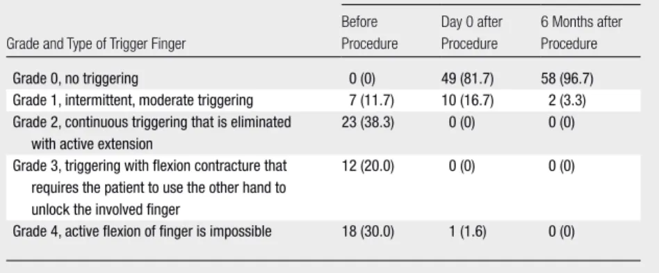
Type of Trigger Finger before the Procedure, Immediately after the Procedure, and 6 Months Later
No. of Each Type of Trigger Finger (n = 60) Grade and Type of Trigger Finger
Before Procedure Day 0 after Procedure 6 Months after Procedure Grade 0, no triggering 0 (0) 49 (81.7) 58 (96.7)
Grade 1, intermittent, moderate triggering 7 (11.7) 10 (16.7) 2 (3.3) Grade 2, continuous triggering that is eliminated
with active extension
23 (38.3) 0 (0) 0 (0)
Grade 3, triggering with flexion contracture that requires the patient to use the other hand to unlock the involved finger
12 (20.0) 0 (0) 0 (0)
Grade 4, active flexion of finger is impossible 18 (30.0) 1 (1.6) 0 (0) Note.—Data in parentheses are percentages.
release; patients were seen again 9 to 15 months later; the mechanical prob-lem had resolved in 100% of cases and the pain had disappeared in 97% (101 of 104) of cases. The persistence of isolated, nonspecific, nondisabling pain in 3% (three of 104) of fingers in the Jou study (16) was also observed in our study (seven of 60 cases).
Possible causes of this moderate re-sidual pain are yet to be determined. However, this residual pain is not limited to this type of procedure, as it has been reported after cortisone injec-tions (19) and after open surgery (20) as well. A concomitant cortisone injec-tion during the release procedure was expected to improve our results as had been demonstrated in one study (21), but this was not confirmed in a meta-analysis (13).
There are limitations to our study. First, a single, experienced radiologist performed all of the US-guided proce-dures; the operator-dependent nature of the procedure was not evaluated— we know this is not an insignificant factor in interventional US. Second, our clinical study did not compare cases treated with standard open sur-gery, which remains the standard or reference, and the follow-up was only 6 months.
In conclusion, US-guided release of the A1 pulley responsible for trig-ger fintrig-ger is feasible with a 21-gauge (0.8-mm) needle. The procedure is quick, painless, risk-free, and low cost, requires almost no time off work, and can be performed on at-risk patients. The trigger digit was resolved immedi-ately and at 6 months in the majority of cases, providing satisfactory results for all patients. If residual triggering is present immediately after the proce-dure, corticosteroid injection improves symptoms, with complete resolution of triggering after 6 months in nearly all patients.
Disclosures of Conflicts of Interest: F.L.
dis-closed no relevant relationships. A.A. disdis-closed no relevant relationships. O.M. disclosed no rel-evant relationships. E.P.B. disclosed no relrel-evant relationships. P.D. disclosed no relevant rela-tionships. C.B. disclosed no relevant relation-ships. S.B. disclosed no relevant relationrelation-ships.
H.C.G. disclosed no relevant relationships. N.S.
The objective improvement in the trigger finger grade is consistent with patients’ feelings after 6 months: The QuickDASH score was less than 21 in 98% (47 of 48 patients) of cases with little to no impact on activities of daily living. The patient with the worst score (score of 38) had concurrent shoul-der problems that interfered with the QuickDASH and was not excluded from the study. Lastly, the procedure is safe: No significant complications were observed in the anatomic or clinical studies.
Beyond the cost of the procedure itself, recovery differs substantially be-tween the standard surgical procedure and percutaneous release. A patient can return to work the day of or the day after a US-guided percutaneous procedure, with no need for nursing care because the needle’s entry point is less than 1 mm long.
Our study used a smaller caliber needle (21 gauge, 0.8 mm) than the one used in similar published studies of pulley release (13,16–18), which makes the procedure less traumatic for the patient, but may also explain why the A1 pulley was not completely divided. In their cadaver study, Smith et al (18) used larger devices and obtained better results: Complete release of A1 pulley was achieved in 32% (eight of 25) of cases with a 19-gauge (1.27-mm) nee-dle and in 88% (22 of 25) of cases with a commercially available hook (HAKI knife; BK Meditech, Seoul, Korea).
Conversely, our clinical outcomes compare well with other recent pub-lished studies of US-guided A1 pul-ley release. In a recent meta-analysis (13) reviewing 2114 procedures with (n = 209) and without (n = 1798) US guidance, the overall success rate was 94% (2004 of 2114). Rajeswaran et al (17) performed a very similar proce-dure to ours, but with a larger needle (19 gauge, 1.27 mm); with a follow-up of 6 months in 35 cases, the trigger finger was completely resolved in 91% (32 of 35) of cases and no complica-tions were observed. Jou et al (16) conducted a larger study (104 fingers) where a specially designed hook (2.5 mm) was used during the US-guided platelet inhibitors and two patients had
resumed use of their hands within an hour of the procedure. These four mi-nor adverse events led us to add a com-pressive dressing after the procedure and to recommend a half day of rest. There were no other complications and no clinical signs of damage to the in-terdigital nerves, flexor tendons, or A2 pulleys and no bowstringing.
The costs of a standard release procedure were compared with those of a US-guided procedure in France (Table E2 [online]). The standard sur-gical treatment requires a doctor’s visit, potentially a diagnostic US, anesthetic nerve block, surgical release of the pulley, at least 10 days off work, and nursing care at home. Our percutane-ous treatment consists of diagnostic US, US-guided release, with or without cortisone injection, and a half day off work.
Discussion
The results of our clinical study were good immediately after the procedure, with 81.7% of trigger finger cases (49 of 60) completely resolved at day 0. Although these results appear contra-dictory with our cadaver study findings, in which the pulley division was incom-plete in 100% of cases, it suggests that even partial release of the A1 pulley can be mechanically effective.
The 10 cases of minor residual trig-gering are probably due to partial in-sufficient release of the A1 pulley. The single case of failed release of the A1 pulley in the thumb can be explained by the fact that the A1 pulley was very thick in this patient (2 mm) and that the procedure is technically much more difficult at the thumb than in the long fingers. The thumb can easily move dur-ing the procedure and cannot be laid completely flat on its dorsal side.
Six months after the procedure, 96.7% (58 of 60 procedures) of cases had an excellent outcome, with the ini-tial trigger finger completely resolved. The additional cortisone injection per-formed at day 0 in the cases with re-sidual triggering likely explains these excellent results at 6 months.
disclosed no relevant relationships. M.F. dis-closed no relevant relationships.
References
1. Guerini H, Pessis E, Theumann N, et al. So-nographic appearance of trigger fingers. J Ultrasound Med 2008;27(10):1407–1413. 2. Sampson SP, Badalamente MA, Hurst LC,
Seidman J. Pathobiology of the human A1 pulley in trigger finger. J Hand Surg Am 1991;16(4):714–721.
3. Sbernardori MC, Bandiera P. Histopathol-ogy of the A1 pulley in adult trigger fingers. J Hand Surg Eur Vol 2007;32(5):556–559. 4. Sbernardori MC, Mazzarello V,
Tranquilli-Leali P. Scanning electron microscopic find-ings of the gliding surface of the A1 pulley in trigger fingers and thumbs. J Hand Surg Eur Vol 2007;32(4):384–387.
5. Gruber H, Peer S, Loizides A. The “dark tendon sign” (DTS): a sonographic indicator for idiopathic trigger finger. Ultrasound Med Biol 2011;37(5):688–692.
6. Akhtar S, Bradley MJ, Quinton DN, Burke FD. Management and referral for trigger finger/thumb. BMJ 2005;331(7507):30–33. 7. Rodgers JA, McCarthy JA, Tiedeman JJ. Functional distal interphalangeal joint splinting for trigger finger in laborers: a re-view and cadaver investigation. Orthopedics 1998;21(3):305–309; discussion 309–310.
8. Patel MR, Bassini L. Trigger fingers and thumb: when to splint, inject, or operate. J Hand Surg Am 1992;17(1):110–113. 9. Dala-Ali BM, Nakhdjevani A, Lloyd MA,
Schreuder FB. The efficacy of steroid injec-tion in the treatment of trigger finger. Clin Orthop Surg 2012;4(4):263–268.
10. Lorthioir J Jr. Surgical treatment of trigger-finger by a subcutaneous method. J Bone Joint Surg Am 1958;40-A(4):793–795. 11. Eastwood DM, Gupta KJ, Johnson DP.
Per-cutaneous release of the trigger finger: an office procedure. J Hand Surg Am 1992; 17(1):114–117.
12. Uçar BY. Percutaneous surgery: a safe pro-cedure for trigger finger? N Am J Med Sci 2012;4(9):401–403.
13. Zhao JG, Kan SL, Zhao L, et al. Percutane-ous first annular pulley release for trigger digits: a systematic review and meta-anal-ysis of current evidence. J Hand Surg Am 2014;39(11):2192–2202.
14. Wang J, Zhao JG, Liang CC. Percutaneous release, open surgery, or corticosteroid in-jection, which is the best treatment method for trigger digits? Clin Orthop Relat Res 2013;471(6):1879–1886.
15. Rojo-Manaute JM, Soto VL, De las Heras Sánchez-Heredero J, Del Valle Soto M, Del Cerro-Gutiérez M, Martín JV. Percutaneous intrasheath ultrasonographically guided first annular pulley release: anatomic study of
a new technique. J Ultrasound Med 2010; 29(11):1517–1529.
16. Jou IM, Chern TC. Sonographically assisted percutaneous release of the a1 pulley: a new surgical technique for treating trigger digit. J Hand Surg [Br] 2006;31(2):191–199. 17. Rajeswaran G, Lee JC, Eckersley R,
Katsar-ma E, Healy JC. Ultrasound-guided percuta-neous release of the annular pulley in trigger digit. Eur Radiol 2009;19(9):2232–2237. 18. Smith J, Rizzo M, Lai JK. Sonographically
guided percutaneous first annular pulley re-lease: cadaveric safety study of needle and knife techniques. J Ultrasound Med 2010; 29(11):1531–1542.
19. Salim N, Abdullah S, Sapuan J, Haflah NH. Outcome of corticosteroid injection ver-sus physiotherapy in the treatment of mild trigger fingers. J Hand Surg Eur Vol 2012; 37(1):27–34.
20. Bruijnzeel H, Neuhaus V, Fostvedt S, Jupiter JB, Mudgal CS, Ring DC. Adverse events of open A1 pulley release for idiopathic trigger finger. J Hand Surg Am 2012;37(8):1650– 1656.
21. Patel MR, Moradia VJ. Percutaneous re-lease of trigger digit with and without cor-tisone injection. J Hand Surg Am 1997; 22(1):150–155.