O
pen
A
rchive
T
oulouse
A
rchive
O
uverte
(OATAO)
OATAO is an open access repository that collects the work of some Toulouse
researchers and makes it freely available over the web where possible.
This is
an author'sversion published in:
https://oatao.univ-toulouse.fr/23096Official URL :
https://doi.org/10.1016/j.jse.2017.12.007
To cite this version :
Any correspondence concerning this service should be sent to the repository administrator:
Bonnevialle, Nicolas and Thélu, Charles Edouard and Bouju, Yves and Vogels, Jérôme and
Agout, Charles and Duriez, Pauline and Azoulay, Vadim Arthroscopic Latarjet procedure with
double-button fixation: short-term complications and learning curve analysis. (2018) Journal of
Shoulder and Elbow Surgery, 27 (6). e189-e195. ISSN 1058-2746
OATAO
Arthroscopic Latarjet procedure with
double-button fixation: short-term complications and
learning curve analysis
Nicolas Bonnevialle, MD, PhD
a,b,*
, Charles Edouard Thélu, MD
c, Yves Bouju, MD
d,
Jérôme Vogels, MD
e, Charles Agout, MD
f, Pauline Duriez, MD
e, Vadim Azoulay, MD
aaDépartement d’Orthopédie Traumatologie du Centre Hospitalier Universitaire de Toulouse, Hôpital Riquet, Toulouse, France
bLaboratoire de Biomécanique, Institut de Mécanique des Fluides de Toulouse- Unité Mixte de Recherche-Le Centre national de la recherche scientifique 5502, Hôpital Riquet, Toulouse, France
cClinique du Sport et de Chirurgie Orthopédique, Marcq-en-Barœul, France dClinique Jeanne d’Arc, Nantes, France
eCentre Ostéo-articulaire Condorcet, Villeurbanne, France
fService d’orthopédie traumatologie, Centre Hospitalier Régional Universitaire de Trousseau, Chambray-les-Tours, France
Background: The arthroscopic Latarjet with double-button fixation is a guided procedure recently
pro-posed to treat anterior shoulder instability with glenoid bone loss. The goal of this study was to report intraoperative and early postoperative complications and to analyze the learning curve.
Methods: This was a prospective, nonrandomized study that included 88 patients. Intraoperative or
post-operative complications as well as adverse events and post-operative time were recorded. Clinical outcomes were evaluated at 2 weeks, 1.5 months, and at the last follow-up. Radiologic analysis was based on an immediate postoperative computed tomography scan.
Results: The intraoperative complications or adverse events rate was 3.3%: 1 conversion to open surgery,
1 bone block fracture, and 1 instrumentation problem. The postoperative complication rate was 6.8%: 4 coracoid migrations, and 2 subluxations. None of these complications occurred beyond the 10th case per-formed. The average operative time significantly decreased with surgical experience (r= −0.8426; 95% confidence interval,−0.9074 to −0.7384; P < .0001) to reach 76 ± 12 minutes (range, 62-95 minutes) at 30 cases. Radiologically, 90% of the bone blocks were flush and subequatorial beyond the 30th case. At a mean follow-up of 12.6 months (range, 6-24 months), Walch-Duplay and Rowe scores were 80 and 81 points, respectively.
Conclusions: At short-term follow-up, the arthroscopic Latarjet procedure with double-button fixation
ex-hibited a low complication rate. Operative time significantly improved with surgical experience and was optimized after 30 cases. Early clinical results confirmed that this procedure can be safe and reliable.
The Hôpitaux de Toulouse Ethics Committee for Research on Human Sub-jects approved this study (No. 01-526).
*Reprint requests: Nicolas Bonnevialle, MD, PhD, Département d’Orthopédie Traumatologie du CHU de Toulouse, Hôpital Riquet, Place Baylac, F-31052 Toulouse, France.
Level of evidence: Level IV; Case Series; Treatment Study
Keywords: Arthroscopie Latarjet; double button; complications; learning curve; bone block; anterior shoulder instability
The Latarjet procedure is indicated to treat anterior shoul-der instability with significant glenoid bone loss: the coracoid transfer provides an adequate bone reconstruction and the con-joint tendon contributes a sling effect in abduction-external
rotation.16.20.26 With an associated Bankart repair, obtaining a "triple locking" effect with an extra-articular bone block position is possible. 8 Despite a low rate of recurrence after
open Latarjet of approximately 5%, the complication rate could
reach 25% in the literature.8•10-12,23,25,26
Arthroscopie techniques have been recently developed to
reduce this complication rate and improve the bone graft position.6
•7•15 Lafosse et aJl5 described a procedure in which the screw fixation of the coracoid was similar to the
origi-nal Latarjet technique. However, some concerns related to
screws (position, length, impingement with soft tissue, place-ment) were reported. 1 In contrast, Boileau et al6 proposed an
innovative double-button fixation method and a guided
ap-proach to transfer the coracoid through the subscapularis
muscle. The procedure remains demanding, and to our
knowl-edge, no other center has reported clinical and radiologie outcomes for this new surgical technique.
The objective of our study was to report early
intraopera-tive and postoperaintraopera-tive complications or adverse events of this new surgical technique and to analyze the learning curve of
this innovative operation. We hypothesized that the
compli-cation rate with this arthroscopie technique would not exceed
the reported rates with the open Latarjet procedure.
Materials and methods
This was a prospective nonrandornized multicenter study con -ducted from April 2015 to October 2016. We included (1) patients with an anterior shoulder instability associated with glenoid bone loss, (2) treated with an arthroscopie Latarjet procedure with double -button bone block fixation, and (3) evaluated with a minimum follow -up of 6 months. Revision operations were excluded. Patients gave their consent for the use of their clinical and radiologie data. Of the 90 patients who fulfilled the criteria for inclusion, 2 were ex -cluded because of revision surgery; therefore, a cohort of 88 patients was available for statistical analysis.
Surgical technique
The 4 surgeons who participated in this study were specialized in shoulder surgery for a mean of 5.5 years (range, 3-9 years) and a mean of343 cases yearly (range, 255--462 cases yearly). They were educated through a specific training program dedicated to this tech -nique, including demonstration step by step by the designer of the technique and cadaveric laboratory assisted procedures.6
A 70° scope and specifically designed instruments (Latarjet Guiding System; Smith & Nephew Inc., Andover, MA, USA) were used. The procedure involved 5 arthroscopie anterior portais (North,
West, South, East, and Northwest) and 5 successive steps accord -ing to Boileau et al6
summarized as follows:
1. Coracoid preparation and osteotomy: pectoralis rninor and coracoacrornial ligament release, abrasion and flattening of the under face, positioning of the peg button, and osteotomy at approximately 1.5 cm from the tip of the coracoid (Fig. 1). 2. Glenoid preparation: abrasion and flattening of the neck of the
scapula, insertion of an anchor at 3 o'clock, and positioning of a posterior-anterior pin with the glenoid drill guide (Fig. 2). 3. Subscapularis split: use of an intra-articular spreader trans
-fixing the subscapularis muscle and opening a "safe window"
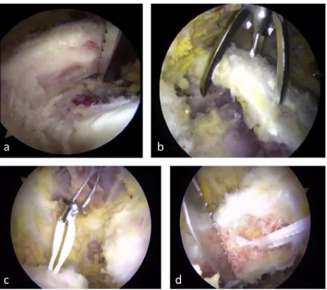
by protecting the visible axillary nerve in front A second spread -er is placed from the east portal to extend the split (Fig. 3). 4. Borre block fixation: transfer of the coracoid through the sub -scapularis split with shuttle suture and definitive fixation with a posterior cortical button fixation ( compression controlled at
100 N) by a Nice knot5 (Fig. 4).
5. Bankart lesion repair with the previously positioned anchor and 1 or 2 sutures.
Postoperatively, an irnmobilization in internai or neutral rotation was established for 4 weeks. Pendular exercises were initiated in week 2 and active mobilization from week 4 by protecting exter -nal rotation up to the week 6. No sports that would place the shoulder at risk were authorized before 3 months.
Intraoperative and postoperative assessment
The learning curve was analyzed through the operative time (from the cutaneous incision toits closure), accuracy of bone block position, and intraoperative complications or adverse events. Postoperative complications with clinical or radiologie effect were also recorded. Clinical monitoring was performed prospectively at 2 weeks, 1.5 months, and every 3 months. Active mobilities were assessed in forward elevation, in external rotation with the elbow at side and in abduction, and in internai rotation ( vertebral level reached by the thumb ). The functional objective assessment was based on the Rowe and Walch-Duplay scores.22.24The preoperative radiologie analysis was based on anteropos -terior and Bernageau views.4 A computed tomography (CT) scan
or arthro-CT scan clarified the presence of a Hill-Sachs lesion and the amount of glenoid bone loss.
Postoperatively, an anteroposterior view was assessed at each clin -ical appointment, and an early CT scan (<15 days) was dedicated to analyze the position of the bone graft. OsiriX (Pixmeo, Geneva, Swit -zerland) irnaging software allowed for the multiplanar reconstruction from the native data.14
,21 The bone block overhang was measured in
the axial plane according to the technique tangent to the subchondral bone over 2 sections ( equatorial and lower one-fourth of the glenoid
cavity). The bone block was thus considered as flush when no medialization or overhang was observed on the 2 levels of analy-sis. The subequatorial coronal position of the bone block was assessed by the coracoid subequatorial length/coracoid total length ratio.
The complication rate and radiologic analysis was assessed by subgroups of 10 successive cases of each surgeon (case 0 to case10, case 11 to case 20, etc) to assess the influence of the surgical ex-perience on these parameters.
Statistical analysis
Statistical tests used SAS 9.3 software (SAS Institute, Inc., Cary, NC, USA). Quantitative variables are described by as average, stan-dard deviation, and maximum and minimum values. The Agostino-Pearson test was used to determine whether the data were normally
distributed. Qualitative variables are described by sample size and percentages. Qualitative variables were compared using theχ2or Fisher exact test. Quantitative variables were compared using the Student t test or the Mann-Whitney test, depending on whether the variable was normally distributed. The Spearman correlation test was used to determine the relationship between 2 variables. The signif-icance threshold was set at 0.05.
Results
Study population
The average age was 25± 7 years (range, 16-60 years), and 86% were men. The dominant side was involved in 57%. Of Figure 1 First step. Posterior viewing portal of a right shoulder. (A) The coracoid process is flattened and abraded with a motorized rasp, (B) a coracoid guide places a Kirschner wire housed inside an outer sleeve, (C) the peg button is positioned over the coracoid with a shuttle suture, and (D) the coracoid is osteotomized with a motorized saw.
Figure 2 Second step. Posterior viewing portal of a right shoulder. (A) The glenoid neck is abraded with a motorized rasp. (B) After an anchor is placed at the 3 o’clock position, a glenoid guide helps to drill the glenoid from posterior to anterior at 5 mm from the rim.
the patient population, 89% participated in sports: 40% in a pure contact sport, 29% in an overhead-contact sport, 16% in a pure overhead sport, and 15% in a sport without a spe-cific risk to the shoulder.
The average instability severity index score was 5± 1.6 points (range, 3-9 points).2
Glenoid bone loss occurred in 100% of cases and a Hill-Sachs lesion in 95%.
Complications
Three (3.3%) complications or adverse events occurred during the procedure: 1 conversion to open surgery because of diffuse soft tissue bleeding despite a controlled low blood pressure, 1 bone block fracture in a 60-year-old patient requiring an additional stabilization with a Hill-Sachs remplissage pro-cedure, and 1 inconsequential glenoid guide pin failure during the step of glenoid drilling. All of these complications or adverse events occurred before the 10th case, regardless of the surgeon. No intraoperative neurologic or vascular lesion was reported.
Six (6.8%) postoperative complications were observed in 5 patients: 2 recurrent subluxations and 4 early bone block migrations (<3 months). The first recurrence was at 2.5 months after the operation while the patient slept on the operated-on shoulder in maximum abductioperated-on positioperated-on. A CT scan
showed the coracoid transfer was nonunited and migrated. The second occurred at 6 months during a contact with another player after resumption of soccer in a young hyperlax patient. The bone graft was united with a slightly too high position on the glenoid rim.
To date, none of these patients required surgical revision and were asked to follow a proprioceptive rehabilitation program. These complications were not observed beyond the 20th case.
No postoperative infectious or neurologic or vascular com-plications were identified in the entire series at the last follow-up.
Surgical time analysis
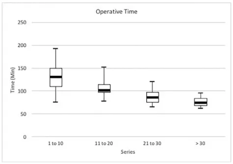
The average operative time of the entire series was 107± 30 minutes (range, 62-192 minutes). A significant inverse cor-relation was found between the operative time and surgical experience noted as number of cases performed (r= −0.8426; 95% confidence interval,−0.9074 to −0.7384; P < .0001). The time decreased by at least 10% every 10 cases, to reach 76± 12 minutes (range, 62-95 minutes) beyond the 30th case (Fig. 5). No significant difference was found among the 4 sur-geons regarding the operative time improvement at any point (P= .3).
Figure 3 Third step. West (anterior subdeltoid space) viewing portal of a right shoulder. The spreader splits the subscapularis muscle and protect the axillary nerve.
Figure 4 Fourth step. West (anterior subdeltoid space) viewing portal of a right shoulder. (A) A shuttle suture through the glenoid sleeve is used to pull the bone block through the subscapularis split. (B) The bone block is positioned parallel to the glenoid rim as flush as pos-sible with an intra-articular view.
Postoperative clinical assessment
For an average follow-up rate of 12.6 months (range, 6-24 months), Walch-Duplay and Rowe scores were 80± 12 points (range, 50-100 points) and 81± 13 points (range, 50-100 points), respectively. The average active mobility was 170°± 11° (range, 140°-180°) in forward elevation, 66° ± 18° (range, 30°-90°) in external rotation with elbow at side, 88°± 6° (range, 70°-100°) in external rotation in abduction, and T12 (range, L1-T7) in internal rotation (Table I). No ap-prehension in 90° of abduction/90° of external rotation was found in 80 patients (90%).
Radiologic assessment
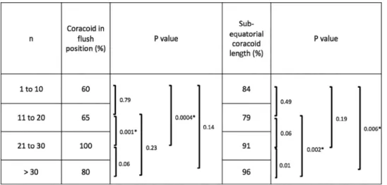
Position of the bone block in the coronal plane
The bone block was flush in 81% of cases, medial in 15%, and lateral in 4%. In the lateral position, the average overhang was 3.6± 1.5 mm (range, 1.2-5 mm). The improved bone block
position was significant beyond the 20th case with a flush bone block up to 90% (Fig. 6).
Position of the bone block in the sagittal plane
The average length of the bone block was 17.3± 2.8 mm (range, 12-25.4 mm). On average, 87%± 18% (range, 33%-100%) of the bone block length was subequatorial. No bone block was fully above the equator. The surgical experience allowed for a significant improvement of subequatorial po-sitioning of the bone block beyond the 30th case (Fig. 6).
Discussion
This study confirmed our initial hypothesis: the intraopera-tive and immediately postoperaintraopera-tive complication rate for the arthroscopic Latarjet procedure with double-button fixation remains low, at approximately 10%, and did not exceed open Latarjet.8,10-12,23,25,26 No major vascular or neurologic
Figure 5 Box-and-whisker plots indicate the surgical time evolution in chronological cohorts of 10 cases. The horizontal line in the middle of each box indicates the median, the top and bottom borders of the box mark the 75th and 25th percentiles, respectively, and the whiskers mark the maximum and minimum of all the data.
Table I Preoperative and postoperative clinical assessments
Variable Preoperative Follow-up P value
Rowe score (100 points total) 30± 12 (20-60) 81± 13 (50-100) <.001
Duplay score (100 points total) 33± 14 (10-70) 80± 12 (50-100) <.001
Active anterior elevation, ° 179± 9 (150-180) 170± 11 (140-180) .29
External rotation, °
Elbow at the side 63± 19 (30-90) 66± 18 (30-90) .90
In abduction 84± 13 (70-100) 88± 6 (70-100) .85
Internal rotation, spine level T8 (T3-T12) T12 (L1-T7) .83
Data are presented as mean± standard deviation (range). T, thoracic; L, lumbar. Operative Time 250 200 c 150 ~
~
è
~ E i= 100 50 0 1 tol0 11 to 20 21 to 30 > 30 Seriescomplications were identified. Moreover, the learning curve analysis demonstrated that the operative time and bone block position significantly improved with surgical experience.
Athwal et al1reported the North American experience for
the first 83 cases using the arthroscopic Latarjet procedure in 5 centers specialized in shoulder surgery. They described 24% adverse events or complications, including neurologic (1%) or vascular (1%) injuries. The fixation of the coracoid transfer with 2 screws was potentially involved in the occur-rence of some complications15: 7% of graft fractures, 3% of
screw backouts, bending or failures, and 4% of revision op-erations for removal of screws.
Studies from the European experience for this same sur-gical technique reported lower complication rates, however. Kany et al13
described 1% neurologic complications and 3% surgical revisions for coracoid fracture or improper screw po-sitions. A recent multicenter study of the French Arthroscopic Society reported by Métais et al18
included 222 patients. Each center that participated was rather experienced in arthro-scopic Latarjet with more than 100 cases already performed before the study. None included their first cases. The imme-diate postoperative complication rate was 4.5% (n= 10), and the revision rate for screw removal was 3% (n= 6).
The device used for graft fixation was not an issue in the double-button technique. Indeed, the device is low profile and adjustable to the patient’s anatomic parameters without im-pingement with surrounding tissue. However, due to its mechanical characteristics in compression, this fixation method does not shield from a bone block fracture in case of de-creased bone density, and we observed 1 intraoperative coracoid fracture in this series.3
Properly positioning the bone graft on the margin of the glenoid cavity is one of the keys of Latarjet procedure. In a position too lateral, residual pain and long-term degenera-tive arthritis of glenohumeral joint was reported, whereas too medial, the failure rate with recurrent shoulder instability increased.17,20,26 In a previous report, after the arthroscopic
Latarjet with screws fixation, Kany et al13noted a bone block
in flush position in 68% and too lateralized in 24%, among their 95 first cases. However, according to a similar surgical technique, Casabianca et al9reported only 32% of bone blocks
flush after 19 procedures. In our study, the flush position was obtained in more than 80% of cases, increasing to 90% beyond the 20th case. Because this had already been proposed in open techniques, the use of a glenoid drill guide seemed to opti-mize the theoretical position of the bone block and miniopti-mized the effects of a learning curve.2,6,19
Thus, the guided approach of arthroscopic Latarjet with double-button fixation offered a double security: a reproduc-ible bone graft position in the coronal plane and a controlled subscapularis split. Indeed, the 2 spreaders protected the ax-illary nerve and avoided any kind of injury during the positioning of the coracoid on the refreshed scapula. This ex-plained why we did not encounter neurologic issues in our series. Not using these specific instruments would increase intraoperative difficulties, expose the patient to potential com-plications, and affect clinical outcomes.
Four early coracoid migrations were observed in our series. One was attributed to the recurrence of instability in a non-compliant patient at less than 3 months postoperatively. The healing of the bone graft was in progress but was insuffi-cient to support the direct constraints of the humeral head and the pullout strain of the conjoint tendon involved in abduction-external rotation. The 3 migrations would be probably conditioned by technical errors when the posterior button was locked with the sliding-locking knot.11Because no
migra-tions were observed beyond the 20th case, we believe that we improved our skills.
This study has some limitations due to its multicenter design. However, we did not identify any significant differ-ences between the surgical time and the rate of complications. In addition, the short-term follow-up constitutes a limit to prop-erly evaluate clinical outcomes, and a minimum of 24 months would be necessary to precisely assess the reliability of this technique. Finally, the rate of nonunion was not reported because it would require further CT scanning.
Figure 6 Analysis of the bone graft position (coronal and sagittal plan) in chronological cohorts of 10 cases.
Coracoid in
Sub-n flush P value equatorial P value
position (%) coracoid
length (%) 1 to 10 60 ] 0.79 84 ] 0.49 11 to 20 65 0.0004• 79 ] 0.001' 0.19 0.14 0.006 0.06 21 to 30 100 ] 0.06 0.23 91 0.002• 0.01 >30 80 96
Conclusions
The arthroscopic Latarjet procedure with double-button fixation exhibits a low rate of intraoperative and postop-erative complications (approximately 10%), including the process of learning the technique. No major neurologic or vascular complications were reported in this series. Thirty cases seem to be necessary to reach a surgical time close to the open procedure and to optimize bone block posi-tion, especially in the sagittal plane. Finally, early clinical assessment confirms that the arthroscopic Latarjet proce-dure with double-button fixation can be a safe and reliable technique.
Disclaimer
Nicolas Bonnevialle, Charles-Edouard Thélu, Jérome Vogels, and Yves Bouju are paid consultants for Smith & Nephew. The other authors, their immediate families, and any research foundations with which they are affiliated have not received any financial payments or other benefits from any commercial entity related to the subject of this article.
References
1. Athwal GS, Meislin R, Getz C, Weinstein D, Favorito P. Short-term
complications of the arthroscopic Latarjet procedure: a North American
experience. Arthroscopy 2016;32:1965-70.http://dx.doi.org/10.1016/
j.arthro.2016.02.022
2. Balg F, Boileau P. The instability severity index score. A simple
pre-operative score to select patients for arthroscopic or open shoulder
stabilisation. J Bone Joint Surg Br 2007;89:1470-7.http://dx.doi.org/
10.1302/0301-620X.89B11.18962
3. Beranger JS, Maqdes A, Pujol N, Desmoineaux P, Beaufils P. Bone
mineral density of the coracoid process decreases with age. Knee Surg
Sports Traumatol Arthrosc 2016;24:502-6.http://dx.doi.org/10.1007/
s00167-014-3483-6
4. Bernageau J, Patte D, Debeyre J, Ferrane J. Value of the glenoid profile
in recurrent luxations of the shoulder. Rev Chir Orthop Reparatrice Appar Mot 1976;62:142-7.
5. Boileau P, Alami G, Rumian A, Schwartz DG, Trojani C, Seidl AJ. The
doubled-suture Nice knot. Orthopedics 2017;40:e382-6.http://dx.doi.org/
10.3928/01477447-20161202-05
6. Boileau P, Gendre P, Baba M, Thélu CÉ, Baring T, Gonzalez JF, et al.
A guided surgical approach and novel fixation method for arthroscopic
Latarjet. J Shoulder Elbow Surg 2016;25:78-89.http://dx.doi.org/
10.1016/j.jse.2015.06.001
7. Boileau P, Thélu CE, Mercier N, Ohl X, Houghton-Clemmey R, Carles
M, et al. Arthroscopic Bristow-Latarjet combined with Bankart repair restores shoulder stability in patients with glenoid bone loss. Clin Orthop
Relat Res 2014;472:2413-24.http://dx.doi.org/10.1007/s11999-014
-3691-x
8. Bouju Y, Gadéa F, Stanovici J, Moubarak H, Favard L. Shoulder
stabilization by modified Latarjet-Patte procedure: results at a minimum 10 years’ follow-up, and role in the prevention of osteoarthritis. Orthop
Traumatol Surg Res 2014;100:213-8.http://dx.doi.org/10.1016/
j.otsr.2014.03.010
9. Casabianca L, Gerometta A, Massein A, Khiami F, Rousseau R, Hardy
A, et al. Graft position and fusion rate following arthroscopic Latarjet.
Knee Surg Sports Traumatol Arthrosc 2016;24:507-12.http://dx.doi.org/
10.1007/s00167-015-3551-6
10. Dauzère F, Faraud A, Lebon J, Faruch M, Mansat P, Bonnevialle N.
Is the Latarjet procedure risky? Analysis of complications and learning
curve. Knee Surg Sports Traumatol Arthrosc 2016;24:557-63.http://
dx.doi.org/10.1007/s00167-015-3900-5
11.Gendre P, Thélu CE, d’Ollonne T, Trojani C, Gonzalez JF, Boileau P.
Coracoid bone block fixation with cortical buttons: an alternative to screw
fixation? Orthop Traumatol Surg Res 2016;102:983-7.http://dx.doi.org/
10.1016/j.otsr.2016.06.016
12. Griesser MJ, Harris JD, McCoy BW, Hussain WM, Jones MH, Bishop
JY, et al. Complications and re-operations after Bristow-Latarjet shoulder stabilization: a systematic review. J Shoulder Elbow Surg 2013;22:286-92.http://dx.doi.org/10.1016/j.jse.2012.09.009
13. Kany J, Flamand O, Grimberg J, Guinand R, Croutzet P, Amaravathi
R, et al. Arthroscopic Latarjet procedure: is optimal positioning of the bone block and screws possible? A prospective computed tomography
scan analysis. J Shoulder Elbow Surg 2016;25:69-77.http://dx.doi.org/
10.1016/j.jse.2015.06.010
14. Kraus TM, Graveleau N, Bohu Y, Pansard E, Klouche S, Hardy P.
Coracoid graft positioning in the Latarjet procedure. Knee Surg Sports
Traumatol Arthrosc 2016;24:496-501.http://dx.doi.org/10.1007/s00167
-013-2651-4
15. Lafosse L, Boyle S, Gutierrez-Aramberri M, Shah A, Meller R.
Arthroscopic Latarjet procedure. Orthop Clin North Am 2010;41:393-405.http://dx.doi.org/10.1016/j.ocl.2010.02.004
16. Latarjet M. Treatment of recurrent dislocation of the shoulder. Lyon Chir
1954;49:994-7.
17. Longo UG, Loppini M, Rizzello G, Ciuffreda M, Maffulli N,
Denaro V. Latarjet, Bristow, and Eden-Hybinette procedures for anterior shoulder dislocation: systematic review and quantitative synthesis of the
literature. Arthroscopy 2014;30:1184-211.http://dx.doi.org/10.1016/
j.arthro.2014.04.005
18. Metais P, Clavert P, Barth J, Boileau P, Broszka R, Nourissat G, et al.
Preliminary clinical outcomes of Latarjet-Patte coracoid transfer by arthroscopy vs. open surgery: prospective multicentre study of 390 cases.
Orthop Traumatol Surg Res 2016;102:271-6.http://dx.doi.org/10.1016/
j.otsr.2016.08.003
19. Meyer DC, Moor BK, Gerber C, Ek ET. Accurate coracoid graft
placement through use of a drill guide for the Latarjet procedure.
J Shoulder Elbow Surg 2013;22:701-8.http://dx.doi.org/10.1016/
j.jse.2012.06.012
20. Mizuno N, Denard PJ, Raiss P, Melis B, Walch G. Long-term results
of the Latarjet procedure for anterior instability of the shoulder.
J Shoulder Elbow Surg 2014;23:1691-9.http://dx.doi.org/10.1016/
j.jse.2014.02.015
21. Rosset A, Spadola L, Ratib O. OsiriX: an open-source software for
navigating in multidimensional DICOM images. J Digit Imaging
2004;17:205-16.http://dx.doi.org/10.1007/s10278-004-1014-6
22. Rowe CR. Evaluation of the shoulder. In: Rowe CR, editor. The shoulder.
New York: Churchill Livingstone; 1988. p. 631-7.
23. Shah AA, Butler RB, Romanowski J, Goel D, Karadagli D, Warner JJ.
Short-term complications of the Latarjet procedure. J Bone Joint Surg
Am 2012;94:495-501.http://dx.doi.org/10.2106/JBJS.J.01830
24. Walch G. Recurrent anterior shoulder instability. Rev Chir Orthop
Reparatrice Appar Mot 1991;77:177-91.
25. Yang JS, Mazzocca AD, Cote MP, Edgar CM, Arciero RA. Recurrent
anterior shoulder instability with combined bone loss: treatment and results with the modified Latarjet procedure. Am J Sports Med
2016;44:922-32.http://dx.doi.org/10.1177/0363546515623929
26. Young AA, Maia R, Berhouet J, Walch G. Open Latarjet procedure for
management of bone loss in anterior instability of the glenohumeral joint.
J Shoulder Elbow Surg 2011;20:S61-9.http://dx.doi.org/10.1016/