O
pen
A
rchive
T
OULOUSE
A
rchive
O
uverte (
OATAO
)
OATAO is an open access repository that collects the work of Toulouse researchers and
makes it freely available over the web where possible.
This is an author-deposited version published in :
http://oatao.univ-toulouse.fr/
Eprints ID : 18545
To link to this article : DOI:10.1007/s00167-015-3900-5
URL :
http://dx.doi.org/10.1007/s00167-015-3900-5
To cite this version : Dauzère, Florence and Faraud, Amélie and
Lebon, Julie and Faruch, Marie and Mansat, Pierre and Bonnevialle,
Nicolas Is the Latarjet procedure risky? Analysis of complications and
learning curve. (2016) Knee Surgery, Sports Traumatology,
Arthroscopy, vol. 24 (n° 2). pp. 557-563. ISSN 0942-2056
Any correspondence concerning this service should be sent to the repository
administrator:
[email protected]
Is the Latarjet procedure risky? Analysis of complications
and learning curve
Florence Dauzère1 · Amélie Faraud1 · Julie Lebon1 · Marie Faruch2 · Pierre Mansat1 · Nicolas Bonnevialle1,3
Conclusion Despite a high rate of post-operative compli-cations, the morbidity of Latarjet procedure remains low. A surgeon’s experience significantly affects the surgery duration and the occurrence of early complications. The main radiological complication is partial lysis of the bone block. After a short learning curve, the clinical outcomes of the Latarjet procedure appear to be satisfactory and reproducible.
Level of evidence IV.
Keywords Anterior instability · Bone block · Latarjet · Learning curve · Complication · Osteolysis · Shoulder
Introduction
Anterior shoulder instability mainly affects young athletes with high functional demands. The coracoid bone block technique (or coracoid process transfer) first described by Latarjet is an alternative to soft tissue re-tensioning, par-ticularly in patients with glenoid rim and/or humeral head bone loss (Hill–Sachs lesion) [1, 16, 19, 21]. A recent North American study reported short-term and medium-term complication rates of up to 25 %, including 6 % infec-tion, 10 % neurological deficit and 10 % recurrence [20]. However, in single-surgeon studies of more than 2000 cases where a standardized technique was followed, the overall complication rate was only 7 %, with a 6 % recurrence rate at 20 years [17, 25]. Although the arthroscopic Latar-jet procedure is becoming more popular, early complica-tions associated with the open Latarjet procedure should be evaluated to determine whether complication rates change as the surgeon becomes more experienced [3, 12]. The pur-pose of this study was to determine the short- and medium-term complication rates in a surgeon’s early cases with the Abstract
Purpose The purpose of this study was to analyse the learning curve and complication rate of the open Latarjet procedure.
Methods The first 68 Latarjet procedures performed by a single surgeon for chronic anterior shoulder instability were reviewed retrospectively. The standard open surgical technique was followed faithfully during each procedure. Post-operative complications were taken from patient med-ical records. Post-operative evaluation consisted of clinmed-ical and radiological assessments.
Results The rate of early (<3 months) clinical compli-cations was 7.4 % (5.9 % haematoma, 1.5 % neurologi-cal deficit), and the delayed complication rate was 7.3 %. Early complication rate, duration of surgery (mean 65 min; 35–135) and hospital stay (mean 3 days; 1–4) were sig-nificantly reduced as experience increased (respectively;
P = 0.03, ρ = − 0.3; P = 0.009, ρ = − 0.3; P < 0.0001,
ρ = − 0.6). On the radiographs, the bone block was healed
and in perfect position in 87 % of cases, with no effect of surgical experience (P = 0.3, ρ = 0.1). The rate of compli-cations on radiographs was 17 %: 11 % partial lysis, 2 % complete lysis and 4 % non-union. No recurrence of insta-bility was found after an average follow-up of 21 months.
* Nicolas Bonnevialle
[email protected]; [email protected]
1 Département de chirurgie Orthopédique, Hôpital Riquet,
CHU de Toulouse, Place Baylac, 31059 Toulouse, France
2 Service de Radiologie, Hôpital Riquet, CHU de Toulouse,
Place Baylac, 31059 Toulouse, France
3 Laboratoire de Biomécanique, IMFT CNRS UMR 5502,
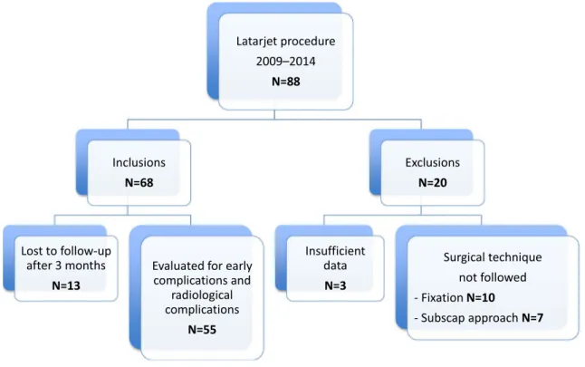
Fig. 1 Study flow chart
Latarjet procedure and to report on the learning curve asso-ciated with this technique. The hypothesis was that a sur-geon’s experience affects the occurrence of complications.
Materials and methods
This was a retrospective study of the initial cases per-formed by a single surgeon using the open Latarjet proce-dure. Patients were included if they met the following crite-ria: (1) operated between January 2009 and January 2014, (2) presented with unidirectional anterior instability of the shoulder with an ISIS score above three points, (3) treated with a primary Latarjet procedure and (4) had a minimum 3-month follow-up [2, 25]. Patients were excluded if they presented with an unstable, painful shoulder with no con-firmed instability episode or if a non-standardized surgical technique was used.
Eighty-eight patients with anterior shoulder instabil-ity were operated with the Latarjet procedure during the inclusion period. Sixty-seven patients (68 shoulders) met the inclusion criteria. Thirteen patients who were lost to follow-up after the third month were not included in the analysis of late complications (Fig. 1). Baseline character-istics of patients are detailed in Table 1.
Table 1 Baseline characteristics of patients
Number of patients included 68
Mean age at surgery (years) 25.5 ± 6.5 (16–40) Sex (male/female) 67/1
Mean body mass index (kg/m2) 24 ± 3.5 (18–35)
Dominant arm involved (number of patients) 42 (62 %) Hyperlaxity (number of patients) 13 (18 %) Smokers (number of patients) 34 (50 %) Type of instability (number of patients)
Subluxation 7 (10 %) Dislocation and subluxation 61 (90 %) Type of sports activities (number of patients)
Contact sport 33 (49 %) Overhead sport 8 (12 %) No-risk sport (for the shoulder) 14 (21 %) No sport 13 (18 %) Competitive level (number of patients) 34 (50 %) Presence of glenoid bone loss (number of
patients)
68 (100 %) Presence of Hill–Sachs lesion (number of
patients)
56 (82 %) Mean ISIS score (points) 5 ± 4.5 (3–10) Revision surgery (number of patients)
Neer capsuloplasty 2 Arthroscopic Bankart 3
Surgical technique [17, 25]
Patients were placed in the beach-chair position with a head rest. General anaesthesia was combined with regional anaesthesia using an interscalenous nerve block. A short 5–6-cm deltopectoral incision was made and then the bone block harvested from the coracoid process using a curved oscillating saw; 1 cm of the acromiocoracoid ligament was preserved on the bone block’s lateral edge, and the pecto-ralis minor muscle was cut on its medial edge. The inferior side of the graft was decorticated, and then two holes were made using a 3.2-mm drill bit. The glenohumeral joint was opened after separating the subscapularis muscle fibres at its upper two-thirds and lower one-third junction. A vertical capsulotomy was performed and the glenoid rim decorti-cated to expose cancellous bone.
The bone block was secured using two bicortical malleo-lar screws. The first inferior screw was placed at 5 o’clock, 7 mm from the glenoid rim. The position of the second screw was determined after making sure the bone block was flush. At the end of the procedure, the capsule was sutured to the acromiocoracoid ligament using two throws of non-absorbable braided suture. The skin was closed after applying a suction drain.
Post‑operative protocol
Patients were immobilized in internal rotation using a sling for 3 weeks. Patients were allowed to start pendulum exer-cises on the first post-operative day. Active-assisted recov-ery of shoulder motion was initiated during the second week, except for external rotation, which was limited to neutral for the first 6 weeks post-operative. Patients were allowed to return to low-risk sports after 3 weeks and then contact or overhead sports after 3 months.
Clinical and radiographic evaluation
The length of hospital stay, surgery duration and length of coracoid harvest were determined from the intraopera-tive data recorded in the patients’ medical record. Clinical assessments were carried out during the follow-up visits at 15 days, 6 weeks, 3 months, 6 months and the final review. During the pre-operative stage and at the final review, active range of motion was evaluated with a goniometer in anterior elevation (AE), external rotation in position 1 (ER1: elbow at body), external rotation in position 2 (ER2: arm abducted 90°) and internal rotation in position 1 (IR1: reach of thumb behind back). Hyperlaxity was defined as ER1 greater than 85°. The anterior apprehension test, the belly-press test and the lift-off test were performed in the
third post-operative month. The Duplay score and subjec-tive shoulder value (SSV) were assessed at the final review [7, 22].
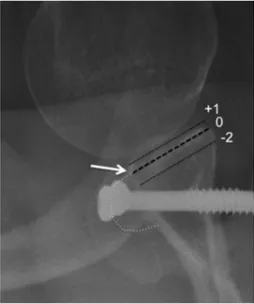
Radiographic analysis was carried out using an anter-oposterior view of the shoulder and a Bernageau view at the third post-operative month. The analysis focussed on the quality of the bone block’s healing and its position. The position was deemed optimal if the bone block was below the equator on the AP view and was flush on the Bernageau view. A flush bone block was defined as a 0 to −2 mm dif-ference between the lateral cortex of the bone block and the glenoid rim; it was defined as lying medially if the differ-ence was less than −2 mm and laterally if the differdiffer-ence was more than 0 (Fig. 2). The screw length was defined as being correct if it did not extend more than 5 mm posteri-orly; the screw position was defined as optimal if the two screws were parallel (±10°).
Clinical complications were classified as either early or late, depending on whether they occurred before or after 3 months post-operative, respectively. Radiological compli-cations (screw back-out, non-union, bone block lysis) were evaluated at the final review. A recurrence of the instabil-ity (subluxation, dislocation) was considered as a failed procedure.
All patients gave written informed consent for their clin-ical and radiologclin-ical data to be reviewed for this study. This study was approved by the Ethical Committee of Toulouse University Hospital (ID number: 01-915).
Fig. 2 Bernageau view used to determine the mediolateral
posi-tion of the bone block: >+1 mm is lateralized, 0 to −2 mm is flush, <−2 mm is medialized. In this example, the bone block (white dotted
Statistical analysis
Statistical tests were carried out using SAS software (ver-sion 9.3, SAS Institute, Cary, NC). Quantitative variables were described by their average, standard deviation, and maximum and minimum values. D’Agostino–Pearson test was used to determine whether the data were normally distributed. Qualitative variables were compared using the Chi-square or Fisher’s exact test. Quantitative variables were compared using Student’s t test or the Mann–Whit-ney test, depending on whether or not the variable was nor-mally distributed. The Spearman correlation test was used to determine the relationship between two variables. The significance threshold was set at 0.05.
Results
Learning curve
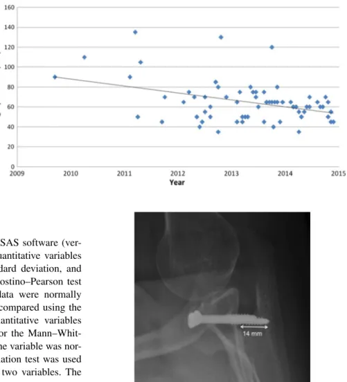
The mean surgery duration was 65 min ± 20.3 (35–135). There was a significant correlation between surgery dura-tion and surgical experience (P = 0.009, ρ = − 0.3; 95 % CI −0.5, −0.08) (Fig. 3).
The mean length of hospital stay was 3 days ± 1.4 (1–4). This variable decreased for the later cases performed by the surgeon (P < 0.0001, ρ = − 0.6; 95 % CI −0.7, −0.4). The length of hospital stay and surgery duration were not significantly correlated with BMI (P = 0.26, ρ = − 0.14; 95 % CI −0.4, 0.1; and P = 0.79, ρ = − 0.03; 95 % CI −0.3, 0.2, respectively).
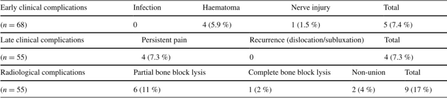
The mean length of the coracoid harvest was 26.2 mm ± 4.2 (22–35) without correlation with surgical experience (P = 0.186, ρ = 0.22; 95 % CI −0.11, 0.51). The position of the bone block on the radiographs was found to be optimal in 87 % of cases, lateralized in 9 % and medialized in 4 %. The screws were parallel in 63 % of
cases and extended beyond the posterior glenoid in 12 % of cases (n = 8) (Fig. 4). The bone block positioning did not improve as the surgeon gained experience with the proce-dure (P = 0.3, ρ = 0.1; 95 % CI −0.1, 0.4).
Clinical complications (Table 2)
The early complication rate was 7.4 % (n = 5). Four patients (5.9 %) had a post-operative haematoma that resolved spontaneously without the need for surgical revi-sion; one case of axillary neuropraxia occurred (1.5 %). Isolated sensory deficit was confirmed on electroneuro-myography, and full recovery was validated at 3 months post-operative. These complications were significantly cor-related with surgeon’s experience (P = 0.03, ρ = − 0.3; 95 % CI −0.5, −0.03). However, they were not signifi-cantly correlated with BMI (P = 0.95, ρ = 0.008; 95 % CI −0.2, 0.2).
Fig. 3 Surgery duration as a
function of the year the Latarjet procedure was performed
Fig. 4 Bernageau view at 3 months post-operative showing posterior
extension of screws by more than 1 cm. This necessitated removal after 7 months post-operative because of persistent posterior pain
The late complication rate was 7.3 % (n = 4). Four patients complained of persistent posterior pain in the infraspinatus fossa. Removal of the screws after an aver-age of 9 months (4–16) completely eliminated these symptoms. In one case, this surgical revision was marked by the occurrence of post-operative periphlebitis of the upper limb, which was treated with curative doses of anticoagulants.
Radiological complications
Among the 55 bone blocks (54 patients) evaluated after an average follow-up of 21 months ± 12 (4–60), 17 % had signs of radiological complications:
• 11 % partial lysis (n = 6) • 2 % complete lysis (n = 1)
• 4 % non-union (n = 2), with the screw having backed out in one case (Fig. 5)
None of these complications required surgical revision, and none had clinical consequences at the last review. Clinical evaluation
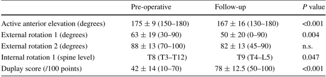
Details of clinical evaluation are reported in Table 3. The average SSV was 89 % ± 8 (70–100). The anterior appre-hension test was only painful in two patients (4 %) and negative in the others; there was no recurrence of instability (subluxation or dislocation) at the last follow-up. Return to sports at pre-injury levels was possible in 74 % of cases. There was no significant clinical difference in the outcomes between patients undergoing a primary procedure and those undergoing a revision procedure.
Discussion
The most important finding of this study is that surgical experience affects the rate of early post-operative clinical complications. These early complications consisted of 6 %
haematoma and 2 % partial neurological deficits, while later complications were mainly related to residual pain (7 %). The morbidity associated with these complications is low, since none of the haematomas required surgical revision, and the neurological deficits resolved spontane-ously. Only the posterior shoulder pain caused by exces-sively long screws required surgical revision to remove the screws.
The published complication rate for the Latarjet pro-cedure ranged from 7 to 30 % [1, 13, 17, 20, 25]. Among the 45 bone block procedures evaluated after an average of 40 months, Shah et al. [20] found a 6 % infection rate, 10 % rate of neurological deficit and 8 % rate of instability recur-rence. The neurological deficits mainly involved the axil-lary nerve and musculocutaneous nerve. As in our study, these were sensory deficits that did not require a new pro-cedure and that resolved spontaneously. Previously, Dela-nay et al. [4] showed through intraoperative monitoring that the highest-risk surgical step for these nerve structures was
Table 2 Clinical and radiological complications following open Latarjet procedure
Early clinical complications Infection Haematoma Nerve injury Total (n = 68) 0 4 (5.9 %) 1 (1.5 %) 5 (7.4 %) Late clinical complications Persistent pain Recurrence (dislocation/subluxation) Total (n = 55) 4 (7.3 %) 0 4 (7.3 %) Radiological complications Partial bone block lysis Complete bone block lysis Non-union Total (n = 55) 6 (11 %) 1 (2 %) 2 (4 %) 9 (17 %)
Fig. 5 Non-union of the bone block with lysis around the inferior
during glenoid exposure with graft positioning, particularly when the surgery duration increased. In the current study, surgery duration was reduced as the surgeon became more experienced, and the rate of early post-operative complica-tions was reduced in the same manner.
In a review of the literature, Griesser et al. [8] found that up to 12 % of bone blocks had failed to heal among the 30 % of cases with complications. However, the stud-ies reviewed did not all use the same surgical technique (e.g. type of fixation, bone block preparation). In our study, despite strict inclusion criteria, the radiological compli-cation rate was 17 %, with 11 % of these complicompli-cations being partial lysis of the bone block. This type of transfor-mation mainly affected the upper part of the graft and has been reported previously. This can probably be attributed to insufficient loading on the upper portion of the bone block [5]. However, there does not seem to be a secondary clinical correlation with partial bone lysis; the hammock effect of the conjoined tendon in the Latarjet procedure is mechanically as important to the anteroinferior stability of the humeral head as the abutment effect [17, 24, 25].
Positioning of the bone block is known to contribute to the shoulder’s stability and the occurrence of arthritic degeneration over time. A bone block placed too medially relative to the glenoid rim or below its equator causes more post-operative recurrences, while overhang of the bone block is a risk factor for osteoarthritis in the long term [9,
10, 17, 18]. Meyer et al. [15] showed that a specially devel-oped instrument can improve the reproducibility of graft positioning. In this study, by meticulously following tech-nical recommendations, more than 80 % of bone blocks were placed in an optimal position, no matter the surgeon’s experience [17, 25]. Arthroscopic techniques currently in development could further improve this positioning, since they provide a better view of the anatomical landmarks and in some cases, the ability to use an alignment guide [3, 12].
We found that more than 10 % of the screws extended more than 5 mm beyond the posterior glenoid; it was necessary to remove the screws in some cases because of pain. Impingement could occur with the infraspinatus muscle and the suprascapular nerve [11, 14]. One reason was related to the type of screw, available only in 5-mm
increments. Another reason was related to the requirement for compressive bicortical screw fixation to ensure union of the bone block [17, 25].
Excessive patient weight is a known risk factor for intra-operative and post-intra-operative complications during shoulder arthroplasty [23]. However, in non-prosthetic surgery of the rotator cuff, BMI does not appear to affect the post-opera-tive course [6]. Despite the technical difficulties potentially encountered in high BMI patients, our study found no sig-nificant effect of BMI on surgery duration or post-operative complications after a Latarjet procedure.
This study had certain limitations. First, the follow-up was not long enough to evaluate the rate of post-operative recurrence of the instability, which some studies list among the procedure’s complications. Second, the occurrence of post-operative glenohumeral osteoarthritis cannot be evalu-ated with radiographs after only 21-month follow-up; a long-term study would be required to determine this. Nev-ertheless, this is the first study, to our knowledge, to report the learning curve for the open Latarjet procedure using a homogeneous cohort of patients undergoing coracoid pro-cess transfer.
Conclusion
This study found a high rate of low-morbidity post-oper-ative clinical complications. The main reason for surgical revision was posterior extension of the screws. A surgeon’s experience significantly affects the surgery duration and the occurrence of early complications. The main radiological complication was partial lysis of the bone block, but this had no effect in the short term. The bone block was opti-mally positioned in more than 80 % of cases; in the other cases, the block was often placed too laterally.
Acknowledgments The authors wish to thank Joanne Archambault,
Ph.D., for editorial assistance in the preparation of the manuscript.
Compliance with ethical standards
Conflict of interest The authors have no conflict of interest to
declare relative to this study.
Table 3 Pre- and post-operative
clinical assessments Pre-operative Follow-up P value Active anterior elevation (degrees) 175 ± 9 (150–180) 167 ± 16 (130–180) <0.001 External rotation 1 (degrees) 63 ± 19 (30–90) 50 ± 20 (0–90) 0.004 External rotation 2 (degrees) 88 ± 13 (70–100) 82 ± 13 (45–90) n.s. Internal rotation 1 (spine level) T8 (T3–T12) T9 (T4–L5) 0.047 Duplay score (/100 points) 42 ± 14 (10–70) 78 ± 12.5 (50–100) <0.001
References
1. Allain J, Goutallier D, Glorion C (1998) Long-term results of the Latarjet procedure for the treatment of anterior instability of the shoulder. J Bone Joint Surg Am 80:841–852
2. Balg F, Boileau P (2007) The instability severity index score a simple pre-operative score to select patients for arthroscopic or open shoulder stabilization. J Bone Joint Surg Br 89:1470–1477 3. Boileau P, Thélu CÉ, Mercier N et al (2014) Arthroscopic
Bris-tow–Latarjet combined with bankart repair restores shoulder sta-bility in patients with glenoid bone loss. Clin Orthop Relat Res 472:2413–2424
4. Delaney RA, Freehill MT, Janfaza DR, Vlassakov KV, Higgins LD, Warner JJ (2014) Neer Award Paper: neuromonitoring the Latarjet procedure. J Shoulder Elbow Surg 23:1473–1480 5. Di Giacomo G, de Gasperis N, Costantini A, De Vita A,
Bec-caglia MA, Pouliart N (2014) Does the presence of glenoid bone loss influence coracoid bone graft osteolysis after the Latarjet procedure? A computed tomography scan study in 2 groups of patients with and without glenoid bone loss. J Shoulder Elbow Surg 23:514–518
6. Fermont AJ, Wolterbeek N, Wessel RN, Baeyens JP, de Bie RA (2015) Prognostic factors for recovery after arthroscopic rotator cuff repair: a prognostic study. J Shoulder Elbow Surg 24:1249–1256
7. Gilbart MK, Gerber C (2007) Comparison of the subjective shoulder value and the Constant score. J Shoulder Elbow Surg 16:717–721
8. Griesser MJ, Harris JD, McCoy BW et al (2013) Complications and re-operations after Bristow–Latarjet shoulder stabilization: a systematic review. J Shoulder Elbow Surg 22:286–292
9. Hovelius L, Sandström B, Olofsson A, Svensson O, Rahme H (2012) The effect of capsular repair, bone block healing, and position on the results of the Bristow–Latarjet procedure (study III): long-term follow-up in 319 shoulders. J Shoulder Elbow Surg 21:647–660
10. Lädermann A, Lubbeke A, Stern R, Cunningham G, Bellotti V, Gazielly DF (2013) Risk factors for dislocation arthropathy after Latarjet procedure: a long-term study. Int Orthop 37:1093–1098 11. Lädermann A, Denard PJ, Burkhart SS (2012) Injury of the
suprascapular nerve during Latarjet procedure: an anatomic study. Arthroscopy 28:316–321
12. Lafosse L, Lejeune E, Bouchard A et al (2007) The arthroscopic Latarjet procedure for the treatment of anterior shoulder instabil-ity. Arthroscopy 23(1242):e1–e5
13. Longo UG, Loppini M, Rizzello G, Ciuffreda M, Maffulli N, Denaro V (2014) Latarjet, Bristow, and Eden-Hybinette pro-cedures for anterior shoulder dislocation: systematic review and quantitative synthesis of the literature. Arthroscopy 30:1184–1211
14. Longo UG, Forriol F, Loppini M et al (2015) The safe zone for avoiding suprascapular nerve injury in bone block procedures for shoulder instability. A cadaveric study. Knee Surg Sports Trau-matol Arthrosc 23:1506–1510
15. Meyer DC, Moor BK, Gerber C, Ek ET (2013) Accurate cora-coid graft placement through use of a drill guide for the Latarjet procedure. J Shoulder Elbow Surg 22:701–708
16. Millett PJ, Clavert P, Warner JP (2005) Open operative treatment for anterior shoulder instability: when and why? J Bone Joint Surg Am 87:419–432
17. Mizuno N, Denard PJ, Raiss P, Melis B, Walch G (2014) Long-term results of the Latarjet procedure for anterior instability of the shoulder. J Shoulder Elbow Surg 23:1691–1699
18. Nourissat G, Delaroche C, Bouillet B, Doursounian L, Aim F (2014) Optimization of bone-block positioning in the Bristow– Latarjet procedure: a biomechanical study. Orthop Traumatol Surg Res 100:509–513
19. Provencher MT, Bhatia S, Ghodadra NS et al (2010) Recurrent shoulder instability: current concepts for evaluation and man-agement of glenoid bone loss. J Bone Joint Surg Am 92(Suppl 2):133–151
20. Shah AA, Butler RB, Romanowski J, Goel D, Karadagli D, Warner JJ (2012) Short-term complications of the Latarjet proce-dure. J Bone Joint Surg Am 94:495–501
21. Streubel PN, Krych AJ, Simone JP et al (2014) Anterior gleno-humeral instability: a pathology-based surgical treatment strat-egy. J Am Acad Orthop Surg 22:283–294
22. Walch G (1991) Recurrent anterior shoulder instability. Rev Chir Orthop Reparatrice Appar Mot 77:177–191
23. Werner BC, Burrus MT, Begho I, Gwathmey FW, Brockmeier SF (2015) Early revision within 1 year after shoulder arthro-plasty: patient factors and etiology. J Shoulder Elbow Surg. doi:10.1016/j.jse.2015.05.035
24. Yamamoto N, Muraki T, An KN et al (2013) The stabilizing mechanism of the Latarjet procedure: a cadaveric study. J Bone Joint Surg Am 95:1390–1397
25. Young AA, Maia R, Berhouet J, Walch G (2011) Open Latarjet procedure for management of bone loss in anterior instability of the glenohumeral joint. J Shoulder Elbow Surg 20:S61–S69