UNIVERSITÉ DU QUÉBEC À MONTRÉAL
STRUCTURE AND FUNCTION STUDIES ON THE HERG POTASSIUM CHANNEL INCLUDING THE DEVELOPMENT AND CHARACTERIZA TION
OF MEMBRANE MIMETICS
DISSERTATION
PRESENTED
AS PARTIAL REQUIREMENT
OF THE DOCTORA TE OF BIOCHEMISTR Y
BY
MAÏWENN BEAUGRAND
Service des bibliothèques
Avertissement
La diffusion de cette thèse se fait dans le respect des droits de son auteur, qui a signé le formulaire Autorisation de reproduire et de diffuser un travail de recherche de cycles supérieurs (SDU-522 - Rév.0?-2011 ). Cette autorisation stipule que «conformément
à
l'article 11 du Règlement no 8 des études de cycles supérieurs, [l'auteur] concèdeà
l'Université du Québecà
Montréal une licence non exclusive d'utilisation et de publication de la totalité ou d'une partie importante de [son] travail de recherche pour des fins pédagogiques et non commerciales. Plus précisément, [l'auteur] autorise l'Université du Québecà
Montréalà
reproduire, diffuser, prêter, distribuer ou vendre des copies de [son] travail de rechercheà
des fins non commerciales sur quelque support que ce soit, y compris l'Internet. Cette licence et cette autorisation n'entraînent pas une renonciation de [la] part [de l'auteur]à
[ses] droits moraux nià
[ses] droits de propriété intellectuelle. Sauf entente contraire, [l'auteur] conserve la liberté de diffuser et de commercialiser ou non ce travail dont [il] possède un exemplaire.••UNIVERSITÉ DU QUÉBEC À MONTRÉAL
ÉTUDE STRUCTURALE ET FONCTIONNELLE DU CANAL POTASSIQUE HERG, INCLUANT LE DÉVELOPPEMENT ET LA CARACTÉRISATION DE
MEMBRANES MODÈLES
THÈSE
PRÉSENTÉE
COMME EXIGENCE PARTIELLE
DU DOCTORAT EN BIOCHIMIE
PAR
MAÏWENN BEAUGRAND
DEDICATION
Je dédicace cette thèse à Erwan Beaugrand
J'espère que tu aurais été .fier de ta petite sœur
"The greatest mistake you can make in !ife is to be continually .fearing you will make one" -Elbert Hubbard (1859-1915)
ACKNOWLEDGEMENTS
Throughout my doctoral studies I have received support from family, friends and co-workers. This acknowledgement section conveys them my full gratitude.
Je tiens tout d'abord à remercier ma directrice de recherche, Professeure Isabelle Marcotte (Université du Québec à Montréal, Canada) pour m'avoir fait confiance et accueillie dans son laboratoire ainsi que pour son soutien tout au long de mon parcours. Merci pour tous tes enseignements ainsi que de m'avoir donné tant d'opportunités d'échanges, congrès, formations et enseignements ici et autour du monde. C'est une superviseure en or et passionnée.
I am lucky to have been co-supervised by Professor Philip T. F. Williamson (University of Southampton, UK). Thank you for welcoming me in your laboratory so many times (from my Master's studies to my PhD, such as a 'boomerang') and for ail your support and advice. Y ou are always available to explain, discuss and think about science and it helps a lot Many thanks for your patience and careness.
La réalisation de ces projets de doctorat a requis l'expertise en résonance magnétique nucléaire du Docteur Alexandre A. Arnold. C'est grâce à tes enseignements que j'ai pu devenir indépendante sur notre capricieux spectromètre RMN 600 MHz et sa version 'relookée', ainsi que sur le 400 MHz. Je te remercie pour tous tes conseils et de répondre présent à ma liste interminable de questions.
Dans cette lignée, je souhaite remercier les professeurs, employés, collègues et amis de l'Université du Québec à Montréal (UQAM) qui m'ont offert de leur temps, de leur savoir et de leur bonne humeur. Tout d'abord, un grand merci à mes collègues,
passés et présents, du laboratoire de résonance magnétique nucléaire des systèmes biologiques complexes: Andrée Grave!, Souryvanh Nirasay, Frédéric Byette, Marc-Olivier Seguin Heine, Delphine Harel, Aline Gambaro, Marwa Laadhari, David Vernon Youmssi, André-Luiz dos Anjos Gomes da Silva, Zeineb Bouhlel, Bertrand Gérard, Antoine Juneau, Abdallah Oukaroum et Catherine Tardy-Laporte. Nous en avons passé des heures ensemble à nous torturer les méninges mais cela aurait été beaucoup plus difficile sans vous. Merci aussi à de formidables étudiants, post-doctorants et employés de cette université: aux biochimistes, Dahmane Ouazia, Audrey Glory, Carole-Anne De Camfel, Germain Lacoste et Mathieu Laporte Wolwertz; aux chimistes, Ngoc Duc Trinh, Danny Chhin, Mathieu Saulnier, Basile Commarieu, Aurore Castets, Chiche Shiao, Jean-Christophe Daigle, David Lepage, Gaëtan Maertens, Melissa Barrera Tomas, André Leblanc, Christian and Sabine Kuss, Sylvain Rocheleau, Marc-André Desjardins, Julien Martel et Elham Akbariromani. Il y a aussi ces professeurs, qui ont pris de leur temps pour rn 'aider et me conseiller, Professeurs Steve Bourgault, Marc Lussier et Jérome Claverie. Sans oublier, les conseils et clarifications administratives des deux directrices successives du programme des études graduées de Biochimie, les professeures Joanne Paquin et Louise Brissette. Merci aussi à l'équipe du personnel de soutien et plus particulièrement: Sonia Lachance, Odette Desrosiers, Mathieu Maurin-Soucy, Luc Arsenault, Chantal Soucy, Sophie Chen, Jacqueline Tieu Hue, Isabelle Rheault, Marie-Josée Crevier, Sylvie Lemieux et Louise Martin-Falstrault. Merci enfin à toute l'équipe du réseau étudiant des cycles supérieurs du secteur des sciences (RECSSS), on faisait une belle brochette: Marie Line Gentes, Sophie Carpentier, Alexandre Terrasa, Anthony François, Dominic Matte et Pierre-Marc Godbout. Je conserverai de bons souvenirs de mon passage entre les murs de I'UQAM grâce à vous tous.
1 will also forever be in debt to my colleague and collaborators at the University of Southampton: James Jarvis, Garrick Foster Taylor, Phedra Marius, Christopher D. Johnson, Neville Wright, Mark S. Friddin, Sumit Kalsi, Rafeel Gomes, Luke S.
IX
Evans, Professors Maurits R. R. De Planque, Morgan Hywel and Declan A. Doyle. I also want to thank Halina Bainbridge, Kelly Hooper, Louise Bolton, John Butler, Nicolas Dalls, Ben Mulcahy, James Beggs, Matt Rodrigues, Professors Stuart Findlow, Marina Carravetta and Anthony Lee. Ali those hours of studying and experimenting in the laboratory would have been unbearable without you all.
Additionnaly, I was very fortunate to have had intemship supervisors that I would like to thank for their guidance, Professor Jason C. Young (McGill University, Canada) and Professor Ansgar B. Siemer (University of Southern Califomia, USA). Each Biochemistry laboratory bas its own tips and tricks and I was glad to leam from them. Many thanks to professor Young for his help with the bERG expression project and to professor Siemer for ali his advice during my PhD.
I would also like to thank the group of Professor Young: Michael Wong, Imad Baaklini, Patrick Kim Chiaw and Yogita Pate!, and the people in the biochemistry department at McGill University: Kristjan Bloudoff, Shane Caldwell and Asparouh Lilov. Un merci tout particulier à Fabien Bergeret qui est devenu très rapidement un très bon ami et m'a aussi donné de précieux conseils et motivation pendant la phase d'écriture. Thanks to al! them, I enjoyed my intemship there.
I want to express my sincere thanks to members at University Southem Califomia and their warm welcoming: Thalia Bajakian, Rachel Service, Ninad Agashe, Silvia Cervantes, Maria Contrad and Sandy Falk, and also to scientists 1 was lucky to met there: Mario Isas, Mark Ambroso, Alan Okada, Nathalie C. Kegulian, Thomas Schmidt and Professor Ralf Langen.
Je tiens à remercier Michèle Auger (Université de Laval, Canada) pour m'avoir donné accès à son laboratoire pour préparer mes échantillons d'infrarouge, ainsi que Jean-François Rioux-Dubé et François Paquet-Mercier pour leur aide technique sur
leur spectromètre infrarouge. J'en profite aussi pour souligner le merveilleux accueil de Mathieu Fillion (même si virtuel), Kim Potvin-Foumier et Thierry Lefevre.
Ces projets ont été rendus possibles grâce à la collaboration de nombreux scientifiques et je tiens donc à remercier pour leur aide deux collaborateurs non mentionnés antérieurement, les Professeurs Dror E. Warschawski et Jérôme Hénin (Université Paris-Diderot, France).
Un grand merci également à ces enseignants qui ont eu une grande influence pendant ma scolarité: l'équipe enseignante de l'école pnmatre de Trémuson et particulièrement, Mme Brigault et Mr Chapelet; Mlle Le Carroux (lycée Eugène Freyssinet), Mme Guillaume et le Professeur Henri Wroblewski (Université Rennes 1 ).
Un remerciement tout spécial à ma famille. Merci à mes parents, Jean-Claude et Marie-Christine Beaugrand pour leur soutien moral sans faille. Merci à mon frère aîné, Gaël Beaugrand, qui a dans mon enfance été mon modèle, qui a toujours cru en moi et m'a donné le goût des mathématiques durant mes premières année de scolarité ainsi qu'à mon frère cadet, Goulwen Beaugrand, avec qui j'ai partagé les jeux d'enfance. J'en profite pour citer les membres étendus de ma famille qui m'ont soutenue durant ces années doctorales: Mikaël Potrat, Pierre, Marie-France et Cécile Chaudot, Nathalie, Pascal, Rémi et Théo Véra, Elizabeth SaJou, Dany Léo, Catherine, Jean-Luc et Stéphane Coquio. La présence de ma famille ainsi que leurs eïïCûUtâgcmcnts dâüS les wvwcü.ts diffici!cs üüïGüt été svü;-~c: de récc:ifcrt '""t de motivation inestimable. Je me considère extrêmement chanceuse de vous avoir et merci de ne pas m'en vouloir d'avoir mis un océan entre nous. Je tien au i à rendre hommage à Suzanne Salau, ma grand-mère et Jean-Pierre Salau, mon oncle, pour qui je n'ai pu être pré ente dan leurs derniers in tants.
Xl
Un profond merci à mes amis, et notamment ceux qui malgré la distance et l'absence sont restés à mes côtés. Tout d'abord mes amis d'enfance: Delphine Calvez, Karen Salmon, Clément Houzé et Guillaume Roussel. Merci à ma partenaire de danse privilégiée, Mushu Le Dragon (alias Aurélie), pour toutes ces années insolites. Merci à Geneviève Martin pour notre amitié. J'aimerais aussi remercier les personnes formidables que j'ai rencontrées à Rennes et avec lesquelles j'ai pu conserver contact: Elisa Marivin, Valentine Genet, Céline Camus, Sophie Rouanet, Phuhai Nguyen, Alexis Saintilan, Eniss Hedhli, Angélique Faramus, Mathieu Le Rouzic, Arnaud le Febvrier, Loïc Cara, Wennaël Deslandes et Audrey Bachatera.
Merci aussi au soutien que j'ai eu pendant cette rude première année de doctorat où j'ai passé près du rapatriement. J'en profite pour remercier ma médecin de famille, la Docteure Janik Le Corveller qui m'a conseillée judicieusement et évité un arrêt d'études non nécessaire. A big thank you to my friends met at Southampton, especially Stefania Fabbri, Vanesa Martinez, Joe Kakande, Asad Ali, Asim Munir et Priyanth Mehta. Gracias a mi mejor amigo, Joaquin Matres Abri! por ayudarme a abrir los ojos en momentos importantes de mi doctorado que permitieron volverme a centrar en mi trabajo. I want to thank my friends from INRS: Romain Dugas, Ivan Cito, Memusc Azodi, Gaston C. Jimenez and Sami Tomoyo. Un agradecimiento para Orlando Trejo por motivarme mientras acababa de escribir esta tesis y, en paralelo, ayudarme a encontrar posiciones post-doctorales. Merci aussi à des personnes fom1idables rencontrées à Montréal: Christopher Wardell, Sébastien Guerry, Manuel Labridy et Jorge Juârez. Un énom1e merci à Huynh Van Du Tran pour son soutien et son aide pour tous les petits tracas technologiques et quotidiens, pour croire en moi ainsi que pour avoir sauvé mes données et phases d'écriture de ce manuscrit face à d'énièmes guignes informatiques. Cam on nhitu, tim cûa em. Sans oublier mes animaux à 4 pattes, Picolo, Wolvie et Ayla, qui méritent également une place dans ma reconnaissance par leur présence, leur affection et tous leurs ronrons réconfortants.
Je tiens à remercier les membres du jury de projet de thèse: Professeurs Richard Desrosiers, Jean Danyluk (UQAM) et Yves Aubin (Carleton University, Canada), ainsi que les membres de mon comité de thèse pour leur temps à lire, évaluer et corriger ce travail de longue haleine: Professeur Joanne Paquin, Marc Lussier (UQAM) et Natalie Goto (University of Ottawa, Canada).
Finally, I want to thank severa! organizations for their financial supports during my PhD studies, laboratory exchanges and conferences: le département de chimie de l'UQAM, le programme des études graduées de Biochimie de l'UQAM, la fondation de l'UQAM, les Fonds à l'accessibilité et à la réussite des études (FARE) de l'UQAM, le syndicat des professeurs et professeures de l'UQAM (SPUQ), le programme de reconnaissance de l'implication étudiante de l'UQAM, le ministère de l'éducation, des loisirs et du spo11 du Québec (MELS), the McGill University Chemical Biology Training Program (ChemBIO) and their coordinator, professor John R. Silvius, the Create Training Program in Bionanomachines (Bionano) and their coordinators, Christopher von Roretz and Anne Noronha, le Groupe de Recherche Axé sur la Structure des Protéine (GRASP) et leur coordinatrice, Annick Guyot, le Centre Québécois des Matériaux Fonctionnels (CQMF) et leur coordinatrice Patricia Basque, the organization committee of South West Structural Biology Consortium (SWSBC) 2012, the organization committee of EUROMAR 2013, the Biophysical Society and the Education Committee for the annual meeting of the Biophysical Society in 2014. This also gave me the opportunity to meet great scientists during meetings, journal r.h1hs ~nrl workshops which enlargerl my network.
AVANT-PROPOS
Par égard pour le fait que ce diplôme de Doctorat en Biochimie a été réalisé dans une université francophone, une traduction en français de chaque résumé ainsi que de cet avant-propos peut être trouvée avant la version en anglais.
Cette thèse se concentre sur 1 'application de la spectroscopie de résonance magnétique nucléaire pour obtenir de l'information sur les systèmes membranaires, telles que les membranes modèles et les protéines membranaires. Le Chapitn~ 1 est l'introduction. Le Chapitre 11 fournit une description théorique des principales techniques utilisées. Les Chapitn~s Ill et 1\' sont des études détaillées sur des membrane modèles, les bicelles. Le but du Chapitre Ill est une caractérisation en profondeur des bicelles diluées constituées de dimyristoylphosphatidylcholine (DMPC ou Dl4PC) et dihexanoyl-phosphatidylcholine (DHPC ou D6PC). Le Chapitr~.· 1 \' décrit et caractérise des bicelles formées à partir de détergent de la famille des monoalkylphosphocholines (MAPCHO). Deux études sur le canal potassique eucaryote human ether-à-go-go related gene (hERG) sont traitées dans les Chapitres V et VI. Le Chap1tre \' est une étude comparative des hélices du pore de hERG et de Kv1.5 dans des bicelles formées à partir de détergent MAPCHO. Le Chapitr~.· VI dépeint un protocole d'expression et de purification du domaine du pore fonctionnel de hERG. Finalement, le C h.tpitrl' \'II regroupe les conclusions et perspectives. Les sections résultats sont présentées sous forme de manuscrits d'articles. Les références sont rassemblées ensemble en une section à la fin de cette thèse.
XIV
Les travaux présentés dans les Chapitres 111, 1\' et \ ont été faits dans le laboratoire
de la Professeure Marcotte (Département de Chimie, Université du Québec à
Montréal - UQAM, Canada), alors que ceux du Chapitn: \'1 ont été réalisés dans le laboratoire du Professeur Williamson (Centre pour les Sciences Biologiques,
University of Southampton, Angleterre).
Publications supplémentaires:
•
Friddin, M. S., Smithers, N. P., Beaugrand, M., Marcotte, 1., Williamson, P. T. F.,Morgan, H., & de Planque, M. R. R. (2013). Single-channel electrophysiology of cell-free expressed ion channels by direct incorporation in lipid bilayers. Analyst, 138, 7294-7298.
• Warschawski, D. E., Arnold, A. A., Beaugrand, M., Grave], A., Chartrand, É., &
Marcotte, 1. (20 11). Choosing membrane mimetics for NMR structural studies of
PREFACE
In respect to achieving this Doctor of Philosophy degree in Biochemistry in a francophone university, a French translation of each abstract and this preface are found prior to the English version.
This dissertation focuses on the application of nuclear magnetic resonance spectroscopy to obtain information on membrane systems, such as mode! membranes and membrane proteins. Chapll:r l is the introduction. Chaplci Il provides a theoretical description of the main techniques used. Chaplcr-; 111 and 1\' are detailed studies on mode! membranes, the bicelles. The purpose of Chaptcr Ill is a deeper characterization of diluted dimyristoylphosphatidylcholine (DMPC or D14PC) 1 dihexanoylphosphatidylcholine (DHPC or D6PC) bi celles. Ch,Iplcr lV describes and characterizes monoalkylphosphocholine (MAPCHO) detergent - based bicelles. Two studies on the eukaryotic potassium channel human ether-à-go-go related gene (hERG) are covered in Chaptcr~ \' and VI. Chaptcr \ is a comparative study of hERG and K v 1.5 pore helices in MAPCHO-based bicelles. C'haptcr VI depicts the expression and purification protocol of a functional hERG pore domain. Finally, Chaplcr \'Il presents the concluding remarks and future perspectives. Results are presented in form of publication manuscripts. References are gathered together ioto one section at the end of this dissertation.
The work presented in C..haplcrs fil,[\' and\' has been canied out in the laboratory of Professor Marcotte (Chemistry department, Université du Québec à Montréal -UQAM, Canada) while the one in C hap!èr \'1 was achieved in the laboratory of
Professor Williamson (Centre for Biological Sciences, University of Southampton, UK).
Additionnai Publications:
• Friddin, M. S., Smithers, N. P., Beaugrand, M., Marcotte, I., Williamson, P. T. F., Morgan, H., & de Planque, M. R. R. (2013). Single-channel electrophysiology of cell-free expressed ion channels by direct incorporation in lipid bilayers. Analyst, 138, 7294-7298.
• Warschawski, D. E., Arnold, A. A., Beaugrand, M., Gravel, A., Chartrand, É., & Marcotte, I. (20 11). Choosing membrane mimetics for NMR structural studies of transmembrane proteins. Biochimica et Biophysica Acta, 1808, 1957- 1974.
TABLE OF CONTENTS
DEDICA TION ... v
ACKNOWLEDGEMENTS ... vii AVANT-PROPOS ... xiii PREFACE ... xv
LIST OF FIGURES ... xxiii LIST OF TABLES ... xxix RÉSUMÉ ... xxxi ABSTRACT ... xxxiii CHAPTER I INTRODUCTION ... 1 1.1 Biological Membrane ... 1 1.1.1 Organisation of the Membrane ... 1 1.1.2 Membrane Lipids ... 3 1.1.3 Membrane Proteins ... 12 1.2 Potassium Channels ... 16 1.2.1 Structure ... l6 1.2.2 Ion Selectivity ... 19 1.2.3 Ga ting ... 19 1.2.4 Subgroups of Potassium Channels ... 21 1.3 Voltage-Gated Potassium Channels ... 22
1.3.1 Overview ofKv channels ... 22
1.3.2 Subfamilies ofKv channels ... 23
1.3.3 Kv 1.5 and hERG ... 23
1.4.1 Mode! Membrane Systems ... 30
1.4.2 Production of Membrane Proteins ... 35
1.5 Objectives ... 39
CHAPTER II THEO RI CAL ASPECTS OF BIOPHYSICAL TECHNIQUES ... .41
2.1 Nuclear Magnetic Resonance ... .41
2.1.1 Nuclear Spin and Lannor Frequency ... .42
2.1.2 Frce Induction Decay ... .44
2.1.3 Nuclear Spin Hamiltonian ... .46
2.1.4 NMR Study of Membrane Systems ... 53
2.2 Circular Dichroism - Study of Peptide and Prote in Secondary Structures ... 62
2.3 Fourier Transform Infrared Spectroscopy- Study ofMiscibility Between Lipids ... 65
CHAPTER III LIPID CONCENTRATION AND MOLAR RATIO BOUNDARIES FOR THE USE OF ISOTROPIC BICELLES ... 71
Foreword ... 72
Résumé ... 72
Abstract. ... 73
3.1 Introduction ... 74
3.2 Materials and Methods ... 77
3.2.1 Materials ... 77
3.2.2 Sample Preparation ... 78
3.2.3 31P Solution NMR Experiments ... 78
'"" ... A T""'.,...T~ .,.-. " • "'70 .J.L.'+ r 1 H\. D.>q.Jt:J llllt:Hl~ ... 1 u 3.2.5 Molecular Dynamics Simulations ... 79
3.3 Results and Di cu sion ... 81 3.3.1 Exact Composition ofBicelles under Dilution: 31P NMR ... 81 3.3.2 Lipid Concentration Threshold to Maintain the Desired q Ratio with Isotropie Bi celles ... 87
XIX
3.3.4 q 2: 1: FTIR Spectra Confirm Lipid Segregation in Isotropie Bicelles .... 89
3.3.5 q :S 0.5: Mo1ecular Dynamics Simulations Show Lipid Mixing in Very Small Bicelles ... 91 3.4 Conclusion ... 94
Note Added in Proof ... 95
Acknowledgments ... 95
Supporting Infom1ation ... 96
CHAPTERIV MAGNETICALL Y -ORIENTED MAPCHO BI CELLES - VERSATILE MIME TICS FOR NMR APPLICATIONS ... 101
Foreword ... 102
Résumé ... l02 Abstract ... 103 4.1 Introduction ... 104
4.2 Materials and Methods ... 107
4.2.1 Materia1s ... 107
4.2.2 Sample Preparation ... 108
4.2.3 Nuclear Magnetic Resonance ... 108
4.2.4 Fourier Transform Infrared Spectroscopy ... 110
4.3 Resu1ts and Discussion ... 112 4.3.1 Versatile Magnetically-Oriented Bicelle with Low Free Surfactant Concentration ... 112 4.3.2 Behaviour as a Function of the Temperature ... 116 4.3 .3 Behaviour as a Function of Mo1ar Ratio q ... 118 4.3.4 Effect ofDetergent-Phospholipid Chain Length Difference on MAPCHO Bicelles ... 120
4.3.5 Miscibi1ity ... 122
4.4 Conclusion ... 125
Acknowledgments ... 126
CHAPTER V
A COMPARATIVE STUDY OF THE STRUCTURE AND INTERACTION OF THE PORE HELICES OF THE HERG AND KY 1.5 POTASSIUM
CHANNELS IN MODEL MEMBRANES ... 133
Foreword ... 134 Résu1né ... 1 34 Abstract ... 135 5.1 Introduction ... 136 5.2 Materia1s and Methods ... 139 5 .2.1 Materia1s ... 139
5.2.2 Peptide Synthesis and Purification ... 139
5.2.3 Samp1e Preparation ... 140
5 .2.4 Circular Dichroism Spectroscopy ... 141
5.2.5 Nuclear Magnetic Resonance (NMR) ... 142 5.3 Results and Discussion ... 142
5.3.1 Secondary Structure (CD) ... 142
5.3.2 Interaction with Membrane (So1id-State NMR) ... 144
5.4 Conclusion ... 152
Acknowledgments ... 153
Supporting Information ... 154
CHAPTER VI EXPRESSION AND FUNCTIONAL CHARACTERIZA TION OF THE PORE DOMAIN FROM THE EUKARYOTIC POTASSIUM CHANNEL, HERG ... 161
Foreword ... 162 Résumé ... 162 Abstract ... 163 6.1 Introduction ... 164
6.2 Materia1s and Methods ... 167 6.2.1 P1asmid Con truction and Protein Expression ... 167 6.2.2 Purification ... 169 6.2.3 Mass Spectrometry ... 170
XXI
6.2.4 Circular Dichroism Spectroscopy ... 171
6.2.5 Reconstitution for the Electrophysiology ... 171
6.2.6 Electrophysiology with Montai Mueller Method ... 172
6.3 Results and Discussion ... 173
6.3.1 Expression of the hERG Pore Domain ... 173
6.3.2 Mass Spectrometry ... 175
6.3.3 Circular dichroism Spectroscopy ... 176
6.3.4 Functional Analysis of the hERG Pore Domain ... 177
6.4 Conclusion ... 179
Ack.nowledgments ... 179
CHAPTER VII CONCLUSION AND OUTLOOK ... 181
7.1 Deve1opment and Characterization of Mode! Membranes ... 181
7.1.1 DMPC and DHPC Bicelles ... 181
7.1.2 Phospholipid and MAPCHO Bicelles ... l82 7.1.3 Pore Helices ofhERG and Kvl.5 ... 184
7.2 Elaboration of a Protocol to Express the HERG Pore Domain ... 185
7.3 Madel Membrane Limitations and Prospects ... 186
LIST OF FIGURES
Figure Page
1.1 Fluid (non random) mosaic model membrane ... 3
1.2 Representation of phospholipid structures ... 5 1.3 Representation of cholesterol structure ... 6
1.4 Representation of the lipid motions in the membranes ... 7
1.5 Gel phase conformation oflipids versus liquid crystalline phase ... 8
1.6 The different shape of lipids associated with their organization in
aqueous medium ... 11 1.7 Representation of integral and peripheral membrane protein classes ... 12
1.8 Representation of detergent structures ... 14
1. 9 Representation of the different types of transmembrane pro teins with a -he li cal segment(s) ... 15
1.10 Structure of the bacterial KcsA potassium channel which constitute a
pore model. ... 17
1.11 Sequence alignment of the hERG pore domain with KcsA, MthK,
KvAP, Kv1.5, Kv2.1, Kv3.1 and Kv4.3 ... 18
1.12 Activation, deactivation, inactivation and reactivation of ion channel s ... 20 1.13 Different activity states of a potassium channel. ... 20 1.14 Predicted topology of K v 1.5 channel... ... 24 1.15 Human atrial and ventricular action potential illustrated with indication
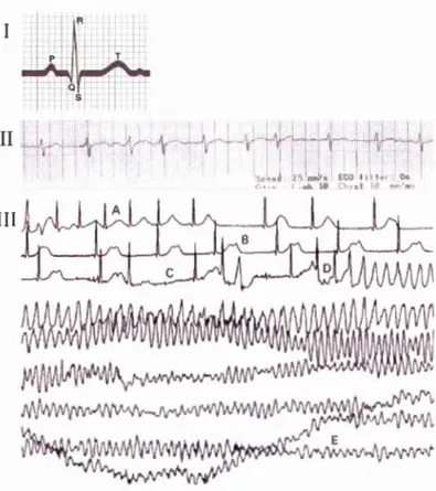
1.16 Electrocardiogram representation for an healthy persan is about 0.8 sec. Electrocardiogram of a 35-year-old patient with Kv 1.5 mutations. Electrocardiogram of a 25-year-old woman with congenital long QT
syndorrne (LQTS) recorded du ring her sleep du ring a 'bad dream' ... 26 1.17 Predicted topology of hERG channel. ... 28 1.18 Representations depicting severa! membrane mimetics with a
representation of the aggregate and a focus on the organisation of
molecule ... 34 2.1 Spin angular momentum of a nucleus and motion of precession of the
spin ... 43
2.2 Directions of spin angular momentum and magnetic moment according
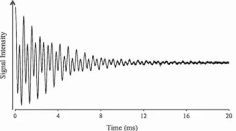
to the sign of the gyromagnetic ratio ... 44 2.3 Example of free induction decay of a binary mixture of phospholipid
and MAPCHO detergent. ... 45 2.4 Representation of the Zeeman effect with energy levels in the presence
of a magnetic field for a hydrogen nucleus (1= 1/2) ... 4 7 2.5 Illustration of energy levels for a spin 1= 1 and 2H-NMR spectra with
Zeeman interaction and with both Zeeman and quadrupolar interactions .... 51 2.6 2H-NMR (spin=l) spectrum and individual transitions in a spheric
symmetric distribution of nuclei ... 52 2.7 Representation of the chemical shift anisotropy cffect on 31P-NMR
spectra of phospholipids ... 54 2.8 Components of the chemical shi ft anisotropy tensor and the ir values
for the phosphorus nucleus of a lipid ... 55 2.9 Illustration of the distribution of lipids in a multilamellar vesicle, example
of three different lipid orientations 8 and the intensity of the corresponding signal with their resonance frequencies on 31P-NMR
spectnun ... 56 2.10 Representation ofthe membrane lipid motions with axial symmetry in a
fluid phase ... 59 2.11 Representation of the chemical shi ft anisotropy effect on 2H-NMR
2.12 2H-NMR spectra of a bicelle with its normal spontaneously oriented perpendicular at the magnetic field, or parallel due to lanthanide ion
xxv
Ytterbium ... 61 2.13 Representation of the origin of the CD signal. ... 63
2.14 Representation of far-UV CD spectra for characteristic secondary
structures ... 64
2.15 Interferogram of a mixture of DPPC-d62 lipid and TPC detergent at molar
ratio q of2, and its corresponding infrared spectrum with framing of
interest bands used in this study ... 67 2.16 Thermotropism curve of a binary mixture of DPPC-d62 lipid and TPC
detergent at molar ratio q of 2 ... 69
3.1 Evolution of the 31 P NMR spectrum of bi celles with q = 1 as a function of dilution ... 82 3.2 Evolution of the critical bicelle concentration as a function of q ... 84 3.3 Evolution of the effective q* for DMPC/DHPC isotropie bicelles as a
function of sample dilution for severa! q ratios. Value of q* as a function of q for different totallipid concentrations ... 86 3.4 Temperature dependence of the wavenumber of the CD2 symmetric
stretching vibration in bicelles with varying q ratios. Determined metting
temperatures for bicelles and calculated for an ideal mixture as a function
of DMPC molar fraction ... 90
3.5 Simulation of a bi celle at q* = 0.25 ... 92 3.6 Lipid mixing and ordering in the simulated low-q* mixture ... 94 S3.1 DMPC/DHPC samples with a molar ratio q of 1.75 at concentrations
ranging from 100 to 2 mM and a fixed concentration of 16 mM and q
ratios ranging from 2 to 0.5 ... 97 S3.2 Variation of the observed 31P chemical shift ofDHPC as a function of the
inverse ofDHPC concentration in DMPC/DHPC bicelle mixtures with
q =
1 ... 984.1 Molecular structures ofphosphatidylcholines and
monoalkylphosphocholine detergents used for the preparation of
4.2 Evolution of the 31P and 2H NMR spectra ofDI6PC 1 TPC14 bicelles
with q = 2 as a function of temperature ... 113 4.3 31P NMR spectra ofDI6PC/TPCI4 bicelles without and with 2.5 mM
of the lanthanide ions Yb3+ ... 114 4.4 Samples of the binary system D 16PC/TPC 14 at a molar ratio q of 2 and
a concentration of- 400 mM and different temperatures ... 117
4.5 Evolution of the 31P NMR spectrum ofDI6PC 1 TPC14 bicelles at 52°C
as a function of the molar ratio q ... 119 4.6 Temperature depcndence of the wavenumber of the CDz symmetric
stretching vibration in D 16PC-d62 and D 14PC-ds4 vesicles and q = 2
bicelles with various detergent. ... 124 S4.1 31P-NMR spectra ofeach oriented systems of mixtures with DPCI2,
TPC14 and HPCI6 at temperature between 7 and
n
·
c. ...
128 S4.2 31P-NMR spectra ofeach oriented systems of mixtures with DPC12,TPC14 and HPC16 at different [phospholipid] 1 [detergent] molar ratio q between 1 and 2.8 ... 130 5.1 Loop connecting the S5 and S6 helices of hERG and Kv 1.5. Frequency
plots ofresidues comprising the pore-helix and selectivity filter. ... 138 5.2 Circular dichroism far-UV spectra of the pore helices from hERG and
Kv 1.5 channe1s reconstituted into DPC micelles, DMPC/DPC bi celles
and DMPC/DMPS/DPC bicelles ... 143 5.3 31P solid-state NMR spectra ofDMPC and DMPC/DMPS liposomes
with pore-helix peptides ... 145 5.4 31P and 2H solid-state NMR spectra ofDMPC/DPC bicelles and
DMPC/DMPS/DPC bicelles with and without pore-helix peptides ... 147 6.1 Schema tic representation of the expression cassette for the His-tagged
fusion of the hERG pore domain comprising the 2 transmembrane
domains (S5-S6) ... 168 6.2 Coomassie stained and Western detected SDS-PAGE showing
expression and purification of the hERG pore domain ... 174 6.3 Electrospray mass spectrum of hERGss-S6· ... 176
XXV li
6.4 CD spectrum of hERGss-sG into sarkosyl revealing the expected a-helical conformation for the two transmembrane domains of hERG pore domain in detergent ... 177 6.5 Representative single channel recording of the hERG pore domain
reconstituted into aperture-suspended POPG bilayers. Histogram analysis of the conduction events for the pore domain of hERG reconstituted into POPG bilayers ... 178
LIST OF TABLES
Table Page
1.1 Composition of lipid, prote in and carbohydrate of membrane from
different sources ... 2 1.2 List of the top ten largest membrane protein families with at !east two
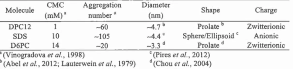
transmembrane segments ... 16 1.3 Critical micelle concentration, aggregation number, size and shape of
different micelles ... 31 2.1 Properties of different isotopes ... 42 2.2 Frequencies of different functional groups of a choline phospholipid
with symmetric and antisymmetric stretching vibration bands ... 68 S3 .l Bi celle disk radius calculated for q= 1 according to equation 4 from the
effective q value q
*
.
....
.
....
.
.
.
.
..
...
.
.
...
..
.
..
.
.
.
...
..
.
.
...
..
.
.
.
....
.
.
...
..
.
....
.
.
.
.
.
.
.
98 S3.2 Minimal total phospholipid concentration recommended to conserve thedesired bi celle molar ratio q ... 99 S3 .3 Comparison of the experimental me !ting temperatures of DMPC-d54 in
DMPC/DHPC bi celles and expected Tm for an ideal mixed micelle at
different q ratios ... 99 4.1 Results compilation of31P solid-state NMR spectra ... l10 4.2 Compilation of molar ratio q and temperature ranges at which
magnetically-oriented systems are formed ... 114 4.3 Critical bicelle concentration ... 115 4.4 Ratio of the hydrophilic-to-hydrophobic volume ratios of the
phospholipid on the detergent or short-chain phospholipid ... 122 4.5 31P resonance integration ratio compared to the molar ratio q ... 125
5.1 Deconvolution of secondary structure contributions to CD spectra of
hERG and Kv 1.5 pore helices in micellar and bicellar environnements ofDMPC/DPC and DMPC/DMPS/DPC. ... 144
5.2 Perpendicular and isotropie chemica1 shifts are given for liposomes of
DMPC and DMPC/DMPS ... 146 5.3 Effect of the hERG and Kvl.5 pore helices on bicelles ofDMPC/DPC and DMPC/DMPS/DPC. ... 148 5.4 Quadrupolar splitting of the plateau and methyl regions of DMPC-d54 into bicelles of DMPC/DPC and DMPC/DMPS/DPC ... 150 5.5 Hydrophobicity, hydrophobie moment and net charge of the pore-helix peptides ... 153 S5. 1 List of a-subunit of human cardiac potassium channels used for the
frequency plot in 1 1gurc 5. 1 B ... 155 S5.2 List of human Kv channe1s used for the frequency plot in
r
1gurc "'.1 ( ... 156 S5.3 List ofhuman Kir channels used for the frequency plot in l1gurc 5.1 D ... 157 S5.4 List ofhuman K2P cham1els used for the frequency plot in 1-tg.ur<..' 5 Il· .... 158 S5.5 List of prokaryotic and eukaryotic channels with known structure usedin the sequence homology for hERG and used for the frequency plot in
f 1gurc "'i 11 ... 159 S5.6 Values for the anisotropie tensor components. The isotropie part and the
RÉSUMÉ
Les protéines membranaires, notamment eucaryotes, sont connues pour représenter un défi à étudier. Dans cette thèse, des méthodes ont été développées pour aider dans
le domaine de recherche des protéines membranaires. Plus précisément, le but de cette thèse était de permettre l'étude de la structure et de la fonction des canaux
potassiques voltage-dépendants par résonance magnétique nucléaire.
Des membranes modèles pour l'analyse des protéines membranaires ont été
développées et caractérisées. Deux types de bicelles ont été étudiés: les bicelles faites avec des phospholipides à courtes chaînes et celles constituées de détergents
monoalkylphosphocholines (MAPCHO). Les bicelles couramment utilisées formées
de dimyristoyl-phosphatidylcholine (DMPC ou Dl4PC) et de dihexanoyl-PC (DHPC ou D6PC) ont été étudiées afin de détem1iner leur morphologie et leur miscibilité
dans des conditions diluées. Les résultats indiquent les concentrations seuils de formation de ces agrégats, la frontière de rapport molaire DMPC 1 DHPC ( q) entre
bicelle et micelle mixte ainsi que les meilleures concentrations à utiliser.
Afin d'améliorer la solubilisation des protéines membranaires et permettre leur étude en systèmes bicellaires, le DHPC a été remplacé par les détergents de la famille MAPCHO, dont la dodécylphosphocholine (DPC ou DPC 12) est connue pour sa capacité à solubiliser les protéines membranaires. Par l'ajout progressif de
phospholipides à des mélanges de tensioactifs et de protéines membranaires, la
protéine membranaire pourrait être reconstituée dans un environnement bicellaire. Une étude systématique a été menée en variant la longueur de chaîne du phospholipide (C 12 à C20) ainsi que du détergent (C 12 à C 16). Les résultats ont indiqué qu'il était possible de fom1er des systèmes orientés magnétiquement pour six
des douze mélanges binaires testés avec différentes épaisseurs de bicouche, à
différents rapports molaires lipides 1 détergents (q) et différentes températures. La
qualité de l'alignement, la miscibilité, les concentrations seuils de formation de ces agrégats ainsi que l'effet de l'ajout des ions lanthanides ou des lipides anioniques ont
également été étudiés. Une tentative d'établir un critère empirique qui pourrait prédire si une paire donnée de tensioactif et de phospholipide formerait des bicelles a été discutée.
Pour évaluer le potentiel de ces membrane modèles comme système de reconstitution directe, des peptides de canaux potassiques ont été solubilisés dans des micelles de
DPC, suivi par l'addition de DMPC. Ce projet a permis d'obtenir des données intéressantes sur la structure et les interactions avec les membranes des hélices du pore de Kvl.5 et du human ether-a-go-go related gene (hERG).
Un obstacle majeur dans la détermination de structure des protéines membranaires est 1 'absence de stratégie d'expression et de purification appropriée. La protéine hERG a été sélectionnée pour son implication dans une cardiopathie associée avec d'importants effets hors-cible de médicaments. Un protocole d'expression et de purification efficace du domaine du pore du hERG a été développé. Cela a pem1is d'obtenir des caractérisations biophysiques et des mesures électrophysiologiques préliminaires.
Ces résultats fournissent des méthodes pour solubiliser les protéines membranaires afin de pouvoir les étudier, ainsi que pour réaliser 1 'expression et la purification du domaine du pore d'un important canal potassique eucaryote: le hERG, qui ouvre la voie à 1' obtention de sa structure.
Mots clés: Protéine membranaire, Canal potassique hERG, Membrane modèle,
ABSTRACT
The study of membrane proteins, especially eukaryotic, is known to be challenging.
In this PhD thesis, methods were developed to aid the membrane protein research
area. More specifically, the aim of this thesis was to allow the study of the structure and function ofvoltage-gated potassium channels using nuclear magnetic resonance.
First, madel membranes for the analysis of membrane proteins were developped and
characterized. Two types of bicelles were investigated; the short-chain phospholipi d-based and the monoalkylphosphocholine (MAPCHO)-based bicelles. The popular dimyristoylphosphatidylcholine (DMPC or D 14PC) 1 dihexanoyl-PC (DHPC or D6PC) bicelles were studied to determine their morphology and miscibility in diluted
conditions. The results provide the concentration thresholds of the formation of the aggregates, the DMPC-to-DHPC molar ratio (q) boundary between bicelle and mixed micelle as well as the recommended concentrations to use.
To improve the solubilization of membrane proteins and their study in bicellar systems, DHPC was replaced by detergents from the MAPCHO family of which dodecylphosphocholine (DPC or DPC12) is known for its ability to solubilize
membrane proteins. By progressively adding phospholipids to the surfacta nt-membrane protein mixture, the membrane protein could be safely reconstituted in a
bicelle bilayer environment. A systematic study was carried out by varying the length
of the phospholipid (C 12 to C20) as well as one of the detergents (C 12 to C 16). The
results indicated a possibility to fom1 magnetically-aligned systems with six binary
mixtures on the twelve tested with different thicknesses of the bilayer, at severa! lipid-to-detergent molar ratios (q) and temperatures. The quality of the alignement, the miscibility, the concentration thresholds of the formation of the aggregates, as weil as the effect of the addition of lanthanide ions or anionic lipids were also
investigated. An attempt to establish an empirical criterion which could predict
whether a given surfactant-phospholipid pair will form bicelles is discussed.
To assess the potential of this system as a direct mode! membrane for reconstitution,
membrane peptides from potassium channels were solubilized into DPC micelles, followed by the addition of DMPC. This project presented interesting data on the structure of the pore helices of K v 1.5 and the hu man ether-a-go-go related gene (hERG) and their interaction with membranes.
A major hurdle when deterrnining the structure of membrane proteins is the absence
of a sui table expression and purification strategy. The protein hERG was selected for its involvement in a cardiopathy associated with important off-target effects of drugs.
An effective protocol for the expression and purification of the hERG pore domain
was developped. This formed the basis of preliminary biophysical characterization
and electrophysiological measurements.
The results in this thesis provide methods to solubilize membrane proteins, allowing
detailed study, as weil as to express and purify the pore domain of an important
eukaryotic potassium channel: the hERG, paving the way to obtain its structure.
Keywords: Membrane protein, Potassium channel hERG, Model membrane, Bicelle, Monoalkylphosphocholine detergent and Nuclear magnetic resonance.
CHAPTERI
INTRODUCTION
In this section, the following elements are introduced: the biological membrane and its components, the potassium chatmels, the voltage-gated potassium channels and lastly, the different mode! membranes and expression systems to study membrane pro teins. The objectives of this thesis are highlighted at the end of this chapter.
1.1 Biological Membrane
Biological membranes encompass the cells and their compartrnents. The plasma membrane is the boundary between the cytoplasm and the extracellular medium. It maintains the homeostasis of the cell and regulates the passage of both information and materials in and out of the cel!. As the ultimate protection from the environment,
this barrier is also the target for viruses, bacteria, or drugs. This section will give an overview of the organisation of the membrane, the lipids and the membrane pro teins.
1. 1.1 Organisation of the Membrane
Cell membranes are composed of a lipid bi layer to which proteins and carbohydrates (glycolipids and glycoproteins) are associated. The proportion of these three
components in membranes are dependant on the type of organism, cell and organelle
( l able 1 1 ).
Table 1.1 Composition (percent of their dry weight) of lipid, protein and
carbohydrate of membrane from different sources.
Lipid Protein Carbohydrate
Species Membrane
(%)
(%)
(%)
Central nervous system myelin" 79 20 1.0
Homo sapiens
E~throc~te 40 60
Bos taurus PeriEheral nervous s~stem m~elin" 76 23 1.0
Muscle skeletal" 35 65 0.0
Rattus
norvegicus Li ver" 40 60 0.0
Liver mitochondria" 27-29 70 l.3
Piswn sativum Thylakoidb 35-37 63-65
Acanthamoeba
Plasma membranec 27 37 37"
casta!fanii
a (Sastry & Tomer, 2008) c (E. D. Korn & Wright, 1973)
b (Chapman et al., 1983) d Phosphoglycan
The membrane structure proposed in 1972 by Singer & Nicolson is composed of a
fluid bilayer (at physiological temperatures) of phospholipids with intercalated
membrane proteins and glycoproteins, this so-called fluid-mosaic mode! membrane
(FMMM). The view of the membrane as a homogeneous mixture of lipids and
proteins has been questioned. The revised version of the FMMM takes into
consideration the constraints of distribution and mobility of some (if not ali) integral
globular membrane proteins and lipids. These phenomena are due to interactions with
membrane components, such as other membrane proteins and lipids as weil as with
the complex network of cytoskeleton and/or carbohydrates (Nicolson, 20 14) (1 t_urc
1 1 ). This mobility limitation of certain molecules in a sea of fluid molecules leads to
specialized nano-, micro- or macro-domains of lipids, proteins and mixture of both,
3
Figure 1.1 Fluid (non random) mosaic mode! membrane with representation of
lipid-protein domains in different colors. Lipids are in green and blue white proteins are colored in pink, red and orange. Extramembrane components, such as cytoskeleton and carbohydrates are represented by brown and purple, respectively.
1.1.2 Membrane Lipids
In humans, membranes are about 4 nm thick and cover a total surface circa 100
km
2(Mouritsen, 2005). They are composed of thousands of different types of lipids (Han
& Gross, 2005). Nevertheless, a given cel! is limited in its lipid composition, allowing a specificity of membrane function, structure and dynamics (Bagatolli et al., 201 0; Cullis & De Kruij ff, 1979).
1.1.2.1 Types ofMembrane Lipids
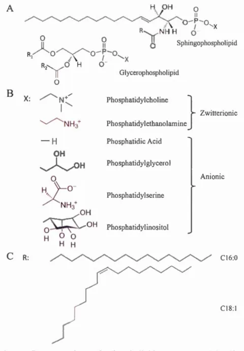
The fundamental building blacks of the bi layer are phospholipids. Phospholipids are amphiphilic (or amphipathic) molecules, with a hydrophilic "head" and a hydrophobie "tai!" groups. Phospholipids are composed of a backbone to which fatty acids are esterified at one or two sites with a headgroup lin.k to the third site via phosphodiester bond (l tgurc l .2).
According to the backbone, two main types of phospholipids can be described - the
glycerophospholipids (or phosphoglycerides) and the sphingophospholipids. In
glycerophospholipids, the backbone is a glycerol, esterified by a phosphate 111
position 'stereospecifically numbered' (sn)-3 and by two fatty acids in position sn-1 and sn-2 (l tgurL· 1.2 \). In sphingo-phospholipids, the backbone is a sphingosine, a long aliphatic amino alcohol N-lin.ked to a fatty acid and 0-lin.ked to a polar group
(l igurc 1 2.\). The structure composed of the sphingosine and the fatty acid is called a ceramide.
Depending on the polar head group associated with the backbone, there are different
classes of phospholipids, such as phosphatidylcholine (PC),
phosphatidyl-ethanolamine (PE), phosphatidylserine (PS) and phosphatidylinositol (PI) for the glycerophospholipids (1 tgurc 1.2B). The addition of a choline polar group to ceramide leads to sphingomyelin (SM). Different polar head groups confer different sizes and charges to the phospholipid (Figure 1.2B).
In membranes, it is possible to fi nd al kyi chains with an even number of carbons from 14 to 24 with or without unsaturation (Cullis et al., 1996; Marsh, 2013; Schechter, 1990). The length of the alkyl chains detennine the thickness of the bi layer with an increase in the length, giving rise to an increase of the bi layer thickness. The number of carbon atoms and unsaturation of the alkyl chains affects the tluidity of the membrane. Increasing the degree of unsaturation and reducing the alkyl chain length
5
of the fatty acid will reduce the melting temperature (Tm) of the lipid. Palmitic acid (16:0) and oleic acid (18:1) are the most abundant fatty acids found in human cell membranes (Watkins, 2005) (h~urc 1 .2C). A H OH 0 '·/ "'- ""..._ ~ .-. ~ __. ~ ·. ~ Il
/ ""-/'
...__,.
.
-..../
-...~...._.., --... "'-...
--
-:-"' -
o
-
P
-
o
,<\ 1 ' X8
C R: R-... _.NH Ho
g
Sphingophospholipid 0 Ilo
-
P
-
o
1'-
x
0-
H
OH~O
H
H,_ 0'}-o
-X NH3+ OH OH 0 H 0 0 H H Glycerophospholipid Phosphatidylcholine } Zwitterionic Phosphatidyletba.nolamine Phosphatidic Acid Phosphatidylglycerol An ionie Phosphatidylserine Phosphatidylinositol CI6:0 CI8:1Figure 1.2 Representation of phospholipid structures. (A) Phospholipid
backbone with X, the headgroup and R, the alkyl chain(s). (B) Hydrophilic headgroups with the associated glycerophospholipid name. (C) Examples of saturated (16:0) and unsaturated (18: 1) alkyl chains.
Another essential lipid found in animal membranes but not present in bacteria and plants is cholesterol. lts structure is based on four rings with a hydrocarbon tai! and a polar hydroxyl group (1 1gur~ 1. ~).
HO
H
Figure 1.3 Representation of cholesterol structure.
1t is found in membranes with similar molar amounts relative to phospholipids (Cooper, 2000; Post et al., 1995). This lipid also hasan effect on membrane fluidity. Due to its rigid steroid ring structure cholesterol maintains membrane fluidity by preventing close interaction between alkyl chains at low temperatures while at higher temperature, it decreases the fluidity by restricting the motion of the alkyl chains (Cooper, 2000; Post et al., 1995).
The most abundant phospholipids in eukaryotic membranes are PC (Warschawski et al., 2011). For example, sarcolemma (plasma membrane of the myocardium cell) are composed of 40-52% PC, 25-36% PE, 6-13% PS and PI, 4-18% SM and a cholesterol/phospholipid molar ratio from 0.33 to 0.59 for mammals (Post et al.,
i 995). The di ·lribui.ion of phosphuiipiùs wiilJÏII iÎ1c cciiui<t~ lllt:J.(JDfëtHê5 is asymmetric. The choline-based phospholipids PC and SM are located on the extemal (luminal) leaflet of the bilayer white the amine-based PE and the anionic phospholipids PS, PI and phosphatidic acid (PA) are found on the eytoplasmie side (Daleke, 2003). This phenomenon is initiated and maintained by lipid transporters (Daleke, 2003; Nicol on, 2014). The asymmetry is physiologically important as demonstrated by the energy consumption spent by eukaryotic cells to generate it and
7
diseases associated with disruption of this process, such as cardiopathy and diabetes (Bengt & Ding, 2009; Daleke, 2003).
1.1.2.2 Lipid Motions in the Bilayer
The membrane is dynamic. Five lipid movements can be described in the bilayer (Blume, 1993; Smith & Magid, 1984): (a) axial rotation where lipids will rotate along their longitudinal axis; (b) oscillation in a cone around this axis; (c) pendulum movement of the alkyl chains due to their flexibility; (d) lateral diffusion; and (e) tlip-flop when a lipid switches to another leaflet (1-tgurc- 1 -.1-).
Figure 1.4 (e) Flip-( flop (a) Axial rotation (d) Lateral diffusion
. -
--+
(g) U ndulation (b) Oscillation (c) Pendulum 'Il 1A lipid might (t) exchange between membranes (or protrusion) according to the curvature of the bi layer (Israelachvili, 2011; Jahnig, 1984) (F1gur~ 1 -l} Additionally,
the membrane can undergo (g) collaborative lipid motions in a wave-like movement
(or undulation) (Brown et al., 1983; Israelachvili, 2011) (FigLIIL' 1.4). All these
movements are enabled by the membrane fluidity.
The free rotation about the carbon-carbon bonds m the fatty acid chains allows
different "phases" of the bilayer according to the temperature. In the gel phase, the
lipids are in the ail-trans conformation (i.e. extended). The alkyl cbains are close and parallel (structure Lp) or inclioed (structure Lp') with respect to the bi layer nonnal
(F1gur~ 1. 'i). This phase occurs at low temperature when thermal motion is restricted.
In the liquid-crystalline fluid phase, the lipids adopt the trans/gauche conformation
(i.e. disordered or melted). The alkyl chains are free to change direction around their
carbon-carbon bonds, thus they are more disordered and less compact (structure La)
(f1gur~ 1. 'i). This phase occurs at temperatures above the main transition temperature of the lipid, the so-called metting temperature (Tm) where the thermal motion is grea ter.
Figure 1.5 phase (right).
Gel phase Fluid phase
9
1.1.2.3 Organization of Lipids
At low concentration tn an aqueous solution, lipids exist as monomers. Above a characteristic concentration, the so-called "critical micelle concentration" (CMC),
these amphiphilic molecules will be driven together into a bilayer organization by the hydrophobie effect and numerous van der Waals interactions. This phenomenon minimizes the contact of the tails with water. The self-assembled bilayer 1s
maintained and thus stabilized by the ability of the hydrophilic head groups to interact with the aqueous environment and one another.
The Hydrophobie Effect
In liquid water, each water molecule can forrn up to four hydrogen bonds, giving rise to a dynamic network. The addition of amphiphilic compounds in an aqueous
medium destabilizes this network since hydrogen bonds can no longer be forrned between water molecules and the lipid alkyl chains. Thus, the solvent forms a cage around the hydrophobie region, giving rise to a reduction of the entropy since the
motions of water molecules are restricted. When many lipids are in solution, the hydrophobie tails are driven together to limit their contact with the aqueous medium white the hydrophilic head groups are exposed to the solvent. The water molecules can forrn hydrogen bonds with the polar groups and are free to move. The association of many lipid molecules gives rise to spontaneous formation of supramolecular aggregates.
Lipid Phases
The overall organization of cell membranes is a lipid bilayer (or lamellar phase). However, sorne membrane lipids when studied in water do not forrn a bilayer. The lipid shape is the main factor in deterrnining the organization of the aggregates. Many phospholipids have a cylindrical shape, i.e. the cross-section of the headgroup is similar to that of the alkyl chains (FtgurL' 1 .ô). These lipids tend to organize m bi layers. For example, PC and PS are bilayer-prone lipids (Grun er et al., 1985). In contrast, nonbi1ayer-prone lipids have different cross-sectional areas between the head and the tails. They are differentiated into two types. The first one hasan inverted conical shape with a larger headgroup in comparison to the alkyl chains (1 t~llt\' 1 ()). These lipids generate a positive spontaneous curvature. They fom1 micelles at low concentrations white they adopt a normal hexagonal Ht phase at higher concentrations. In H1 phase, lipids forrn cylinders with the hydrophobie tails towards the center of the cylinder. These cylinders are organized into hexagons. For example, lysoPC (one alkyl chain) is an inverted conical shaped lipid, which forrns micelles in
solution (Gruner et al., 1985).
The second nonbilayer-prone lipid type exhibits a conical shape with a smaller headgroup in comparison to the alkyl chains (Figure 1 .6), such as PE (Gruner et al., 1985). They induce a negative spontaneous curvature and adopt a reverse hexagonal H11 phase. In this phase, lipids are organized in hexagonally arranged cylinders with the hydrophobie ta ils towards the outside of the cylinder.
The cubic phases are more complex organizations of lipids. It is a bicontinuous phase with a three dimensional symmetry, since the water regions are separated by the bilayer. This bilayer has positive and negative curvatures (Mouritsen, 2005) (1 tgurc
1 1
The cell membranes contain bi1ayer- and nonbi1ayer-prone 1ipids. By modu1ating the
ratio between the different lipid types, membrane curvatures cao be induced. This membrane phenomenon can influence cellular processes, such as membrane fusion
and fission as weil as the activity of membrane proteins (Carsten & Unger, 20 12).
Non bi layer-prone
...
----
.... Invetted cone Micelle Bi layer-prone Cylinder illt.~ ~ ~ ~ ~ Lamellar bilayer Cu bicNonbilayer-prone
Co ne
~
Inverted micelleFigure 1.6 The different shape of 1ipids associated with their organization in
aqueous medium. A representation of the complex cubic phase is in an inset. This
1.1.3 Membrane Proteins
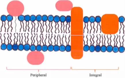
The lipid bilayers of ce li membranes are embedded with intrinsic (or integral) and extrinsic (or peripheral) proteins (Ftgun:: 1.1 ). These proteins have roles in cell-to-cell communications or interactions and molecular transport. For example, ion channels allow the flow of ions through the membranes. Membrane proteins are the components giving the specificity of a cell (Koolman & Roehm, 20 13). They arc essential drug targets to treat many pathologies, such as cystic fibrosis and cancer (Clunes & Boucher, 2007; Prevarskaya et al., 2007). About 20 to 30% of the genome is predicted to encode for membrane proteins (Stevens & Arkin, 2000; Wallin & von Heijne, 1998).
1.1.3 .1 Classification of Membrane Proteins
Membrane proteins can be defined in three classes according to thcir proccss of extraction and their interactions with the components of the membranes (f tgurl''- 1 1 and 1.').
Peripheral Integral
Figure 1.7 Representation of integral (orange) and peripheral (light red) membrane protein classes. Membrane-associated proteins are not represented.
13
Peripheral Membrane Proteins and Membrane-Associated Proteins
The first class of membrane proteins comprises extrinsic (or peripheral) membrane proteins that are localized at the surface/hydrophilic part of the membrane (F1gurc 1. 7). Their adhesion to the membrane can be via electrostatic or ionie interactions
with integral membrane protein(s) or with the headgroup of lipid(s). Sorne peripheral membrane proteins are linked to a hydrophobie motif, a fatty acid or a lipid (e.g. glycophosphatidylinositol anchored proteins). Their structures are mainly extended ~ sheet (Nicolson, 2014 ).
The second class of membrane proteins are the membrane-associated proteins which do not have direct interactions with the membrane (Nicolson, 2014). They can have interactions with peripheral protein(s), the cytoskeleton or the carbohydrates (1"1gurc 1. 1 ).
These two classes of membrane proteins are mainly soluble in aqueous environment
and removable from a cell membrane without affecting its bilayer structure (Cooper, 2000; Nicolson, 20 14). An exception to this extraction "rule" can happen with
proteins anchored to the membrane by their hydrophobie motif, fatty acid or lipid covalently linked. The ir purification process is cl oser of the one of integral membrane proteins.
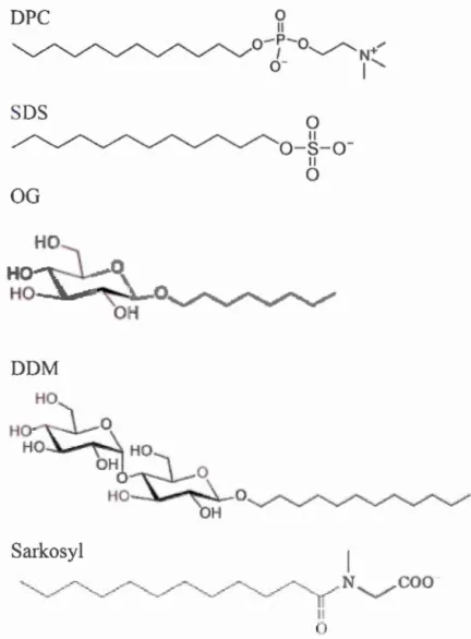
Integral Membrane Proteins
The third class of membrane proteins are the intrinsic (or integral) membrane proteins and they are embedded into the hydrophobie part of the phospholipid bilayer (rigurc
1 .7). They interact with both the polar heads and the apolar tails of the lipids. Most of
proteins. To extract integral membrane proteins from the membrane, it is necessary to disrupt the bilayer by using surfactants, also called detergents (f 1.~urc 1
.x.
Scl'lion1 .4 .1 ).
SDS
0 Ilo
-
s
-
o
-11 0OG
HO
Ho~~~~~-o~DDM
0Sarkosyl
IlFigure 1.8 Representation of detergent structures. SDS, DPC, OG, DDM are
sodium dodecyl sulfate, dodecylphosphocholine, octylglucoside and
dodecylmaltoside, respectively.
Most of the transmembrane proteins have their transmembrane domains made of
15
membrane, the a-helix portion needs to be 20-25 amino-acid long (Cooper, 2000;
Koolman & Roehm, 2013). These proteins with a-helical segment(s) can be
subdivided into five types according to their orientation and the number of segment(s)
as detailed in Figurè 1 .9. An exception is the porin family, which structure consists in
a ~-barrel (Benz, 1994). These integral membrane pro teins are fou nd in bacteria and
in the mitochondria (Benz, 1994).
C N
Type 1 ·rype Il Type Ill Type IV
Monotopic l:litopic Polytopic Oligomeric
Figure 1.9 Representation of the different types of transmembrane proteins
with a-helical segment(s). N and C are for the NH2- and COOH-terminus, respective! y.
1.1.3.2 Major Families oflntegral Membrane Proteins
In 2006, it has been estimated that about 4075 transmembrane protein families exist with at )east two transmembrane domains (Oberai et al., 2006). The top ten of the
largest membrane protein families is listed in Table 1 2 and comprises proteins such as G-protein-coupled receptors (GPCR), ATP-binding cassette (ABC) transporters
and cytochrome b (Oberai et al., 2006). Potassium channels, which two members
were studied in this thesis (Chapkr'> \" and \'1), are ranked sixth with 1153 members
Table 1.2 List of the top ten largest membrane protein families with at !east two transmembrane segments (Oberai et al., 2006).
Family 1. GPCR #1 (Rhodopsin-like)
2. Major facilitator
3. ABC transporter #1 (amino acid, phosphate, ferric, nitrate, nickel,
taurine, sugar)
4. GPCR #2 (serpentine)
5. ABC transporter #2 (multidrug resistance) 6. Potassium channel
7. Receptor protein kinase
8. Transporter #1 (cationic acid, aromatic amino acid, choline, K+ uptake) 9. Cytochrome b#l (N-terminal part) 1 O. Cytochrome b#2 CC-terminal part) 1.2 Potassium Channels Number of members 5520 3680 2469 1311 1179 1153 1150 1082 1030 942
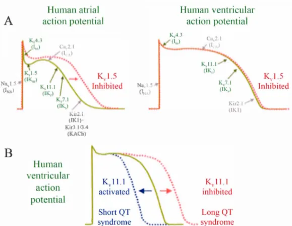
A major objective of this thesis was to understand the function of potassium channels. These channels are ubiquitous and highly specialized for the selective conduction of potassium ions down their electrochemical gradient. Potassium channels are weil known for their role in excitable cells (i.e. neurons and ca.rdiomyocytes). They shape the action potential and regulate the membrane potential. This section will give an overview of the structure, the ion selectivity, the gating and the subgroups of the potassium channels.
1.2.1 Structure
Despite significant difference in K+ channels, they consist of a well-conserved pore domain with four subunit (identical or different). Each subunit consists of two a
-17
helical transmembrane domains linked by a loop (Figure 1. 1 OA-U). This P-loop is composed of (i) an extracellular loop connecting S5 to the pore helix also called the
turret of S5P-linker, (ii) a short pore helix, (iii) a selectivity filter and a (iv) pos
t-selectivity filter loop (Capener et al., 2002; Doyle et al., 1998) (r1gun.::-. 1.10 and
1 1 1 ). The K+ channel signature sequence TXTTVGYG con tains the highly conserved motif of the selectivity fil ter. In a tetramer, this sequence has a crucial role
in the pem1eation and the selectivity mechanism of the K+ channel (Capener et al.,
2002).
A
c
Extracellula.r37Â
1 \
lntracellularFigure 1.10 Structure of the bacterial KcsA potassium channel which constitute a pore mode!. (A-C) Ribbon representation of the KcsA channel based on the KcsA
crystal structure (PDB number: 2QTO). (A) Side view and (B) above view with each
subunit highlighted in a different color. (C) Side view with only two of the four
subunits are displayed for the sake of clarity. The horizontal !ines show the probable
limits between the hydrophobie and hydropholic parts of the phospholipids.
Potassium ions are represented in purple. Representations (A-C) are generated with
UCSF Chimera software. (D) Sculpture of the KcsA channel where the internai pore and the aqueous cavity are highlighted with an iron and yellow glass structure,
HERG KcsA MthK KvaP Kvl.5 Kv2.1 Kv3.1 Kv4.3
55P lin ker 1 turret 55
540DRYSEYGAAV LFLLMCTFAL IAHWLACIWY AIGNMEQPHM DSRIGWLHNL GDQIGKPYNS SGLGGPSI!ill609 20HGSALHWRAA GAATVLLVIV LLAGSYLAVL AERGAPG--- - - - --- - - - - ---AQLI60
11HLPRVLKVPA TRILLLVLAV IIYGTAGFHF IEG--- -154IADAADKIRF YHLFGAVMLT VLYGAFAIYI
VEYPDPN---425KTLQASMREL GLLIFFLFIG VILFSSAVYF AEADNQG---322FTLRRSYNEL GLLILFLAMG IMIFSSLVFF AEKDEDD -345FLLLIIFLAL GVLIFATMIY YAERIGAQPN DPSASEH--
-312YTLKSCASEL GFLLFSLTMA IIIFATVMFY
AEKGSSA---5F oiu_J _ _ P_H:...__--1)~ ,.,
Il
56 ---E44 ---SSIK194 ---THFS465 - - - -TKFK362 - - - -THFK'as ---SKFT352HERG 61 °KYVTALYFTF SSLTSVGFGN VSPNTNSEKI FSICVMLIGS LMYASIFGNV SAIIQRLYSG TARY613
KcsA 61TYPRALWWSV ETATTVGYGD LYPVTLWGRL VAVVVMVAGI TSFGLVTAAL ATWFVGREQE RRGH124
MthK 45SWTVSLYWTF VTIATVGYGD YSPSTPLGMY FTVTLIVLGI GTFAVAVERL LEFLINREQM KLMG108
KvaP 195SVFDALWWAV VTATTVGYGD VVPATPIGKV IGIAVMLTGI SALTLLIGTV SNMFQKILVG EPEP258 Kvl.5 466SIPDAFWWAV VTMTTVGYGD MRPITVGGKI VGSLCAIAGV LTIALPVPVI VSNFNYFYHR ETDH529
Kv2.1 363SIPASFWWAT ITMTTVGYGD IYPKTLLGKI VGGLCCIAGV LVIALPIPII VNNFSEFYKE QKRQ424 Kv3 .1 386NIPIGFWWAV VTMTTLGYGD MYPQTWSGML VGALCALAGV LTIAMPVPVI VNNFGMYYSL AMAK449
Kv4.3 353SIPASFWYTI VTMTTLGYGD MVPKTIAGKI FGSICSLSGV LVIALPVPVI VSNFSRIYHQ NQRA416
Figure 1.11 Sequence alignment of the hERG pore domain (UniProt Accession
Number: Ql2809) with KcsA (POA334), MthK (027564), KvAP (Q9YDF8), Kvl.S (P22460), Kv2.1 (Ql4721), Kv3.1 (P48547) and Kv4.3 (Q9UK17). Transmembrane
helices (S5-S6) and pore helix (PH) are represented with grey cylinders while the selectivity filter (SF) sequence is shown by an arrow. The well-conserved glycine hinge is in red.
The first and second transmembrane helices of the pore domain are tenned outer and inner helix, respectively. They are localized on the exterior and the inside side of the
pore, respectively. Both stabilize the orientation of the pore helix and the selectivity filter. Ail together, they are organized in a cone shape (Doyle et al., 1998) (t tg ure
l.IOC).
From this base core topoiogy, some heiicai lransmembrane segmenls, nun-membraue do mains and subunits that control the ir ga ting can be added (Ca pener et al., 2002).
Tlu·ee types of subunits can be defined; (i) the primary (a) subunits forming the pore,
(ii) accessory (~) subunits which might associate with the a-subunits regulating the