HAL Id: dumas-01962507
https://dumas.ccsd.cnrs.fr/dumas-01962507
Submitted on 20 Dec 2018HAL is a multi-disciplinary open access
archive for the deposit and dissemination of sci-entific research documents, whether they are pub-lished or not. The documents may come from teaching and research institutions in France or abroad, or from public or private research centers.
L’archive ouverte pluridisciplinaire HAL, est destinée au dépôt et à la diffusion de documents scientifiques de niveau recherche, publiés ou non, émanant des établissements d’enseignement et de recherche français ou étrangers, des laboratoires publics ou privés.
Distributed under a Creative Commons Attribution - NonCommercial - NoDerivatives| 4.0 International License
Confirmation of the colonization path of Hymenoscyphus
fraxineus from leaves to shoots in Fraxinus excelsior
Astrid Koehl
To cite this version:
Astrid Koehl. Confirmation of the colonization path of Hymenoscyphus fraxineus from leaves to shoots in Fraxinus excelsior. Life Sciences [q-bio]. 2018. �dumas-01962507�
Confirmation of the colonization path of
Hymenoscyphus fraxineus from leaves to shoots in
Fraxinus excelsior
Par : Astrid KOEHL
Soutenu à Rennes le 28 juin 2018
Devant le jury composé de :
Président : Antoine GRAVOT Maître de stage : Thomas KIRISITS Tuteur : Antoine GRAVOT
Rapporteur : Francisco CABELLO HURTADO Examinateur : Régine DELOURME
Les analyses et les conclusions de ce travail d'étudiant n'engagent que la responsabilité de son auteur et non celles d’AGROCAMPUS OUEST et l’université de Rennes 1
Ce document est soumis aux conditions d’utilisation
« Paternité-Pas d'Utilisation Commerciale-Pas de Modification 4.0 France » disponible en ligne http://creativecommons.org/licenses/by-nc-nd/4.0/deed.fr
AGROCAMPUS OUEST
CFR Angers CFR Rennes
Année universitaire : 2017-2018 Master Biologie, Agrosciences Parcours Amélioration, Production, Valorisation du végétal
Option Physiologie Moléculaire et Adaptations aux Stress
Rapport de stage
d’Ingénieur de l’Institut Supérieur des Sciences agronomiques, agroalimentaires, horticoles et du paysage
de Master de l’Institut Supérieur des Sciences agronomiques, agroalimentaires, horticoles et du paysage
Fiche de confidentialité et de diffusion du mémoire
Confidentialité
Non Oui si oui : 1 an 5 ans 10 ans
Pendant toute la durée de confidentialité, aucune diffusion du mémoire n’est possible (1). Date et signature du maître de stage (2) :
A la fin de la période de confidentialité, sa diffusion est soumise aux règles ci-dessous (droits
d’auteur et autorisation de diffusion par l’enseignant à renseigner).
Droits d’auteur
L’auteur(3) Koehl Astrid
autorise la diffusion de son travail (immédiatement ou à la fin de la période de confidentialité)
Oui Non
Si oui, il autorise
la diffusion papier du mémoire uniquement(4)
la diffusion papier du mémoire et la diffusion électronique du résumé la diffusion papier et électronique du mémoire (joindre dans ce cas la fiche de conformité du mémoire numérique et le contrat de diffusion)
accepte de placer son mémoire sous licence Creative commons CC-By-Nc-Nd (voir Guide du mémoire Chap 1.4 page 6)
Date et signature de l’auteur : 13/07/2018
Autorisation de diffusion par le responsable de spécialisation ou son
représentant
L’enseignant juge le mémoire de qualité suffisante pour être diffusé (immédiatement ou à la fin de la période de confidentialité)
Oui Non
Si non, seul le titre du mémoire apparaîtra dans les bases de données. Si oui, il autorise
la diffusion papier du mémoire uniquement(4)
la diffusion papier du mémoire et la diffusion électronique du résumé la diffusion papier et électronique du mémoire
Date et signature de l’enseignant :
(1) L’administration, les enseignants et les différents services de documentation d’AGROCAMPUS OUEST s’engagent à respecter cette confidentialité.
(2) Signature et cachet de l’organisme
(3) Auteur = étudiant qui réalise son mémoire de fin d’études
(4) La référence bibliographique (= Nom de l’auteur, titre du mémoire, année de soutenance, diplôme, spécialité et
Dépôt numérique de mémoire
ATTESTATION DE CONFORMITE DE LA VERSION NUMERIQUE
Je, soussigné(e),
Nom : Koehl
Prénom : Astrid
Ci-après désigné « l’Auteur »
Atteste que la version numérique de mon mémoire de fin d’études dans sa version définitive (incluant les corrections demandées par le jury de soutenance),
Intitule
Confirmation of the colonization path of Hymenoscyphus fraxineus from leaves to shoots in Fraxinus excelsior
correspond à la version imprimée du document, déposé à la bibliothèque générale d’AGROCAMPUS OUEST (CFR de référence)
A Rennes le 13/07/2018
Dépôt numérique de mémoire
CONTRAT DE DIFFUSION NUMERIQUE DE MEMOIRE
Entre
AGROCAMPUS OUEST, Institut supérieur des sciences agronomiques, agroalimentaires, horticoles et du paysage dont le siège est basé 65 rue de Saint-Brieuc, 35042 RENNES, représenté par son Directeur Général, Grégoire THOMAS
et
L’auteure du mémoire : Nom : Koehl
Prénom : Astrid
Adresse personnelle : 16, rue de Plaisance 35310 MORDELLES
Intitulé du mémoire : Confirmation of the colonization path of Hymenoscyphus fraxineus
from leaves to shoots in Fraxinus excelsior
Ci-après désigné auteur,
Article 1
Le présent contrat ne concerne que les mémoires de fin d’études des cursus de formation d’AGROCAMPUS OUEST, déposés suite à la soutenance dans leur version validée par le jury. La diffusion de ces mémoires est conditionnée au visa du responsable de spécialisation/ option, garantissant la prise en compte de l’avis du jury.
Article 2
L’auteur autorise AGROCAMPUS OUEST à diffuser le mémoire sur le site Internet de l’établissement ou sur les plateformes choisies par AGROCAMPUS OUEST en conformité avec la fiche de diffusion correspondante. Le présent contrat a pour objet de permettre à AGROCAMPUS OUEST de diffuser le mémoire dans le respect des droits de propriété intellectuelle de son auteur.
Le présent contrat n’implique pas l’obligation pour AGROCAMPUS OUEST de faire usage de l’autorisation qui lui est donnée. La diffusion effective, tout comme son éventuelle suppression, n’implique en aucun cas une appréciation au bénéfice de l’auteur ou des tiers et n’est pas source de responsabilité à l’égard des tiers.
Article 3
L’auteur demeure responsable du contenu de son œuvre. L’auteur garantit à AGROCAMPUS OUEST qu’il détient tous les droits nécessaires à la diffusion de son œuvre, en particulier les autorisations écrites des titulaires des droits sur les œuvres reproduites, partiellement ou intégralement. En cas de non respect de cette clause, AGROCAMPUS OUEST se réserve le droit de refuser, suspendre ou arrêter la diffusion des parties du mémoire intégrant des documents ou parties de documents pour lesquels les droits de reproduction et de représentation n’auraient pas été acquis.
AGROCAMPUS OUEST ne pourra être tenu responsable de représentations illégales de documents, pour lesquels l’auteur n’aurait pas signalé qu’il n’en avait pas acquis les droits.
Article 4
L’auteur pourra à tout moment retirer l’autorisation de diffusion qu’il accorde par le présent contrat. Pour cela, il devra en aviser formellement AGROCAMPUS OUEST par lettre recommandée avec accusé de réception. AGROCAMPUS OUEST aura alors l’obligation de retirer l’œuvre lors de la plus
L’auteur autorise AGROCAMPUS OUEST à procéder, le cas échéant, au reformatage de son mémoire en vue de l’archivage, de la diffusion ou de la communication dans le respect des autorisations de diffusion définies par lui précédemment.
Article 6
Les autorisations de diffusion données à AGROCAMPUS OUEST n’ont aucun caractère exclusif et l’auteur conserve toutes les autres possibilités de diffusion de son mémoire.
Article 7
L’auteur autorise, à titre gracieux, la cession des droits de diffusion, concernant le mémoire qui lui appartient. Cette autorisation, dans la durée maximale définie par le droit patrimonial, est strictement réservée à la diffusion du mémoire à des fins pédagogiques et de recherche.
Fait à Rennes le 13/07/2018
Pour AGROCAMPUS OUEST, L’auteur,
Pour Le Directeur Général Et par délégation,
Acknowledgments
I would like to express my deep gratitude to Thomas Kirisits for his guidance, open-mindedness and advices during this research work. I would also like to thank Susanne Krumböck, for her useful critiques and exceptional assistance in keeping my progress on schedule. My grateful thanks are also extended to Christian Stauffer for his help and Florian Kunz in helping me understanding my raw data. Furthermore, I gratefully acknowledge Thomas N. Sieber and Andrin Gross (ETH Zürich, Switzerland) for providing the microsatellite primers for my work. Andrin Gross also gave valuable advice on the microsatellite analyses.
I would also like to extend my thanks to all the wonderful people at the institute who made my stay so special, especially Waltraud Pleyl, Christa Schafellner and Axel Schopf.
Table of Contents
1. Introduction ... 1
1.1. History of ash dieback ... 1
1.2. Impact on Fraxinus excelsior ... 1
1.3. Host-pathogen interactions ... 2
1.4. Symptoms of ash dieback ... 3
1.5. Life cycle of Hymenoscyphus fraxineus ... 3
1.6. Objectives... 4
2. Material and Methods ... 5
2.1. Collection sites and ash sampling ... 5
2.2. Fungal isolation and identification ... 5
2.3. DNA extraction ... 6
2.4. Mating type determination ... 6
2.4.1. Multiplex PCR ... 6 2.5. Microsatellite markers ... 7 2.5.1. Multiplex PCR ... 7 2.5.2. Fragment analysis ... 7 2.6. Data analysis ... 7 3. Results ... 8
3.1. Isolation of H. fraxineus and other fungi ... 8
3.2. Differentially encoded alleles ... 8
3.3. Genetic profiles ... 8
3.4. Genotypic continuity ... 9
3.5. Genotypic diversity ... 9
4. Discussion ... 10
4.1. Association of H. fraxineus with symptoms of ash dieback ... 10
4.2. Confirmation of the leaf petiole-shoot infection path for H. fraxineus ... 10
4.3. Occurrence of identical genotypes on different trees ... 11
4.4. Genotypic diversity in single petioles and single shoots ... 12
4.5. Methodological considerations... 12
5. Conclusions ... 14
1. Introduction
1.1. History of ash dieback
European ash dieback (ADB) is a highly destructive disease killing ash trees (Fraxinus
excelsior) in large parts of Europe. After its first observation in Poland and Lithuania in the
early to mid-1990s, ADB has spread across Europe over the past 25 years from east to west (Kowalski, 2006; Timmermann et al., 2011; Kjær et al., 2012). The disease has now been reported in most of Europe and was even detected in the British Islands and in Ireland in 2012.
The pathogenic agent responsible for the disease was identified in 2006 and named Chalara
fraxinea (Kowalski, 2006), based on the morphology of its asexual structures. However, with
the aid of molecular methods, the fungus was later assigned to the genus Hymenoscyphus. For a short while, it was confused with its cryptic sister species Hymenoscyphus albidus but was then recognized as a separate taxon (now known under the name Hymenoscyphus
fraxineus, see below). Actually, H. albidus is an avirulent species closely related to the
pathogen but there are well established species barriers (Wey et al., 2016). In fact, co-occurrence has been reported but no hybridization had been found in nature between the two species so far (Husson et al., 2011; Queloz et al., 2011). H. albidus is an indigenous European foliage colonizer which decomposes Fraxinus excelsior leaves and is non-pathogenic on F.
excelsior (Baral & Bemmann, 2014; Kowalski et al., 2015). It has been hypothesized that the
endemic H. albidus could be replaced by the alien species causing ADB which could lead to extinction of the former species through competitive exclusion (McKinney et al., 2012). Molecular differentiation led to the recognition that the two Hymenoscyphus species were indeed distinct, and hence a new name was given to the pathogenic species: Hymenoscyphus
pseudoalbidus (Queloz et al., 2011). However, the scientific name of the pathogen has been
changed recently and is now Hymenoscyphus fraxineus (Baral et al., 2014).
In its natural habitat, H. fraxineus is a nearly harmless leaf endophyte found in Eastern Asia (Japan, Korea, northeast of China, Russian Far East) on Fraxinus mandshurica, the Manchurian ash, and F. chinensis spp. Rhynchophylla, the Korean ash (Cleary et al., 2016; Zhao et al., 2012; Gross et al. 2014). However, Drenkhan et al. (2017) reported the association of H. fraxineus with symptoms on leaves and possibly also on twigs of both Manchurian and Korean ash in Russian Far East. This indicates that the fungus was an overlooked weak pathogen in its natural environment prior to having received attention due to the ash dieback epidemic in Europe. A recent study suggests that the European population was founded by two individuals from a large diverse population originating from East Asia (McMullan et al., 2018).
1.2. Impact on Fraxinus excelsior
Common ash (F. excelsior) is an important forest tree in Europe. Its timber is mainly used for floors and furniture. However, its ecological value is even more substantial. For example, in the British Islands, almost a thousand species are associated with F. excelsior and forty are obligate associates. Furthermore, if a shift of woodland composition happens towards other tree species as a result of ADB, it would change nutrient cycling, carbon storage, and soil formation, which would drive shifts in the soil community (Mitchell et al., 2014).
In Sweden, about 60 species associated with common ash (and F. excelsior itself) have been put on the Sweden red-list of threatened species because of damages caused by H. fraxineus. Finally, the disease also has a cultural impact. Indeed, trees are not only affected in forests, but also in parks and gardens.
This devastating pathogen is capable of colonizing every part of F. excelsior: leaves, shoots, main stems and even roots (Kirisits et al., 2009; Schumacher et al., 2010; Husson et al. 2012; Gross et al., 2014; Schwanda & Kirisits, 2016). H. fraxineus is an aggressive invasive pathogen which is particularly damaging on young ash trees, often killing them within one growing season after infection. Older trees can survive initial infections, but become chronically diseased and tend to succumb after several seasons due to secondary diseases such as root and butt rot caused by Armillaria species (Skovsgaard et al., 2010; Chandelier et al., 2016).
On a European scale, current levels of H. fraxineus may infect and kill 95% of all European ash trees (McMullan et al., 2018).
Many studies have evaluated in detail the damage levels caused by H. fraxineus on F.
excelsior in different European countries. For example, in a young ash plantation in Germany,
73% of the saplings were dead five years after planting (Langer et al., 2015). In Norway, a mortality rate of 57% was observed on monitoring plots in ash forests in 2016 (Timmermann
et al., 2017). Furthermore, a relationship between the severity of infection and the age of the
trees was detected. In the natural forests monitoring in Norway, 80% of young trees were dead in 2016, whereas for older trees, the mortality was only around 20% (Timmermann et al., 2017). In France and Belgium, survey found an annual mortality rate of 35% in younger ash stands and only 3% in older stands (Marçais et al., 2017).
1.3. Host-pathogen interactions
It is estimated than less than 5% of ash trees are partially resistant or tolerant to ADB (Kjær et
al., 2012; Sollars et al., 2017). Overall comprehension of the metabolic pathways of the
interaction between F. excelsior and H. fraxineus (both on the host side and pathogen side) is still incomplete but new insights start to unravel some parts of this metabolic network.
The level of susceptibility of F. excelsior to H. fraxineus has been recently associated with the abundance of iridoid glycosides, which are part of a well-known anti-herbivore defense mechanism in the Oleaceae, also capable of enhancing fungal growth (Sollars et al., 2017). Thus, their findings suggest that there might be a trade-off between ADB susceptibility and herbivore susceptibility.
Some of the pathogen effectors have been described (McMullan et al., 2018). They present an NPP1 (necrosis-inducing Phytophthora protein) domain, which is present in fungal proteins that induce hypersensitive-reaction-like cell death when infiltrating plant leaves. Their presence seems to be common among necrotrophic parasites, to which H. fraxineus also belongs. Furthermore, H. fraxineus has a large number of predicted secreted Cytochrome P450 proteins, which might play a role in pathogenesis (McMullan et al., 2018). Cytochrome P450s’ modes of action are usually via the monooxygenase reaction. Potential roles for P450s in H.
fraxineus include i) destruction of ash aromatic compounds with antifungal properties, ii)
penetration of ash tree tissues through degradation of hydrocarbon compounds, the main constituents of the cuticle and iii) secretion of toxins during invasion of ash tree tissues.
Figure 1: Symptoms of ash dieback on Fraxinus excelsior. (a) necrotic spots on a leaflet; (b),(c) necrotic lesions on leaflet midribs and adjacent leaf blade tissues; (d) leaf wilting distal to a necrotic lesion on the rachis; (e),(f) necrotic bark lesion on a shoot originating from an infected side leaf and twig; (g) extensive bark necrosis leading to shoot dieback; (h) wood discoloration beneath a necrotic bark lesion on a shoot; (i),(j) tongue-shaped bark lesions at the base of F. excelsior trunks; (k) wedge-shaped wood discoloration beneath a root collar lesion.
Also, it has been described that H. fraxineus produce secondary metabolites such as viridian, a mycotoxine, and its dihydroderivative viridiol, a phytotoxine (Grad et al., 2009). Viridiol might be involved in the development of ADB, especially with the induction and expansion of necrotic spots.
Even if progress still need to be made in order to understand H. fraxineus metabolic pathways and how F. excelsior defend itself against the pathogen, the symptoms of ash dieback are well-known and allow fast and accurate detection.
1.4. Symptoms of ash dieback
H. fraxineus causes necrotic lesions on leaves and lesions, cankers and wood discoloration in
the bark and on the root collar (figure 1). Initially, small necrotic spots and necrotic lesions appear on leaves (Gross et al., 2014). The latter usually form along leaflet veins, from where they expand into petioles. Leaf infections often result in premature shedding of leaflets and entire leaves. From a small portion of these infections, H. fraxineus is supposed to penetrate into shoots, twigs and branches where, after an incubation period, necrotic lesions and perennial cankers form. Necrotic lesions and cankers often lead to girdling of woody parts, resulting in wilting of the foliage and crown dieback. Trees respond to infections by prolific production of epicormic shoots leading to a short- to medium term partial restoration of the crown. Necrosis in shoots is assumed to develop mainly after infection through leaf petioles. First appearance of bark lesions from infections in the same year occurs during late summer, but most lesions emerge during autumn and winter, and they can even become visible as late as in late spring or early summer of the next year (Kirisits et al., 2009, 2012; Gross et al., 2014).
Besides crown damage, serious symptoms of ash dieback are necrotic lesions and wood discolorations on the trunk base and root collar (figure 1). These are caused by H. fraxineus, likely by germinating ascospores penetrating the intact bark via lenticels (Husson et al., 2012; Chandelier et al., 2016). Such lesions act as entrance courts for Armillaria species and other secondary pathogens which cause root an butt rot, destabilizes trees and predisposes them to fall over (Husson et al., 2012; Chandelier et al., 2016). The attacks also drive mortality of ash trees (Marçais et al., 2017).
Accumulating knowledge on the symptoms of ash dieback and their timing, together with overall knowledge on the biology of H. fraxineus, and ascomycetes in general, permitted to develop a hypothetic life cycle for the pathogen and a disease cycle of ash dieback.
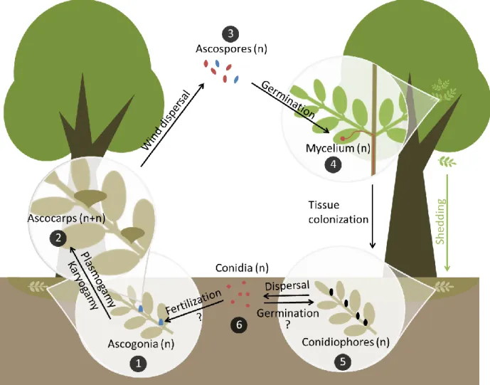
1.5. Life cycle of Hymenoscyphus fraxineus
Some well-known parts of the life cycle in ascomycetes have not yet been described for H.
fraxineus. This is why some interactions are supposed, but need to be verified. All the main
steps are summarized in figure 2.
By early summer, ascogonia develop on old fallen leaf rachises and are probably fertilized by contact with conidia or by mycelium of germinated conidia, if strains of opposite mating types are present. Sexual reproduction in ascomycetes is governed by the mating type locus MAT, harboring different MAT genes. In heterothallic fungi, like H. fraxineus, the MAT genes are separated in a bipolar manner (MAT1-1 and MAT1-2) and only individuals with complementary
Figure 2: Hypothetical life cycle of Hymenoscyphus fraxineus. (1) By early summer, ascogonia develop on old fallen leaf rachises and are probably fertilized by contact with conidia or by the mycelium of germinated conidia, if strains of opposite mating types are present. (2) After plasmogamy, ascocarps form. Karyogamy and then meiosis takes place, resulting in the formation of ascospores. (3) Ascospores are actively released and dispersed by wind. They can land on ash leaves from early summer onwards. (4) With the help of an appressorium, the ascospore penetrates and colonizes susceptible ash leaflet tissues and can then spread to all tree parts. (5) Infected leaves are shed in late summer or early autumn, the fungus overwinters in the leaf litter and develops conidiophores in the same year and during the next season, which produce conidia. (6) Conidia could lead both to sexual and asexual reproduction as they can maybe germinate and encounter ascogonia or germinate and produce other conidiophores. Interrogation marks indicate interactions known in ascomycetes’ life cycle which have not described for
H. fraxineus. The colors red and blue correspond to the two different mating types (1 or MAT1-2).
After plasmogamy, ascocarps are formed. Karyogamy and then meiosis takes place, resulting in the formation of ascospores which are actively released and dispersed by wind. They can land on ash leaves in early summer. H. fraxineus spreads by producing airborne ascospores on a massive scale, indicating that ascospores play a significant (and probably exclusive) role in infection and long-distance dispersal and that propagule pressure might be a major strategy for colonization success (Gross et al., 2012a, 2012b; Drenkhan et al., 2017). Indeed, it has been shown that initiation of ascospore production by H. fraxineus after overwintering is followed by pathogen accumulation in asymptomatic leaves (Cross et al., 2016).
When in contact with a compatible host, a susceptible Fraxinus species, an ascospore from H.
fraxineus will develop an appresorium on the leaf surface to penetrate an epidermal cell. If
penetration is successful, the pathogen is able to expand in leaf tissues and some infections spread towards the shoot and further into woody parts. Remarkably, H. fraxineus is able to colonize all cell types in all parts of common ash trees. This unusual behavior can be explained by the lack of co-evolution between the alien invasive pathogen and its naïve European host. When infected leaves are shed in late summer or early autumn, the fungus overwinters on the leaf petioles and rachises in the leaf litter, and develops conidiophores in the same year and during the next season. On these asexual fruiting structures conidia are produced. They could both facilitate sexual reproduction, by acting as fertilizing spermatia (directly by contact or indirectly by germination and hyphal contact with ascogonia), and lead to asexual reproduction through germination and hyphal proliferation and production of further conidiophores. They could even be infectious (Fones et al., 2016), although most studies agree that the asexual spores of H. fraxineus do not germinate, and are thus exclusively spermatia, not playing a role in the infection biology of the pathogen (Kirisits et al., 2009, 2013; Gross et al., 2012b, 2014; Haňáčková et al., 2017). Clearly, more research is needed to unravel the definite role of conidia in the biology of the ash dieback pathogen.
1.6. Objectives
The main objective of this work was to provide new insights on the life cycle of H. fraxineus and to the disease cycle of ash dieback, more specifically, on the colonization path of the pathogen from leaves to shoots of F. excelsior.
Here, we used 18 microsatellites markers to analyze 84 isolates obtained from petiole and shoot fragments of 42 F. excelsior saplings. The specific goals of the work were to i) identify differentially encoded alleles, ii) establish genetic profiles to characterize individuals and iii) thereby confirm the progression of H. fraxineus from petioles to adjacent shoots.
In this work, we chose to use microsatellite markers over internal transcribed spacers (ITS), which are largely used in fungal barcoding, because they are not capable of discriminating genotypes at the individual level. A microsatellite is a tract of DNA motifs repeated in tandems. All of the microsatellites used here are intergenic (non-coding) DNA in the nuclear genome (Gross et al., 2012a; Haňáčková et al., 2015). In order to be used as markers, they need to be selectively neutral, not linked among loci, inherited according to Mendel’s laws and polymorphic. Their sequence can be altered through replication slippage, which will result in a gain or a loss of an entire repeat motif, or through point mutations (indels), which affect only one single nucleotide.
Table 1: Collection sites, years of sampling and number of leaf petiole-shoot pairs with fresh symptoms of ash dieback (originating from infections in the year of collection) from which fungal isolations were made.
Site Geographic coordinates Elevation (m) Year1 N° of samples2
Hernals / site 1 48.24839 N, 16.26868 E 332 2016 4 / 4 Hernals / site 2 48.24153 N, 16.27634 E 280 2016 12 / 8 Hernals / site 3 48.24128 N, 16.27559 E 278 2016 2 / 1 Hernals / site 4 48.24082 N, 16.27667 E 276 2016 15 / 12 Hernals / site 5 48.24043 N, 16.27660 E 276 2016 1 / 0 Hernals / site 6 48.24096 N, 16.27857 E 277 2016 3 / 3 Hernals / site 7 48.24134 N, 16.27707 E 281 2016 5 / 5 Hernals / site 8 48.23923 N, 16.27517 E 281 2016 4 / 4 Hernals / site 9 48.23123 N, 16.28754 E 344 2017 2 / 23 Hernals / site 10 48.24127 N, 16.27876 E 281 2017 4 / 24 Hernals / site 11 48.23788 N, 16.27206 E 294 2017 1 / 1 Zagersdorf / site 12 47.75394 N, 16.53356 E 255 2016 1 / 1
1Sampling and fungal isolation was done in late August, September and October 2016 as well as in October 2017; 2Leaf
petiole-shoot pairs; the first number refers to the number of pairs from which fungal isolations were made, the second number refers to pairs from which H. fraxineus was both isolated in leaf petiole and the adjacent shoot. These latter isolates were examined in detail in this work; 3From one petiole-shoot pair at site 9 not only one but three shoot isolates obtained from three different positions
of a single shoot were included; 4Three shoot isolates obtained from three different positions of a single shoot at site 10 were
additionally included in the microsatellite analysis although H. fraxineus was not isolated from the corresponding petiole.
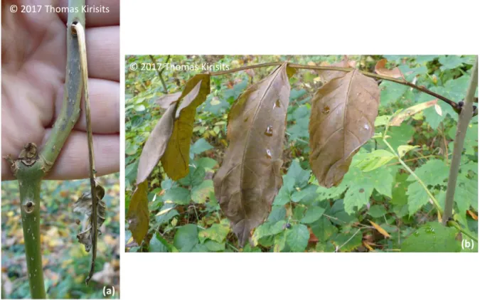
Figure 3: Example of general state of leaves and shoots before sampling on tree n°58 and tree n°52; (a) necrotic lesion is visible on bark and the leaf is dry and ready to fall; (b) no necrotic lesion is visible on bark but the leaf is wilting and the petiole is necrotic.
© 2017 Thomas Kirisits
2. Material and Methods
2.1. Collection sites and ash sampling
The samples for this work were collected in late August, September and October 2016 as well as in October 2017 in the 17th district of Vienna (Hernals), Austria, at 11 different sites (table 1). The maximum distance between two sites was about 2.25km, but many sites were located within short distances (20-100m) to each other. In addition, one leaf petiole-shoot pair was collected in Zagersdorf (Austrian province of Burgenland; table 1). Each site was woodland of broadleaved trees (various species) with natural regeneration of common ash (F. excelsior) in the understorey. The sites were non-systematically chosen based on observations of ash saplings showing appropriate symptoms for this study.
At each sampling date, ash seedlings were carefully inspected for necrotic lesions caused by
H. fraxineus on the leaf petiole close to the leaf node where it was suspected that the pathogen
could have already progressed into the adjacent shoot/seedling stem via the petiole-shoot junction. On some shoots, a necrotic lesion on the bark surface adjacent to a symptomatic leaf petiole was already visible. If this was not the case, one or several small incisions were carefully cut into the bark of the shoot, in order to check whether the phloem around a leaf node was necrotic, indicating a fresh infection by the ADB pathogen. Shoots where several overlapping infections were suspected to occur (indicated by extensive bark necrosis) were not considered for sampling. When necrosis of tissues was confirmed both on a leaf petiole and an adjacent shoot, this leaf petiole-shoot pair was sampled, placed individually in an envelope or plastic bag and transported to the laboratory. Samples were refrigerated (at 4 to 6°C) until they were processed for fungal isolation.
In total, 54 leaf petiole-shoot pairs were collected, 46 in 2016 and 8 in 2017 (table 1). The ash saplings from where samples were taken ranged in height from about 20cm to about 2.5m, and the shoot samples had diameters between 1 and 9mm. The condition of the sampled leaf petioles was variable. While some were totally necrotic and dry but still attached to the shoot, others were still alive and partly green but showed distinct necrotic lesions (figure 3).
2.2. Fungal isolation and identification
From 49 leaf petiole-shoot pairs, fungal isolation was made within two days after collection, from two pairs three days after collection and from three pairs 18 or 19 days after collection. Isolations were done under sterile conditions in a laminar flow. Isolation methods were similar to those described by Kirisits (2017). Malt extract agar (MEA; 20 g DiaMalt malt extract [Hefe Schweiz AG, Stettfurt, Switzerland], 16 g Becoagar agar [W. Behrens & Co., Hamburg, Germany], 1000 ml tap water, 100 mg streptomycin sulphate [Calbiochem; Merck KGaA, Darmstadt, Germany], added after autoclaving) poured in 5.2cm diameter plastic Petri dishes was used as medium.
For fungal isolation, leaf petiole and shoot sections, about 5-8cm long, were surface sterilized; leaf petioles for 30sec in 96% ethanol, 1min in 4% NaClO and 30sec in 96% ethanol, and shoot samples for 1min in 96% ethanol, 3min in 4% NaClO and 30sec in 96% ethanol. The surface sterilized plant parts were then dried for a few minutes, in order to let the ethanol evaporate. From a leaf petiole, the epidermis was carefully scraped off with a sterile scalpel, and an
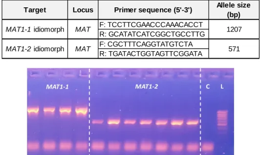
Table 2: Primers for MAT multiplex PCR. F stands for forward and R for reverse primer. The allele size refers to the expected size of the PCR products visible on the gel (Gross et al., 2012b).
Figure 4: Gel-electrophoresis showing amplified fragments of Hymenoscyphus fraxineus
MAT1-1 and MATMAT1-1-2 idiomorphs. In this example, positive results from several samples are shown. L stands
for ladder and C for negative control.
Target Locus Primer sequence (5'-3') Allele size (bp) F: TCCTTCGAACCCAAACACCT R: GCATATCATCGGCTGCCTTG F: CGCTTTCAGGTATGTCTA R: TGATACTGGTAGTTCGGATA MAT MAT 1207 571 MAT1-1 idiomorph MAT1-2 idiomorph
4-6 small fragments were then cut consecutively from the petiole and placed onto a single MEA plate. Similarly, from a shoot section, the outer bark was cut off superficially with a sterile scalpel, and 4-6 small shoot discs (comprising wood and phloem) were cut and put onto one MEA plate. Removal of discs started at or near the proximal edge of the phloem necrosis and adjacent wood discoloration. From two shoots, samples were each taken at three different positions: i) close to the leaf scar where the infection likely originated, ii) at the proximal lesion margin and iii) at a position in between. Petiole fragments and shoot discs removed for fungal isolation were 1-2mm (only occasionally up to 3mm) wide, and samples from a single petiole or shoot were placed well separated from each other onto a single MEA plate, so that well-defined individual fungal colonies could develop. The consecutive order of the pieces along a petiole or shoot was, however, not marked on the isolation plates.
The Petri dishes with the primary isolations were incubated at room temperature and diffuse daylight in the laboratory, and repeatedly inspected for the growth of H. fraxineus and other fungi during a two months period. The ADB pathogen was identified based on the morphology of its colony and morphological characteristics of its asexual stage (phialophores, phialids and conidia; Kowalski 2006; Gross et al. 2014). Colonies of other fungi were recorded, but they were not identified. H. fraxineus isolates were sub-cultured on ash leaf malt extract agar (AMEA; 20g DiaMalt malt extract, 16g Becoagar agar, 50g frozen F. excelsior leaflets [which were removed after autoclaving], 1000mL tap water), which greatly enhances the fungus’s growth (Kirisits et al., 2013; Gross et al., 2014). Pure cultures and the majority of the primary isolation plates were stored at 4-6°C until further use for this thesis from January 2018 onwards.
2.3. DNA extraction
H. fraxineus isolates were grown on AMEA (prepared with frozen leaflets of F. mandshurica
seedlings) for three to four weeks. Genomic DNA from H. fraxineus isolates was extracted using DNeasy Plant Mini Kit (Qiagen) with minor changes from manufacturer’s instructions. Mycelium was scraped and grinded in 70µL of Buffer AP1 with a 4mm steel ball using a Mixer Mill MM 2000 (Retsch). Then, after adding 330µL of Buffer AP1 and 4µL of RNase A (100mg/mL), samples were incubated for 3h at 55°C. Samples were finally eluted in 70µL two times.
2.4. Mating type determination
2.4.1. Multiplex PCREach reaction was set up in 10µL volumes containing 4.7µL of PCR water, 1x of S-Buffer (PeqLab), 1mg.mL-1 of Bovine Serum Albumin (BSA), 800µM of dNTPs, 0.5µM of each primer (table 2), 0.5U of Taq polymerase (PeqLab) and 1µL of the template DNA (or PCR water for negative control). PCR conditions were 3min at 94°C and continued with 34 cycles at 94°C for 30sec, 55°C for 45sec, 72°C for 45sec then followed by a final extension at 72°C for 5min. PCR amplification was verified on 1.5% agarose gel stained with GelRed Nucleic Acid Gel Stain (Biotum). Gel-electrophoresis was accomplished with 1.7µL of PCR product mixed with 6.2µL of loading buffer. The amplified fragments were visualized on a UV-transilluminator (UVP) after 20min (figure 4). A 100bp-1kb DNA ladder (Biozym) was included in each gel.
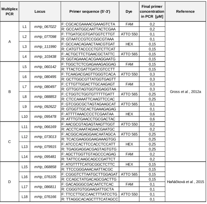
Table 3: Primers for microsatellite markers’ multiplex PCR. F stands for forward and R for reverse primer. Each locus is given a short code name (L1-L18) corresponding to their order in this table.
Multiplex
PCR Primer sequence (5'-3') Dye
Final primer concentration in PCR [µM] Reference F: CGCACGAAAACGAAAGTCTA FAM 0,2 R: GCCAATGGCAATTACTCGAA 0,2 F: TTGATGCGTGATGGTCTTGT ATTO 550 0,1 R: GTAATCCGTCCGGCGTAAA 0,1 F: GCCAACAGAACTAACGTGAT HEX 0,15 R: CATGTTACCCCTGTCTTCAT 0,15 F: ACTGCTTCTGAACGCTATTC ATTO 565 0,15 R: GGTAGAAACACGAAGGAATG 0,15 F: TGGCTCTCGAGAAAGAGGAG FAM 0,15 R: TTACTCGATTGATCGTCCTT 0,15 F: TCAAGACGAGTTGGGTCACA ATTO 550 0,3 R: GCTTGGCGTTATGGTGAGTT 0,3 F: CTTGTTGGACTTGCAAGAGT FAM 0,3 R: GTTGGTAGTGGTGGAGGTAA 0,3 F: CTGGTCTGGTGTTTTTGATT ATTO 565 0,25 R: CTCCAAAATTCAAGTTCCAC 0,25 F: GTCGGCGCTAGTAGAAGCAT ATTO 565 0,1 R: GTGGTTGCACTGAAAGAGAG 0,1 F: ATTTTAAACCCCTCGAATAA HEX 0,6 R: ATTTGTGAACCTGCGACTAC 0,6 F: AACGCGTAGAGTAAGTTGGT ATTO 550 0,2 R: ACCTCAAATAGAACGAATGC 0,2 F: ACGGCAGAGGAACAATAGCA ATTO 565 0,25 R: TCACGAAGGGAAGAAAGTGG 0,25 F: ATCCCACTTCCACCTCCATT HEX 0,25 R: TGAGGAGGACGAGTAGTGTG 0,25 F: AGCTTGGTTGTAGCCCAGAG FAM 0,2 R: TATTCCAAGCAGCCGATTCT 0,2 F: ATGTTTTCATGCGGCTCTTC HEX 0,15 R: TTCCGGGAAACAATTACGC 0,15 F: CGGGTCTTAATGCTTGGAGAT ATTO 565 0,15 R: CCAGCTATGACAGCGACTTG 0,15 F: GACAGGGCGACAATCTCAC FAM 0,1 R: CGGGTGTGGAAGATTGCTA 0,1 F: TTCCTTGCCAACTTTATCCTG ATTO 550 0,1 R: TTAGGCACAGCTTTCATAGCC 0,1
Gross et al. , 2012a
Haňáčková et al. , 2015 A B L11 L12 L9 L10 L6 L7 L8 L1 L2 L3 L4 L5 L13 mHp_060142 mHp_080495 mHp_080497 L18 C D L16 L17 L14 L15 mHp_079915 mHp_095481 mHp_068858 mHp_076105 mHp_086811 Locus mHp_067022 mHp_077098 mHp_111990 mHp_103438 mHp_076166 mHp_088853 mHp_092622 mHp_095478 mHp_066169 mHp_073013
2.5. Microsatellite markers
2.5.1. Multiplex PCREach reaction was set up in 10µL volumes containing 1x of S-Buffer (PeqLab), 800µM of dNTPs, 0.5U of Taq polymerase (PeqLab) and 1µL of the template DNA (or PCR water for negative control). 18 microsatellite markers (Gross et al., 2012a; Haňáčková et al., 2015) were amplified in four multiplex PCRs (A, B, C and D). Each one includes 4 to 6 primer pairs in various concentrations (table 3). Every multiplex reaction was completed with PCR water. PCR conditions were 3min at 94°C and continued with 32 cycles at 94°C for 30sec, 56°C for 30sec, 72°C for 90sec then followed by a final extension at 72°C for 5min. PCR amplification was verified on 1.5% agarose gel stained with GelRed Nucleic Acid Gel Stain (Biotum). Gel-electrophoresis was accomplished with 1.7µL of PCR product mixed with 6.2µL of loading buffer. Amplified fragments were visualized on a UV-transilluminator (UVP) after 20min and a 100bp-1kb DNA ladder (Biozym) was included in each gel. This visualization step was only used for determining the dilution for the fragment analysis.
2.5.2. Fragment analysis
Fragment analysis was performed on an ABI 3130xl Genetic Analyser at the Comprehensive Cancer Center DNA Sequencing Facility, University of Chicago (Chicago, USA). Genotypes were scored by analyzing fragment sizes using the software Peak Scanner version 2.0 (Applied Biosystems). Basic statistics were done using GenAlEx (Peakall & Smouse, 2006, 2012).
2.6. Data analysis
For analysis purposes, we assumed that record of the same genotype in sample pairs (petiole and shoot) resulted from the growth of the given genotype from petiole to shoot, rather than from a direct infection. Furthermore, we used 18 species-specific microsatellites with high genotypic diversity. They have a high resolution power which allowed proper differentiation of all strains in the studies by Gross et al. (2012a) and Haňáčková et al. (2015). The quality of these markers has been assessed in these articles by showing their selective neutrality, their absence of linkage among loci and their Mendelian inheritance. This is why we are postulating here that each genotype corresponds to one individual. Thus, they are referred to as distinguished multilocus genotypes (MLGs).
Isolates of H. fraxineus from 42 leaf petiole-shoot pairs were used for the microsatellite analyses. A pair was excluded because of genotyping delays and, for the remaining 11 petiole-shoot pairs, isolation from the petiole failed in four cases and from the petiole-shoot in seven cases, and these pairs could thus not be included in the investigations. However, from one shoot (tree n°59) three isolates obtained from three different positions were investigated as well, despite isolation of H. fraxineus from the corresponding petiole was unsuccessful. Similarly, from tree n°52 three shoot isolates, also obtained from different positions, were examined, in addition to the petiole isolate. For 39 of the remaining 42 pairs, only one petiole and one shoot isolate (out of up to six which had grown from the different petiole fragments and shoot discs) were chosen for the main data set of the study. Additionally, all six available isolates from three saplings (n°29, n°54 and n°56) from both the petiole and the shoot (36 samples) were investigated. They were chosen because they were among 6 pairs where petiole and shoot isolates had opposite mating type results. The other 3 of such pairs are not included in the presentation of
Table 4: Identification of differentially encoded alleles of polymorphic microsatellite loci of
Hymenoscyphus fraxineus. Each locus is given a short code name corresponding to their order in
table 3. L2 was excluded from further analyses due to its lack of polymorphism. Motif size corresponds to the number of nucleotides repeated, according to Gross et al., 2012a and Haňáčková et al., 2015.
Figure 5: Allelic frequencies of 17 polymorphic microsatellite loci of Hymenoscyphus fraxineus. Allele size corresponds to all identified alleles per locus, summarized in table 4.
Locus Motif size Number of alleles Allele size range (bp) L1 3 3 247-253 L2 3 1 170 L3 3 2 230-236 L4 3 2 213-216 L5 3 2 164-170 L6 2 2 148-154 L7 3 2 245-254 L8 3 2 280-300 L9 3 2 111-117 L10 2 2 233-250 L11 4 2 248-262 L12 2 2 185-240 L13 3 2 184-201 L14 4 3 137-146 L15 3 2 120-129 L16 4 2 197-201 L17 3 2 151-160 L18 3 3 155-187
3. Results
3.1. Isolation of H. fraxineus and other fungi
H. fraxineus was isolated from 50 out of the 54 (92.6%) investigated symptomatic leaf petioles
of F. excelsior, 36 times (66.7%) in pure culture and 14 times (26%) together with other fungi. Other fungi, various, unidentified species, were obtained from one third of the petioles (18), but only from four petioles they were isolated alone, without H. fraxineus. Likewise, the pathogen was recovered from 47 out of the 54 (87.0%) examined shoots with necrotic lesions in the phloem, in 44 cases (81.5%) as the sole fungus and in three cases (5.6%) in mixture with other fungi. In total, other fungi were isolated from six shoots (11.1%), but only from three shoots (5.6%) without H. fraxineus. Four shoots (7.4%) did not yield any fungal growth.
3.2. Differentially encoded alleles
Among the 18 microsatellite markers used in the genotyping of H. fraxineus strains, we identified 38 different alleles in our population. One microsatellite locus (L2) did not show any polymorphism and thus did not contribute to the overall allelic variability of the investigated fungal population, slightly decreasing the initial discriminatory power of this set of microsatellites markers (table 4).
Generally, we observed low allelic polymorphism. Indeed, 14 microsatellite loci showed a bi-allelic polymorphism and only three were tri-bi-allelic. Moreover, the mean number of effective alleles was around 1.745±0.070. However, the mean Shannon’s information index was 0.604±0.033, indicating that even with a low polymorphism the markers were still quite diverse.
More precisely, nine alleles had a frequency below 0.3 and six allelic frequencies were above 0.7 (figure 5). As locus L2 was not taken into account, due to its monomorphic nature, we had 22 out 37 alleles (59%) at mean frequencies of 0.5±0.2, which shows that there is a good allelic repartition in the population and that this microsatellite marker set has still a suitable discriminatory power.
3.3. Genetic profiles
We decided to take into account all genotyped isolates (135 samples), even if some were not used in further analyses (figure 6 and 7), in order to determinate the overall genotypic richness of our H. fraxineus population.
Out of the total data set from 135 isolates, 81 different genotypes were detected and assigned to H. fraxineus MLGs. If we just take the main data set into account, consisting of 84 isolates (42 petiole-shoot isolate pairs from 42 trees; see figure 6), 63 MLGs were identified. Considering the second data set, obtained from six isolates each from a petiole and corresponding shoot of three seedlings (36 isolates in total), only nine MLGs were detected. As expected, there was less genotypic richness in this set, as in all cases more than one isolate, and often many, from the same petiole-shoot pair belonged to the same genotype.
Figure 6: Conformity of distinguished multilocus genotypes (MLGs) of H. fraxineus between petiole and shoot samples. It was calculated with frequencies of cases where the MLG of a petiole isolate was identical (“Same genotype”) or different (“Different genotype”) to the MLG of the corresponding shoot isolate. In 21 trees the same genotype was detected both in the petiole and the shoot, while in 21 trees the petiole and shoot isolates were different genotypes.
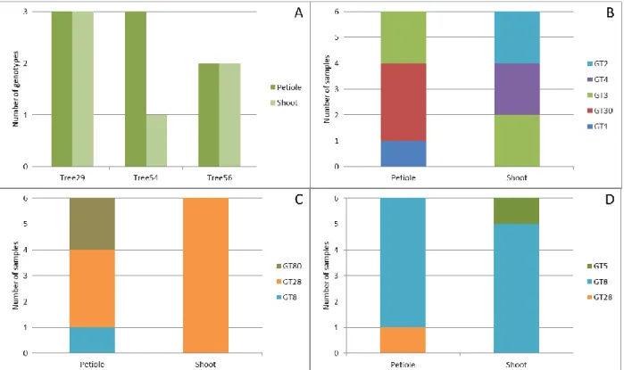
Figure 7: Number of distinct multilocus genotypes (MLGs = GT) of H. fraxineus among six isolates each from a petiole and corresponding shoot of each of three different common ash trees. Total number of MLGs present in the petiole and shoot, respectively, of all three trees (A). Number of MLGs present in tree n°29 (B), in tree n°54 (C) and in tree n°56 (D).
3.4. Genotypic continuity
In 21 pairs, the petiole and shoot isolates were confirmed to be genetically identical, whereas in the remaining 21 pairs, they belonged to different genotypes, leading to a perfect 50-50% ratio of cases of genotype continuity or discontinuity in the examined petiole-shoot isolate pairs (figure 6).
3.5. Genotypic diversity
Among the isolate pairs where different genotypes were detected in the petiole and in the corresponding shoot (figure 6), three trees were investigated in more detail, by genotyping further five isolates each from the same petiole and the same shoot (figure 7). The first part of figure 7 (A) shows an overview of the number of genotypes identified among 6 petiole isolates and 6 shoot isolates per tree. It is showing a varying diversity of H. fraxineus genotypes in the single petiole and corresponding shoot of the three respective trees, which is even more emphasized in the other three parts of the figure (B-D). In the petiole, three different genotypes were detected in two trees (29 and 54), and two genotypes in one tree (56; figure 7A). In the shoot, the number of different genotypes ranged from one to three (figure 7A).
Tree n°29 had the highest H. fraxineus genotypic diversity (figure 7B). Indeed, three MLGs were detected both in the petiole and in the shoot. Initially, GT30 was identified in the petiole and GT3 in the shoot, resulting in a “different genotype” result according to figure 6. Although GT30 was the most frequently isolated genotype (from three fragments) in the petiole, only genotype GT3 was detected both in the petiole and in the shoot, (both two times). In the petiole, a third genotype (GT1), isolated once, was detected, and two further genotypes, GT2 and GT4 (each isolated twice), were detected in the shoot. The two latter genotypes were, however, not confirmed in the petiole.
Tree n°54 had the lowest H. fraxineus genotypic diversity in the shoot, as all six isolates were confirmed to belong to a single MLG (GT28; figure 6C). Genotype GT8 was identified in the petiole in the initial analysis, and this pair was thus assigned as “different genotype” pair (figure 6), even though the prevailing genotype in the petiole (3 isolates) was also GT28. Moreover, a third genotype, GT80, was isolated twice.
Finally, tree n°56 showed the highest genotype agreement between petiole and shoot isolates, as MLG GT8 was recovered five times from both the petiole and the shoot (figure 7D). Nevertheless, the isolates used in the initial analysis represented two different genotypes: GT28 in the petiole and GT5 in the shoot. The subsequent did not detect genotype GT28 in the shoot, and GT5 was not recorded on the petiole.
In addition, genotypes GT8 and GT28 were recorded (each one once) in two different trees, n°54 and n°56 (figure 7C; figure 7D).
4. Discussion
4.1. Association of H. fraxineus with symptoms of ash dieback
In this work, H. fraxineus was isolated at high frequencies from symptomatic organs of
F. excelsior, from 93.6% of the leaf petioles and 87.0% of the shoots, while other undetermined
fungi were obtained much more rarely (from 33.3% of the petioles and 11.1% of the shoots). Moreover, the pathogen was frequently isolated in pure culture, from 66.7% of the petioles and 81.5% of the shoots. These isolation results therefore reinforce, in agreement with previous investigations (e.g. Kowalski 2006; Bakys et al., 2009b; Schumacher et al., 2010; Kowalski et
al., 2016; Schwanda & Kirisits 2016), the association of H. fraxineus with symptoms of ADB
on leaves and woody parts of common ash.
In early studies on ADB, when the cause of the disease was still unknown or debated,
H. fraxineus was not at all (Przybyl 2002) or at low frequencies isolated from necrotic shoots
and twigs of F. excelsior (Bakys et al., 2009a). This was likely because in these investigation isolations were made from ash samples showing late stages of disease. In contrast, in the present work, leaves and shoots showing early and fresh symptoms originating from infections in the year of collection were examined, which greatly increases the likelihood to recover the pathogenic but slow growing H. fraxineus. Scraping off the epidermis from petioles and the outer bark from shoots had likely also a positive effect on the detection of the pathogen and selected against other fungi, especially endophytic species.
4.2. Confirmation of the leaf petiole-shoot infection path for H. fraxineus
Out of the 42 pairs used in the main data set for the microsatellite analyses, 21 presented continuity between petiole and shoot isolates, meaning the same MLG was recorded in both plant organs. Thus, we were able to confirm the colonization path of H. fraxineus from leaves to shoots, through the leaf petiole-shoot junction, in 50% of the cases. The number of pairs where different genotypes were detected in the petiole and corresponding shoot was, however, unexpectedly high, as great care was taken to collect plant samples where symptom appearance clearly suggested pathogen progression from the leaf into the shoot (figure 3).
For three trees where different genotypes were initially detected in the petiole and the shoot (with mating type analysis), the subsequent analysis of all six petiole and all six shoot isolates always led to the identification of one genotype occurring in both plant organs (figure 7). This confirmed the progression of the respective genotypes from the petiole to the shoot in these three cases as well. The results of the complete analysis of all isolates from the three trees suggest that more cases of genotypic continuity probably would have been discovered, if more petiole and shoot isolates had been examined for the pairs that gave inconsistent results in the initial microsatellite analysis (figure 6). As H. fraxineus is a very slow growing fungus, time was too short to prepare a larger number of isolates from individual petioles and shoots for such more comprehensive analyses.
It is planned to do further analyses in the near future, to strengthen knowledge on the infection biology of the ash dieback pathogen. In fact, the petiole-shoot isolate pair assemblage partly investigated here is a special collection because in most cases infected ash leaves drop prematurely, before necrotic bark lesions become visible (Schwanda & Kirisits, 2016).
Collecting corresponding petiole-shoot isolate pairs for this study was only possible by careful inspection of a large number of seedlings for appropriate symptoms at various dates (T. Kirisits, personal communication). Fixing and careful marking of leaves is an alternative approach, allowing assigning fallen leaves to particular shoot parts (Haňáčková et al., 2017).
In two of the three comprehensively analyzed petiole-shoot isolate pairs, one (tree n° 29) or two (n° 56) additional genotypes, besides the one that was confirmed to have entered from the petiole into the shoot, were detected in the shoot but were not found in the adjacent leaf petiole (figure 7). The origin of these genotypes thus remains unknown. Likely, isolation of these genotypes from the petiole failed or they occurred in the most basal part of the leaf node which was cut away in the course of fungal isolation. They could also represent direct shoot infections or infections from another petiole. The latter is, however, rather unlikely because of the careful inspection of seedlings during sampling. Of course, in pairs where different genotypes were recorded in the petiole and the shoot, alternative infection pathways into the shoot (direct shoot infection, different leaf petiole) may also be possible.
Leaf petioles have been suggested as main infection path of H. fraxineus into shoots since the discovery that the fungus’ sexual fruiting bodies are formed on leaf petioles and rachises in the leaf litter (Kirisits et al., 2009; Kowalski & Holdenrieder, 2009). Another indication was the frequent appearance of fresh necrotic lesions around leaf scares (figure 3; Kirisits et al., 2009; Bengtsson et al., 2014). Furthermore, H. fraxineus proved to be able to infect shoots via artificially inflicted leaf scars (Kräutler et al., 2014). However, the present work is only the second study after the one of Haňáčková et al. (2017), where this infection path has been definitely proven.
4.3. Occurrence of identical genotypes on different trees
In the examination of several isolates from the same petiole and shoot of three trees, two MLGs (GT8 and GT28) were found on two different trees (n°54 and n°56), which were growing on the same site (n°10; table 1), relatively close to each other. This finding can have several explanations. First of all, the ascospores responsible for the infections could have had the same parental genotypes, although this is rather unlikely. Another possibility would be that H.
fraxineus conidia are capable of causing infections, as it has been proposed by Fones et al.
(2016).
Finally, this result could also question the discriminative power of our set of microsatellite markers to accurately distinguish individuals, as in the study by Haňáčková et al. (2017), although we used 6 more microsatellite markers than they did in their study. Also, even though our population showed low allelic polymorphism and no microsatellite locus displayed more than a tri-allelic polymorphism (table 4), this was expected and already taken into account in the design of the combination of markers (Gross et al., 2012a; Haňáčková et al., 2015, 2017). Moreover, compared to Gross et al. (2012a), we had 2 loci (L1 and L14) which were more polymorphic (tri-allelic instead of di-allelic), even though we had one monomorphic locus (L2), showing one less allele than expected. In conclusion, the set of microsatellite markers should have been powerful enough to assign different genotypes accurately.
Besides all suppositions discussed above, I am more inclined to think that the occurrence of the two identical genotypes on two different trees could be due to a mistake. In fact, I have high suspicions that I mismatched the two samples leading to this result during the DNA extraction step, as their codes are very alike (54a-1 and 56a-1). If this were the case and the two isolates can in fact be assigned to the respective other tree, genotype GT8 would be the only one occurring in the petiole of tree n°56, and genotype GT28 would be even more frequent in the petiole of tree n°54 (figure 7).
Furthermore, in the initial analysis of 42 petiole-shoot isolate pairs, we detected 63 MLGs out of the 84 isolates tested. Knowing that in exactly half of our pairs (21) the petiole isolate belongs to the same MLG and that in the other half the petiole and the shoot isolate represent two different MLGs, it means that no identical MLGs occurred on different trees.
42 𝑀𝐿𝐺𝑠 + 42 𝑀𝐿𝐺𝑠
2 = 63 𝑀𝐿𝐺𝑠
This result tends to invalidate the earlier argument questioning accuracy of the microsatellite markers and supports the view of a possible human mistake. Thus, it would be better to be cautious regarding the occurrence of identical genotypes on different trees (figure 7) and repeat DNA extraction and genotyping for isolates 54a-1 and 56a-1 before drawing any definite conclusions.
4.4. Genotypic diversity in single petioles and single shoots
Up to 3 MLGs were observed within a width of approximately 6 to 12mm (6 consecutive cuts of 1-2mm each), both in petioles and shoots. Furthermore, in some additional sets of isolates, we detected up to 6 MLGs in an approximately 12mm-long section of a petiole (data not shown).
Even if it has previously been described that several genotypes can be found in a single petiole and shoot (Gross et al., 2012a; Bengtsson et al., 2014; Haňáčková et al., 2017), such a small-scale diversity of genotypes as observed here was initially not expected and has not been reported before. Rather, when fungal isolations for this study were made in 2017 and 2018, it was supposed that just a single genotype occurs in the small sampled areas of petioles and shoots.
4.5. Methodological considerations
The fact that only one isolate per plant organ (petiole, shoot) was examined for the majority of isolate pairs (39 out of 42) created an important bias, as in many cases several individuals seem to be able to co-inhabit small areas of tissue. Indeed, the results from just three trees from which not only one but all six isolates per plant organ were analyzed (figure 7) indicate that there may have been a lot of false negative results for the remaining 18 petiole-shoot isolate pairs where different genotypes were recorded (figure 6). Our results (figure 7) demonstrated that analyzing several isolates from each organ (petiole and shoot) is necessary, in order to diminish false negative results in the case of several overlapping MLGs occurring in close physical proximity in petiole and shoot tissues. For the isolate pairs where the genotype in the petiole and the shoot was found to be identical (figure 6), it would be intriguing to know as well whether several genotypes co-inhabit small areas of tissue.
It would also have been desirable to mark the consecutive order of the plant tissue fragments in the course of fungal isolation, as this would have allowed inferring about the spatial distribution of different genotypes along petiole and shoot.
Finally, without substantial statistical analysis, it would be better to stay cautious in interpreting all the results presented here, even if they are largely in agreement with the current understanding of the infection biology and life cycle of H. fraxineus. They were supposed to be done but last results of the microsatellite analyses were obtained as late as on the 5th of June only, because of technical difficulties, experimental reworks and delays in genotyping.
In future work, we could use the analysis of molecular variance (AMOVA), considering petiole samples and shoot samples as two sub-populations, in order to see if diversity is significantly different. Such analyses could, for example, clarify whether the passage from the leaf to the shoot via the petiole represents a strong genetic bottleneck for the pathogen as proposed in the study of Haňáčková et al. (2015).
5. Conclusions
Using isolation of H. fraxineus from symptomatic leaf petioles and adjacent shoots of F.
excelsior at multiples sites coupled with microsatellite analysis, we were able to confirm the
infection route of H. fraxineus from a single leaf petiole to the shoot in 50% of the cases. However, it is very likely that a big proportion of our results for the cases where the genotype in the petiole was not identical to that in the shoot represent false negatives. Indeed, we could see in a small sub-set of isolate pairs where genotypes were different in the initial analysis that one genotype occurred in both the petiole and the shoot, when a larger number of isolates per single petiole and shoot was analyzed. These analyses also highlighted the occurrence of several genotypes in small areas of a petiole and shoot. In order to have a more accurate appraisal for the infection path of H. fraxineus from a petiole into the adjacent shoot, all pairs should be investigated again using more isolates per plant organ instead of only one.
One of the pitfalls of our study could be the spatial and temporal effects, which we could not evaluate properly. It would have definitely been an improvement of our experiment to include these aspects. If the consecutive order of the fragment cuts for fungal isolation had been marked, it would have been possible to map the location of each genotype in a petiole and shoot on a very fine scale. Also, by doing the same experiment before the appearance of bark necrosis, we could have compared the occurrence and diversity of H. fraxineus genotypes in the shoots.
As said earlier, H. fraxineus may infect and kill 95% of all common ash trees in Europe. This is why it is very important to enhance our understanding of the disease and the pathogen at all levels, in order to find ways to stop its spread and mitigate its impact. We would then maybe assist to a “Rising out of the ashes” (Muñoz et al., 2016).
6. References
Bakys R, Vasaitis R, Barklund P, Thomsen IM & Stenlid J (2009a) Occurrence and pathogenicity of fungi in necrotic and non-symptomatic shoots of declining common ash (Fraxinus excelsior) in Sweden. European Journal of Forest Research 128: 51–60. Bakys R, Vasaitis R, Barklund P, Ihrmark K & Stenlid J (2009b) Investigations concerning the
role of Chalara fraxinea in declining Fraxinus excelsior. Plant Pathology 58: 284–292. Bengtsson SBK, Barklund P, von Brömssen C & Stenlid J (2014) Seasonal pattern of lesion
development in diseased Fraxinus excelsior infected by Hymenoscyphus pseudoalbidus.
PLoS ONE (9) e76429: 1–9.
Baral H-O & Bemmann M (2014) Hymenoscyphus fraxineus vs. Hymenoscyphus albidus – A comparative light microscopic study on the causal agent of European ash dieback and related foliicolous, stroma-forming species. Mycology 5: 228–290.
Baral HO, Queloz V & Hosoya T (2014) Hymenoscyphus fraxineus, the correct scientific name for the fungus causing ash dieback in Europe. IMA Fungus 5(1): 79–80.
Chandelier A, Gerarts F, San Martin G, Herman M & Delahaye L (2016) Temporal evolution of collar lesions associated with ash dieback and the occurrence of Armillaria in Belgian forests. Forest Pathology 46: 289–297.
Cleary M, Nguyen D, Marčiulynienė D, Berlin A, Vasaitis R & Stenlid J (2016) Friend or foe? Biological and ecological traits of the European ash dieback pathogen Hymenoscyphus fraxineus in its native environment. Scientific Reports 6: 21895.
Cross H, Sønstebø JH, Nagy NE, Timmermann V, Solheim H, Børja I, Kauserud H, Carlsen T, Rzepka B, Wasak K, Vivian‐Smith A & Hietala AM (2016) Fungal diversity and seasonal succession in ash leaves infected by the invasive ascomycete Hymenoscyphus
fraxineus. New Phytologist 213(3): 1405–1417.
Downie JA (2017) Ash dieback epidemic in Europe: How can molecular technologies help?
PLoS Pathogens 13(7): e1006381.
Drenkhan R, Solheim H, Bogacheva A, Riit T, Adamson K, Drenkhan T, Maaten T & Hietala AM (2016) Hymenoscyphus fraxineus is a leaf pathogen of local Fraxinus species in the Russian Far East. Plant Pathology 66: 490–500.
Fones HN, Mardon C & Gurr SJ (2016) A role for the asexual spores in infection of Fraxinus excelsior by the ash-dieback fungus Hymenoscyphus fraxineus. Scientific Reports 6: 34638.
Grad B, Kowalski T & Kraj W (2009) Studies on secondary metabolite produced by Chalara fraxinea and its photytoxic influence on Fraxinus excelsior. Phytopathologia 54: 61–69. Gross A, Grünig CR, Queloz V & Holdenrieder O (2012a) A molecular toolkit for population
genetic investigations of the ash dieback pathogen Hymenoscyphus pseudoalbidus.