A R T I C L E
OBC
www.rsc.org/obc
Design, synthesis and evaluation of graftable thrombin inhibitors for
the preparation of blood-compatible polymer materials
Claudio Salvagnini,a Catherine Michaux,bJulie Remiche,cJohan Wouters,bPaulette Charlierc
and Jacqueline Marchand-Brynaert*a
aUnit´e de Chimie Organique et M´edicinale, Universit´e catholique de Louvain, Bˆatiment
Lavoisier, place Louis Pasteur 1, B-1348, Louvain-la-Neuve, Belgium. E-mail: marchand@chim.ucl.ac.be; Fax: +32-10-474168; Tel: +32-10-472740
bLaboratoire de Chimie Biologique Structurale, Facult´es Universitaires Notre Dame de la Paix,
rue de Bruxelles, B-5000, Namur, Belgium
cLaboratoire de Cristallographie des Prot´eines, Universit´e de Li`ege, Bˆatiment B5, All´ee de la
chimie, B-4000, Sart-Tilman, Li`ege, Belgium
Received 20th July 2005, Accepted 3rd October 2005
First published as an Advance Article on the web 19th October 2005
Piperazinyl-amide derivatives of N-a-(3-trifluoromethyl-benzenesulfonyl)-L-arginine (1) were synthesized as graftable thrombin inhibitors. The possible disturbance of biological activity due to a variable spacer-arm fixed on the N-4 piperazinyl position was evaluated in vitro, against human a-thrombin, and in blood coagulation assay. Molecular modelling (in silico analysis) and X-ray diffraction studies of thrombin-inhibitor complexes were also performed. The fixation of bioactive molecules on poly(butylene terephthalate) (PBT) and poly(ethylene terephthalate) (PET) membranes was performed by wet chemistry treatment and evaluated by XPS analysis. Surface grafting of inhibitor
1d improved the membrane hemocompatibility by reducing blood clot formation on the modified surface.
1.
Introduction
The design of biomaterials endowed with high blood compat-ibility (i.e. preventing clot formation) represents a substantial challenge.1The usual strategies to improve hemocompatibility of material devices consist of selective surface treatments like the grafting of zwitterionic components as biomembrane mimics,2 coating with heparin3 or thrombomodulin,4 two natural an-ticoagulants, and the immobilization of extracellular matrix proteins or active small peptides for promoting the growth of endothelial cells.5 Another approach, less exploited till now, makes use of synthetic drugs that are active in preventing blood clotting. For instance, a thrombin inhibitor derived from Argatroban (MD-805)6was graft-polymerized on the surface of polyurethane membranes.7,8A p-amino-benzamidine derivative was covalently immobilized on maleic anhydride copolymer thin films by nucleophilic substitution via a spacer.9
In our laboratory, we are interested in the biocompatibi-lization of polymer materials by their surface derivatization with biologically active molecules of synthetic origin, designed on the basis of medicinal chemistry data. Polyesters, such as poly(ethylene terephthalate) (PET) and poly(butylene tereph-thalate) (PBT) were chosen as model substrates. For instance, the surface grafting of peptidomimetics of the Arg-Gly-Asp sequence on PET membranes resulted in materials showing interesting cell adhesion properties.10,11 Now, we consider the possibility to improve the blood compatibility of polyesters by the covalent grafting of thrombin inhibitors. For that purpose, we have synthesized and evaluated a novel family of thrombin inhibitors equipped with a spacer arm for polymer surface anchorage. This study reports on the design, preparation, surface grafting and in vitro evaluation of the inhibitory activity and anticoagulant effect of this family of compounds based on the L-arginine template. Taking into account possible disturbance due to the spacer arm, in silico evaluation of the interaction with the protein active site has been performed, as well as the X-ray diffraction study of one representative thrombin-inhibitor complex.
2.
Results
2.1 Design of the thrombin inhibitors
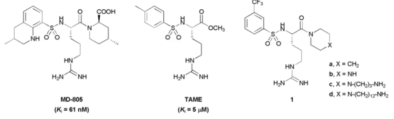
Clot formation results from a complex sequence of biochemical events including the blood coagulation cascade in which throm-bin plays a central role. Thromthrom-bin is a serine protease involved in the conversion of soluble fibrinogen into insoluble fibrin and in the activation of platelet aggregation.12This key protein has become the principal target in the discovery of novel antithrom-botic agents.13,14 Historically, two research lines have been developed in parallel: (i) the steric inhibitors (non-covalently bound) based on two simple lead compounds, namely N-tosyl-L-arginine methyl ester (TAME, Fig. 1),15and benzamidine;16 (ii) the electrophilic inhibitors (covalently bound) derived from the tripeptide motif (D)-Phe-Pro-Arg,17with the peptide arginal Efegatran18and the chloromethylene ketone PPACK,19as leads. The only two synthetic thrombin inhibitors, Argatroban20and Ximelagatran,21available at the moment in the market for the treatment of coagulation complications were developed from the first class of inhibitors.
In our context of biomaterials, the surface bound inhibitor is not allowed to be processed by the enzyme. We thus selected one representative of the first class as a model compound. We considered a simplified structure of Argatroban 1 (Fig. 1) featuring the following characteristics: (i) the guanidyl function of theL-arginine skeleton, for specific ionic interaction with Asp-189 in the S1 enzyme pocket—since oral bioavailability14is not a relevant problem in the field of hemocompatible materials, this strongly basic function could be maintained as a recognizing function; (ii) the lipophilic sulfonamide moiety, for interaction with the S2–S3 binding sites—this substituent was also used to introduce a trifluoromethyl group as a useful spectroscopic tag22for XPS (X-ray photoelectron spectroscopy) analysis of the biomaterial surface and quantification of the amount of grafted inhibitor; (iii) the piperidinyl amide moiety (X= CH2), stable
towards processing by the active serine of thrombin and blood esterases—it has been proven that the acid function placed on the piperidine ring of Argatroban is not absolutely necessary for
DOI
Fig. 1 Thrombin inhibitors.
activity.14Moreover, by removing the carboxylic acid and methyl group in the piperidine ring of Artagroban, we also avoided the control of two chiral centers.
We further transformed the piperidine ring into a piperazine (X = NH) with the view to introduce a versatile anchorage point for different spacer arms, without creating a new chiral center. Thus, we chose to fix the spacer arm on the amide substituent; this was also the strategy selected by Ito and Imanishi23 who transformed the acid function of Argatroban into a polymerizable acrylamide residue.
The effect of the piperazine motif on the activity, and the effect of the spacer arm on the positioning in the enzyme cavity, had to be controlled before using molecules 1 in our biomaterial application. Accordingly, we prepared the reference compound
1a, the parent compound 1b, and two derivatives 1c, and 1d,
bearing respectively a short and a long spacer. The terminal function of the spacer is a primary amine for coupling to polymers displaying activated hydroxyl or carboxyl functions, following previously established protocols.22,24
2.2 Chemistry
The target molecules 1 were constructed from protected argi-nine 2 (N-a-Boc-N-x-nitro-L-arginine) in five steps outlined in Scheme 1. After activation of the carboxyl function, piperidine (X= CH2), N-(benzyloxycarbonyl)piperazine (X= NCO2Bn)
and piperazines 8 and 13 (X= N–spacer–NH–CO2PNB) were
coupled to give the corresponding amides 3a–d (Table 1). After deprotection of the tert-butoxycarbonyl group (Boc), reaction of the free amine 4 with m-(trifluoromethyl)benzenesulfonyl chloride led to compounds 5a–d (Table 1). The final compounds
1a–d (Table 1) were isolated as acetate salts after cleavage of the
protective groups by catalytic hydrogenation.
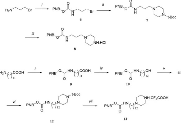
Piperazines 8 and 13 equipped with the protected amino-propyl and aminododecyl spacers were independently pre-pared according to Scheme 2, by using classical chemistry. The piperazine carrying the C3 spacer was prepared from 3-bromopropylamine by first protecting the amino group, then substitution of the bromine with Boc-piperazine, and finally Boc deprotection. For the preparation of the piperazine carrying the C12 spacer, the sequence starting from 12-aminododecanoic
Table 1 Yields of inhibitors and intermediates
Xa 3, (%) 4, (%) 5, (%) 1, (%) CH2 a, 70 a, quant. a, 70 a, 95 N–CO2Bn b, 63 b, quant. b, 70 — N–H — — — b, 90 N–(CH2)3–NH–CO2–PNB c, 60 c, quant. c, 75 — N–(CH2)3–NH2 — — — c, 90 N–(CH2)12–NH–CO2–PNB d, 65 d, quant. d, 75 — N–(CH2)12–NH2 — — — d, 90 aBn= CH 2Ph; PNB= CH2–Ph–p-NO2.
Scheme 1 General synthesis of compounds 1a–d: (i) iBuO2CCl, Et3N,
THF,−20◦C, 15 min; (ii) azacyclohexane, THF,−20◦C to 20◦C, 1 h; (iii) TFA–CH2Cl21 : 1, 20◦C, 2 h; (iv) 3-trifluoromethyl-benzenesulfonyl
chloride, Et3N, CH2Cl2, 0◦C to 20◦C, 4 h; (v) H2, Pd/C, EtOH–AcOH
(3 : 1), 50◦C, 12 h.
acid involved the protection of the amino group, the reduction of the carboxylic acid to the corresponding alcohol, and its activation for the substitution with Boc-piperazine.
Intermediates and final products of Schemes 1 and 2 were fully characterized by IR, NMR and Mass spectroscopy (see Experimental).
2.3 Biological activity
The inhibition of human thrombin (Ki) by compounds 1a–
d was measured spectrophotometrically using as a
competi-tor the chromogenic substrate H-D-phenylalanyl-L-pipecolyl-L -arginine-p-nitroanilide dihydrochloride (S-2238). The thrombin activity was evaluated from the hydrolysis rate of S-2238 measured at 405 nm (production of p-nitroaniline). All the compounds were active at the micromolar level, similarly to TAME (Table 2). The presence of a basic function on the amide substituent, and the use of this function to fix an amino-alkyl chain, did not significantly modify the activity.
The effect of compounds 1a–d on clot formation on polymer materials surface was also evaluated with human whole blood. Two representative substrates were considered, a cell culture support made of PET and a blood filtration membrane made of
Scheme 2 Synthesis of the piperazine linked spacer arms: (i) NaOH 1 N, p-nitro benzylchloroformate, THF, 0–5◦C; (ii) Boc-piperazine, CH2Cl2,
DIPEA, 60◦C; (iii) HCl 1 N, HOAc; (iv) BH3·THF, MeOH, HCl; (v) MeSO2Cl, pyridine, CH2Cl2, 0–5◦C; (vi) NaI, Boc-piperazine, DIPEA, CH3CN,
85◦C; (vii) TFA–CH2Cl21 : 1.
Table 2 Inhibition of human thrombin
Compound Ki/lMa 1a 4–8 1b 17–24 1c 8–18 1d 25–39 TAME 5–6
aReported values are the result of two independent measurements.
PBT. The weight of clot was measured on native polymers and on polymers coated with inhibitors, under standardized conditions, using heparin (Hep) as a control (Table 3).
Compounds 1a–d were found to be active anticoagulants in our experimental conditions (about 100 nmol cm−2of inhibitor
on apparent support surface) reducing the weight of clot on both substrates by about 25%, while TAME was similarly active on PBT only.
Table 3 Inhibitor coating effect on blood clot formation
Sample Clot weight/mga
PET 27.5± 1.5 PET Hep 11.4± 0.5 PET 1a 20.1± 1.4 PET 1c 19.6± 4.2 PET 1d 21.6± 3.0 PET TAME 27.0± 2.0 PBT 29.9± 1.0 PBT Hep 8.0± 3.2 PBT 1a 20.5± 1.0 PBT 1c 22.0± 3.2 PBT 1d 23.8± 1.5 PBT TAME 23.0± 3.1
aResults are the mean of 3 independent experiments ± standard
deviation.
2.4 Docking study
Our purpose was to localize, by molecular modelling, the pos-sible positions of the spacer arm of compounds 1 in the thrombin cavity. Indeed, after grafting on a solid support via the spacer terminus, the inhibitor should still be able to enter and fit in the enzyme active site.
Thrombin is formed by an A chain of 36 amino acids and a B chain of 259 amino acids connected by a disulfur bridge. This enzyme contains the catalytic triad (Asp-102, His-57, Ser-195) characteristic of the chymotrypsin family. Moreover, three important binding pockets have been identified: the specific pocket S1 with Asp-189, the hydrophobic proximal pocket S2 (also called P-pocket) and a larger hydrophobic distal pocket S3 (also called D-pocket).25,26
In our docking study, the coordinates of the human thrombin crystal structure complexed with Argatroban (1DWC in PDB) were used.27 In order to test the two docking algorithms (Autodock28 and Gold29), Argatroban was firstly docked in thrombin. The resulting complexes are in good agreement with the crystal structure (data not shown).
The most active compound of the series, 1c, was then docked using the two methods. To account for amino acids flexibility of the active site, the solutions common to the two programs were then refined with the Discover3 module.30Only the three water molecules close to the catalytic triad in the crystal complex were considered during the run. The geometry of the ligand was optimized in vacuo using quantum mechanics and particularly the HF/6-31+G(d,p). method and the 6-31+G** basis set (Gaussian98 program).31 The guanidyl and terminal amine groups of 1c were considered as protonated at physiological pH. The theoretical pKaof the piperazine moiety was calculated
with the PALLAS program (CompuDrug Chemistry), and gave a value of 7.6 for the alkylamine. That means that about 50% of the ligand is not protonated. Here, we have considered this form but the conclusions are the same for the protonated state of the piperazine moiety.
Two binding modes, being the two best ranked solutions of both programs, were identified (Fig. 2). In the first mode (A,
Fig. 2 Two potential binding modes of 1c into the binding site of human thrombin, as deduced by docking and energy minimization studies. Water molecules and hydrogen atoms were omitted for clarity.
“internal”), the spacer points into the binding site of the enzyme, while in the second one (B, “external”), the spacer points toward the surface of the enzyme.
In the “internal” mode, compound 1c approximately lay in the same orientation as Argatroban. The guanidyl function was close to Asp-189 in the S-pocket and interacted by H-bonding with Gly-219 and a water molecule. The lipophilic sulfonamide moiety lay in the D-pocket and the sulfone group interacted with Gly-219. The piperazine group and its spacer arm were buried in the P-pocket where H-bond was observed between Ser-195 and the amine group. The amide carbonyl moiety was also involved in H-bonding with Gly-216.
In the “external” mode, the lipophilic sulfonamide and piperazine moieties were located in the P- and D-pockets respectively. The guanidyl function still lay in the S-pocket. The sulfone group was involved in H-bonding with Ser-195 and His-57, and the amide carbonyl with Gly-219. The trifluoromethyl group interacted with Lys-60f and a water molecule; the protonated amine interacted with Glu-97a and was close to the enzyme surface. Moreover, p–p stacking interaction was observed between Trp-60d and the phenyl group.
The two complexes were found to be stable and have very close binding energies,−79.2 and −79.6 kcal mol−1respectively.
Thus, the docking studies clearly showed that both orientations are possible from the energetic point of view.
2.5 X-Ray diffraction
Several crystal structures27,32–36 of thrombin complexed with covalent and non -covalent inhibitors have been disclosed, since the seminal work of Bode et al.19A single crystal of the novel compound 1c and human a-thrombin was prepared by using the hanging drop vapour diffusion technique, in the presence of hirudin peptide fragment; this crystal diffracted to 1.65 A˚ resolution.
In the X-ray complex structure, a well-defined electron density for the enzyme at the catalytic site and for the inhibitor was found (Fig. 3A). Not surprisingly, the binding mode of compound 1c
to the different pockets of the active site of a-thrombin was very similar to that of inhibitor MD-805 (Argatroban) as was described in structure 1DWC27and was close to the “internal mode” described above (see superimposition in Fig. 4). Such experimental observation validates our theoretical approach.
The orientation of compound 1c in the active site of a-thrombin is described in Fig. 3B and the compared distances in inhibitor binding are listed in Table 4. The guanidyl group (N22, N21 and N19) was oriented to the S-pocket but was unable to form H-bonds with the OD1 and OD2 groups of the Asp-189 side chain, as it is in the a-thrombin–Argatroban complex or in the “internal mode”. The sulfonamide moiety lay in the D-pocket with the O24 atom of the sulfone group H-bonded to the amide backbone of Gly-219. The carbonyl O15 and amide N12 were both H-bonded to the peptidic backbone of Gly-216, as described in the a-thrombin–Argatroban complex. In the “internal mode”, Gly-216 was slightly shifted. Its carbonyl backbone is moved about 1 A˚ from its position observed in the a-thrombin–Argatroban complex and further from the inhibitor amide N12 atom, resulting in the loss of the hydrogen bond. The loss of one hydrogen bond induced a different positioning of the piperazine cycle, bound in the P-pocket, in comparison with the two X-ray structures where it makes a hydrophobic interaction with the catalytic His-57. The spacer with its amine terminal group (N9) also had a different orientation in the X-ray structure compared to the model. While in the “internal mode” structure we observed H-bonding between the OG hydroxyl function of the active serine and the OE1 and OE2 groups of the Glu-192 side chain, the X-ray structure revealed an unexpected interaction with the NZ terminal amine group of the Lys-60f side chain. Since this spacer does not exist in Argatroban, this kind of interaction was unpredictable by theoretical methods.
2.6 Inhibitors grafting on surface
Transformation of hydroxyl group into sulfonic ester is a well-known process in organic synthesis for alcohol activation towards nucleophilic substitution. This strategy was already successfully
Fig. 3 (2Fo–Fc) electron density map of compound 1c, at the active site of the human a-thrombin complex, at 1.0r contour level (A). Interactions
of compound 1c, at the active site of the human a-thrombin complex (B). The inhibitor is in the same orientation as in Fig. 2A. The CPK colour code has been used for both protein and inhibitor atoms, with compound 1c atoms drawn in larger spheres. Shortest contacts with the protein within a distance of less than 4.1 A˚ are shown with dotted lines.
Fig. 4 Superimposed active sites of crystal structure of the a-thrombin complex with compound 1c (in CPK color code), the “internal” binding mode model (Fig. 2A) (in magenta) and the crystal structure of the a-thrombin complex with compound MD-805 (Argatroban) from structure 1DWC (in cyan). Compound 1c and MD-805 atoms are drawn in larger sphere.
used in our laboratory for polymer functionalization.22 Chain-end hydroxyl groups of PET and PBT membranes were activated with p-toluenesulfonyl chloride (TsCl) and pyridine. The incu-bation of activated membranes with inhibitor solutions should provide sulfonyl ester displacement and covalent fixation of the active molecules on the surfaces (Scheme 3). The efficiency of the binding was verified by XPS analysis. Blank samples were prepared by processing samples without the activating agent
p-toluenesulfonyl chloride. The F detection on blank samples
could be attributed to unspecific adsorption. The corrected
values of F/C (difference between activated and blank samples) allowed the quantification of grafted inhibitors (Table 5). The fixation of compound 1c on both surfaces was not successful: on PET the difference in F detection on blank and activated samples (act) was not significant, while on PBT no F was detected. On the other hand, compound 1d resulted in a good level of fixation, with a higher ratio on PET than on PBT. From the F/C corrected values we could determine the concentration of grafted signals in the interface domain analyzed by XPS (50–100 A˚ depth or about 10 atomic layers). These values (% derivatization) were
Table 4 Compared distances in inhibitor binding Distance/A˚ Inhibitor/protein atom
identification 1DWC 1W7G Internal mode
XaN22 Asp-189 OD1 3.05 3.58 3.33 Asp-189 OD2 2.93 4.08 2.93 Ala-190 O 3.63 4.12 2.97 Trp-215 O 4.06 3.31 3.80 Gly-219 O 4.50 4.93 2.95 XaN21 Asp-189 OD1 5.07 5.37 4.28 Asp-189 OD2 5.11 6.11 2.73 Ala-190 O 4.36 5.19 3.97 Trp-215 O 3.76 3.44 4.62 Gly-219 O 5.48 6.43 5.01 XaN19 Ala-190 O 3.55 3.26 3.11 XaO24 Gly-219 N 3.76 3.21 3.02 XaO15 Gly-216 N 3.01 3.02 3.29 XaN12 Gly-216 O 2.85 2.73 5.05 XaO34 Ser-195 OG 2.99 — — XaN9 Ser-195 OG — 5.21 2.96 His-57 O — 3.50 4.71 Lys-60f NZ — 2.81 4.48 Glu-192 OE1 — 6.59 2.99 Glu-192 OE1 — 4.80 2.63
aThe inhibitor name is abbreviated by X and represents compound
MD-805 (Argatroban) in structure 1DWC and compound 1c (this study) in structure 1W7G and “internal mode”, respectively. Atom numbers refer to Fig. 4 atom labels.
transformed into pmol cm−2as previously described for PET.37 Indeed this membrane is dense, regular and semi-crystalline.
Scheme 3 Inhibitors grafting on polyesters surface: (i) TsCl, pyridine, acetone, 20–60◦C, 1 h; (ii) NH2R (inhibitors), PBS (pH 7.2)–CH3CN 1 :
1, 2 h, 20◦C. PET: n= 2; PBT: n = 4.
Table 6 Inhibitor grafting effect on blood clot formation Clot weight/mga PET 1d blank 29.7± 0.4 PET 1d act 27.5± 1.5 PET 1c blank 31.3± 3.4 PET 1c act 31.1± 5.7 PBT 1d blank 28.5± 4.1 PBT 1d act 21.7± 4.5 PBT 1c blank 29.3± 1.4 PBT 1c act 29.7± 0.6
aResults are the mean of 3 independent experiments ± standard
deviation.
The PBT membrane is tortuous and highly porous; the lack of crystallographic and density data in this case did not allow a similar calculation of surface molecular concentration.
The different morphology of the two polymers may explain the systematically lower fixation ratios observed in the web-like PBT membrane in comparison with the smooth PET membrane. Grafting phenomena occur in the solvent accessible surface of the membrane (open surface) that, in the case of the particular morphology of the considered PBT membrane, may not coincide with the XPS analyzed portion of the sample. Indeed, grafted signals, mostly present in the polymer matrix for instance, could be underestimated by the spectroscopic analysis.
The evaluation of materials hemocompatibility was per-formed by measuring the weight of clot per-formed on the supports after static incubation with blood in standardized conditions. The blood clot formation on samples grafted with compound
1d was found to be systematically lower than on blank samples
(Table 6). The decrease in blood clot formation was more important on PBT (around 24%) than on PET (7%). No effect on blood clot formation was observed on samples treated with compound 1c. This finding was not surprising as low fixation of
1c was detected on PET and no fixation at all was observed on
PBT.
3.
Conclusion
Although significantly less active than Argatroban, the designed inhibitors 1 were found to be sufficiently active for our bio-material application. The lack of condensed piperidine ring on the N-terminal sulfonyl benzene group, and carboxyl and methyl groups on the C-terminal piperidine ring, in fact caused a weaker inhibition potency. However the activities were found to be similar to amide derivatives of N,a-substituted-L-arginine precursors of Argatroban15and benzamidine derivatives devel-oped for material hemocompatibilization.9
Our novel thrombin inhibitors were designed to be grafted on the surface of biomaterials via a spacer arm, either short (compound 1c) or long (compound 1d). From docking and X-ray studies, two potential binding modes of 1c in thrombin were identified. The docking studies clearly showed that both orientations are possible, from an energetic point of view. Indeed,
Table 5 Atomic surface composition of modified polymers analyzed by XPS
C 1s O 1s F 1s N 1s Si 2p F/C Corr F/C Der%a pmol cm−2b
PET 1c blank 72.1 27.0 0.2 0.2 0.5 0.003 PET 1c act 73.4 25.6 0.3 0.5 0.3 0.004 0.001 0.4 11 PET 1d blank 69.0 27.7 0.7 1.5 1.1 0.010 PET 1d act 69.5 25.5 1.3 1.9 1.8 0.019 0.009 3.3 94 PBT 1c blank 73.5 26.2 0.0 0.2 0.1 0 PBT 1c act 74.5 25.2 0.0 0.2 0.1 0 0 0 0 PBT 1d blank 72.1 24.2 0.2 0.8 2.8 0.003 PBT 1d act 71.2 25.2 0.6 1.0 2.1 0.008 0.005 1.9 —
the two complexes are stable and have very similar binding energies. The second mode, called “external mode”, where the molecular spacer is close to the enzyme surface, showed that the grafted inhibitor would still be able to bind the active site of thrombin. Docking studies of analogues of 1c bearing longer spacers ((CH2)4and (CH2)5) were also performed and confirmed
the possibility of observing both the A (internal) and B (external) modes of binding (data not shown). Interestingly, for those longer spacers, even in the so-called “internal mode” observed experimentally, the spacer was also able to join the enzyme surface by displacing Trp-60d (data not shown). The same situation should occur with compound 1d bearing a (CH2)12
spacer arm. Moreover, from biological data, we observed that the substitution of a CH2group with a NH function in position
4 of the piperidine ring, as well as the introduction in the same position of a spacer arm, does not change significantly the activity of the inhibitor (Table 1).
All the experimental and theoretical results confirmed our initial hypothesis that an inhibitor carrying a suitable spacer arm in position 4 of the piperazinyl-amide moiety should be able to enter and fit the active site of thrombin providing the specific biological activity that we required for the development of blood-compatible materials. The grafting of our synthetic thrombin inhibitors on a polyester surface was performed and validated by XPS thanks to the incorporation of a fluorine tag into their structures: fixation of compound 1d with a long spacer was more efficient than the grafting of 1c equipped with a short arm. Moreover, due to the morphology of the polymers chosen as models for this study, the ratio of PET surface derivatization was higher than that of PBT.
The grafting of compound 1d on both PET and PBT membranes was found to reduce significantly the blood clot formation. The inhibitor bound on the surface is still active and its surface concentration of about 90 pmol cm−2 seems
to be sufficient to inhibit the coagulation cascade. Modifi-cation of the grafting protocol and other surface activation techniques are under investigation to increase the surface concentration of bioactive signals and hopefully improve the materials performance. Nevertheless, the proposed technique appeared to be a very promising method for the development of hemocompatible polymer materials, by mild, non-aggressive wet-chemistry treatments.
4.
Experimental
4.1 Chemistry
Reagents and solvents were purchased from Acros Chim-ica, Aldrich or Fluka. Tetrahydrofuran was dried over sodium/benzophenone, then distilled. Column chromatography was carried out with silica gel 60 (70–230 mesh ASTM) supplied by Merck. The IR spectra were recorded with a Perkin-Elmer 1710 instrument, only the most significant adsorption bands being reported. The mass spectra were obtained with a Finnigan MAT TSQ-70 instrument. The high resolution mass spectra (HRMS) were performed at the University of Mons, Belgium (Professor R. Flammang). The microanalyses were performed at the Christopher Ingold Laboratories of University College, London (Dr A. Stones). The melting points were determined with an Electrothermal microscope and are uncorrected. The1H
and13C NMR spectra were recorded on Varian Gemini 300 (at
300 MHz for proton and 75 MHz for carbon) or Bruker AM-500 spectrometers (at AM-500 MHz for proton and 125 MHz for carbon); the chemical shifts were reported in ppm (d) downfield from tetramethylsilane (TMS).
4.1.1 General procedure for the preparation of 5-(3-nitrogua-nidino)-2(
S)-tert-butoxycarbonylamino-1-(azacyclohexan-1-yl)-pentan-1-one (3a–c). N-a-Boc-N-a-nitro-L-arginine (1.00 g, 3.13 mmol, 1 equiv) was dissolved in dry THF (10 mL) and Et3N (0.32 g, 3.13 mmol, 1 equiv). Isobutyl chloroformate
(0.43 g, 3.13 mmol, 1 equiv) was then added dropwise at−20 to−25◦C. After 30 min azacyclohexane (3.13 mmol, 1 equiv) in dry THF (2 mL) was added dropwise at−20◦C and stirred for an additional 20 min. The solution was allowed to warm up to room temperature and stirred for 1 h. The solvent was then evaporated and the solid residue partitioned between EtOAc and brine. The organic layer was dried over MgSO4and
concentration under vacuum gave crude 3 that was purified by column chromatography on SiO2(EtOAc) with a yield of about
65%.
5-(3-Nitroguanidino)-2(
S)-tert-butoxycarbonylamino-1-(piper-idin-1-yl)pentan-1-one (3a). White amorphous solid; Rf = 0.5
(SiO2, EtOAc, UV); IR mmax(CH2Cl2liquid film on NaCl) 3292,
2932, 2857, 1700, 1624, 1522, 1445, 1366, 1254, 1165 cm−1; dH (300 MHz, CDCl3, Me4Si) 1.42 (9H, s), 1.56 (2H, m), 1.65 (6H, m), 1.74 (2H, m), 3.27 (1H, m), 3.36 (2H, m), 3.52 (1H, m), 3.59 (2H, m), 4.58 (1H, m), 5.90 (1H, d, J= 7.0 Hz), 7.87 (2H, s), 8.95 (1H, s); dC (50 MHz, CDCl3, Me4Si) 24.2, 24.3, 25.3, 26.2, 28.3, 31.3, 40.1, 43.2, 46.4, 48.5, 80.3, 156.6, 159.3, 169.5; MS (ESI) m/z 387.0 (M+, 100%); HRMS (ESI) m/z: 425.1912 (calcd for C16H30N6O5K= 425.1915). 5-(3-Nitroguanidino)-2( S)-tert-butoxycarbonylamino-1-[4-(ben-zyloxycarbonyl)piperazin-1-yl]pentan-1-one (3b). White amorphous solid; Rf = 0.1 (SiO2, EtOAc, UV); IR mmax(KBr)
3305, 2979, 2932, 1700, 1635, 1430, 1251, 1231, 1165 cm−1; dH (300 MHz, CDCl3, Me4Si) 1.42 (9H, s), 1.64 (2H, m), 1.72 (2H, m), 3.20 (1H, m), 3.30–3.60 (8H, m), 3.65 (1H, m), 4.57 (1H, m), 5.13 (2H, s), 5.80 (1H, m), 7.34 (5H, m), 7.74 (2H, m), 8.87 (1H, m); dC (50 MHz, CDCl3, Me4Si) 24.5, 28.3, 30.7, 40.5, 41.9, 43.6, 43.7, 45.2, 49.2, 67.5, 80.2, 127.8, 128.1, 128.5, 136.3, 154.9, 156.1, 159.5, 170.5; MS (APCI) m/z 522.0 (M+, 100%),
465.9 (50%), 422.1 (7%); Anal. calcd. for C23H35N7O7·0.5H2O:
C, 52.18; H, 6.63; N, 17.98%. Found: C, 52.32; H, 6.85; N, 18.04%.
{(S)-1-[4-(3-(4-Nitrobenzyloxycarbonyl)aminopropyl)pipera-zine-1-carbonyl]-4-nitroguanidino-butyl}carbamic acid tert-butyl ester (3c). Beige amorphous solid; Rf = 0.1 (SiO2,
isopropanol–CH2Cl210 : 90, UV); IR mmax(CH2Cl2liquid film
on NaCl) 3305, 2926, 2857, 1704, 1629, 1521, 1445, 1347, 1254 cm−1; d H(500 MHz, CDCl3, Me4Si) 1.43 (9H, s), 1.63 (2H, m), 1.71 (4H, m), 2.42 (2H, m), 2.46 (2H, m), 2.50 (2H, m), 3.25 (1H, m), 3.30 (2H, m), 3.43 (2H, m), 3.61 (3H, m), 4.56 (1H, m), 5.18 (2H, s), 5.58 (1H, t, J= 5.7 Hz), 5.81 (1H, d, J = 8.3 Hz), 7.50 (2H, d, J= 8.4 Hz), 7.59 (2H, m), 8.21 (2H, d, J= 8.4 Hz), 8.68 (1H, m); dC (75 MHz, CDCl3, Me4Si) 24.2, 26.0, 28.2, 31.2, 40.1, 42.0, 45.3, 48.6, 52.4, 52.9, 56.1, 64.9, 80.4, 123.6, 128.0, 144.1, 147.4, 155.8, 156.5, 159.3, 169.8; MS (APCI)
m/z 624.2 (M+, 100%); Anal. calcd. for C
26H41N9O9·H2O: C,
48.66; H, 6.76; N, 19.65%. Found: C, 48.62; H, 6.16; N, 19.05%.
{12-[4-((S)-2-tert-Butoxycarbonylamino-5-nitroguanidino-pentanoyl)piperazin-1-yl]-dodecyl}carbamic acid 4-nitrobenzyl ester (3d). Yellow amorphous solid; Rf = 0.5 (SiO2, EtOAc,
UV); IR mmax(CH2Cl2liquid film on NaCl) 3354, 2928, 2855,
1708, 1650, 1523, 1437, 1347, 1254 cm−1; dH(300 MHz, CDCl3, Me4Si) 1.26 (12H, m), 1.43 (9H, s), 1.49 (4H, m), 1.64 (2H, m), 1.75 (2H, m), 2.36 (2H, t, J= 7.2 Hz), 2.45 (4H, m), 3.19 (2H, q, J= 6.6 Hz), 3.26 (1H, m), 3.46 (2H, m), 3.64 (3H, m), 4.56 (1H, m), 4.85 (1H, m), 5.19 (2H, s), 5.85 (1H, d, J= 8.2 Hz), 7.51 (2H, d, J= 8.7 Hz), 7.80 (2H, m), 8.21 (2H, d, J = 8.7 Hz), 8.84 (1H, m); dC(50 MHz, CDCl3, Me4Si) 26.8, 27.5, 28.0, 29.3, 29.6, 29.9, 41.2, 43.6, 52.9, 58.7, 65.0, 71.5, 123.6, 128.0, 144.3, 147.6, 155.5, 155.7, 159.8, 168.0; MS (APCI) m/z 750.3 (M+, 100%); Anal. calcd. for C
35H59N9O9·0.5H2O: C, 55.39; H,
7.97; N, 16.61%. Found: C, 55.70; H, 8.00; N, 16.14%.
4.1.2 General procedure for the preparation of 2(
S)-amino-5-nitroguanidino-1-(azacyclohexan-1-yl)pentan-1-one (4). Prod-uct 3 (1.25 mmol) was dissolved in a 1 : 1 mixture of TFA–CH2Cl2
(10 mL) and stirred at room temperature for 3 h. The solution was concentrated at reduced pressure and the obtained orange gel was suspended and triturated in Et2O until precipitation
of the trifluoroacetate salt as a crystalline solid occurred. The precipitate was washed with Et2O and used directly in the
next reaction (quantitative yield). For spectroscopic analysis the product was converted to the free amine by dissolution in NaOH 1 N, and extraction of the aqueous solution with EtOAc (about 90% product recovery).
2(S)-Amino-5-nitroguanidino-1-(piperidin-1-yl)pentan-1-one
(4a). White amorphous solid; IR mmax(KBr) 3292, 2937, 2857,
1617, 1439, 1257 cm−1; d H(300 MHz, D2O) 1.47 (2H, m), 1.64 (6H, m), 1.91 (2H, m), 3.29–3.42 (4H, m), 3.51 (1H, m), 3.67 (1H, m), 4.57 (1H, t, J = 5.7 Hz); dC (50 MHz, D2O) 23.9, 25.2, 25.6, 26.3, 27.5, 30.6, 40.4, 44.4, 47.2, 50.7, 159.1, 167.1; MS (ESI) m/z 287.1 (M+, 100%); HRMS (ESI) m/z: 309.1643 (calcd for C11H22N6O3Na= 309.1651). 4-(2-(
S)-Amino-5-nitroguanidino-pentanoyl)piperazine-1-car-boxylic acid benzyl ester (4b). White solid; IR mmax(KBr) 3290,
3055, 2938, 1700, 1638, 1430, 1265, 1232 cm−1; d H (300 MHz, CDCl3, Me4Si) 1.47 (2H, m), 1.70 (4H, m), 3.30 (2H, m), 3.35–3.65 (8H, m), 3.82 (1H, m), 5.15 (2H, s), 7.36 (5H, m), 7.80–8.40 (3H, br); dC (50 MHz, acetone, Me4Si) 25.1, 29.1, 41.4, 43.6, 44.7, 44.9, 46.6, 51.9, 68.2, 129.1, 129.2, 129.7, 138.5, 156.2, 161.2, 170.5; MS (APCI) m/z 422.4 (M+, 100%); HRMS
(ESI) m/z: 422.2172 (calcd for C18H28N7O5= 422.2152).
{3-[4-(2-(S)-Amino-5-nitroguanidino-pentanoyl)piperazin-1-yl]propyl}carbamic acid 4-nitrobenzyl ester (4c). Brown
amorphous solid; IR mmax(KBr) 3600–3200, 1701, 1638, 1551,
1329 cm−1; d H(300 MHz, D2O) 1.66 (2H, m), 1.90 (4H, m), 3.00 (1H, m), 3.20 (5H, m), 3.27 (3H, m), 3.59 (4H, m), 4.05 (1H, m), 4.53 (1H, m), 5.17 (2H, s), 7.53 (2H, d, J = 8.1 Hz), 8.19 (2H, d, J = 8.1 Hz); dC (75 MHz, CDCl3, Me4Si) 25.1, 26.1, 29.7, 40.3, 40.7, 42.3, 45.2, 50.7, 52.7, 53.3, 56.3, 65.0, 123.6, 128.0, 144.2, 147.4, 155.8, 159.6, 173.2; MS (APCI) m/z 524.2 (M+, 100%); Anal. calcd. for C
21H33N9O7: C, 48.18; H, 6.35; N,
24.08%. Found: C, 48.14; H, 6.43; N, 24.15%.
{12-[4-((S)-2-Amino-5-nitroguanidino-pentanoyl)piperazin-1-yl]dodecyl}carbamic acid 4-nitrobenzyl ester (4d). Yellow
amorphous solid; IR mmax(CH2Cl2 liquid film on NaCl) 3300,
2928, 2855, 1708, 1690, 1521, 1347, 1256 cm−1; d H (300 MHz, D2O) 1.18 (16H, m), 1.20 (2H, m), 1.43 (2H, m), 1.69 (4H, m), 1.91 (2H, m), 3.11 (6H, m), 3.29 (2H, m), 3.64 (4H, m), 4.55 (1H, m), 5.17 (2H, s), 7.54 (2H, d, J= 8.1 Hz), 8.22 (2H, d, J = 8.1 Hz); dC(50 MHz, CDCl3, Me4Si) 24.2, 24.3, 24.4, 27.3, 27.5, 40.3, 40.8, 41.6, 43.3, 51.2, 51.6, 51.8, 51.9, 57.3, 57.5, 59.3, 65.1, 73.1, 124.4, 129.0, 145.2, 146.7, 156.8, 162.0, 168.7; MS (APCI) m/z 650.5 (M+, 100%); HRMS (ESI): 650.4001 (calcd
for C30H52N9O7= 650.3990).
4.1.3 General procedure for the preparation of 3-trifluoro-methyl-benzenesulfonic acid [4-guanidino-1(
S)-(azacyclohexan-1-ylcarbonyl)butyl]amide (5). Product 4 (0.54 mmol, 1 equiv) was dissolved CH2Cl2(5 mL) and Et3N (0.1688 g, 1.67 mmol,
3.1 equiv). Then 3-trifluoromethyl-benzenesulfonyl chloride (0. 1527 g, 0.64 mmol, 1.2 equiv) in CH2Cl2 (1 mL) was added
dropwise with stirring at 0–5◦C. The solution was stirred for additional 2 h at room temperature, then the mixture was washed with brine, dried over MgSO4, and evaporated under reduced
pressure. The residue was purified by column chromatography on SiO2(EtOAc) with a yield of about 75%.
3-Trifluoromethyl-benzenesulfonic acid [4-nitroguanidino-1(
S)-(piperidin-1-ylcarbonyl)butyl]amide (5a). White amorphous solid; Rf= 0.2 (SiO2, EtOAc, UV); IR mmax(KBr) 3228, 2942,
2857, 1628, 1430, 1326, 1256, 1164, 1129 cm−1; d H (300 MHz, CDCl3, Me4Si) 1.23 (2H, m), 1.51 (4H, m), 1.65 (2H, m), 1.86 (2H, m), 3.16 (3H, m), 3.37 (3H, m), 4.11 (1H, m), 6.53 (1H, m), 7.61 (2H, m), 7.68 (1H, t, J = 7.5 Hz), 7.83 (1H, d, J = 7.5 Hz), 8.06 (1H, d), 8.07 (1H, s), 8.79 (1H, m); dC (50 MHz, CDCl3, Me4Si) 23.9, 24.6, 25.2, 26.2, 30.1, 40.5, 43.5, 46.4, 52.6, 123.2 (JCF = 272.6 Hz), 124.2 (JCF = 3.8 Hz), 129.4 (JCF = 3.4 Hz), 130.0, 130.8, 131.5 (JCF = 33.3 Hz), 140.9, 159.5,
168.3; MS (APCI) m/z 495.2 (M+, 100%); Anal. calcd. for
C18H25N6O5SF3·0.5H2O: C, 42.94; H, 5.40; N, 16.69; S, 6.37%.
Found: C, 42.78; H, 4.97; N, 16.23, S, 6.77%.
3-Trifluoromethyl-benzenesulfonic acid {4-nitroguanidino-1(S)-[4-(benzyloxycarbonyl)-piperazin-1-yl-carbonyl]butyl}amide
(5b). White amorphous solid; Rf = 0.2 (SiO2, EtOAc, UV);
IR mmax(KBr) 3234, 2927, 2863, 1699, 1636, 1429, 1327, 1231, 1166, 1132 cm−1; dH (300 MHz, CDCl3, Me4Si) 1.60 (2H, m), 1.78 (2H, m), 3.0–3.6 (10H, m), 4.18 (1H, m), 5.08 (2H, s), 6.77 (1H, br s), 7.30 (5H, m), 7.40 (2H, m), 7.60 (1H, t, J= 7.6 Hz), 7.78 (1H, d, J= 7.6 Hz), 8.02 (1H, d, J = 7.6 Hz), 8.04 (1H, s), 8.60 (1H, br s); dC(50 MHz, CDCl3, Me4Si) 24.2, 29.9, 40.4, 41.8, 43.0, 43.5, 44.9, 52.6, 67.5, 123.1 (JCF = 273 Hz), 124.0, 127.7, 127.8, 128.1, 128.4, 129.2, 129.9, 130.6, 131.4 (JCF = 33 Hz), 136.0, 140.7, 154.8, 158.9, 168.8; MS (APCI) m/z 630.1 (M+, 100%); Anal. calcd. for C
25H30N7O7SF3: C, 47.69; H, 4.80;
N, 15.57; S, 5.09%. Found: C, 47.52; H, 4.85; N, 15.13; S, 5.13%.
(3- {4-[(S)-5-Nitroguanidino-2-(3-trifluoromethyl-benzenesul-fonylamino)pentanoyl]piperazin-1-yl}propyl)carbamic acid 4-nitrobenzyl ester (5c). Beige amorphous solid; Rf= 0.5 (SiO2,
CH2Cl2–isopropanol 9 : 1, UV); IR mmax(CH2Cl2liquid film on
NaCl) 3308, 2946, 1716, 1635, 1608, 1522, 1436, 1348, 1327, 1263, 1166 cm−1; dH (300 MHz, CDCl3, Me4Si) 1.66 (2H, m), 1.80 (2H, m), 1.97–2.06 (2H, m), 2.30–2.38, (2H, m), 2.34 (4H, m), 3.26 (5H, m), 3.34 (3H, m), 4.15 (1H, m), 5.18 (2H, s), 5.66 (1H, t), 6.62 (1H, br s), 7.51 (2H, d, J= 8.7 Hz), 7.66 (1H, t, J= 8.1 Hz), 7.81 (1H, d, J = 8.1 Hz), 8.04 (1H, d, J = 8.1 Hz), 8.06 (1H, s), 8.21 (2H, d, J= 8.7 Hz); dC(75 MHz, CDCl3, Me4Si) 25.3, 26.0, 29.8, 40.0, 42.0, 45.1, 52.2, 52.7, 52.8, 55.9, 64.8, 123.0 (JCF= 273 Hz), 123.5, 124.0 (JCF= 4 Hz), 126.4 (JCF= 4 Hz), 127.9, 129.8, 130.7, 131.1 (JCF = 32 Hz), 144.4, 146.1, 147.2, 155.8, 159.4, 168.2; MS (ESI) m/z 732.2 (M+, 100%);
Anal. Calcd. for C28H36N9O9SF3: C, 45.96; H, 4.96; N, 17.23%.
Found: C, 46.06; H, 5.09.
(12- {4-[(S)-5-Nitroguanidino-2-(3-trifluoromethyl-benzenesul-fonylamino)pentanoyl]piperazin-1-yl}dodecyl)carbamic acid 4-nitrobenzyl ester (5d). Orange amorphous solid; Rf = 0.4
(SiO2, CH2Cl2–isopropanol 9 : 1, UV); IR mmax(CH2Cl2liquid
film on NaCl) 3314, 2928, 2854, 1707, 1632, 1523, 1431, 1347, 1327, 1263, 1166, 1134 cm−1; d H(300 MHz, CDCl3, Me4Si) 1.24 (16H, m), 1.39 (2H, m), 1.50 (2H, m), 1.62 (2H, m), 1.85 (2H, m), 1.98 (2H, m), 2.22 (2H, m), 2.35 (2H, m), 3.19 (2H, q, J= 6.6 Hz), 3.25 (2H, m), 3.36 (4H, m), 4.10 (1H, m), 4.91 (1H, m), 5.18 (2H, s), 6.60 (1H, m), 7.50 (2H, d, J= 8.7 Hz), 7.68 (1H, t, J= 7.5 Hz), 7.81 (1H, d, J = 7.5 Hz), 8.04 (1H, d, J = 8.7 Hz), 8.07 (1H, s), 8.20 (2H, d, J = 8.4 Hz); dC (50 MHz, CDCl3, Me4Si) 24.7, 25.6, 26.8, 27.0, 27.6, 29.5, 29.8, 30.1, 40.4, 41.4, 42.5, 45.5, 52.5, 52.6, 53.1, 58.4, 65.2, 123.2 (JCF = 273 Hz), 123.8, 124.4 (JCF = 4 Hz), 128.2, 129.7 (JCF = 4 Hz), 130.2, 130.9, 131.6 (JCF = 34 Hz), 140.5, 144.4, 146.6, 147.6, 155.9, 159.3, 168.0; MS (APCI) m/z 858.5 (M+, 100%); HRMS (ESI):
880.3596 (calcd for C37H54N9O9SF3Na: 880.3615).
4.1.4 General procedure for the preparation of 3-(trifluoro-methyl)benzenesulfonic acid [4-guanidino-1(
S)-(azacyclohexan-1-ylcarbonyl)butyl]amide (1). Product 5 (1.01 mmol, 1 equiv) was dissolved in a 3 : 1 mixture of EtOH–AcOH (10 mL). The flask was purged with nitrogen and the catalyst Pd/C 10% (0.091 g, 0.086 mmol, 0.085 equiv) was added. The reaction mixture was then stirred at 50 ◦C for 12 h under hydrogen atmosphere. The catalyst was filtered off and the solution was concentrated under reduced pressure. The solid residue was crystallized from a solution of H2O–MeOH 1 : 1 and washed
with Et2O. The solvent was evaporated and the product was
isolated as acetate salt (about 90% yield).
3-Trifluoromethyl-benzenesulfonic acid [4-guanidino-1(
S)-(piperidin-1-yl-carbonyl)butyl]amide (1a). White amorphous solid; IR mmax(KBr) 3333, 3200, 2944, 2853, 1639, 1325, 1163, 1122 cm−1; d H(300 MHz, CD3OD) 1.23 (2H, m), 1.43 (4H, m), 1.61 (4H, m), 3.01–3.21 (6H, m), 4.18 (1H, m), 7.67 (1H, t, J= 8.1 Hz), 7.83 (1H, d, J= 7.8 Hz), 7.99 (1H, d), 8.00 (1H, s); dC (50 MHz, CD3OD) 25.1, 25.9, 26.4, 27.5, 31.2, 41.9, 44.3, 47.5, 53.7, 124.7 (JCF= 276.6 Hz), 125.3 (JCF= 3.8 Hz), 130.5 (JCF= 3.6 Hz), 131.3, 132.1, 132.4 (JCF= 33.1 Hz), 143.1, 158.8, 170.1; MS (APCI) m/z 450.3 (M+, 100%); HRMS (ESI) m/z: 450.1762 (calcd for C18H27N5O3SF3: 450.1787).
3-Trifluoromethyl-benzenesulfonic acid [4-guanidino-1(
S)-(piperazin-1-ylcarbonyl)butyl]amide (1b). White amorphous solid; IR mmax (KBr) 3500–3000, 1657, 1549, 1423, 1322, 1163 cm−1; dH (300 MHz, CD3OD) 1.37 (2H, m), 1.44 (2H, m), 2.66 (1H, m), 2.79 (2H, m), 2.92 (3H, m), 3.26 (2H, m), 3.48 (2H, m), 4.10 (1H, m), 7.56 (1H, t, J= 7.5 Hz), 7.69 (1H, d, J = 7.5 Hz), 7.88 (1H, d), 7.89 (1H, s); dC (50 MHz, CD3OD) 25.1, 30.9, 40.9, 41.8, 44.5, 44.9, 45.1, 53.6, 125.2, 130.5, 131.6, 132.0, 132.8, 143.5, 158.8, 171.0 (CF3 not
visi-ble); MS (APCI) m/z 451.3 (M+, 100%); Anal. calcd. for
C17H25N6O3SF3·CH3COOH·4H2O: C, 39.18; H, 6.40; N, 14.4%.
Found: C, 38.85; H, 6.12; N, 13.97%.
N -
{(S)-1-[4-(3-Aminopropyl)piperazine-1-carbonyl]-4-guani-dino-butyl}-3-trifluoromethyl-benzenesulfonamide (1c). Beige
amorphous solid; IR mmax(CH2Cl2liquid film on NaCl) 3395,
1636, 1558, 1412, 1328, 1165 cm−1; d H(500 MHz, CD3OD) 1.61 (6H, m), 2.16 (2H, m), 2.22 (1H, m), 2.29 (3H, m), 2.63 (2H, t, J = 7.4 Hz), 3.13 (2H, m), 3.27–3.61 (4H, m), 4.00 (1H, m), 7.60 (1H, t, J= 8.1 Hz), 7.69 (1H, d, J = 8.1 Hz), 8.00 (1H, d, J = 8.1 Hz), 8.04 (1H, s); dC (125 MHz, CDCl3) 26.9, 30.5, 32.9, 41.0, 42.2, 42.7, 46.2, 53.8, 54.3, 57.1, 58.4, 124.5 (JCF= 270 Hz), 125.1 (JCF= 4 Hz), 130.0 (JCF= 4 Hz), 131.4, 132.0, 132.1 (JCF = 37 Hz), 149.2, 158.7, 170.4; MS (ESI) m/z 508.2 (M+, 100%);
Anal. calcd. for C20H32N7O3SF3·2CH3COOH·1.5H2O: C, 44.03;
H, 6.62; N, 14.98%. Found: C, 44.37; H, 6.32; N, 14.68%. N-
{(S)-1-[4-(12-Aminododecyl)piperazine-1-carbonyl]-4-guani-dino-butyl}-3-trifluoromethyl-benzenesulfonamide (1d).
Orange amorphous solid; IR mmax(KBr) 3500–2800, 1647, 1560,
1407, 1328, 1280, 1166, 1134 cm−1; d H(300 MHz, CD3OD) 1.24 (16H, m), 1.41 (3H, m), 1.58 (6H, m), 1.92 (1H, m), 2.04 (1H, m), 2.22 (2H, t, J= 7.5 Hz), 2.37 (4H, m), 2.82 (2H, t, J = 7.5 Hz), 3.14 (3H, m), 3.34 (2H, m), 4.23 (1H, m), 7.68 (1H, t, J= 7.5 Hz), 7.81 (1H, d, J= 7.5 Hz), 8.02 (1H, d, J = 7.5 Hz), 8.05 (1H, s); dC(50 MHz, CD3OD) 26.0, 27.6, 28.6, 28.8, 30.4, 30.7, 30.8, 31.2, 40.8, 41.8, 42.8, 46.3, 53.7, 54.3, 59.5, 59.7, 125.1 (JCF = 260 Hz), 125.2 (JCF= 4 Hz), 130.5 (JCF= 4 Hz), 131.6, 131.8 (JCF= 32 Hz), 132.3, 143.4, 158.9, 170.5; MS (ESI) m/z 634.4
(M+, 100%); Anal. calcd. for C
29H50N7O3SF3·3CH3COOH: C,
51.65; H, 7.68; N, 11.21%. Found: C, 52.17; H, 7.76; N, 12.05%.
4.1.5 Preparation of propyl spacer arm moiety.
(Bromopropyl)-carbamic acid 4-nitrobenzyl ester (6).
3-Bromopropyl amine (2 g, 9 mmol, 1 equiv) was dissolved in water (20 mL), THF (20 mL) and NaOH 1 N (9 mL, 9 mmol, 1 equiv). 4-Nitrobenzyl chloroformate (1.97 g, 9 mmol, 1 equiv) dissolved in cold THF (5 mL) was added dropwise to the reaction mixture at 0–5◦C, simultaneously with NaOH 1 N (10 mL, 10 mmol, 1.1 equiv). The solution was then allowed to warm up at room temperature and stirred for 2 h, checking the pH to be above 10. The solvent was then removed by evaporation at reduced pressure, the residue was dissolved in EtOAc and washed twice with brine. The aqueous phases were extracted once with fresh EtOAc. The organic phases were combined and dried over MgSO4and evaporated at reduced pressure to give pure
com-pound 6 (2.85 g) as white solid (98% yield). Mp= 71.9–72.3◦C;
Rf = 0.5 (SiO2, cyclohexane–EtOAc 5 : 4, UV); IR mmax(KBr)
3417, 3339, 2938, 2848, 1705, 1607, 1521, 1347, 1245, 859 cm−1; dH (300 MHz, CDCl3, Me4Si) 2.1 (2H, quint, J = 6.4 Hz), 3.37 (2H, t, J= 6.4 Hz), 3.46 (2H, t, J = 6.4 Hz), 5.07 (1H, s), 5.20 (2H, s), 7.51 (2H, d, J= 8.5 Hz), 8.22 (2H, d, J = 8.5 Hz); dC(50 MHz, CDCl3, Me4Si) 30.5, 32.3, 39.5, 65.1, 123.6, 128.0, 143.8, 147.6, 155.7; MS (APCI) m/z 318.9 (M81Br+, 80%), 316.9
(M79Br+, 80%); Anal. calcd. for C
11H13N2O4Br: C, 41.66; H, 4.13;
N, 8.83%. Found: C, 41.80; H, 4.13; N, 8.81%.
4-[3-(4-Nitrobenzyloxycarbonylamino)propyl]piperazine-1-carboxylic acid tert-butyl ester (7). Boc-piperazine (1.372 g,
7.22 mmol, 1 equiv) and compound 6 (2.517 g, 7.94 mmol, 1.1 equiv.) were dissolved in dry CH2Cl2(40 mL) and DIPEA
(4 mL, 20.4 mmol, 3 equiv). The mixture was heated at reflux for 2 h. The solvent was then evaporated under reduced pressure and the gel obtained was extracted with EtOAc. The organic layer was then washed with brine, dried over MgSO4 and
evaporated under reduced pressure. The residue was purified by column chromatography to obtain pure compound 7 (2.72 g), as a yellow gel (84% yield). Rf= 0.3 (SiO2, EtOAc–acetone 2 :
1, UV); IR mmax(CH2Cl2liquid film on NaCl) 2947, 1719, 1685,
1523, 1349, 1241 cm−1; d H(300 MHz, CDCl3, Me4Si) 1.47 (9H, s), 1.72 (2H, quint, J= 6.5 Hz), 2.40 (4H, t, J = 4.8 Hz), 2.47 (2H, m), 3.32 (2H, m), 3.44 (4H, t, J= 4.8 Hz), 5.20 (2H, s), 6.09 (1H, t), 7.52 (2H, d, J= 8.8 Hz), 8.22 (2H, d, J = 8.8 Hz); dC (50 MHz, CDCl3, Me4Si) 25.7, 28.4, 40.6, 43.6, 52.9, 56.8, 64.8, 79.7, 123.5, 127.9, 144.2, 147.3, 154.5, 155.7; MS (APCI)
m/z 423.2 (M+, 100%); Anal. calcd. for C
20H30N4O6·H2O: C,
54.53; H, 7.32; N, 12.72%. Found: C, 54.57; H, 7.36; N, 12.65%.
(3-Piperazin-1-yl-propyl)carbamic acid 4-nitrobenzyl ester (8). A solution of HCl 1 N in AcOH (56 mL, 8 equiv)
was added to product 7 (2.95 g, 7 mmol, 1 equiv) and the reaction mixture was stirred at room temperature for 2 h. The solution was concentrated by evaporation at reduced pressure to obtain a brown gel. This residue was several times triturated and suspended in Et2O until a beige solid suspension was
observed. The solvent was then evaporated to obtain product
8 as hydrochloride salt (quantitative yield). To isolate the free
amine, the product was dissolved in water and the solution was brought to pH 10 with NaOH 1 N. The aqueous solution was then extracted 5 times with EtOAc, the organic phases were combined, dried over MgSO4and evaporated to give compound
8 (2.28 g) as a free amine (92% product recovery). IRmmax(KBr)
3316, 3214, 2944, 2801, 1718, 1607, 1521, 1347, 1260 cm−1; d H (200 MHz, CDCl3, Me4Si) 1.63 (2H, quint, J= 6.4 Hz), 1.94 (1H, s), 2.37 (6H, m), 2.83 (4H, t, J= 4.9 Hz), 3.23 (2H, q, J = 6.0 Hz), 5.12 (2H, s), 6.36 (1H, t, J = 6.0 Hz), 7.44 (2H, d, J= 8.8 Hz), 8.14 (2H, d, J = 8.8 Hz); dC (75 MHz, CDCl3, Me4Si) 25.9, 41.5, 46.5, 54.7, 58.2, 65.3, 124.1, 128.4, 144.8,
147.8, 156.2; MS (APCI) m/z 323.3 (M+, 100%); Anal. calcd.
for C15H22N4O4·1.2HCl: C, 49.21; H, 6.34; N, 15.31%. Found:
C, 49.50; H, 6.19; N, 14.80%.
4.1.6 Preparation of dodecyl spacer arm moiety.
12-(4-Nitrobenzyloxycarbonylamino)dodecanoic acid (9).
12-Aminododecanoic acid (250 mg, 1.16 mmol, 1 equiv) was dissolved in THF (3 mL), water (3 mL) and NaOH 1 N (1.14 mL, 1 equiv). 4-Nitrobenzyl chloroformate (246 mg, 1.16 mmol, 1 equiv) in THF (1 mL) and NaOH 1 N (1.3 mL, 1.1 equiv) were added simultaneously to the reaction mixture at 0◦C. The solution was stirred at 0 ◦C for 10 min and then for 2 h at room temperature, checking pH was above 10. HCl 1 N was added to the reaction mixture to reach pH 1, and the solution was then extracted twice with EtOAc. The organic layers were combined, washed with HCl 1 N and water, dried over MgSO4, and evaporated under reduced pressure to give a yellow
solid that was purified by column chromatography on SiO2to
obtain pure product 9 (0.345 g) as a white solid (75% yield). Mp = 107.9–108.9 ◦C; Rf = 0.3 (SiO2, EtOAc–cyclohexane
3 : 5, UV); IR mmax(CH2Cl2liquid film on NaCl) 3367, 2920, 2851, 1725, 1689, 1613, 1527, 1351, 1256, 1246 cm−1; d H (300 MHz, CDCl3, Me4Si) 1.27 (14H, m), 1.52 (2H, m), 1.64 (2H, m), 2.36 (2H, t, J = 7.1 Hz), 3.21 (2H, m), 4.84 (1H, m), 5.20 (2H, s), 7.52 (2H, d, J= 8.6 Hz), 8.22 (2H, d, J = 8.6 Hz); dC(75 MHz, CDCl3, Me4Si) 24.7, 26.7, 29.0, 29.1, 29.2, 29.3, 29.4, 29.9, 33.8, 41.3, 65.1, 123.7, 128.0, 144.1, 150.2, 155.7, 178.2; MS (APCI)
m/z 395.0 (M+, 100%); Anal. calcd. for C
20H30N2O6: C, 60.90;
H, 7.65; N, 7.10%. Found: C, 60.55; H, 7.45; N, 6.76%.
(12-Hydroxydodecyl)carbamic acid 4-nitrobenzyl ester (10).
Compound 9 (0.220 g, 0.56 mmol, 1 equiv) was dissolved in dry THF (10 mL) and the solution was cooled down to 0◦C. BH31 M
in THF (1.11 mL, 1.11 mmol, 2 equiv) was added dropwise to the solution that was then stirred for additional 2 h at 0◦C. The reaction mixture was quenched by dropwise addition of AcOH– MeOH 1 : 9. The mixture was then concentrated and the residue dissolved in EtOAc. The organic phase was washed with water, dried over MgSO4, and concentrated under reduced pressure.
The residue was further purified by column chromatography on SiO2to give pure 10 (0.193 g) as a white solid (91% yield). Mp=
103.7–104.7 ◦C; Rf = 0.6 (SiO2, EtOAc–cyclohexane 4 : 5,
UV); IR mmax (CH2Cl2liquid film on NaCl) 3400, 2987, 2865,
1688 cm−1; d H (300 MHz, CDCl3, Me4Si) 1.27 (16H, m), 1.55 (4H, m), 3.21 (2H, q, J= 6.6 Hz), 3.65 (2H, t, J = 6.6 Hz), 4.83 (1H, m), 5.20 (2H, s), 7.52 (2H, d, J= 8.7 Hz), 8.22 (2H, d, J = 8.7 Hz); dC (75 MHz, CDCl3, Me4Si) 25.8, 26.7, 29.2, 29.3, 29.4, 29.5, 29.6, 29.9, 32.8, 41.3, 63.1, 65.0, 123.7, 128.0, 144.1, 150.2, 155.7; MS (APCI) m/z 381 (M+, 100%); Anal. calcd. for
C20H32N2O5: C, 63.14; H, 8.48; N, 7.36%. Found: C, 63.76; H,
8.78; N, 7.02%.
Methanesulfonic acid 12-(4-nitrobenzyloxycarbonylamino)-dodecyl ester (11). Compound 10 (10.24 g, 26.91 mmol, 1 equiv)
was dissolved in CH2Cl2(200 mL). Pyridine (4.22 g, 53.83 mmol,
2 equiv) and methanesulfonyl chloride (4.63 g, 40.37 mmol, 1.5 equiv) were added dropwise at 0◦C. The solution was stirred at 0◦C for 3 h, and for an additional 12 h at room temperature. The mixture was then washed with water, and HCl 1 N, dried over MgSO4and concentrated at reduced pressure. The residue
was purified by column chromatography on SiO2 to give pure
compound 11 (9.33 g) as a white solid (76% yield). Mp= 80.9– 82.9◦C; Rf = 0.3 (SiO2, CH2Cl2–EtOAc 99 : 1, UV); IR mmax
(CH2Cl2 liquid film on NaCl) 3370, 2921, 2851, 1690, 1614, 1531, 1355, 1264, 1167 cm−1; d H(300 MHz, CDCl3, Me4Si) 1.27 (14H, m), 1.37 (2H, m), 1.52 (2H, m), 1.75 (2H, tt, J= 7.0 Hz), 3.01 (3H, s), 3.21 (2H, q, J= 6.9 Hz), 4.23 (2H, t, J = 6.8 Hz), 4.83 (1H, br s), 5.20 (2H, s), 7.51 (2H, d, J= 8.6 Hz), 8.22 (2H, d, J = 8.6 Hz); dC (50 MHz, CDCl3, Me4Si) 25.4, 26.6, 28.9, 29.1, 29.2, 29.3, 29.4, 29.5, 29.6, 29.9, 37.3, 41.2, 64.9, 70.2, 123.7, 128.0, 144.2, 147.6, 155.8; MS (APCI) m/z 459.1 (M+,
100%); Anal. calcd. for C21H34N2O7S: C, 55.00; H, 7.47; N, 6.11;
S, 6.99%. Found: C, 54.85; H, 7.40; N, 6.19; S, 6.46%.
4-[12-(4-Nitrobenzyloxycarbonylamino)dodecyl]piperazine-1-carboxylic acid tert-butyl ester (12). Compound 11 (0.100 g,
0.22 mmol, 1 equiv), NaI (0.033 g, 0.22 mmol, 1 equiv), and Boc-piperazine (0.041 g, 0.22 mmol, 1 equiv) were dissolved in CH3CN (5 mL) and water (0.2 mL). The reaction mixture
was stirred at 80 ◦C for 30 min, then ethyldiisopropylamine (0.028 g, 0.22 mmol, 1 equiv) was added. After 12 h at 80◦C the solvent was removed by evaporation and the residue dissolved in CH2Cl2. The organic phase was washed with water, dried
over MgSO4, and concentrated at reduced pressure. The residue
was purified by column chromatography on SiO2 to give pure
compound 12 (0.108 g) as a yellow solid (90% yield). Mp = 62.6–63.6◦C; Rf= 0.5 (SiO2, CH2Cl2/isopropanol 96 : 4, UV);
IR mmax(CH2Cl2liquid film on NaCl) 3320, 2930, 2855, 1696,
1601, 1523, 1413, 1347, 1252, 1163 cm−1; d H(300 MHz, CDCl3, Me4Si) 1.24 (16H, m), 1.44 (13H, m), 2.30 (2H, m), 2.34 (4H, m), 3.17 (2H, q, J= 6.6 Hz), 3.42 (4H, m), 5.02 (1H, m), 5.17 (2H, s), 7.49 (2H, d, J = 8.1 Hz), 8.19 (2H, d, J = 8.7 Hz); dC (50 MHz, CDCl3, Me4Si) 26.9, 27.7, 28.6, 29.4, 29.7, 29.9, 30.1, 41.5, 43.8, 53.2, 58.9, 65.2, 79.7, 123.8, 128.2, 144.4, 147.8, 154.9, 156.1; MS (APCI) m/z 549.9 (M+, 100%); Anal. calcd.
for C29H48N4O6·0.3H2O: C, 62.84; H, 8.95; N, 10.11%; Found:
C, 62.76; H, 8.85; N, 10.07%.
(12-Piperazin-1-yl-dodecyl)carbamic acid 4-nitrobenzyl ester (13). Compound 12 (0.497 g, 0.91 mmol, 1 equiv) was dissolved
in a 1 : 1 mixture of TFA–CH2Cl2 (7 mL) and the solution
was stirred at room temperature for 2 h. The solution was concentrated under reduced pressure to give a brown gel that was triturated and suspended in Et2O until precipitation of a brown
solid occurred. The solvent was evaporated and compound 13 (0.510 g) was recovered as the trifluoroacetate salt (quantitative yield). Mp= 79.5–80.5◦C; IR mmax(CH2Cl2liquid film on NaCl)
3354, 2920, 2850, 1690, 1612, 1528, 1350 cm−1; dH(300 MHz, CDCl3, Me4Si) 1.25 (16H, m), 1.49 (4H, m), 2.33 (2H, t, J= 7.7 Hz), 2.47 (4H, m), 2.96 (4H, m), 3.19 (2H, q, J= 6.7 Hz), 3.99 (1H, m), 4.93 (1H, m), 5.18 (2H, s), 7.50 (2H, d, J= 8.4 Hz), 8.21 (2H, d, J= 8.7 Hz); dC(50 MHz, CDCl3, Me4Si) 25.3, 26.6, 26.9, 29.1, 29.3, 29.4, 29.6, 29.9, 30.7, 41.2, 42.6, 49.3, 57.9, 65.0, 123.6, 128.0, 144.3, 147.6, 155.9; MS (APCI) m/z 449.4 (M+,
100%); HRMS (ESI) m/z 449.3119 (calcd. for C24H41N4O4=
449.3128).
4.2 Polymer surface modification
The polymer materials considered were poly(ethylene terephtha-late) (PET) track-etched micro-porous membrane (Whatman S.A., thickness 12 lm, density 1.39 g cm−3, pore diameter
0.49 lm, pore density 1.45× 10⁶pore cm−2) and poly(butylene
terephthalate) (PBT) melt blown filtration membrane (Johns Manville, thickness 133 lm, mean flow pore 5 lm, basis weight 82 gsm). The surface chemical composition of the derivatized polymers was determined by XPS with a SSX 100/206 photo-electron spectrometer from Surface Science Instruments (USA) equipped with a monochromatized microfocus Al X-ray source (powered at 20 mA and 10 kV). The pressure in the analysis chamber was around 10−6 Pa. The angle between the surface
normal and the axis of the analyser lens was 55◦. The analysed area was approximately 1.4 mm2and the pass energy was set
at 150 eV. In these conditions, the resolution determined by the full width at half maximum (FWHM) of the Au 4f7/2peak
was around 1.6 eV. A flood gun set at 10 eV and a Ni grid placed 3 mm above the sample surface were used for charge stabilization. The binding energies were calculated with respect to the C–(C,H) component of the C 1s peak fixed at 284.8 eV. Data treatment was performed with the CasaXPS program (Casa Software Ltd, UK). Molar fractions were calculated using peak areas normalised on the basis of acquisition parameters and sensitivity factors provided by the manufacturer.
4.2.1 Activation and coupling. Native polyester mem-branes (14) were cut into disks of 13 mm of diameter. The polymer samples (10 disks) were stirred at 20 ◦C (PBT) or 60◦C (PET) for 1 h into a mixture of acetone (50 mL), pyridine (1.06 mL) and p-toluenesulfonyl chloride (2.5 g). The samples were washed successively with acetone (50 mL, 5 min) and water (50 mL, 5 min). The resulted activated samples (15) were individually immersed in 1 mL of a 10−3M solution of inhibitor
in a phosphate buffer (PBS, pH 7.2)–CH3CN mixture (1 : 1)
and incubated for 2 h at 20◦C with shaking. The samples were washed with PBS–CH3CN (1 mL, 2× 10 min), water (1 mL,
2× 5 min), 5 × 10⁻³M HCl (1 mL, 2× 5 min) and water (1 mL, 2× 10 min). The resulting inhibitor grafted membranes (16) were finally dried over filter paper and analysed by XPS within 24 h. The blank samples were prepared in the same way but without addition of p-toluenesulfonyl chloride in the activation step.
4.2.2 XPS analysis. The surface molar fractions were measured by XPS analysis. The concentration of F tagged
molecules on the surface was calculated from the F/C ratio. Considering for instance the polymer repeat unit of PET – CO–C6H4–CO2–CH2CH2–O– (C10H8O4) and the derivatized
(by grafting of compound 1d) chain unit –CO–C6H4–CO2–
CH2CH2–1d (C39H57F3N7O6S) we calculated the percentage of
derivatized units as follows. For a mixture of 96.7% (C10H8O4)
and 3.3% (C39H57F3N7O6S), F/C× 100 = 9.9 × 100/1095.7 =
0.904 (experimental value 0.900). Similarly, the corrected F/C value of 0.005 for compound 1d on PBT gave a derivatization ratio of 1.9%. We previously calculated on the basis of PET crystallographic data and simple geometrical considerations37 an average of 1.72 × 10¹⁵ PET monomer units per cm2 of
surface covering 10 atomic layers (the depth analysed by XPS) or about 2850 pmol cm−2. Thus, 3.3% of derivatized
surface monomer units corresponds to about 90 pmol cm−2of
fixed compound 1d. In the same way we calculated a surface concentration of compound 1c of about 10 pmol cm−2 for a
corrected F/C value of 0.001 (0.4% derivatization). The lack of crystallographic data on the PBT membrane did not allow an analogue calculation of signal surface concentration.
4.3 Biological assays
4.3.1 Enzyme kinetics. Substrate H-D-phenylalanyl-L -pipecolyl-L-arginine-p-nitroanilide dihydrochloride (S2238) was obtained from Chromogenix. Human thrombin was obtained from Hypen BioMed. Enzyme kinetics studies were performed on a Cary210 spectrophotometer.
The inhibition activities were measured by adapting the protocol developed by Lottemberg et al.:38 100 lL of the solution of substrate S2238 (0.1 mM in water) and 100 lL of water solution of the tested compound (50–200 lM final concentration) were diluted in 2 mL of Tris-Hepes Buffer (Tris 0.01 M, Hepes 0.01 M, NaCl 0.5 M, PEG 6000 0.1% m/v, pH 7.8). Finally 10 lL of water solution of thrombin (10 NIH mL−1, 75.2 nM) were added to start the reaction. The
appearance of the substrate hydrolysis product (p-nitroaniline) was measured at 405 nm as a function of time. Plots of V /Vi
versus inhibitor concentration (ratio of hydrolysis in the absence
and in the presence of inhibitors) gave the inhibition constants indicated in Table 2.
4.3.2 Clot formation. For the evaluation of material hemo-compatibility we used a simple test based on the weight measurement of the blood clot formed on given substrates (polymer squares of 4× 4 cm fixed on a cylindrical glass support of 2.6 cm internal diameter) after blood contact and incubation. Human blood was withdrawn from a healthy volunteer, collected in citrated tubes and used within a day. For coating experiments 200 lL of a 10−3M solution of the tested compound were placed on the polymer surface that was then dried overnight. After weighing the samples, 200 lL of human blood were placed on the surface, 20 lL of CaCl2 were added to start coagulation
and the samples were incubated statically for 1.5 h at 37◦C in a saturated water atmosphere and 5% CO2. The samples were
then washed with water (3× 2 mL), the clot was fixed with formaldehyde (2 mL of a 5% solution in water) and then washed with water (2 mL). The samples were dried in oven (37◦C) until constant weight. The sample weight differences gave the weight of the formed clot. Native polymer coated with heparin (Leo, 5000 I.E./U.I./mL, 200 lL) was used as anti-clotting reference sample.
4.4 Docking study
Molecular modelling studies were performed on a Silicon Graphics Octane2 workstation.
Gold implements a genetic algorithm allowing the protein–
ligand docking with full ligand and partial protein flexibility. Indeed, conformation of some amino acids (Ser, Thr and Lys) are optimized during the run. The energy function is partly based
on conformational and non-bonded contact information from the CSD database. Parameters: Popsiz= 100; maxops = 100000; niche size= 2.
Autodock uses a hybrid method called Lamarckian Genetic
Algorithm (genetic algorithm coupled with a local search) to pre-dict the interaction of flexible ligands with rigid macromolecular targets. The scoring function includes van der Waals, coulombic electrostatic, directional hydrogen bonding, entropy of ligand binding and desolvation contributions. Parameters: Runs= 200; Population size= 50; Number of generations = 27000.
Discover3 uses the molecular mechanics to optimize the
conformation of the ligand–protein complex and evaluate the interaction energy (the enthalpic contribution of the binding) which is the sum of coulombic and van der Waals terms (DEcb
and DEvdw). The backbone is moved following force constants;
side chains and water molecules move freely.
Forcefield: CVFF; Dielectric constant: 1*r; Criteria conver-gence: 10 kcal mol−1for the Steepest Descent algorithm, 0.01
for the Conjugate Gradient one.
The thrombin amino acid residues were numbered by the chymotrypsin(ogen) numbering system as suggested by Bode
et al.19
4.5 Crystallization, data collection and structure refinement
Human a-thrombin was obtained from Kordia Life Sciences (Leiden, The Netherlands) and its anion-binding exosite in-hibitor, the hirudin peptide fragment (54–65), was obtained from Bachem (Bubendorf, Switzerland). Crystals of human a-thrombin were grown in the presence of hirudin and compound
1c using the hanging drop vapour diffusion technique at
293 K. Prior to the crystallization, the protein was solubilized to approximately 10 mg ml−1in 0.36 M NaCl, 96 mM pH 6.5 citrate
buffer, containing hirudin at a concentration of 3 mM. Drops were prepared by mixing 1 ll of protein solution with 1 ll of compound 1c at a final concentration of 2.9 mM to 1 ll of reservoir solution. The reservoir solution contained 20% PEGMME 5000, 0.36 M NaCl in 0.1 M HEPES buffer pH 7.5. Compound 1c was additionally further diffused into the crystal for 72 h from a 44 mM solution containing 22% PEGMME 5000 and 0.36 M NaCl to a final concentration of 16 mM.
One crystal was flash frozen in liquid nitrogen after rapid soaking in a cryoprotectant solution containing about 50% glycerol in the crystallization buffer. Data were collected at ESRF (European Synchrotron Radiation Facility, Grenoble, France) on beamline BM30a at a wavelength value of 0.9797 A˚ . All data were recorded on a MarCCD detector, and intensities were indexed and integrated using MOSFLM version 6.01.39 This single crystal, belonging to space group C2 with unit cell dimensions a= 70.51 A˚, b = 71.34 A˚, c = 72.56 A˚ and b = 100.59◦, diffracted to 1.65 A˚ resolution. The scaling of the intensity data was accomplished with SCALA of the CCP4 program suite,40 and all corresponding statistics are given in Table 7.
The phasing procedure for solving the structure was the molecular replacement method using the AMoRe package.41 The structure of the complex human a-thrombin/hirugen (PDB code 1HTG)42was used as the search model for the rotation and translation searches. Model building with TURBO-FRODO43 and refinement with CNS 1.144gave final overall crystallographic
R factors of 24.3% (working) and 27.2% (free), with values in
the outer shell of 27.5% and 31.7%, respectively, for 2333 protein atoms, 34 inhibitor atoms and 187 water molecules. The statistics of refinement are summarized in Table 7. The inhibitor density was very clear as shown in Fig. 3A: the refined temperature factors for the inhibitor range from 15 (O15 atom) to 30 A˚2.
Co-ordinates and structure factors have been deposited in the Protein Data Bank (accession code 1W7G). Figures were prepared with MOLSCRIPT/RASTER3D.45