HAL Id: hal-02698404
https://hal.inrae.fr/hal-02698404
Submitted on 1 Jun 2020
HAL is a multi-disciplinary open access
archive for the deposit and dissemination of
sci-entific research documents, whether they are
pub-lished or not. The documents may come from
teaching and research institutions in France or
abroad, or from public or private research centers.
L’archive ouverte pluridisciplinaire HAL, est
destinée au dépôt et à la diffusion de documents
scientifiques de niveau recherche, publiés ou non,
émanant des établissements d’enseignement et de
recherche français ou étrangers, des laboratoires
publics ou privés.
The hrpB and hrpG regulatory genes of Ralstonia
solanacearum are required for different stages of the
tomato root infection process
Jacques Vasse, Stéphane Genin, Pascal Frey, C. Boucher, B. Brito
To cite this version:
Jacques Vasse, Stéphane Genin, Pascal Frey, C. Boucher, B. Brito. The hrpB and hrpG regulatory
genes of Ralstonia solanacearum are required for different stages of the tomato root infection process.
Molecular Plant-Microbe Interactions, American Phytopathological Society, 2000, 13 (3), pp.259-267.
�hal-02698404�
MPMI Vol. 13, No. 3, 2000, pp. 259–267. Publication no. M-2000-0103-01R. © 2000 The American Phytopathological Society
The hrpB and hrpG Regulatory Genes
of Ralstonia solanacearum Are Required for Different
Stages of the Tomato Root Infection Process
Jacques Vasse, Stéphane Genin, Pascal Frey, Christian Boucher, and Belen Brito
Laboratoire de Biologie Moleculaire des Relations Plantes-Microorganismes. INRA-CNRS, BP27, 31326 Castanet-Tolosan Cedex, France
Accepted 26 November 1999.
hrp genes, encoding type III secretion machinery, have
been shown to be key determinants for pathogenicity in the vascular phytopathogenic bacterium Ralstonia
cearum GMI1000. Here, we show phenotypes of R. solana-cearum mutant strains disrupted in the prhJ, hrpG, or hrpB regulatory genes with respect to root infection and
vascular colonization in tomato plants. Tests of bacterial colonization and enumeration in tomato plants, together with microscopic observations of tomato root sections, re-vealed that these strains display different phenotypes in planta. The phenotype of a prhJ mutant resembles that of the wild-type strain. An hrpB mutant shows reduced infec-tion, colonizainfec-tion, and multiplication ability in planta, and induces a defense reaction similar to a vascular hypersen-sitive response at one protoxylem pole of invaded plants. In contrast, the hrpG mutant exhibited a wild-type level of infection at secondary root axils, but the ability of the in-fecting bacteria to penetrate into the vascular cylinder was significantly impaired. This indicates that bacterial multi-plication at root infection sites and transit through the en-dodermis constitute critical stages in the infection process, in which hrpB and hrpG genes are involved. Moreover, our results suggest that the hrpG gene might control, in addi-tion to hrp genes, other funcaddi-tions required for vascular colonization.
Additional keywords: bacterial wilt, vascular coating.
Ralstonia (formerly Pseudomonas) solanacearum
(Yabuu-chi et al. 1995) is a vascular pathogen responsible for the bacterial wilt disease that causes severe losses in many agronomically important crops (Hayward 1991). As in other major groups of Gram-negative, phytopathogenic bacteria, R.
solanacearum hrp genes have been identified as essential
de-terminants for disease development on compatible hosts and for elicitation of the hypersensitive response (HR) on resistant plants (Boucher et al. 1985; for a review see Lindgren 1997). Homology observed between certain hrp gene products and structural components of the type III secretion machinery of
animal pathogens as well as evidence of proteins secreted in an hrp-dependent manner have confirmed that hrp genes are involved in the biogenesis of a secretion apparatus (Mudgett and Staskawicz 1998). In R. solanacearum, the hrp gene cluster comprises more than 20 genes organized in at least seven transcriptional units (Arlat et al. 1992; van Gijsegem et al. 1995). Expression of hrp genes during growth in minimal medium or in co-culture with plant cells is dependent on the transcriptional activator HrpB (Genin et al. 1992; Marenda et al. 1998). Recently, a specific pathway for hrp gene induction in the presence of plant cells has been characterized (Marenda et al. 1998; Brito et al. 1999). Plant cell signals are sensed by the putative outer membrane receptor protein PrhA and trans-duced to the prhJ regulatory gene. Upon co-culture with plant cells PrhJ activates hrpG gene expression, which, in turn, regulates expression of hrpB and the remaining hrp genes in minimal medium and in plant cell co-culture. Although the HrpB, HrpG, and PrhJ transcriptional activators are integrated in the same regulatory cascade, their corresponding mutants differ in their interactions with plants. The hrpG and the hrpB mutant strains are nonpathogenic toward tomato plants, while a prhJ mutant strain is hypoaggressive on this host (Genin et al. 1992; Brito et al. 1999).
The different stages of the pathogenic interaction between tomato roots and wild-type strains of R. solanacearum have been studied (Vasse et al. 1995; Saile et al. 1997; Araud-Razou et al. 1998). These bacteria infect tomato plants through root tips and lateral root cracks, and rapidly develop within intercellular spaces of the inner cortex. Intercellular infection extends to the vascular parenchyma to finally invade protoxylem vessels via cell wall degradation. Observations with the pathogenic strain GMI1000 indicate that colonization of a few xylem vessels in each hypocotyl vascular bundle is sufficient to induce wilting symptoms and plant death (Vasse et al. 1995). The characterization of the infection process of an
hrcV (formerly hrpO) mutant strain affected in a structural
component of the secretion machinery has also been reported (Frey et al. 1994; Etchebar et al. 1998). The nonpathogenic
hrcV mutant strain GMI1556 is still able to naturally infect
tomato roots, but shows a reduced colonization ability and bacterial multiplication in planta.
The recent identification of two additional transcriptional activators, PrhJ and HrpG, with distinct regulatory roles, and whose corresponding mutants display different pathogenic
Corresponding author: Belen Brito; Current address: Laboratorio de Microbiologia, E.T.S. Ingenieros Agronomos, Ciudad Universitaria s/n, 28040 Madrid, Spain; Telephone: 34-91-336 5759; Fax: 34-91-336 5757; E-mail: [email protected]
phenotypes on plants, prompted us to study in more detail the different roles played by PrhJ, HrpG, and HrpB during the interaction between R. solanacearum and tomato plants. To do this, we investigated the root infection and vascular coloniza-tion phenotypes of mutant strains affected in each of these genes. This work has revealed distinct phenotypes for the
prhJ, hrpG, and hrpB mutant strains, showing that different hrp regulatory genes are involved in different and critical
stages of the infection process. We also report two types of plant defense responses observed in plants infected with these strains. Furthermore, our results suggest that, in addition to
hrp gene regulation, hrpG might be involved in the activation
of other genes required for efficient plant colonization.
RESULTS
Ability of R. solanacearum prhJ and hrp mutant strains to colonize and to multiply within tomato plants.
We had previously reported that the pathogenic R.
solana-cearum strain GMI1485 displayed a wild-type phenotype for
infection and colonization of tomato roots (Vasse et al. 1995). GMI1485 carries the Tn5-B20 insertion 1485, in which the
lacZ reporter gene is constitutively and highly expressed
(Arlat et al. 1992) (Fig. 1). The histochemical analysis of the resulting β-galactosidase activity facilitates bacterial detection during the infection process. We took advantage of insertion 1485 and interrupted the prhJ, hrpG, or hrpB genes in strain GMI1485 by inserting an Ω-Sp interposon (for details see Mate-rials and Methods). The resulting strains GMI1584 (prhJ::Ω), GMI1583 (hrpG::Ω), and GMI1541 (hrpB::Ω) (Genin et al. 1992) showed a constitutive β-galactosidase activity associated with insertion 1485 (data not shown) and phenotypes in patho-genicity tests similar to those of the corresponding hrp and
prh single mutants (Genin et al. 1992; Brito et al. 1999).
We then studied the colonization ability of strains GMI1485, GMI1584 (prhJ::Ω), GMI1583 (hrpG::Ω), and GMI1541 (hrpB::Ω) in tomato plants grown in soil. The non-pathogenic mutant strain GMI1556 (hrcV::Ω) (Frey et al. 1994) was included in these experiments as a control Hrp–
strain. The proportion of plants showing bacterial invasion at different levels of the stem is shown in Figure 2 and is ex-pressed as a percentage of inoculated plants. Ten days after inoculation, strains GMI1485, GMI1584 (prhJ::Ω), GMI1541 (hrpB::Ω), and GMI1556 (hrcV::Ω) colonized 100% of plants at the collar level (Fig. 2A). In contrast, the hrpG mutant strain GMI1583 was only detected in 60% of plants. At the epicotyl and middle stem levels, we obtained comparable re-sults. The hrpG mutant strain was the most impaired in vas-cular colonization ability (Fig. 2A). Observations carried out
18 days post inoculation (dpi) gave a similar pattern of coloni-zation (Fig. 2B). Only 60 and 40% of plants were colonized by GMI1583 (hrpG::Ω) at collar and epicotyl levels, respec-tively, while the remaining strains invaded all plants tested. We observed that the colonization by GMI1541 (hrpB::Ω) and GMI1556 (hrcV::Ω) was reduced at the middle stem level, but the percentage of invasion was still higher than in strain GMI1583 (hrpG::Ω). This reduced ability of hrp mutants to colonize higher aerial parts of infected tomato plants at 18 dpi (when sampling is done at a higher distance from the cotyle-dons) has already been reported (Frey et al. 1994).
Bacterial enumeration at the collar, epicotyl, and middle stem levels of successfully colonized plants was also carried
Fig. 2. Bacterial colonization of Ralstonia solanacearum hrp and prhJ
mutants in tomato plants. Relative colonization ability of strains GMI1485 (wild type) (white bars), GMI1584 (prhJ::Ω) (dotted bars) GMI1556 (hrcV::Ω) (black dotted bars), GMI1541 (hrpB::Ω) (gray bars), and GMI1583 (hrpG::Ω) (black bars) was assessed at (A) 10 days post inoculation (dpi) and (B) 18 dpi, and expressed as a percentage of plants colonized at collar, epicotyl, and middle stem level.
Fig. 1. Transcriptional and genetic organization of the hrp gene cluster. Horizontal black arrows indicate hrp transcriptional units. Open arrows represent
out (Table 1). At 10 and 18 dpi, densities of GMI1584 (prhJ::Ω) were comparable to those observed in the wild-type strain GMI1485. In contrast, values for GMI1583 (hrpG::Ω), GMI1541 (hrpB::Ω), and GMI1556 (hrcV::Ω) were reduced by between 1 and 2 orders of magnitude relative to densities obtained for GMI1485. The difference observed between mu-tants GMI1541 and GMI1556, and the wild-type strain GMI1485, was enhanced at middle stem level 18 days after inoculation. Densities of GMI1583 remained as high as for GMI1485 at collar and epicotyl levels and a 2-log-reduction was observed at the middle stem level.
Taken together, these results show that mutant strains af-fected in the prhJ, hrpG, or hrpB regulatory genes show dis-tinct behaviors in planta. Although the prhJ mutant strain is hypoaggressive, the phenotypes of colonization and develop-ment in planta of this mutant resemble those observed for the wild-type strain GMI1485. The hrpB mutant is a nonpatho-genic strain that is apparently not affected in colonization, but its ability to multiply in planta appears to be reduced. This phenotype is reminiscent of that described for the hrcV mutant strain (Frey et al. 1994; Etchebar et al. 1998), which has also been reproduced in our experiments. Finally, the nonpatho-genic hrpG mutant strain is remarkably impaired in plant colonization. However, when invasion occurs, bacterial densi-ties can reach levels comparable to those obtained in the wild-type strain.
Cytological analysis of tomato root infection and vascular colonization by R. solanacearum prhJ and hrp mutants.
In view of the above results, we further characterized the different stages of interaction between tomato roots and R.
solanacearum hrp mutants with light and electron
micros-copy. Tomato plants grown in hydroponic conditions were in-oculated with strains GMI1485, GMI1584 (prhJ::Ω), GMI1583 (hrpG::Ω), GMI1541 (hrpB::Ω), and GMI1556 (hrcV::Ω). Plants were examined at 6, 12, and 18 days after bacterial inoculation. Bacteria were localized by detecting the
β-galactosidase activity associated with the constitutively ex-pressed insertion 1485. For each strain, we present the most advanced stage of root infection and ultrastructural observa-tions of relevant events.
The prhJ mutant strain displays a wild-type phenotype for root infection and vascular invasion.
The root infection phenotype of mutant GMI1584 (prhJ::Ω) is characterized by intercellular infection at the axil of numer-ous secondary roots 6 days after inoculation. Such intercellu-lar infection progresses longitudinally into the root cortex along the central cylinder of primary and secondary roots (Fig. 3A). Twelve days after inoculation, bacterial invasion of
xylem vessels is accompanied by a weak vascular coating de-velopment, which is visualized as a suberin-like material forming bubbles in some noninvaded xylem vessels (Fig. 3B,D). At this stage, ultrastructural observations confirmed that no large vascular coating occurs and that bacterial mor-phology in xylem vessels is not altered (Fig. 3D). A transverse cut of the primary root shows an effective bacterial coloniza-tion of the vascular cylinder 18 days after inoculacoloniza-tion (Fig. 3C).
From these observations, we consider that the root infection and vascular colonization phenotypes of the prhJ mutant GMI1584 are qualitatively similar to those observed for the wild-type strain GMI1000 (Vasse et al. 1995).
An hrpB mutant strain is both poorly infective and weakly invasive.
The microscopic studies carried out with mutant GMI1541(hrpB::Ω) revealed that this strain is significantly affected in the intercellular infection process. Bacteria were found at lateral root cracks in the intercellular spaces of the external crown of cells surrounding the basis of the secondary roots (Fig. 4A). Six days after inoculation, only one vascular system out of 12 root systems observed was colonized by the
hrpB mutant strain. Bacteria invading xylem vessels were
ob-served in 8 out of 18 root systems studied 18 dpi. Observation of transverse, semithin sections of colonized root segments revealed that invasion of protoxylem vessels occurs accompa-nied by a limited bacterial infection of the neighboring inter-cellular spaces (Fig. 4B). This invasion is also associated with the death of vascular parenchyma cells adjacent to the outer protoxylem vessels (Fig. 4B). In addition, we observed a low level of bacterial exudation from the vascular cylinder, com-pared with that of strains GMI1485 (not shown) and GMI1584 (prhJ::Ω) (compare Figure 3C and Figure 4C). This suggests that few xylem vessels were invaded by GMI1541 and that bacterial multiplication was reduced inside the vascular cylin-der. Ultrastructural observations showed that intercellular bacteria proximal to protoxylem vessels had degenerated and confirmed the dead nature of the adjacent parenchyma cells (Fig. 4D). This simultaneous degeneration of both partners resembles a vascular HR (VHR). In addition, a large produc-tion of vascular coating (a suberin-like material visualized as osmiophilic and electronically dense multilamellar globules) was observed in xylem vessels (Fig. 4D), and bacteria inside these vessels were usually embedded in a globular material (Fig. 4E). Strain GMI1556 (hrcV::Ω) showed a similar phe-notype (data not shown).
In conclusion, the phenotypes of both hrpB and hrcV mutant strains are characterized by a poor infection at root axils and a limited vascular colonization. Interestingly, this phenotype is
Table 1. Bacterial populations of Ralstonia solanacearum hrp and prhJ mutants in tomato plantsa
10 days post inoculation 18 days post inoculation
Bacterial strains Genotype Collar Epicotyl Middle stem Collar Epicotyl Middle stem
GMI1485 Wild type 10.94 (0.02) 11.08 (0.09) 10.51 (0.11) 10.54 (0.13) 10.95 (0.01) 10.14 (0.01) GMI1584 prhJ -10.16 (0.05) 10.78 (0.05) 10.85 (0.28) 10.63 (0.15) 10.75 (0.03) 10.76 (0.23) GMI1583 hrpG -8.67 (0.17) 9.88 (0.05) 9.84 (0.03) 10.20 (0.02) 10.01 (0.08) 8.29 (0.04) GMI1541 hrpB -8.85 (0.24) 8.70 (0.18) 9.06 (0.18) 9.81 (0.07) 8.95 (0.03) 6.21 (0.51) GMI1556 hrcV -9.62 (0.05) 9.51 (0.09) 8.64 (0.32) 8.67 (0.1) 8.30 (0.07) 6.51 (0.15)
associated with a reaction that causes the simultaneous degen-eration both of intercellular bacteria and of adjacent paren-chyma cells. In addition, the development of an extensive vas-cular coating defense reaction was observed.
An hrpG mutant is highly infective but significantly impaired in vascular colonization.
Microscopic studies confirmed the phenotype of mutant GMI1583 (hrpG::Ω). Infection at secondary root axils was observed in most root systems 6 days after inoculation. The level of infection was extremely high and bacteria extended longitudinally in the cortex along the vascular cylinder (Fig. 5A). Transverse, semithin sections from infected secondary root axils showed that this mutant strain develops extensively in the intercellular spaces adjacent to the endodermis (Fig. 5B). Twelve days after inoculation, infection of the root cortex is so extensive that all cortical cells appear plasmolyzed and destroyed (data not shown). Surprisingly, none of the 15 root systems studied presented vascular colonization at this time. Invasion of the vascular cylinder was only noticed in one out of 18 root systems analyzed 18 days after inoculation. Ac-cordingly, bacterial exudation from a transverse cut of a
non-invaded root was not associated with vascular colonization (Fig. 5C).
Ultrastructural observations of highly infected root axil segments revealed plant reactions at different histological lev-els. The periclinal outer cell walls of endodermal cells ap-peared to be reinforced by apposition of a multilamellar mate-rial similar to suberin. Bacteria proximal to this cell wall become coated with an electron dense and globular material and degenerate (Fig. 5D). Plasmolysis of xylem parenchyma cells (Fig. 5E) and a vascular coating reaction that developed at a certain distance from the infecting bacteria were also ob-served (Fig. 5F).
In conclusion, data from microscopic studies have revealed that mutant GMI1583 (hrpG::Ω) is remarkably impaired in vascular invasion, but not in its root infection ability. This is the first report for such a phenotype in an hrp mutant strain.
DISCUSSION
During the infection process of tomato roots, R.
solana-cearum enters through secondary root axils and root tips and
develops in the intercellular spaces of the cortex. At this stage,
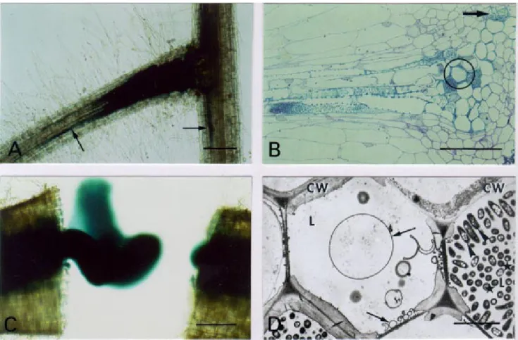
Fig. 3. Microscopy of tomato root infection and vascular colonization phenotype of the Ralstonia solanacearum prhJ mutant strain GMI1584. A–C,
Bacteria were localized and visualized by histochemical staining of the β-galactosidase activity in whole root. A, Intercellular infection along the vas-cular cylinder of secondary and primary roots (arrows) observed in a secondary root axil 6 days after inoculation. Bar: 200 µm. B, Bacterial invasion of several xylem vessels, including protoxylem (arrow), observed on a semithin section of a colonized secondary root axil 12 days after inoculation. Serial ultrastructural observation of circled zone is shown in D. Bar: 50 µm. C, Vascular colonization of the upper primary root is demonstrated by bacterial exudation from the vascular cylinder after roots were cut transversally 18 days after inoculation. Bar: 200 µm. D, TEM (transmission electron micros-copy) observation of xylem colonization reveals a weak development of vascular coating forming bubbles (arrows) in some noncolonized xylem vessels. Bacteria against the cell wall (CW) are oriented in a polar fashion in the invaded xylem vessel (arrowhead) while most bacteria in the vessel lumen (L) are transversally sectioned (stars). Bar: 5 µm.
a pathogenic strain is able to overcome plant defense mecha-nisms and pass through the endodermis to colonize the vas-cular system. Then, bacteria multiply extensively inside xylem vessels, resulting in the development of wilting symptoms that culminate in plant death (Vasse et al. 1995). We have found that, compared with this pathogenic phenotype, R.
solana-cearum mutant strains disrupted in prhJ, hrpG, or hrpB genes
have different capacities to develop in planta, to induce vas-cular defense reactions, and to invade the vasvas-cular system.
Observations of the tomato root infection process by the R.
solanacearum hrpB or hrcV mutant strains revealed that these
strains are both poorly infective and weakly invasive. They show limited multiplication in the intercellular spaces of the root cortex as well as in the vascular system of colonized plants. The similar phenotype of the hrpB (GMI1541) and the
hrcV (GMI1556) mutant strains is consistent with the fact that
the hrp secretion system is blocked in both strains, and indi-cates that functional hrp genes are needed for bacterial devel-opment in planta. This is in agreement with the results of Hirano et al. (1999), who report the role of the hrp system in growth of Pseudomonas syringae pv. syringae on host plants. In addition, similar vascular defense mechanisms were ob-served in plants infected by the hrpB or the hrcV mutant strains. The barrier exerted by the vascular defense reactions and the low bacterial multiplication in the intercellular spaces of the cortex could explain the poorly invasive phenotype of these two mutant strains, and also the reduced number of colonized plants at middle stem 18 dpi.
Two different defense reactions were observed in plants in-fected with the hrpB (GMI1541) and the hrcV (GMI1556)
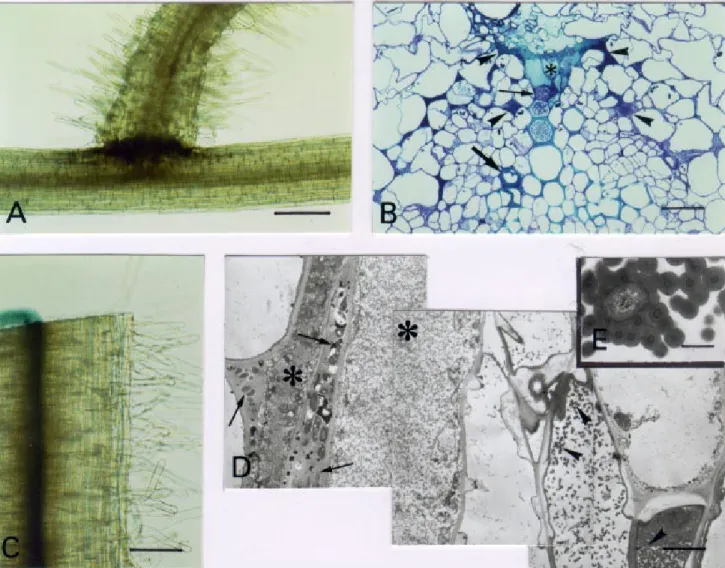
Fig. 4. Microscopy of tomato root infection and vascular colonization phenotypes of the Ralstonia solanacearum hrpB mutant strain GMI1541. A and C, Bacteria were localized and visualized by histochemical staining of the β-galactosidase activity in whole root. A, Bacterial infection 18 days after inoculation at secondary root axils is restricted to outer cortical cells of the primary root. Bar: 200 µm. B, Transverse, semithin section of a colonized root system 18 days after inoculation. Bacterial colonization only affects protoxylem vessels (thin arrow). Neighboring parenchyma cells appear degen-erated (asterisk) and infected intercellular spaces intensely stained (arrowheads). The thick dark blue reaction of a xylem vessel wall is characteristic of the metachromatic staining of vascular coating by toluidine blue (thick arrow). Bar: 25 µm. C, Transverse section of an invaded root 18 days after in-oculation shows a weak bacterial exudation from the vascular cylinder. Bar: 200 µm. D, Dead bacteria within infected intercellular spaces (arrows) adja-cent to degenerated vascular parenchyma cells (asterisks) are revealed by TEM (transmission electron microscopy) observations o n ultrathin longitudinal sections of invaded roots 18 days after inoculation. Abundant globular material associated with vascular coating (arrowheads) i s observed in adjacent xylem vessels. Bar: 2.5 µm. E, Detail of a bacterium embedded and degenerated into a globular and multilamellar material inside a non-invaded xylem vessel. Bar: 0.5 µm.
mutants. First, we observed the accumulation of suberin-like material characteristic of a vascular coating (Robb et al. 1991). Vascular coating is a nonspecific plant defense reaction apparently induced in response to intercellular infection, and has been described as a physical barrier to xylem colonization by vascular pathogenic fungi (Robb et al. 1987). This re-sponse has also been observed following abiotic stress (Robb et al. 1980). Vascular coating is observed in tomato roots
colonized by the wild-type strain GMI1485 and the prhJ mu-tant GMI1584, and appears to be amplified in plants infected with the hrpB and hrcV mutant strains. Localized cell wall-associated defense reactions have been described in plants in-fected with different hrp mutants of Xanthomonas campestris pv. vesicatoria (Brown et al. 1995). The reduced bacterial multiplication in planta of the hrpB and hrcV mutant strains could be either the cause or the consequence of their apparent
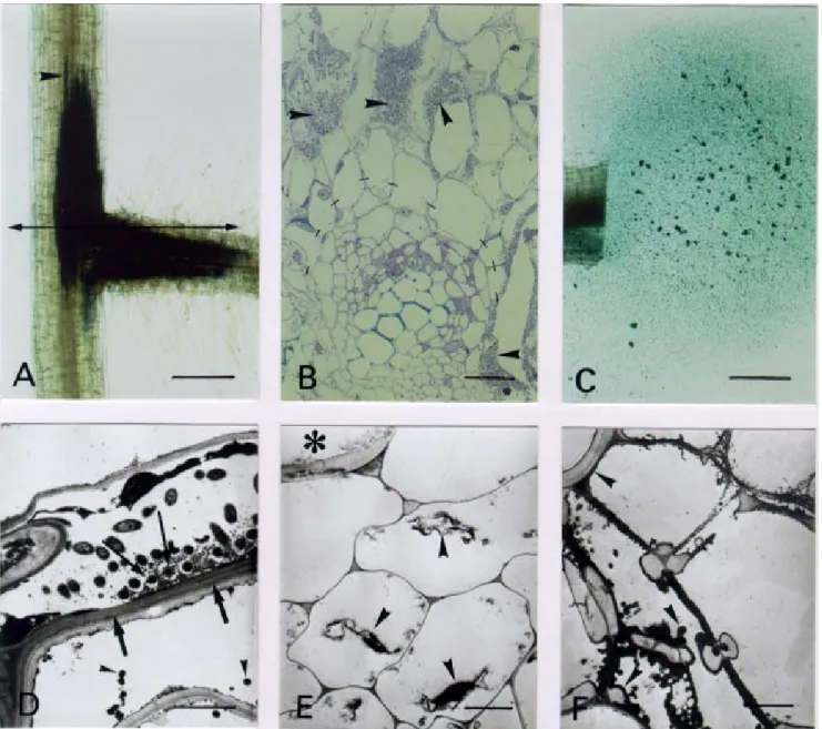
Fig. 5. Microscopy of tomato root infection and vascular colonization phenotypes of the Ralstonia solanacearum hrpG mutant strain GMI1583. A and C, Bacteria were localized and visualized by histochemical staining of the β-galactosidase activity in whole root. A, High level of infection at a secon-dary root axil 6 days after inoculation. Bacteria progressing along the vascular cylinder are indicated (arrowhead). Bar: 200 µm. B, Semithin sections at the infection site as indicated in A. Bacterial multiplication in intercellular spaces of the inner root cortex (arrowheads). Bacteria do not pass beyond the endodermis (dashes) and the vascular cylinder remains uninvaded. Lumens observed in some intercellular spaces correspond to bacteria drained away during embedding preparation. Bar: 25 µm. C, Bacterial exudation from a transversal root section is clearly not related to the vascular cylinder. Root material was taken 12 days after inoculation. Bar: 200 µm. D, Bacterial invasion of a destroyed inner cortical cell adjacent to the endodermis 18 days after inoculation. TEM (transmission electron microscopy) observations show an electronically dense and plasmolyzed cytoplasm of a degenerated cor-tical cell. The outer periclinal cell wall of the endodermis is reinforced by apposition of a multilamellar suberin-like material (thick arrows), and electron dense globules are visualized in the vacuole (arrowheads). Degenerated bacteria against the outer endodermis cell wall are embedded in a globular mate-rial (thin arrow). Bar: 2 µm. E, Vascular parenchyma cells, adjacent to an uninvaded xylem vessel (asterisk), are degenerated and present a plasmolyzed cytoplasm (arrowheads). Bar: 2 µm. F, Numerous electron dense and globular particles (arrowheads) forming vascular coating in xylem vessels at a vas-cular junction between a primary and a secondary root 18 days after inoculation. Bar: 2 µm.
inability to overcome this plant response. The second type of defense mechanism is the simultaneous degeneration of in-fecting bacteria and parenchyma cells adjacent to the colo-nized protoxylem pole. This is consistent with a VHR. In contrast to vascular coating, the VHR seems to be a plant de-fense reaction modulated by the function of bacterial hrp genes. This specific plant response has also been reported in the incompatible interaction between crucifers and X.
cam-pestris pv. camcam-pestris mutant strains affected in the hrpX
lo-cus (Kamoun et al. 1992). Moreover, it has been proposed that
hrpXc, the hrpB homologous gene in this bacterium, might
function to suppress defense responses in compatible hosts (Kamoun et al. 1992). These previous reports support our data suggesting a role for hrp genes in suppression of the VHR in-duced by the wild-type strain in the tomato root infection process. In this context, it is tempting to speculate that this suppressor effect might be exerted by proteins secreted or in-jected into plant cells through the hrp machinery.
In contrast to the hrpB or the hrcV mutants, the prhJ (GMI1584) and hrpG (GMI1583) mutant strains showed an optimal development in the early stages of the root infection process and later inside the vascular system. This was an un-expected result because the PrhJ and HrpG regulators act up-stream of HrpB in the same regulatory cascade (Fig. 6). Ge-netic evidence supports the fact that hrp genes can be activated in vitro through prhJ-independent pathways, for ex-ample, in minimal medium (Brito et al. 1999). In addition, it
has been reported that there is a residual level of hrp gene ex-pression in minimal medium in an hrpG mutant strain (Brito et al. 1999). If we hypothesize that signals activating hrp genes in minimal medium also exist and induce hrp genes in the plant, then these two previous observations could explain the phenotypes of the prhJ and hrpG mutants. Another expla-nation for the high multiplication of the hrpG mutant could be related to the presence of plasmolyzed cortical and xylem pa-renchyma cells in root systems infected with this mutant strain. Plasmolyzed cells could release nutrients into the apoplast that favor development of this mutant in planta.
Despite its highly infective phenotype, the hrpG mutant is remarkably impaired in its ability to pass through the endo-dermis. This result suggests that additional functions required for vascular invasion could be specifically controlled by HrpG, but not by HrpB. The phenotype of GMI1583 (hrpG::Ω) slightly resembles those of two genetically defined
exo mutants of R. solanacearum affected in acidic
exopoly-saccharide production. These exo mutants showed a reduced infection ability and were unable to colonize the vascular system of hydroponically grown tomato plants (Araud-Razou et al. 1998). Production of exopolysaccharides and hrp gene expression in R. solanacearum are controlled by independent and complex regulatory networks (Huang et al. 1995; Schell 1996). This indicates that the endodermal crossing is probably a multifactorial process requiring several bacterial functions. On the other hand, the reduced vascular colonization ability of the hrpG mutant might be due to a deficient production of certain virulence factors involved in plant cell wall degrada-tion. Enzymatic assays indicate that polygalacturonase and carboxymethylase production is not significantly impaired in the hrpG mutant strain (B. Brito, unpublished results). How-ever, as R. solanacearum possesses three polygalacturonases and additional degradative enzymes (Allen et al. 1997), we cannot exclude a possible interconnection between the hrp system and virulence factor production in R. solanacearum and this possibility is currently being investigated.
In conclusion, this study provides evidence that hrp genes are involved in the early steps of the interaction between R.
solanacearum and tomato roots, acting at different stages of
the infection process. The HrpB and HrcV products seem to facilitate bacterial development in planta and to suppress the VHR. In contrast, our results indicate that HrpG is required for bacterial transit through the endodermis. This result opens new perspectives for the identification of additional HrpG-regulated genes involved in endodermal transit.
MATERIALS AND METHODS
Bacterial strains and culture conditions.
Strains GMI1556, GMI1541, GMI1583, and GMI1584 are derivatives of R. solanacearum GMI1485 (Arlat et al. 1992). GMI1556 and GMI1541 carry an Ω interposon (Prentki and Krisch 1984) in the hrcV and hrpB genes, respectively (Genin et al. 1992; Etchebar et al. 1998). GMI1583 and GMI1584 are disrupted in the hrpG and prhJ genes, respectively (this work). For plant inoculations, R. solanacearum strains were grown in B medium for 20 h at 28°C (Boucher et al. 1985). DH5α Escherichia coli strain was cultivated in Luria-Bertani medium (Sambrook et al. 1989). When required, antibiotics were added to the media at the following final concentrations
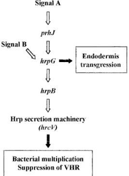
Fig. 6. Schematic representation of the hrp gene regulatory cascade and
the specific hrp gene functions required in the interaction between to-mato roots and Ralstonia solanacearum. The PrhJ activator induces
hrpG gene expression in response to signals (Signal A) derived from
plant cell recognition. In turn, HrpG regulates hrpB and the remaining
hrp genes. Additionally, signals (Signal B) from the bacterial nutritional
status can also activate hrp genes through HrpG (Brito et al. 1999). The function of HrpB and HrcV (and consequently the Hrp secretion system) is to favor bacterial development in planta and to suppress the vascular hypersensitive response (VHR). In addition, HrpG appears to control other bacterial determinants required for endodermal crossing.
(mg · l–1): ampicillin (Ap) 100; spectinomycin (Sp) 25 (E.
coli), 40 (R. solanacearum); and kanamycin (km) 25 (E. coli),
50 (R. solanacearum).
Construction of R. solanacearum hrp mutants.
Generation of GMI1583 and GMI1584 was achieved by transformation of strain GMI1485 with DNA from plasmids pBBL102Ω and pBBL103Ω (Brito et al. 1999). pBBL103Ω is a pBluescriptKS containing a 1.8-kb EcoRI-HindIII DNA fragment with an Ω-Sp insertion in the SalI restriction site of the hrpG gene. Plasmid pBBL102Ω carries a 3.3-kb
EcoRI-KpnI fragment with an Ω-Sp cassette in the XhoI re-striction site of the prhJ gene. DNA (5 µg) from plasmids pBBL102Ω and pBBL103Ω was linearized with XbaI and used for transformation of strain GMI1485. Transformation was performed as previously described (Boucher et al. 1985). Recombinant clones were selected in Bacto-Agar and glucose (BG) medium for resistance to spectinomycin and kanamycin. Correct marker exchange was confirmed by Southern blot experiments with total genomic DNA from recombinant strains hybridized with an 1.8-kb
EcoRI-HindIII probe encompassing the hrpG and prhJ genes (Brito
et al. 1999). Standard methods were used for plasmid ex-traction, restriction-fragment analysis, and DNA cloning (Sambrook et al. 1989). Double mutants were checked for HR inducing ability on tobacco leaves and disease develop-ment on tomato plants (see below, Pathogenicity tests). Phe-notypes were similar to those of the corresponding single mutants (Genin et al. 1992; Brito et al. 1999).
β-Galactosidase assays.
β-Galactosidase activities were assayed in bacterial cultures grown in minimal medium at 28°C for 18 h as previously (Arlat et al. 1992). Activities were also measured upon co-culture with Arabidopsis thaliana At-202 cell suspensions following the procedure described by Marenda et al. (1998).
Plant material, growth conditions, and root inoculations.
Lycopersicon esculentum cv. Supermarmande plants were
grown in test tubes under axenic conditions as well as in soil. For growth in hydroponic conditions, seeds were surface ster-ilized and germinated as previously described (Boucher et al. 1985). Germinated seeds were then transferred to large, ster-ile, test tubes and grown under previously described condi-tions (Vasse et al. 1995). At the first-leaf stage (2 weeks after pregermination), tomato seedlings were inoculated by adding 5 × 10⁸ CFU per tube to the nutrient solution.
Tomato plants grown in soil were inoculated at the second-leaf stage (3 weeks after transplanting) with 50 ml per pot of a bacterial suspension adjusted to 108 CFU ml–1.
Pathogenicity tests.
The HR-inducing ability of R. solanacearum strains was tested by infiltration of bacterial suspensions at 108 CFU ml–1
into tobacco leaves (cv. Bottom Special) as previously de-scribed (Boucher et al. 1985). Wilting development on whole tomato plants inoculated with R. solanacearum strains was analyzed for 21 days and scored with an arbitrary disease in-dex ranging from 0 (no leaves wilted) to 4 (75 to 100% leaves wilted) (Arlat and Boucher 1991). Two independent tests, with 20 tomato plants per experiment, were carried out.
Bacterial colonization in tomato plants at 10 or 18 dpi was determined as previously described (Etchebar et al. 1998). Middle stem level corresponds to 10 cm above cotyledons af-ter 10 dpi and 20 cm afaf-ter 18 days afaf-ter inoculation. Briefly, lateral roots and leaves of inoculated tomato plants were dis-carded. Stems were then surface sterilized and cut in 2-cm-long segments. Each segment was imprinted on a selective B agar medium supplemented with kanamycin or kanamycin and spectinomycin as required. Plates were incubated at 28°C for 2 days. Scoring represents the percentage of infected stem segments found at a defined stem level with the population studied.
For measuring bacterial numbers in colonized stem segments, the 2-cm stem segments corresponding to collar, epicotyl, and middle stem levels were incubated overnight at 10°C in 40 ml of sterile, distilled water to allow diffusion of infecting bacteria out of the plant tissues. Bacterial suspensions were then serially di-luted and plated with a Spiral Plate Maker (Interscience, Saint Nom la Breteche, France) onto selective agar medium contain-ing kanamycin or kanamycin and spectinomycin. Dry weight from stem segments was also measured. Assays were per-formed with a group of 20 plants for each bacterial strain and bacterial densities were expressed as a log CFU per gram of dry weight–1 of colonized stem tissue.
Microscopic methods.
Microscopic studies were performed on whole root systems from seedlings processed 6, 12, and 18 days after bacterial inoculation and on root sections as previously described (Vasse et al. 1995; Araud-Razou et al. 1998). To localize bac-teria, whole root systems were fixed and histochemically stained to specifically reveal the bacterial β-galactosidase ac-tivity associated with the constitutively expressed fusion 1485. For cytological observations, semithin and ultrathin sections were obtained from the whole root systems already stained. To do this, selected segments of roots were fixed again, dehydrated, embedded in epoxy resin, and sectioned with an ultramicrotome (Ultracut E; Reichert-Jung, Nussloch, Germany). Semithin sections (1 µm thick) were stained with a mixture of toluidine blue and methylene blue in an aqueous solution of sodium borate. Ultrathin sections (70 to 90 nm thick) were contrasted with uranyl acetate 2.5% (vol/vol) in 50% ethanol then with lead citrate. Whole root and semithin sections were observed by bright-field microscopy with a light microscope (Zeiss; Axiophot, Jena, Germany), while ultra-structural observations were performed with a transmission electron microscope (EM 600 Hitachi) operating at 75 kV.
ACKNOWLEDGMENTS
We thank Dominique Douilhac and Jean Luc Pariente for material, media, and plant preparation. We also thank Patrick Barberis for the HR tests. We are in debt to Clare Gough for critical reading of the manu-script. This work was founded by projects BIO4-CT-97-2244 from the European Commission and AIP-188 Microbiologie from the Institut National de la Recherche Agronomique. B. B. was the recipient of a Marie Curie TMR postdoctoral grant from the European Commission.
LITERATURE CITED
Allen, C., Gay, J., and Simon-Buela, L. 1997. A regulatory locus, pehSR, controls polygalacturonase production and other virulence functions
in Ralstonia solanacearum. Mol. Plant-Microbe Interact. 10:1054-1064.
Araud-Razou, I., Vasse, J., Montrozier, H., Etchebar, C., and Trigalet, A. 1998. Detection and visualization of the major acidic exopolysaccha-ride of Ralstonia solanacearum and its role in tomato root infection and vascular colonization. Eur. J. Plant Pathol. 104:795-809. Arlat, M., and Boucher, C. 1991. Identification of a dsp DNA region
controlling aggressiveness of Pseudomonas solanacearum. Mol. Plant-Microbe Interact. 4:211-213.
Arlat, M., Gough, C. L., Zischek, C., Barberis, P. A., Trigalet, A., and Boucher, C. A. 1992. Transcriptional organization and expression of the large hrp gene cluster of Pseudomonas solanacearum. Mol. Plant-Microbe Interact. 5:187-193.
Boucher, C., Barberis, P., Trigalet, A., and Demery, D. 1985. Transposon mutagenesis of Pseudomonas solanacearum: Isolation of Tn5-induced avirulent mutants. J. Gen. Microbiol. 131:2449-2457. Brito, B., Marenda, M., Barberis, P., Boucher, C., and Genin, S. 1999.
prhJ and hrpG two new components of the plant signal-dependent
regulatory cascade controlled by PrhA in Ralstonia solanacearum. Mol. Microbiol. 31:237-252.
Brown, I., Mansfield, J., and Bonas, U. 1995. hrp genes in Xanthomonas
campestris pv. vesicatoria determine ability to suppress papilla
depo-sition in pepper mesophyll cells. Mol. Plant-Microbe Interact. 8:825-836.
Etchebar, C., Trigalet-Demery, D., van Gijsegem, F., Vasse, J., and Tri-galet, A. 1998. Xylem colonization by an HrcV– mutant of Ralstonia solanacearum is a key factor for the efficient biological control of
tomato bacterial wilt. Mol. Plant-Microbe Interact. 11:869-877. Frey, P., Prior, P., Marie, C., Koutoujansky, A., Trigalet-Demery, D., and
Trigalet, A. 1994. Hrp- mutants of Pseudomonas solanacearum as potential biocontrol agents of tomato bacterial wilt. Appl. Environ. Microbiol. 60:3175-3181.
Genin, S., Gough, C. L., Zischek, C., and Boucher, C. 1992. Evidence that the hrpB gene encodes a positive regulator of pathogenicity genes from Pseudomonas solanacearum. Mol. Microbiol. 6:3065-3076. Hayward, H. C. 1991. Biology and epidemiology of bacterial wilt
caused by Pseudomonas solanacearum. Annu. Rev. Phytopathol. 29: 65-67.
Hirano, S. S., Charkowski, A. O., Collmer, A., Willis, D. K., and Upper, C. D. 1999. Role of the Hrp type III protein secretion system in growth of Pseudomonas syringae pv. syringae B728a on host plants in the field. Proc. Natl. Acad. Sci. USA 96:9851-9856.
Huang, J., Carney, B. F., Denny, T, Weissinger, A. K., and Schell, M. A. 1995. A complex network regulates expression of eps and other viru-lence genes of Pseudomonas solanacearum. J. Bacteriol. 177:1259-1267.
Kamoun, S., Kamdar, H. V., Tola, E., and Kado, C. I. 1992. Incompatible
interactions between crucifers and Xanthomonas campestris involve a vascular hypersensitive response: Role of the hrpX locus. Mol. Plant-Microbe Interact. 5:22-33.
Lindgren, P. B. 1997. The role of hrp genes during plant-bacterial inter-actions. Annu. Rev. Phytopathol. 35:129-152.
Marenda, M., Brito, B., Callard, D., Genin, S., Barberis, P., Boucher, C., and Arlat, M. 1998. PrhA controls a novel regulatory pathway re-quired for the specific induction of Ralstonia solanacearum hrp genes in the presence of plant cells. Mol. Microbiol. 27:437-453.
Mudgett, M. B., and Staskawicz, B. J. 1998. Protein signalling via type III secretion pathways in phytopathogenic bacteria. Curr. Opin. Mi-crobiol. 1:109-114.
Prentki, P., and Krisch, H. M. 1984. In vitro insertional mutagenesis with a selectable DNA fragment. Gene 29:303-313.
Robb, J., Busch, L., and Rauser, W. E. 1980. Zinc toxicity and xylem vessel wall alterations in white beans. Ann. Bot. 46:43-50.
Robb, J., Lee, J. S., Mohan, R. and Kolattukudy, P. E. 1991. Chemical characterization of stress-induced vascular coating in tomato. Plant Physiol. 97:528-538.
Robb, J., Powell, D. A., and Street, P. F. S. 1987. Vascular coating: A barrier to colonization by the pathogen Verticilium wilt of tomato. Can. J. Bot. 67:600-607.
Saile, E., McGarvey, J. A., Schell, M. A. and Denny, T. P. 1997. Role of extracellular polysaccharide and endoglucanase in root invasion and colonization of tomato plants by Ralstonia solanacearum. Phytopa-thology. 87:1264-1271.
Sambrook, J., Fritsch, E. F., and Maniatis, T. A. 1989. Molecular Clon-ing: A Laboratory Manual. 2nd ed. Cold Spring Harbor Laboratory, Cold Spring Harbor, NY.
Schell, M. A. 1996. To be or not be: How Pseudomonas solanacearum decides whether or not to express virulence genes. Eur. J. Plant Pa-thol. 102:459-469.
van Gijsegem, F., Gough, C. L., Zischek, C., Niqueux, E., Arlat, M., Genin, S., Barberis, P., German, S., Castello, P., and Boucher, C. 1995. The hrp locus of Pseudomonas solanacearum, which controls the production of type III secretion system, encodes eight proteins re-lated to components of the bacterial flagellar biogenesis complex. Mol. Microbiol. 15:1095-1114.
Vasse, J., Frey, P., and Trigalet, A. 1995. Microscopic studies of inter-cellular infection and protoxylem invasion of tomato roots by
Pseu-domonas solanacearum. Mol. Plant-Microbe Interact. 8:241-251.
Yabuuchi, E., Kosako, Y., Yano, Y., Hotta, H., and Nishiuchi, Y. 1995. Transfer of two Burkholderia and an Alcaligenes species to Ralstonia gen. Nov.: Proposal of Ralstonia pickettii (Ralston, Palleroni and Doudoroff 1973) comb. nov., and Ralstonia solanacearum (Smith 1896) comb. Nov. and Ralstonia eutropha (Davis 1969) comb. nov. Microbiol. Immunol. 39:897-904.