UNIVERSITÉ DE MONTRÉAL
STRESS RELAXATION OF GROWTH PLATE TISSUE UNDER UNIFORM
COMPRESSIVE LOAD: RELATIONSHIP BETWEEN MECHANICAL
RESPONSE AND EXTRACELLULAR MATRIX BIO-COMPOSITION AND
STRUCTURE
SAMIRA AMINI
INSTITUT DE GÉNIE BIOMÉDICAL ÉCOLE POLYTECHNIQUE DE MONTRÉAL
THÈSE PRÉSENTÉE EN VUE DE L’OBTENTION DU DIPLÔME DE PHILOSOPHIAE DOCTOR
(GÉNIE BIOMÉDICAL) DÉCEMBRE 2011
UNIVERSITÉ DE MONTRÉAL
ÉCOLE POLYTECHNIQUE DE MONTRÉAL
Cette thèse intitulée:
STRESS RELAXATION OF GROWTH PLATE TISSUE UNDER UNIFORM
COMPRESSIVE LOAD: RELATIONSHIP BETWEEN MECHANICAL
RESPONSE AND EXTRACELLULAR MATRIX BIO-COMPOSITION AND
STRUCTURE
présentée par : AMINI Samira
en vue de l’obtention du diplôme de : Philosophiae Doctor a été dûment acceptée par le jury d’examen constitué de : M. AUBIN Carl-Éric, Ph.D., président
Mme VILLEMURE Isabelle, Ph.D, membre et directrice de recherche Mme HOEMANN Caroline , Ph.D., membre et codirectrice de recherche M. BUSCHMANN Michael, Ph.D, membre
DEDICATION
To Parham, for his unconditional love,
ACKNOWLEGEMENTS
I would like to convey my sincere gratitude to those who helped me to complete this study. First and foremost, I am genuinely grateful to my supervisor, Professor Isabelle Villemure, for her friendly guidance, continuous and substantial support, precious advice, scientific rigor and in particular her total involvement in the monitoring of this project. It was a great pleasure for me to work under her supervision. She was an endless source of encouragement to me throughout this period. Her exceptional high standards inspired me to improve my skills throughout my research at École Polytechnique de Montréal. I am deeply indebted to her for her availability at any time for consultations and discussions on my research.
I would like to thank my co-supervisor, Professor Caroline D Hoemann, for her friendly quidance, continuous support, wealth of advice, and insightful guidance on the biochemical aspect of my project. Her passion for science inspired me throughout my doctoral studies and it was an honor to work with her.
I also wish to thank Dr. Martin Lévesque, collaborator and associate professor of mechanical engineering at the Ecole Polytechnique de Montreal, for his involvement in providing advice on my third publication.
I take great pleasure in acknowledging all present and former members of the Laboratory of Pediatric Mechanobiology (LMP) of Professor Isabelle Villemure and of course my good friends. Thank you to Annie Bélisle-L’Anglais, Daniel Veilleux, Maria Laitenberger, Irene Londono, Anne-Laure Ménard, Farhad Mortazavi, Kim Sergerie, Barthélémy Valteau, Yaroslav Wakula, and Roxanne Wosu, who created lots of memorable moments for me during these years and helped me to integrate into the group. I would also like to extend my thanks to Irene Londono for being a great help during hard times and Anne-Laure Ménard for the editorial revision of the French Résumé.
I like to extend my thanks to the technical and administrative staff of the Applied mechanics section of Mechanical Engineering Department. Special thanks to Mr. Thierry Lafrance and Mr. Benedict Besner for their marvellous work on my experimental set-up.
I would like to thank present and former members of Biomaterials and Cartilage Laboratory of Professor Caroline Hoemann. I take great pleasure to specifically thank Adele Changoor,
Gaoping Chen, Anik Chevrier, Viorica Lascau-Coman, and Jun Sun for their help in the biochemical aspect of my project.
I made many friends in Montreal, and it gives me great pleasure to acknowledge the support and love I received from them. They were with me in sad and happy moments and made all these years memorable for me. I refrain from acknowledging them individually because that would take many pages, and for fear that I may inadvertently miss some of them. However, I can not help mentioning the name of my best friends, Mahnaz, Ravid, Azin, Naser, Arezou, Alireza, Maryam, Farhad, Sara, and Ali, who were with me during the most tragic moments of my life when I lost my grand father and my father-in-law.
I owe special gratitude to my mother, Maman Parvin, and my father, Baba Hossein, for their prayers, unconditional love, support, and encouragement throughout my entire life. I am deeply indebted to them for every success I have ever achieved. I would also thank my brother, Ali, for his support, love, and cheerfulness. He provided a great deal of encouragement to me during my studies and I am grateful to him.
I would like to send a heartfelt acknowledgement to my Family-in-law for the support and love I received from them. I can’t help mentioning my Father-in-law who left us too early to join the invisible. He was an endless source of encouragement and love during my studies. May his soul rest in peace.
I would also like to acknowledge Canada, the province of Québec, and École Polytechnique de Montréal for giving me the opportunity to continue my studies. It is with great pleasure that I express my thanks to Montrealers for their kindness.
I would like to convey my gratitude to Professor Carl-Eric Aubin, Professor Michael Buschmann, and Professor Eve Langelier for accepting to participate to my Ph.D. Jury.
This project was funded by the Canada Research Chair in Mechanobiology of the Pediatric Musculoskeletal System (I.V.), the Board of Natural Sciences and Engineering Research Council of Canada (NSERC), and the MENTOR program of the Canadian Institute of Health Research (CIHR, S.A.).
Last, but most important, I would like to express my special gratitude to my lovely husband, Parham Eslami Nejad, for his patience, love, encouragment and support. He was an endless
source of inspiration to me for achieving my goals. He was genuinely caring during these years and I am deeply indebted to him for every single second of our life.
RÉSUMÉ
La modulation mécanique de la croissance osseuse a des implications dans la progression des déformations musculo-squelettiques telles que la scoliose idiopathique adolescente. Ce processus présente aussi un intérêt croissant pour le développement et l'amélioration des approches minimalement invasives de correction de ces déformations musculo-squelettiques en modulant localement la croissance osseuse, tout en préservant la croissance normale ainsi que les fonctions et mobilités segmentaires. La croissance longitudinale des os longs et des vertèbres s’effectue au droit des plaques de croissance, qui sont divisées en trois zones histologiques distinctes (réserve, proliférative et hypertrophique). La matrice extracellulaire de la plaque de croissance est principalement composée d'eau et de protéoglycanes imbriquées dans des fibrilles de collagène de type II, qui sont considérées comme des composants déterminants des propriétés biomécaniques des tissus cartilagineux. Des études antérieures ont étudié le comportement biomécanique des plaques de croissance, mais aucune étude n’a à ce jour analysé le comportement mécanique en compression de la plaque de croissance et de ses zones in situ à l'égard de sa composition biochimique et de l'organisation de ses fibres de collagène. L'objectif principal de cette étude était de déterminer les caractéristiques histomorphologiques et le comportement mécanique des plaques de croissance aux niveaux cellulaire et tissulaire, d'évaluer la composition biochimique et l'orientation des fibres de collagène de la plaque de croissance dans ses trois zones distinctes, puis d'établir des associations entre le comportement mécanique et la composition biochimique de la plaque de croissance.
Cinq groupes d’explants de plaques de croissance provenant de porcs âgés de quatre semaines ont été utilisés dans ce projet. Le premier groupe d’explants (N=12) a été utilisé pour caractériser l’histomorphologie 3D de la plaque de croissance in situ aux niveaux cellulaire (volume, aire surfacique, sphéricité, et rayons mineur/majeur) et tissulaire (ratio cellule/matrice extracellulaire), en utilisant un marquage fluorescent du cytoplasme cellulaire (Calcéine AM) couplé à la reconstruction 3D des coupes sériées d’images numériques confocales (logiciel IMARIS). Afin de caractériser le comportement mécanique sous compression de la plaque de croissance et de ses cellules constitutives en 3D, un second groupe d’explants de cartilage de croissance (N=6), dont les cytoplasmes cellulaires ont été marqués à la Calcéine AM, a été testé sous compression semi-confinée en relaxation de contraintes à l’aide d’un montage combinant un appareil de
micro-chargement fixé sur un microscope confocal inversé. Ces explants ont été soumis à une déformation totale de 15% (5% pré-chargement et 10% de déformation) à un taux de 1.7x10⁻³ s-1 jusqu’à l’obtention de l’équilibre, suivant un critère de relaxation de 8E-6 N/sec. Des coupes sériées d’images numériques des cytoplasmes cellulaires marqués ont été acquises par microscopie confocale avant chargement et après relaxation du tissu. Des reconstructions 3D des chondrocytes dans les conditions pré- et post-chargement ont été complétées à partir d’images prises séparément dans les trois zones des plaques de croissance. Différents paramètres morphométriques au niveau cellulaire (volume, aire surfacique, sphéricité et rayons mineur/majeur) et au niveau tissulaire (ratio cellule/matrice extracellulaire) ont été évalués (logiciel IMARIS) puis comparés à l’aide de tests statistiques pour les trois zones de la plaque de croissance. Afin de caractériser le comportement mécanique sous compression de la plaque de croissance au niveau tissulaire, un troisième groupe d’explants de cartilage de croissance (N=7), dont les noyaux cellulaires ont été marqués au Syto-17, a été testé sous compression semi-confinée en relaxation de contraintes à l’aide du même montage et des mêmes paramètres de chargement. Des images numériques des noyaux des chondrocytes marqués ont été acquises par microscopie confocale avant chargement et après relaxation du tissu, puis les champs de déformation 2D ont été déterminés à l’aide d’un algorithme de corrélation d'images appliqué à des paires d'images d’un même explant. Au niveau biochimique, un quatrième groupe d’explants (N=7) du même modèle animal a été analysé pour obtenir leurs contenus en eau, en glycosaminoglycanes sulfatés (S-GAG) et en hydroxyproline (OH-Pro), comme une mesure de leurs contenus en collagène, dans les trois zones histologiques de la plaque de croissance. La teneur en eau a été déterminée par pesée des tissus avant et après lyophilisation. Les contenus en collagène et en GAG ont été quantifiés à l'aide des essais d'hydroxyproline et de bleu de diméthylméthylène (DMMB), respectivement. Finalement, dans un cinquième groupe d’explants (N=7) du même modèle animal, l’organisation des fibres de collagène a été évaluée dans les trois zones histologiques de la plaque de croissance en utilisant la microscopie en lumière polarisée (PLM).
Les caractéristiques histomorphologiques de la plaque de croissance aux niveaux tissulaire et cellulaire sont hétérogènes et dépendent de la zone de la plaque de croissance. Des variations significatives de la morphologie des chondrocytes ont été observées entre les différentes zones histologiques. Les volumes et les aires surfaciques maximaux des chondrocytes ont été trouvés
dans la zone hypertrophique par rapport à ceux des zones de réserve et proliférative. Le volume et l’aire surfacique des chondrocytes ont augmenté d'environ cinq et trois fois respectivement en se rapprochant de la jonction chondro-osseuse à partir de la zone de réserve. Les chondrocytes de la zone proliférative ont été les cellules de formes les plus discoïdes entre les trois différentes zones histologiques. Des différences significatives ont aussi été observées concernant le ratio cellule/matrice extracellulaire entre les trois zones. Le minimum et le maximum des ratios cellule/matrice extracellulaire ont été identifiés dans les zones de réserve et proliférative, respectivement.
Les analyses morphologiques tridimensionnelles aux niveaux tissulaire et cellulaire sous compression, basées sur des explants marqués à la Calcéine AM, des coupes sériées d’images numériques des cytoplasmes cellulaires et des reconstructions 3D des chondrocytes dans les conditions pré- et post-chargement, démontrent que la plaque de croissance subit des déformations non uniformes sous chargement tant au niveau tissulaire qu’au niveau cellulaire, et également au niveau du ratio cellule/matrice extracellulaire. De plus grandes déformations cellulaires (changement de volume cellulaire normalisé au volume initial) ont été trouvées dans les zones proliférative et hypertrophique. Inversement, les plus faibles déformations se sont développées dans la zone de réserve. Suite à la compression, le ratio cellule/matrice extracellulaire a diminué dans les zones de réserve et hypertrophique alors qu’il a augmenté dans la zone proliférative.
L’analyse biomécanique au niveau tissulaire, basée sur des explants marqués au Syto-17, l’imagerie 2D et la corrélation numériques d'images (DIC), a démontré un comportement mécanique hétérogène dépendamment de la zone de la plaque de croissance considérée. Des déformations tissulaires axiales supérieures se sont développées dans la zone proliférative par rapport aux deux autres zones histologiques. Par ailleurs, des déformations transverses plus élevées ont été principalement trouvées dans les zones proliférative et hypertrophique par rapport à la zone de réserve. Enfin, les déformations transverses et axiales les plus faibles et les plus homogènes se sont principalement développées dans la zone de réserve.
Les analyses biochimiques ont indiqué que la zone de réserve a un contenu en collagène plus élevé par rapport aux zones proliférative et hypertrophique. Cependant, les contenus en eau et en GAG ont été évalués identiques dans les trois zones histologiques.
Les caractérisations en microscopie polarisée ont montré que les fibres de collagène de la zone de réserve sont principalement orientées parallèlement à l’interface entre la plaque de croissance et l’os, soit perpendiculairement à l'axe longitudinal des os. Toutefois, certaines fibres orientées longitudinalement ont également été observées dans cette zone. A l'inverse, les fibres de collagène ont été trouvées alignées presqu'exclusivement selon l'axe longitudinal de l'os, soit dans la direction de croissance, pour les zones proliférative et hypertrophique.
Au niveau histomorphologique, les hétérogénéités marquées de la taille des cellules à travers les différentes zones histologiques de la plaque de croissance sont cohérentes avec celles des études antérieures sur la morphologie des chondrocytes à l'aide de l'histologie conventionnelle et des méthodes stéréologiques. Les chondrocytes subissent les changements de forme tout en progressant de la zone de réserve à la jonction chondro-osseuse. Les chondrocytes hypertrophiques et réserves sont ronds par rapport aux chondrocytes aplatis de la zone proliférative, tel que confirmé par les valeurs significativement plus faibles de sphéricité des chondrocytes prolifératifs par rapport aux zones de réserve et hypertrophique. La morphologie tissulaire et cellulaire peut avoir des contributions notables sur le comportement de la plaque de croissance durant le processus de croissance. La capacité d’obtenir la morphométrie cellulaire in situ et de surveiller les changements dans la direction de la croissance pourrait améliorer notre compréhension des mécanismes par lesquels la croissance anormale est déclenchée.
Deuxièmement, les chondrocytes et leur matrice extracellulaire environnante subissent des changements morphologiques significatifs avec la compression, mais le niveau de déformation dépend de la zone histologique. Ces déformations variables sont probablement liées aux propriétés mécaniques hétérogènes des trois zones, où la zone de réserve a été trouvé plus rigide que les zones proliférative et hypertrophique dans les directions parallèle et perpendiculaire à l'axe de compression. Dans notre étude, les chondrocytes hypertrophiques ont montré les plus grandes déformations parmi les trois zones histologiques; ils pourraient ainsi être davantage susceptibles de déclencher des messages biologiques altérés, via un étirement plus important de leur membrane cellulaire, ce qui modulerait l'activité des ARN-messagers et pourrait éventuellement provoquer une décélération de croissance sous compression mécanique.
Le contenu en collagène de la plaque de croissance et l'orientation de ses fibres de collagène sont également non uniformes à travers l'épaisseur de la plaque de croissance. La dispersion aléatoire
des chondrocytes dans la zone de réserve et la disposition en colonnes des chondrocytes dans les zones proliférative et hypertrophique sont en corrélation avec l'orientation des fibres de collagène observées dans ces zones. En outre, nos données corroborent les données existantes sur le contenu biochimique de la plaque de croissance.
Enfin, le comportement biomécanique de la plaque de croissance sous compression est lié à son contenu en collagène et à l'organisation des fibres de collagène. La zone de réserve, moins sensible aux déformations comparé aux zones proliférative et hypertrophique, contient le contenu maximum en collagène avec des fibres alignées perpendiculairement à la direction de croissance. A l'inverse, les zones proliférative et hypertrophique, où un contenu en collagène inférieur et des fibres de collagène organisées longitudinalement ont été trouvés, s’avèrent plus sensibles aux déformations, tant aux niveaux cellulaire que tissulaire. La zone de réserve, plus rigide, pourrait jouer un rôle plus significatif de support mécanique comparativement aux zones proliférative et hypertrophique, qui seraient plus susceptibles d’être impliquées dans le processus de modulation de la croissance. Ces données s'ajoutent à notre compréhension de la relation entre les forces de compression subies par les chondrocytes de la plaque de croissance et de leur environnement extracellulaire.
Les principales limites de ce projet de recherche comprennent l'utilisation d'un modèle animal unique sans tenir compte des variations pouvant exister entre sites osseux et avec les stades de développement. La petite taille de l’échantillonnage ainsi que des analyses statistiques limitées pour établir des relations entre le comportement mécanique et les caractéristiques structurelles constituent aussi des limites de la présente étude. En contrepartie, cette étude est la première à offrir des informations importantes et complémentaires sur le comportement mécanique et les caractéristiques morphologiques et structurelles de la plaque de croissance ainsi que sur leurs relations pour un modèle animal avec un taux de croissance plus faible qui s'apparente davantage à celui des humains.
Les trois hypothèses de cette étude stipulaient que: 1) les trois zones de la plaque de croissance présentent différentes caractéristiques histomorphologiques, 2) les déformations cellulaires et les champs de déformation au niveau tissulaire sous compression sont non uniformes dans les trois zones de la plaque de croissance, et 3) le comportement biomécanique de la plaque de croissance en compression est relié à la composition biochimique de sa matrice extracellulaire (le contenu en
GAG et en collagène), sa teneur en eau ainsi que l’organisation de ses fibres de collagène type II dans les trois zones de la plaque de croissance. Basé sur les résultats obtenus, ces hypothèses sont vérifiées pour 1) et 2) et en partie confirmées pour 3).
En conclusion, les zones histologiques les plus activement impliquées dans la croissance longitudinale osseuse (proliférative et hypertrophique) seraient plus sensibles aux contraintes de compression aux niveaux cellulaire et tissulaire dû à leurs caractéristiques histomorphologiques et structurelles, et donc davantage susceptibles d’être impliquées dans la progression des déformations musculo-squelettiques infantile et juvénile. Une connaissance combinée de la mécanique et mécanobiologie de la plaque de croissance est essentielle afin de mieux comprendre les mécanismes par lesquels la croissance anormale est déclenchée et, à plus long terme, afin d'améliorer les approches de traitement minimalement invasive des malformations squelettiques progressives, qui exploitent directement le processus de modulation de croissance pour corriger ces déformations.
ABSTRACT
Mechanical loading has key implications in the progression of infantile and juvenile musculoskeletal deformities. Furthermore, the mechanical modulation of growth is of growing interest in the development and improvement of minimally invasive approaches that aim at modulating local bone growth while preserving the natural growth and functions of bone and bone segments. Longitudinal growth of long bones and vertebrae occurs in growth plates, which are divided into three distinct histological zones (reserve, proliferative and hypertrophic). Growth plate extracellular matrix is composed of water, large aggregating proteoglycans embedded within type II collagen fibrils, which are believed to be a critical determinant of tissue biomechanical competence. Previous studies have investigated the biomechanical behaviour of growth plates but no study up to date has comprehensively analyzed the zonal growth plate compressive mechanical behaviour in situ with respect to its biochemical composition and collagen fiber organization. The main objective of this study was to characterize the histomorphological characteristics and mechanical behaviour of growth plates at both cellular and tissue levels and to evaluate the biochemical composition and collagen fiber orientation of growth plate tissue in the three functionally distinct zones and to further establish associations between zonal mechanical behavior and biochemical composition of the growth plate.
Five groups of growth plate explants from 4-week old swine were used in this project.The first group of explants (N=12) was used to characterize the 3D zonal histomorphology of in situ growth plate at the cellular (volume, surface area, spherecity, minor/major radii) and tissue (cell/matrix volume ratio) levels using fluorescent labeling of cell cytoplasm (Calcein AM) coupled with 3D reconstruction of serial confocal sections (IMARIS software). In order to characterize the three-dimensional compressive mechanical behaviour of the growth plate tissue and its constitutive cells, a second group of growth plate explants (N = 6), labeled with Calcein AM for cell cytoplasm, were tested in semi-confined compression under stress relaxation using a loading apparatus mounted on the stage of an inverted confocal microscope. These explants were subjected to 15% compressive strain (5% pre-strain and 10% strain) at a rate of 1.7x10⁻³s-1 until equilibrium. Serial sections of Calcein AM loaded explants were taken at two time points: prior to compression loading and after tissue relaxation. Three dimensional reconstruction of the serial sections taken pre-and post loading were completed from images taken separately in three zones
of growth plates. Morphometric parameters at cellular level (volume, surface area, sphericity, and the minor/major radii) and at tissue level (cell/extracellular matrix ratio) were evaluated (IMARIS software) and compared for the three zones of the growth plate using statistical tests. In order to characterize the compressive mechanical behaviour of the growth plate at tissue level, a third group of growth plate explants (N=7), labeled with Syto-17 for cell nuclei, were tested in semi-confined compression under stress relaxation using the same loading apparatus mounted on the stage of an inverted confocal microscope and the same loading parameters. Single images of Syto-17 loaded explants were taken at two time points: prior to compression loading and after tissue relaxation. Digital image correlation (DIC) was performed on 2D image pairs to obtain strain distribution through the growth plate thickness using a costum-designed image correlation algorithm. At the biochemical level, a fourth group of explants (N=7) from the same animal model were assayed for water content, total sulfated glycosaminoglycan (S-GAG) content and hydroxyproline (OH-Pro), as a measure of collagen content, in the three distinct histological zones. Water content was determined by weighing the tissue before and after lyophilisation. Collagen and GAG content was quantified using hydroxyproline assay and dimethylmethylene blue (DMMB) assay, respectively. Finally, in a fifth group of explants (N=7), collagen fiber organization was evaluated in the three histological zones of growth plate using polarized light microscopy.
Histomorphological analyses of growth plate at tissue and cellular levels revealed the heterogeneous and zone-dependent morphological state of the growth plate. Significant variation in the chondrocytes morphology was observed within different histological zones. Maximum chondrocytes volume and surface area were found in the hypertrophic zone compared to the reserve and proliferative zones. Chondrocyte volume and surface area increased about five- and three-fold respectively as approaching the chondro-osseous junction from the pool of reserve cells. Chondrocytes from the proliferative zone were the most discoidal cells among three different histological zones. Significant differences were also observed in cell/matrix volume ratio between the three zones. Minimum and maximum cell/matrix volume ratios were identified in the reserve and proliferative zone, respectively.
Three-dimensional morphological tissue and cellular analyses under compression, based on Calcein AM loaded explants, serial sections and quantitative morphological evaluations, prior to loading and after relaxation indicated zone-dependent biomechanical behaviour. Greater
chondrocyte bulk strains (volume decrease normalized to the initial cell volume) were found in the proliferative and hypertrophic zones, with lower chondrocyte bulk strains in the reserve zone. Following compression, the cell/matrix volume ratio decreased in the reserve and hypertrophic zones whereas it increased in the proliferative zone.
Tissue level biomechanical analyses, based on Syto-17 loaded explants, 2D imaging and digital image correlation (DIC), resulted in heterogenous and zone-dependent mechanical behaviour of growth plate. Higher axial strains arose in the proliferative zone compared to the two other histological zones. Moreover, higher transverse strains were mainly found in the proliferative and hypertrophic zones compared to the reserve zone. On the contrary, lower and more homogenous axial as well as transverse strains developed primarily within the reserve zone.
Biochemical analyses indicated that the reserve zone contains higher collagen content compared to the proliferative and hypertrophic zones. However, similar contents in water and GAG were obtained for all three histological zones.
Polarized microscopy investigation showed that fibers in the reserve zone were organized mainly horizontally (parallel to the growth plate/bone interface) in a radial fashion. However, some fibers were also observed as aligned in other directions in this zone. Collagen fibers were aligned almost exclusively vertically (parallel to the growth direction) in the proliferative and hypertrophic zones.
First of all, at the histomorphological level, the marked heterogeneity in cell size through the different histological zones of the growth plate observed in this study are consistent with previous studies on chondrocyte morphology using conventional histology and stereological methods. Chondrocytes undergo spatial shape changes while progressing from the reserve zone to the chondro-osseuse junction. Reserve and hypertrophic chondrocytes were round relative to the flattened proliferative chondrocytes. This was confirmed by the significantly lower sphericity values of proliferative chondrocytes, as compared to reserve and hypertrophic zones. Tissue and cellular morphology may have noteworthy contribution to the growth plate behavior during growth process.Thus, the ability to obtain in situ cell morphometry and monitor the changes in the growth direction could improve our understanding of the mechanisms through which abnormal growth is triggered.
Secondly, chondrocytes and their surrounding extracellular matrix undergo significant zone-dependent morphological changes with compression, most probably due to heterogeneous mechanical properties characterizing the three zones, where the reserve zone was found stiffer along and perpendicular to the compression axis. In our study, hypertrophic chondrocytes showed the greatest deformations among chondrocytes of the three histological zones. Hence, hypertrophic chondrocytes could be more prone to trigger altered biological messages, potentially through cell membrane stretch, which is believed to modulate second messenger activity, and eventually cause growth deceleration under mechanical compression.
The growth plate collagen content and collagen fiber orientation were also non uniform through the growth plate thickness. Random dispersion of chondrocytes in the reserve zone and the columnar arrangement of chondrocytes in proliferative and hypertrophic zones correlate with the observed orientation of collagen fibers in these zones. Moreover, our data corroborates existing data on growth plate bio-composition.
Finally,the zone-dependent biomechanical behavior of the growth plate under compression is related to its collagen content and collagen fiber organization. Reserve zone, which was less susceptible to strains compared to proliferative and hypertophic zones, contained the maximum collagen content with fibers aligned perpendicular to growth direction. Conversely, lower collagen content and longitudinally oriented collagen fibers were detected in the proliferative and hypertrophic zones with high proneness to strains. Overall, the proliferative and hypertrophic zones, where lower collagen levels and longitudinally organized collagen fibers were found, could be more susceptible to compressive strains at both cellular and tissue levels. The more rigid reserve zone could play a more significant role of mechanical support compared to the proliferative and hypertrophic zones, which would be more likely to be involved in the process of growth modulation. These data add to our understanding of the relationship between compressive forces experienced by growth plate chondrocytes and their extracellular environment.
The main limits of this research project include the use of a single animal model without considering the variations with site and developmental stage, the limited sample size as well as limited statistical analyses for establishing relationships between mechanical behaviour and structural characteristics. In return, this study was the first to offer significant information on the
growth plate mechanical behavior and morphological and structural characteristics as well as their relationships in an animal model with a lower growth rate that most resembles human. The three hypotheses of this study, stating that: 1) different histomorphometrical characteristics are found within the three zones of growth plate, 2) cell deformation and strain distribution at tissue and cellular levels are non uniform within the three zones of the growth plate under uniform compressive stress, and 3) strain distribution is related to the biochemical composition of the extracellular matrix (GAG and collagen contents), the water content as well as type II collagen organization within the three zones of the growth plate, are therefore confirmed for 1) and 2) and partly confirmed for 3).
Overall, histological zones most actively involved in longitudinal bone growth (proliferative and hypertrophic) would be more susceptible to compressive strains both at cellular and tissue levels due to their histomorphological and structural characteristics, and hence more prone to be involved in the progression of infantile and juvenile musculoskeletal deformities. A combined improved knowledge of growth plate mechanics and mechanobiology is essential to better understand the possible mechanisms through which abnormal growth is triggered and to eventually improve the minimally invasive treatment approaches of progressive skeletal deformities, which directly exploit the process of growth modulation to correct these deformities.
TABLE OF CONTENTS
DEDICATION ... III ACKNOWLEGEMENTS ... IV RÉSUMÉ ... VII ABSTRACT ... XIII TABLE OF CONTENTS ... XVIII LIST OF TABLES ... XXIII LIST OF FIGURES ... XXIV LIST OF ABBREVIATIONS ... XXIX
INTRODUCTION ... 1
General organization of the thesis ... 3
CHAPTER 1 BACKGROUND AND REVIEW OF THE LITTERATURE ... 5
1.1 Longitudinal bone growth and growth plate ... 5
1.1.1 Longitudinal bone growth ... 5
1.1.2 Endochondral bone growth ... 5
1.1.3 Growth plate form and site ... 10
1.1.4 Growth plate composition ... 10
1.1.5 Growth plate ultrastructure and function ... 14
1.2 Growth plate mechanobiology ... 17
1.2.1 Mechanical modulation of bone growth ... 18
1.2.2 Musculoskeletal pathologies and treatments involving longitudinal bone growth .... 18
1.2.3 Effect of compression on bone growth rate ... 24
1.2.4 Effect of compression on growth plate histomorphometry ... 26
1.3 Growth plate mechanical behavior ... 29
1.3.1 Mechanical characteristics of cartilaginous tissue ... 29
1.3.2 Biphasic stress-relaxation response of growth plate in compression ... 31
1.3.3 Experimental compression tests ... 32
1.3.4 Quantitative tissue and cell compressive biomechanics of growth plate ... 35
1.4 Molecular and biochemical assays of cartilaginous tissue constituents ... 39
1.4.1 Commonly used biochemical assays for cartilaginous tissue components ... 39
1.4.2 Evaluation methods of growth plate bio-composition used in this thesis ... 40
1.4.3 Growth plate biochemical content evaluation up to date ... 43
1.5 Quantitative laser scanning confocal microscopy ... 44
1.5.1 Basic principles of the laser scanning confocal microscopy ... 44
1.5.2 Implications for cell and tissue biomechanics of cartilaginous tissue ... 48
1.6 Polarized light microscopy ... 49
1.6.1 Basic principles of polarized light microscopy ... 49
1.6.2 Applications in evaluation of extracellular matrix organization of hyaline cartilage .... ... 50
CHAPTER 2 PROJECT RATIONALE, HYPOTHESES AND SPECIFIC OBJECTIVES .. 52
2.1 Rationale ... 52
2.2 Thesis hypotheses and objectives ... 54
CHAPTER 3 SCIENTIFIC ARTICLE #1: THREE-DIMENSIONAL IN SITU ZONAL MORPHOLOGY OF VIABLE GROWTH PLATE CHONDROCYTES: A CONFOCAL MICROSCOPY STUDY ... 57
3.1 Abstract ... 58
3.2 Keywords ... 58
3.4 Methods ... 62
3.4.1 Specimen preparation ... 62
3.4.2 Fluorescent labeling ... 63
3.4.3 Imaging protocol ... 64
3.4.4 Three-dimensional visualization and quantitative analysis ... 65
3.4.5 Statistical analysis ... 65
3.5 Results ... 66
3.6 Discussion ... 70
3.7 Acknowledgements ... 73
3.8 References ... 73
CHAPTER 4 SCIENTIFIC ARTICLE #2: TISSUE AND CELLULAR MORPHOLOGICAL CHANGES IN GROWTH PLATE EXPLANTS UNDER COMPRESSION ... 78
4.1 Abstract ... 79
4.2 Keywords ... 79
4.3 Introduction ... 79
4.4 Methods ... 81
4.4.1 Animal Model and Specimen Preparation ... 81
4.4.2 Imaging and Loading Protocol ... 83
4.4.3 Three-dimensional Reconstruction and Quantitative Analysis ... 84
4.4.4 Statistical Analysis ... 84
4.5 Results ... 85
4.6 Discussion ... 90
4.7 Conflict of interest statement ... 93
4.8 Acknowledgements ... 93
CHAPTER 5 SCIENTIFIC ARTICLE #3: STRESS RELAXATION OF SWINE GROWTH PLATE IN SEMI-CONFINED COMPRESSION: DEPTH DEPENDANT TISSUE DEFORMATIONAL BEHAVIOR VERSUS EXTRACELLULAR MATRIX COMPOSITION
AND COLLAGEN FIBER ORGANIZATION ... 97
5.1 Abstract ... 98
5.2 Keywords ... 99
5.3 Abbreviations ... 99
5.4 Introduction ... 99
5.5 Methods ... 101
5.5.1 Animal model and tissue processing ... 101
5.5.2 Histological study ... 103
5.5.3 Imaging and mechanical loading protocols ... 103
5.5.4 Quantification of strain patterns using Digital image correlation ... 104
5.5.5 Biochemical analyses of water, collagen and GAG content ... 105
5.5.6 Collagen fiber organization using polarized light microscopy ... 106
5.5.7 Statistical analysis ... 107
5.6 Results ... 107
5.6.1 Swine growth plate zonal proportion ... 107
5.6.2 Strain distributions throughout growth plate thickness ... 108
5.6.3 Biochemical contents of 4-week old swine growth plate ... 112
5.6.4 Collagen fiber organization through the thickness of 4-week old swine growth plate .. ... 113
5.7 Discussion ... 114
5.8 Acknowledgements ... 119
CHAPTER 6 GENERAL DISCUSSION ... 124 6.1 Growth plate histomorphology ... 124 6.2 Growth plate mechanical behavior under compression ... 128 6.2.1 Three-dimensional in situ deformation under compressive loading ... 128 6.2.2 Two-dimensional in situ strain distribution through growth plate under compression .. ... 129 6.3 Growth plate structural characteristics ... 131 6.4 Global discussion and overall limits of the research project ... 133 CONCLUSIONS AND RECOMMENDATIONS ... 135 BIBLIOGRAPHY ... 139
LIST OF TABLES
Table 1-1 : Studies on mechanical properties of growth plate tissue using stress relaxation tests. ... 37 Table 3-1 : Cell and tissue level morphometric analyses for the three histological zones of the
growth plate (means ± standard deviations) ... 68 Table 4-1 : Cell and tissue level morphological parameters in three histological zones of growth plate at platen-to-platen compression of 0% and 15%. (mean values ± standard deviations) ... ... 88 Table 5-1 : Mean zonal thickness proportions of the reserve, proliferative and hypertrophic zones of 4-week old ulnar growth plates. ... 108 Table 5-2 : (a) Average developed strains along the bone axis ( yy) and (b) average developed strains perpendicular to the bone axis ( xx) within the reserve, proliferative and hypertrophic zones of 4-week old ulnar swine growth plates under 10% uniform compressive strain. ... 111 Table 5-3 : Water, GAG and collagen contents in three histological zones of 4-week old swine
distal ulna growth plates (mean ± standard deviation). Both GAG and collagen contents are normalized to wet weight of the tissue. ... 112
LIST OF FIGURES
Figure 1-1 : Endochondral ossification process in long bones, adapted from Marieb et al. (2006) . . ... 5 Figure 1-2 : A typical confocal section of porcine growth plate (chondrocytes cytoplasm labeled using Calcein-AM) showing its ultrastructure and three histological zones. ... 6 Figure 1-3 : Relative contribution of proliferative and hypertrophic zones in daily longitudinal
growth of four different bone types from 28-day old rats at the chondro-osseous junction, adapted from Wilsman et al. (1996). ... 7 Figure 1-4 : Schematic representation of different scoliotic patterns (obtained on 22 June 2011
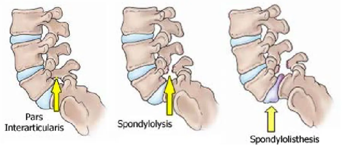
from http://www.niams.nih.gov/Health_Info/Scoliosis/default.asp) ... 19 Figure 1-5 : Schematic representation of Spondylolisthesis and Spondylolysis (obtained on 22
June 2011 from http://orthoinfo.aaos.org/topic.cfm?topic=a00053) ... 20 Figure 1-6 : Representation of tibia vara (obtained on 3 November 2010 from
http://knee-replacement-india.blogspot.com/2010/06/tibia-vara-causing-oa-knee.html) ... 21 Figure 1-7 : Representation of genu varum and valgum (obtained on 8 September 2010 from
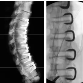
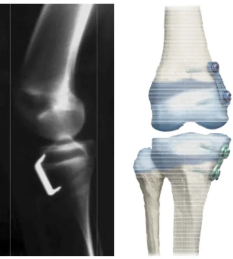
http://sports.jrank.org/pages/8251/genu-valgum.html) ... 21 Figure 1-8 : Screws and ligaments inserted at different levels between vertebral bodies (Newton et al., 2005) ... 22 Figure 1-9 : Staples implanted at different levels between vertebral bodies: left panel (Wall et al., 2005) and right panel (Guille et al., 2007). ... 23 Figure 1-10 : (left) Staple inserted in on proximal tibial growth plate to correct idiopathic genu
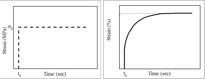
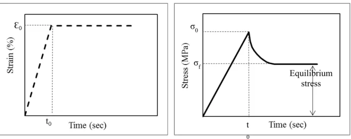
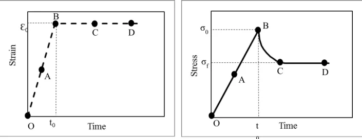
valgum (Courvoisier et al., 2009), (right) screw and plate (eight-plate) inserted on the distal femoral and proximal tibial growth plates (obtained on 22 June 2011 from http://www.eight-plate.com/glossary.php). ... 24 Figure 1-11 : Creep schematic. ... 30 Figure 1-12 : Stress relaxation schematic. ... 31
Figure 1-13 : Controlled ramp displacement applied on a cartilaginous tissue and the stress-relation response in the uniaxial compression experiment, adapted from (Mow et al., 2005). ... 32 Figure 1-14 : Schematic representation of confined uniaxial compression of cartilaginous tissue.
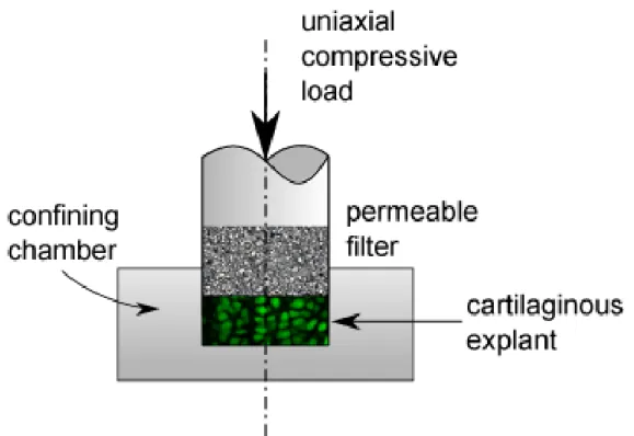
... 33 Figure 1-15 : Schematic representation of unconfined uniaxial compression of cartilaginous
tissue. ... 34 Figure 1-16 : Schematic representation of indentation of cartilaginous tissue. ... 35 Figure 1-17 : Schematic representation of confocal microscope light path (dash lines show the
out-of-focus light and simple lines represent the in-focus light). ... 45 Figure 1-18 : Simplified light path of confocal microscope. ... 46 Figure 3-1 : (Right) Porcine ulnar growth plate shown as a light purple line at the physis/bone
junction (inside the rectangular box). (Left) Histological section of the growth plate showing its three zones: reserve (R), proliferative (P), and hypertrophic (H). ... 59 Figure 3-2 : (A) Growth plate location on the porcine radius-ulna complex. (B) Punching of the ulna perpendicular to the growth plate/bone junction for recovery of the 4 mm diameter sample. (C) Recovered growth plate sample. (D) Growth plate semicylindrical samples cut parallel to the sample longitudinal axis ... 62 Figure 3-3 : Calcein AM concentration and cellular viability. (A) Confocal section of growth
plate chondrocytes loaded with 0.5 mM Calcein AM and 1 mM EthD-1 and showing very good cell viability (B) Confocal section of growth plate chondrocytes loaded with 5 mM Calcein AM and 1 mM EthD-1 and showing decreased cellular viability, as confirmed with EthD-1 taken up by the chondrocytes. ... 64 Figure 3-4 : Projected three-dimensional image of Calcein AM loaded in situ growth plate
chondrocytes obtained from a four-week old porcine distal ulna (10×). ... 66 Figure 3-5 : Chondrocyte shape and size showed zonal heterogeneity in growth plate. The
Chondrocytes are aligned along the growth direction except the cell marked by *, which is perpendicular to growth direction. ... 67 Figure 3-6 : Cell level morphometrical parameters: (A) chondrocytes volume, (B) chondrocytes
surface volume, (C) chondrocytes minor radius, (D) chondrocytes major radius, (E) chondrocytes sphericity and tissue level morphometrical parameter (F) cell/matrix volume ratio evaluated in 4-week old swine growth plates (mean values ± standard deviations). Significant differences among the three histological zones are shown with thick connecting lines (—) and significant differences between two histological zones are shown with thin connecting lines (—). (cell level morphological parameters: *p-value≤0.05, tissue level parameter: **p-value≤0.001). ... 69 Figure 4-1 : (Right) Typical confocal section of a fluorescently labelled porcine growth plate
showing its three histological zones: reserve (R), proliferative (P) and hypertrophic (H). (Left) Growth plate cylindrical plug extracted using a 6 mm biopsy punch. (Center) The growth plate site is located between the metaphysis and secondary ossification center of the epiphysis. ... 82 Figure 4-2 : Experimental set up: (A) custom-made loading apparatus mounted on the stage of an inverted laser scanning confocal microscope (B) Schematic of the loading apparatus. ... 83 Figure 4-3 : Typical experimental stress relaxation curves of growth plate samples undergoing
semi-confined compression at 5% pre-strain (A) followed by 10% strain (B) at a strain rate of 1.7E-3 s-1. ... 85 Figure 4-4 : Typical three-dimensional reconstructed chondrocytes in each histological zone
before compression (intact) and after compression and relaxation ... 86 Figure 4-5 : Morphometrical changes at cellular level ((a) volume, (b) surface area, (c) minor
radius, (d) major radius and (e) sphericity) and tissue level ((f) cell/matrix volume ratio) in response to 15% uniform tissue compression. Significant differences between pre-compression and post-relaxation parameters are shown with asterisks (p-value ≤ 0.05). ... 89 Figure 5-1 : (A) Growth plate site located between the metaphysis and secondary ossification
center of the epiphysis, where the longitudinal growth occurs. (B) Typical confocal section of a swine growth plate fluorescently labeled using Syto-17 showing the three histological
zones: reserve zone (RZ), proliferative zone (PZ) and hypertrophic zone (HZ). (C) Growth plate cylindrical sample extracted using a 6 mm biopsy punch perpendicular to the growth plate/bone junction. ... 102 Figure 5-2 : (A) Experimental set-up consisting of a custom-made loading apparatus mounted on the stage of an inverted laser scanning confocal microscope. (B) Growth plate semi-cylindrical sample being sandwiched between the two platens of the loading apparatus in a bath of HBSS. (C) Schematic of the experimental set-up. ... 103 Figure 5-3 : Image correlation results for a swine ulnar growth plate sample (GP#4) undergoing 10% uniaxial compressive strain between initial and final images: (A) initial 512×512 confocal image (before loading); (B) final 512×512 confocal image (after loading and relaxation); (C) region of interest (ROI) covering three histological zones of growth plate, which was cropped out from the final image and used in DIC algorithm; (D) calculated strain map along loading direction ( yy); (E) calculated strain map perpendicularly to the loading direction ( xx). ... 109 Figure 5-4 : Variations in strain developed across swine growth plates under 10% uniform
compressive strain, normalized to growth plate zone thicknesses. The strains were averaged on a pixel by pixel basis to compute the “average” strains in each histological zone. Each point on each curve represents an average of yy distributions as observed along the x axis (perpendicular to loading direction) of the imaged growth plates. Each curve stands for the average of strain distribution along the y axis (in loading direction). 0-1, 1-2, and 2-3 on the abscissa represent the reserve zone, the proliferative zone, and the hypertrophic zone, respectively. ... 110 Figure 5-5 : Biochemical composition of the three histological zones of 4-week old swine growth plates. Significant differences between the histological zones are shown with connecting lines (*p-value≤0.05 and **p-value≤0.005)... 113 Figure 5-6 : A typical presentation of collagen orientation in birefringent swine growth plate at
(a) 45°, (b) 90° and (c) 135° relative to the analyzer. At 45° vertically oriented collagen network is present in the proliferative and hypertrophic zones. At 135°, a mainly horizontally oriented collagen network is present in RZ. Panel (d) shows an adjacent section stained with Safranin-O/Fast-Green, with the histological zones as indicated (RZ: reserve
zone, PZ: proliferative zone, HZ: hypertrophic zone, and MB: metaphyseal bone. The analyzer direction (A) is marked on the panel (a). All angels are relative to the analyzer filter. The scale bar is 250µm. ... 114 Figure 6-1 : Frequency distribution of chondrocytes volume within the three different zones of
porcine growth plate. Polynomial curves were fitted to the data. ... 126 Figure 6-2 : Superposition of 2D confocal images of chondrocytes labeled with Calcein AM and Ethidium homodimer-1 through the growth plate thickness. ... 127 Figure 6-3 : Changes in chondrocytes volume within three histological zones of growth plate with two repeated confocal scans. ... 129 Figure 6-4 : Typical experimental stress relaxation curve of growth plate samples undergoing a
LIST OF ABBREVIATIONS
ADAMTS Aggrecanases, adamalysin-thrombospondins ANOVA analysis of varianceCO2 Carbone dioxyde
DIC Digital image correlation
DMMB 1,9-dimethylmethylene blue ECM Extracellular matrix
FGFs Fibroblast growth factors GAG Glycosaminoglycan
GH Growth hormone
HBSS Hank's balanced salt solution HZ Hypertrophic zone
IGF-I, II Insulin-like growth factor –I and –II IGFs Insulin-like growth factor
IHC Immunohistochemistry MMP Metalloproteinases
O2 Oxygene
OH-Pro Hydroxyproline PLM Polarized light microscopy
PG Proteoglycan
PZ Proliferative zone RA Retinoic acid ROI Region of interest
S-GAG Sulfated glycosaminoglycan TGFβs Transforming growth factor VEGF Vascular endothelial growth factor
INTRODUCTION
The longitudinal growth of bones is synchronized by hormones, genetics, nutrition, and mechanical factors, which regulate the normal development of bones (Ballock & O'Keefe, 2003b; Bonnel, Dimeglio, Baldet, & Rabischong, 1984; LeVeau & Bernhardt, 1984; I. A. Stokes, Mente, Iatridis, Farnum, & Aronsson, 2002). It has been clinically shown that mechanical loads are essential for normal growth. However, when excessive, these loads are involved in the progression of musculoskeletal deformities such as adolescent idiopathic scoliosis, neuromuscular disease, spondylolisthesis, the genu varum/valgum, tibia vara and other deformities (Bonnel et al., 1984; Frost, 1990; LeVeau & Bernhardt, 1984), through a phenomenon called the mechanical modulation of bone growth. The mechanical modulation of bone growth is also of growing interest in the development and improvement of minimally invasive approaches aiming to exploit local modulation of growth to correct the deformities while preserving the natural growth of bone as well as the functions and mobility of the bone segments. Growth plates are the site of the longitudinal bone growth, which occurs through a process of interstitial expansion and endochondral ossification of growth plate cartilage. These cartilaginous disks at the ends of long bones and vertebrae are responsible for bone growth until complete ossification of the growth plate at maturity (C. E. Farnum & N. J. Wilsman, 1998; Fujii et al., 2000; LeVeau & Bernhardt, 1984). The shape, size and arrangement of the constitutive cells of the growth plate, the chondrocytes, as well as biochemical composition of the extracellular matrix define three distinct morphological zones: the reserve zone, the proliferative zone and the hypertrophic zone (Breur, VanEnkevort, Farnum, & Wilsman, 1991; Buckwalter et al., 1985). Differentiation and hypertropy of chondrocytes are the result of a complex spatio-temporal process that occurs through these three histological zones, where the columns of chondrocytes are bone growth functional units (Alvarez et al., 2000; Hunziker & Schenk, 1989).
Growth plate extracellular matrix is composed of water, large aggregating proteoglycans embedded within type II collagen fibrils as well as short chain type X collagen, exclusively in the hypertrophic zone, which all provide the growth plate with its mechanical stiffness (Mwale, Tchetina, Wu, & Poole, 2002) and functions such as promoting/synchronizing the cell differentiation. Type II collagen, one of the major extracellular components of growth plate, forms a highly organized fibrilar network, which is believed to be a critical determinant of tissue
biomechanical competence (Speer, 1982). This collagen network has the ability to entrap large, hydrophilic proteoglycan molecules.
The mechanisms by which growth plate chondrocytes modulate longitudinal bone growth are still not well understood. Hypertrophy (changes in cell volume and height), proliferation of chondrocytes as well as matrix synthesis have been identified as the most important factors in both normal and mechanically modulated growth of long bones (Ballock & O'Keefe, 2003a; I. A. Stokes, Clark, Farnum, & Aronsson, 2007; I. A. Stokes et al., 2002; Villemure & Stokes, 2009; Wilsman, Farnum, Green, Lieferman, & Clayton, 1996; Wilsman, Farnum, Leiferman, Fry, & Barreto, 1996). Furthermore, one of the important mechanisms through which chondrocytes may respond to changes in their environment in both normal and mechanically modulated growth is directly through deformation of the cellular membrane. However, in situ three-dimensional visualization and zonal characterization of growth plate morphometry at both cellular and tissue levels have not been documented yet.
The growth plate mechanobiology, i.e. the effect of mechanical stimuli on the tissue biological responses, was studied in vivo in different animal models (Alberty, Peltonen, & Ritsila, 1993; Apte & Kenwright, 1994; Farnum et al., 2000; I. A. Stokes, Aronsson, Dimock, Cortright, & Beck, 2006; I. A. Stokes et al., 2002). There is convincing evidence that static forces alter the longitudinal bone growth, increased pressure on the growth plates retards growth and a reduced pressure accelerates it (Bonnel et al., 1984; I. A. Stokes et al., 2006). Many authors refer to this phenomenon by designating the Hueter-Volkmann law. Different approaches have been used to interpret the role of compressive loading in regulating growth plate and chondrocytes activity ranging from studies at the tissue and extracellular level to experiments at the cell level (Bachrach, 1995; Cohen, Chorney, Phillips, Dick, & Mow, 1994; Cohen, Lai, & Mow, 1998; Radhakrishnan, Lewis, & Mao, 2004; Sergerie, Lacoursiere, Levesque, & Villemure, 2009; Villemure, Cloutier, Matyas, & Duncan, 2007; Wosu, Sergerie, Levesque, & Villemure, 2011). However, up to date, no study has comprehensively analyzed the zonal growth plate compressive mechanical behavior at cellular and tissue levels with respect to the biochemical composition and type II fibrilar collagen organization of growth plates.
The main objective of the present research project was to characterize histomorphological (cell and tissue morphology) and structural (biochemical composition, fibrilar collagen organization)
characteristics as well as mechanical behaviour (in terms of developed strains) of growth plate at cellular and tissue levels in its three distinct histological zones. To do this, different techniques such as confocal microscopy combined with fluorescent labelling, polarized microscopy, digital image correlation, and biochemical assays have been used.
General organization of the thesis
This dissertation includes six chapters and is submitted as a "thesis by articles". The first chapter includes a litterature review required to achieve a sufficient understanding of the issues and methods used in this research project. The topics covered are the anatomy and basic physiology of growth plates, the mechanical modulation of growth and associated pathologies, the state of knowledge on mechanobiology of the growth plate and the state of knowledge on the mechanical behaviour of growth plate tissue. Finally, confocal microscopy and polarized light microscopy concepts were described as the techniques which were used in the present project.
The second chapter describes the hypotheses and clarifies the objectives of the research project including the methodological framework. The body of this doctoral thesis is composed of three principal articles presented in Chapters 3 to 5. Chapter 3 presents the first article entitled: “Three-dimensional in situ zonal morphology of viable growth plate chondrocytes: a confocal microscopy study” which was published in Journal of Orthopaedic Research. This scientific article addresses the three-dimensional zonal morphology of viable growth plate at cellular and tissue levels and presents a confocal microscopy approach combined with fluorescent labeling of the tissue. This chapter answers the first objective of this research project. The fourth chapter includes the second scientific article entitled: “Tissue and Cellular Morphological Changes in Growth Plate Explants under Compression” which was published in Journal of Biomechanics. This study addresses three-dimensional deformations of chondrocytes and growth plate tissue in all histological zones of growth plates under compression using stress relaxation tests. This chapter answers the first step of the second objective of the research project. The fifth chapter presents the last article entitled “Stress relaxation of swine growth plate in semi-confined compression: depth dependant tissue deformational behavior versus extracellular matrix composition and collagen fiber organization” which revision was recently submitted to Biomechanics and Modeling in Mechanobiology. The growth plate mechanical behaviour evaluated using the digital image correlation (DIC) technique as well as corresponding biochemical
composition and collagen fiber orientation in its three functionally distinct zones are presented in this chapter. This chapter deals with the second step of the second objective as well as the third, fourth and fifth objectives of this thesis. The final chapter (Chapter 6) contains a general discussion on the research project followed by a conclusion and recommendations for future related projects.
CHAPTER 1
BACKGROUND AND REVIEW OF THE LITTERATURE
1.1 Longitudinal bone growth and growth plate
1.1.1 Longitudinal bone growth
The longitudinal growth of long bones and vertebrae occurs through the process of endochondral ossification, i.e. through the synthesis of a cartilaginous growth plate, which then transforms into bone. Although in humans the growth rate decreases until skeletal maturity at around 18-20 years, periods of accelerated growth are observed in childhood and adolescence. The closure of the growth plate marks the completion of longitudinal growth, usually in late adolescence (Villemure & Stokes, 2009).
1.1.2 Endochondral bone growth
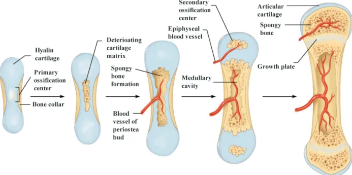
Ossification is always done through another tissue whether fibrous connective tissue or cartilaginous tissue. The ossification taking place primarily through a fibrous connective tissue is called intramembranous ossification, whereas the ossification through a hyaline cartilage is called endochondral ossification. Figure 1-1 illustrates the process of endochondral ossification of long bones, also referred to as longitudinal bone growth.
Longitud through t hypertrop longitudi Figure 1-using Cal In the gr die. This column a Hunziker epiphysis osseous j endochon Proliferat Buckwalt Farnum, process o dinal bone g three zones phic (Figure nal growth o -2 : A typica lcein-AM) s rowth plate, process of and in a pa r & Schenk s pushing old junction, ost ndral ossifica tion and hy ter, Mower, Green, et al of longitudin growth is the of the cart e1-2)), wher of long bone al confocal se howing its u chondrocyt interstitial g articular, syn k, 1989). Ch
der cells tow teoblasts oss ation. ypertrophy o Ungar, Sch l., 1996; Wil nal growth. S e result of a tilaginous gr re the colu es (Alvarez e ection of por ultrastructure tes proliferat growth takes nchronized hondrocytes ward the diap sify them to of chondroc haeffer, & G lsman, Farnu Synthesis and a complex s rowth plate umns of cho et al., 2000; H rcine growth e and three h te, hypertrop s place in co and predefi are constan physis. Whil o form new ytes (Breur Ginsberg, 198 um, Leiferm d degradatio spatio-tempo (the reserv ondrocytes Hunziker & h plate (chon histological z phy, synthes onjunction w ined order ( ntly dividin le chondrocy bone tissue et al., 199 86; Hunzike man, et al., 1 on of the ext oral process ve, the proli are the fun Schenk, 198 ndrocytes cyt zones. size extrace with chodroc (Farnum & ng and piled ytes degener . This pheno 91; Buckwal r & Schenk, 996) play a tracellular m s, which ope iferative, an nctional uni 89). toplasm labe llular matrix cytes of the Wilsman, d in front o ate at the ch omenon is c lter et al., , 1989; Wils major role i matrix are als
erates nd the its of eled x and same 1993; of the hodro-called 1985; sman, in the o key
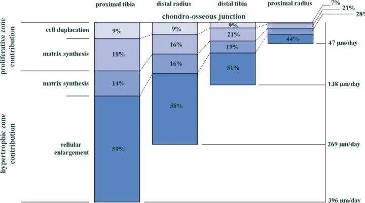
elements in longitudinal growth of long bones (Alvarez et al., 2000; Breur et al., 1991; Buckwalter et al., 1986; Cowell, Hunziker, & Rosenberg, 1987; Hunziker, 1994; Hunziker & Schenk, 1989; Wilsman, Farnum, Leiferman, et al., 1996). Indeed, the matrix remodeling can regulate cell shape and volume, and is hence involved in the spatio-temporal sequence of proliferation, hypertrophy and chondrocytes migration (Alvarez et al., 2000). The relative contribution of proliferative and hypertrophic zones in the daily elongation of bones is shown in Figure 1-3 for four different bones from rats (Wilsman, Farnum, Leiferman, et al., 1996).
Overall, Wilsman et al. (Wilsman, Farnum, Leiferman, et al., 1996) showed that the largest contribution to the daily growth in all bones came from cellular hypertrophy (40-60%) and extracellular matrix synthesis in the hypertrophic zone (15-30%), which together represents almost 75% of the total contribution. Matrix synthesis in the proliferative zone (15-20%) and especially chondrocyte proliferation (10%) had a minimal, although significant influence on longitudinal growth, concluding that hypertrophy remains the most important factor for longitudinal growth.
Figure 1-3 : Relative contribution of proliferative and hypertrophic zones in daily longitudinal growth of four different bone types from 28-day old rats at the chondro-osseous junction, adapted from Wilsman et al. (1996).
1.1.2.1 Biological regulation of bone growth
Biological mechanisms involved in the growth process are regulated by various factors including: ; Systemic hormones including growth hormone GH (Isaksson, Jansson, & Gause, 1982;
Russell & Spencer, 1985; Thorngren & Hansson, 1974; Wit, Kamp, & Rikken, 1996), , IGF-I (insulin-like growth factor I) (Baker, Liu, Robertson, & Efstratiadis, 1993; Schoenle, Zapf, Humbel, & Froesch, 1982), thyroid hormones T3 and T4 (Bassett, Swinhoe, Chassande, Samarut, & Williams, 2006; Leger & Czernichow, 1989) and androgenic steroids during puberty and estrogens (Cassorla et al., 1984; Coxam et al., 1996; Cutler, 1997; Takano, Aizawa, Irie, Kokubun, & Itoi, 2007; Zung et al., 1999). They are necessary to allow bone to grow at the same pace on the left and right sides of the body (Rauch, 2005);
; PTHrP protein (parathyroidhormone-related protein), which plays an important role in endochondral ossification by increasing the synthesis of extracellular matrix in the growth plates. The expression of PTHrP protein is also influenced by mechanical loads (Tanaka et al., 2005).
; Growth factors are polypeptides known to affect growth and cell differentiation in bone and cartilage. There are several main types of growth factors: IGFs (insulin-like growth factors) and TGFβs (transforming growth factors) (Zerath et al., 1997) The FGFs (fibroblast growth factors) (Hutchison, Bassett, & White, 2007) and VEGF (vascular endothelial growth factor) (van der Eerden, Karperien, & Wit, 2003) are also involved in the regulation of longitudinal bone growth. Finally, it has also been shown that RA (retinoic acid) is associated with chondrocyte maturation during endochondral ossification (Iwamoto et al., 1993; W. Wang & Kirsch, 2002).
; Transmembrane proteins, such as the integrins, which allow communication between cells and matrix (Egerbacher & Haeusler, 2003; Hausler, Helmreich, Marlovits, & Egerbacher, 2002);
; The nutrients (Hunziker, 1994).
Certain biological factors influence the production of various matrix components which are important for growth and remodeling. Indeed, it has been shown that IGF-I (Demarquay et al., 1990; Wroblewski & Edwall-Arvidsson, 1995), TGF-β1 (Ballock et al., 1993; Thorp, Anderson, & Jakowlew, 1992) and GH (Monsonego, Halevy, Gertler, Hurwitz, & Pines, 1995) stimulate the expression of type II collagen. The expression of this same component is however inhibited by bFGF (Wroblewski & Edwall-Arvidsson, 1995) and by retinoic acid (Yoshida et al., 2001). IGF-I and IGF-II as well as retinoic acid stimulate the expression of collagen type X (Yoshida et al., 2001) The expression of collagen type X is inhibited by TGF-β1 (Ballock et al., 1993) and PTHrP (O'Keefe et al., 1997). The expression of proteoglycans is also stimulated by IGF-I and IGF-II (Leach, Richards, Praul, Ford, & McMurtry, 2007), however, it is inhibited by retinoic acid (Yoshida et al., 2001) and bFGF (Makower, Wroblewski, & Pawlowski, 1988). It should be noted that extracellular matrix components are also influenced by the enzymes, particularly metalloproteinases (or MMPs) and aggrecanases (Abbaszade et al., 1999; Keeling & Herrera, 2008; M. D. Tortorella et al., 1999).
1.1.2.2 Environmental regulation of bone growth
In addition to biological factors already mentioned such as hormones, growth factors, nutrients and genetic factors, the mechanical environment also affects the behavior of the growth plate. The mechanical environment is a very important factor of influence on the functioning of the growth plate; however, its role in growth is not yet clearly defined (Arriola, Forriol, & Canadell, 2001; Farnum et al., 2000; Niehoff, Kersting, Zaucke, Morlock, & Bruggemann, 2004; Ohashi, Robling, Burr, & Turner, 2002; I. A. Stokes, 2002; I. A. Stokes et al., 2006; I. A. Stokes et al., 2007; I. A. Stokes, Gwadera, Dimock, Farnum, & Aronsson, 2005). The known effects of mechanical loading on the growth process is presented in section 1.2. Vascularization of the growth plate is also a significant factor in the growth regulation (Trueta & Trias, 1961). It is generally accepted that growth plate is vascularized during early childhood, even if this theory does not reach consensus (Jaramillo et al., 2004; Wirth et al., 2002). In the subsequent growth, growth plate regulatory elements like growth factors and systemic hormones come from three vascularization systems: that of the metaphyseal junction, of the epiphyseal junction, and of the perichondrium (Trueta & Trias, 1961; Williams, Zipfel, Tinsley, & Farnum, 2007; Wirth et al., 2002). The transport of molecules from the epiphyseal and metaphyseal junctions is done partly
by diffusion and partly by convection (Williams et al., 2007). To allow convection, a pressure gradient is therefore required between the bone and growth plate cartilage. That is the reason why the mechanical loading of the growth plate could interfere with this transport. A suppression of the first vascularization system (metaphyseal junction) seems to induce a thickening of proliferative and hypertrophic zones of growth plate and an abnormal endochondral ossification (Trueta & Little, 1960). The chondrocytes terminal differentiation was distrupted and led to accumulation of chondrocytes instead of disappearing (Trueta & Trias, 1961). Suppression in the second vascularization system (epiphyseal junction) seems to induce a large cellular disorganization and disruption in chondrocytes proliferation (Brashear, 1963; Trueta & Little, 1960; Trueta & Trias, 1961).
1.1.3 Growth plate form and site
Growth plates are cartilaginous discs at the ends of long bones and vertebrae. In the long bones, they are located between the epiphysis and the metaphysis, whereas in human vertebrae, where there is no epiphysis, they are located between the intervertebral disc and the vertebral body. There are also growth plates in the posterior parts of the vertebrae, called the neurocentral junctions. The following sections describe the structure and function of the growth plate and its respective zones, as well as their composition.
1.1.4 Growth plate composition
1.1.4.1 Chondrocytes
Chondrocytes are the constitutive cells that are found specifically in cartilage. Their main function is the synthesis and degradation of the extracellular matrix components, i.e. collagens and proteoglycans, as well as the secretion of enzymes that degrade these components (Mow, Gu, & Chen, 2005; Mow & Hung, 2001). The shape and arrangement of chondrocytes in the growth plate differ from one zone to another (Ballock & O'Keefe, 2003b; Bonnel et al., 1984; Buckwalter et al., 1985; C. E. Farnum & N. J. Wilsman, 1998).
1.1.4.2 Extracellular matrix
The extracellular matrix consists of a network of collagen fibrils embedded in a complex, highly hydrated proteoglycans and hyaluronic acid aggregate whose concentrations vary from one
histological zone to another (Alvarez et al., 2000; Poole, Matsui, Hinek, & Lee, 1989; Sandell, Sugai, & Trippel, 1994).
Collagen
Collagen, the most common protein in the human body, is the main structural component of the growth plate extracellular matrix. Collagen is formed by the polymerization of five tropocollagen into fibrils, a molecule composed of three alpha chains arranged in a helix (Mow & Hung, 2001; Ratcliffe & Mow, 1996). Collagen forms small fibrils with an architectural arrangement that varies through the hyaline cartilage depth. For example, in articular cartilage, collagen fibrils are organized in parallel to the articular surface in the superficial zone while they are randomly oriented in the middle zone and are perpendicular to the articular surface in the deep zone. Such a structure gives the cartilage the ability to withstand loads in tension, and retain glucosaminogylcanes (GAG) into a coherent wholesome. This structure also provides a permeability effect producing a frictional drag of the fluid, which provides the extracellular matrix the ability to withstand compression. Although several types of collagen exist, hyaline cartilage contains types II, IX, X and XI, and of these, the concentration of type II collagen is the highest, except in the hypertrophic zone. This high concentration in the reserve and proliferative zones helps the prevention of premature calcification and efficient distribution of loads (Mow & Hung, 2001; Niehoff et al., 2004; Radhakrishnan et al., 2004; Ratcliffe & Mow, 1996). Type IX collagen is also a fibrillar collagen. It is still composed of three α chains, but also a proteoglycan glycosaminoglycan chain which is covalently bound to its α chains (Bruckner, Vaughan, & Winterhalter, 1985; van der Rest & Mayne, 1988). It is covalently bound to the fiber surface of type II collagen (Diab, Wu, & Eyre, 1996; Eyre, Apon, Wu, Ericsson, & Walsh, 1987; Ruggiero et al., 1993; van der Rest & Mayne, 1988). It is highly expressed in the growth plate (Ballock & O'Keefe, 2003a), although in much smaller amounts than type II collagen (Balmain et al., 1995). Ballock et al. (Ballock & O'Keefe, 2003a) postulated that collagen type IX allows type II collagen to interact with other components of the extracellular matrix and is also likely to participate in growth control and thickness of type II collagen fibers (Balmain et al., 1995). Type XI collagen is another fibrillar collagen of the extracellular matrix and is composed of three α chains (Francomano, 1995; Sutmuller, Bruijn, & de Heer, 1997). It is linked to type II collagen by structural links (Pihlajamaa et al., 1999; Ruggiero et al., 1993; Sandell et al., 1994). Although it is less important than type II collagen (Balmain et al., 1995; Sandell et al., 1994), it is also