Université de Montréal
Development and Characterization of Long
Circulating Emulsions to Target Solid Tumors
par banna Rossi
Sciences pharmaceutiques Faculté de pharmacie
Mémoire présenté à la Faculté des études supérieures en vue de l’obtention du grade de Maître en sciences (M.Sc.)
en sciences pharmaceutiques option technologie pharmaceutique
Août, 2006 © Joanna Rossi, 2006
C
Q
Université t
de Montréal
Direction des bibliothèques
AVIS
L’auteur a autorisé l’Université de Montréal à reproduire et diffuser en totalité ou en partie, par quelque moyen que ce soit et sur quelque support que ce soit, et exclusivement à des fins non lucratives d’enseignement et de recherche, des copies de ce mémoire ou de cette thèse.
L’auteur et les coauteurs le cas échéant conservent la propriété du droit d’auteur et des droits moraux qui protègent ce document. Ni la thèse ou le mémoire, ni des extraits substantiels de ce document, ne doivent être imprimés ou autrement reproduits sans l’autorisation de l’auteur.
Afin de se conformer à la Loi canadienne sur la protection des renseignements personnels, quelques formulaires secondaires, coordonnées ou signatures intégrées au texte ont pu être enlevés de ce document. Bien que cela ait pu affecter la pagination, il n’y a aucun contenu manquant. NOTICE
The author of this thesis or dissertation has granted a nonexclusive license allowing Université de Montréal to reproduce and publish the document, in part or in whole, and in any format, solely for noncommercial educational and research purposes.
The author and co-authors if applicable retain copyright ownership and moral rights in this document. Neither the whole thesis or dissertation, flot substantial extracts from it, may be printed or otherwise reproduced without the author’s permission.
In comptiance with the Canadian Privacy Act some supporting forms, contact information or signatures may have bean removed from the document. While this may affect the document page count, t does flot represent any loss of content from the document
Faculté des études supérieures
Ce mémoire intitulé
Development and Characterization of Long-Circulating Emulsions to Target Solid Tumors
présenté par: Joanna Rossi
a été évalué par un jury composé des personnes suivantes:
Dr. Emmanuelle Roux, président-rapporteur Dr. Jean-Christophe Leroux, directeur de recherche
Dr. Christine Allen, co-directrice Dr. François-Xavier Lacasse, membre du jury
11
Résumé
Des émulsions furtives ont été préparées à l’aide d’excipients approuvés pour une application pharmaceutique et ont été évaluées in vivo pour leur capacité à cibler des tissus néoplasiques. Les émulsions étaient composées d’une phase interne de triglycéride et émulsifiées avec un tensioactif synthétique (polysorbate 80) et un co émulsifiant lipidique. Afin de produire des émulsions furtives, de la sphingomyéline d’oeuf (ESM) a été choisie comme co-émulsifiant et différents dérivés de 1,2-distéaryl-sn-glycero-3 -phosphatidyléthanolamine-poly(éthylène glycol) (DSPE-PEG) ont été greffés à l’interface de l’émulsion. Les dérivés sélectionnés de DSPE-PEG étaient la DSPE-PEG 2000, la DSPE-PEG 5000, et la DSPE-N-{pentaérythritol polyoxyéthylène glutaryl] (DSPE-4armPEG), (MM 2000). Les effets de l’ESM et de la DSPE-PEG (la concentration et la structure) sur la prolongation du temps de circulation et sur l’accroissement de l’accumulation dans les tissus néoplasiques ont été évalués sur des souris inoculées du mélanome B16 et de l’adénocarcinome du côlon C26 en sous-cutané.
Dans cette étude, nous rapportons que des émulsions furtives ont été obtenues en enrobant la surface des gouttelettes avec de la DSPE-PEG 2000 ou 5000. L’accroissement du temps de circulation n’a pu être atteint ni avec la DSPE-4-armPEG malgré le segment de PEG de masse molaire de 2000 ni avec l’ESM. Le temps de circulation accru des émulsions enrobées de PEG 2000 ou 5000 s’est traduit par une accumulation plus élevée dans les tumeurs C26, mais pas dans les tumeurs B16. Ces émulsions pouvaient améliorer la sélectivité d’agents anticancéreux
lipophiles pour certains tissus néoplasiques et aussi augmenter leur index thérapeutique.
Mots-clés : Émulsions furtives, poly(éthylène glycol), biodistribution, pharmacocinétique, vecteurs de médicaments
iv
Abstract
Long-circulating emulsions were prepared using pharmaceutically acceptable excipients and evaluated in vivo for their ability to target neoplastic tissues. The emulsions were composed of a triglyceride internai phase and emulsified with a synthetic surfactant (polysorbate $0) and a lipid emulsifier. Attempts made to produce long-circulating emulsions included adding egg sphingomyetin (ESM) as a coemulsifier and graffing various 1
,2-distearoyl-sn-glycero-3-phosphatidylethanolamine-poly(ethylene glycol) (D$PE-PEG) derivatives into the emulsion interface. The DSPE-PEG derivatives selected were D$PE-PEG 2000, DSPE-PEG 5000 and DSPE-N-[pentaerythritol polyoxyethylene) glutaryl] (D$PE-4-armPEG), (MW 2000). The effect of ESM and DSPE-PEG concentration and structure in prolonging circulation time and enhancing accumulation into neoplastic tissues was assessed in mice bearing subcutaneously implanted B 16 melanoma or C26 colon adenocarcinoma.
In this study, we report that long-circulating emulsions were obtained by coating the droplet surface with single chain DSPE-PEG 2000 or 5000. Circulation Ïongevity could flot be achieved with DSPE-4-armPEG despite the 2000 MW PEG segment nor with ESM. Enhanced circulation time of emulsions grafted with PEG 2000 or 5000 translated into higher accumulation into C26 tumors but flot into B16. These emulsions can potentially enhance the specificity of lipophilic anticancer drugs towards neoplastic tissues and enhance the therapeutic index.
Keywords: Long-circulating emulsions, poly(ethylene glycol), biodistribution, pharmacokinetics, drug carriers
Table of Contents
RÉSUMÉ .11
ABSTRACT .1V
LIST 0F TABLES VIII
LIST 0F FIGURES IX
ABBREVIATIONS XV
ACKNOWLEDGEMENTS xx
CHAPTER 1: COLLOIDAL DRUG CARRIERS I
1. INTRODUCTION 2 2. LIPOSOMES 5 3. MICELLES 7 4. MACROMOLECULAR PRODRUGS 9 5. POLYMERIC NANOPARTICLES 10 6. EMULSIONS 11 7. REFERENCES 12
CHAPTER 2: PRINCIPLES IN THE DEVELOPMENT 0F INTRAVENOUS LIPID
EMULSIONS 15 1. INTRODUCTION 16 2. EMULSION STABILITY 17 2.1. DESTABILIzAT;oN PROCESSES 18 2.2. ELEcTR0sTATIc STABILIZATION 20 2.3. STERIc STABILIZATION 22
vi
3. EUMINATION MECHANISMS FOR LIPID EMULSIONS .22
3.1. LIPID EMULSIONS METABOLIZED AS ENDOGENOUS CHYLOMICRONS 23 3.2. ELIMINATION BY THE MONONUCLEAR PHAGOCYTE SYSTEM 24
4. BIODISTRIBUTION 0F LIPID EMULSIONS 25
4.1. EFFECT0F LIPID EMULSION SIZE 25
4.2. EFFECT 0F LIPID EMULSION COMPOSITION AND EMULSIFIERS 30
4.3. EFiECT 0F SURFACE CHARGE 32
4.4. LONG-CIRCULATING LIPID EMULSIONS 33
4.5. ACTIvE TARGETING 0F SELECTED CELLS 37
4.6. DRUG LEAKAGE FROM EMULSIONS 39 5. PREPARATION 0F EMULSIONS FOR INTRAVENOUS ADMINISTRATION 40 5.1. ExCIPIENT AND FORMULATION CONSIDERATIONS 41
5.2. EMULSI0N PREPARATION 47
5.3. EMULS1ON CHARACTERIZATION 49
5.4. STABILITY MEASUREMENT 50
6. LIPID EMULSIONS FOR THE DELIVERY 0F NUCLEIC ACID-BASED DRUGS 53
7. CONCLUSIONS 55
8. ACKNOWLEDGEMENTS 56
9. REFERENCES 57
CHAPTER 3: PRESENTATION 0F THE ARTICLE 7$
I. ABSTRACT 79
2. INTRODUCTION 80
3. MATERIALS AND METHODS 82
3.1. MATERIALS 82
3.3. PREPARATI0N AND CHARACTERIZATION 0F EMULSIONS $4
3.4. BIODI5TRIBUTI0N STUDIES 85
4. RESULTS AND DISCUSSION 87
4.1. COMPRESSION ISOTHERMS $7 4.2. BIODtSTRIBUTION STUDIES 90 5. ACKNOWLEDGEMENTS 96 6. REFERENCES 97 CHAPTER 4: DISCUSSION 107 1. FORMULATION OPTIMJZATION 108 1.1. EMULSION COMPOSITION 108 1.2. SONICATION PARAMETERS 111 2. EMULSION STABILITY 113 3. COMPRESSION ISOTHERMS 115 4. BIODISTRIBUTION STUDIES 116 5. REFERENCES 119 CHAPTER 5: CONCLUSION 121 1. REFERENCES 124 APPENDIX I 125 APPENDIX II 126
VII’
List of Tables
Table 1: $everal commercially avaïlable emulsions for intravenous injection 66
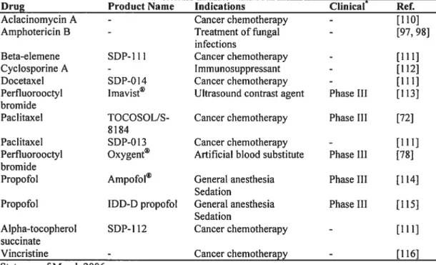
Table 2: Some intravenously injectable emulsions in development and in clinical
trials 66
Table 3: Antitumor activity of RS-1541 emulsion formulations against M5076
sarcoma at the MTD 67
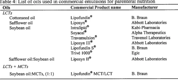
Table 4: List ofoils used in commercial emulsions for parenteral nutrition 67
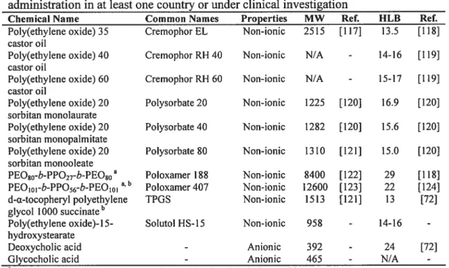
Table 5: Several non-phospholipid surfactants approved for intravenous administration in at least one country or under clinical investigation 68
Table 6: Properties of the lipid emulsions administered to mice bearing either
B16-F10 melanoma or C26 colon adenocarcinoma 101
Table 7: Stability ofseveral non-PEGylated emulsions over time 114
List of Figures
Figure 1: Capillary structure of normal tissues with a continuous endothelial lining (A) and tumors possessing enhanced vascular permeability (B) 3
Figure 2 : The total potential energy of interaction between two droplets as a fimction of separation distance (electrical double layer repulsion and van der Waals attraction).
69
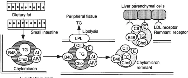
Figure 3 $ Absorption and metabolism of dietary fat. Dietary fats are metabolized and
incorporated into chylomicrons in the small intestine. Then, chylomicrons enter the blood circulation via the thoracic duct. During circulation, the triglycerides of chylomicrons are rapidly hydrolyzed via lipoprotein lipase (LPL) on endothelial surfaces, then chylomicron remnants are produced. Finally, chylomicron remnants are cleared by the liver by the LDL or remnant receptors. 1G, triglyceride; Chol, cholesterol; AI, apolipoprotein AI; AIV, apolipoprotein AIV; 348, apolipoprotein B4$; CII, apolipoprotein CII; E, apolipoprtoein E. Reprinted with permission from
ElsevierRef. [13] Copyright 2000 70
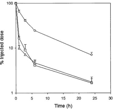
Figure 4 : Effect of particle size on the clearance of cholesteryl oleate (CO) label from plasma as a function of time afier intravenous administration into mice. The emulsions were composed of triolein (TO): 1
,2-dipalmitoyl-sn-glycero-3-phosphatidylcholine (DPPC):polysorbate $0: polyethylene glycol modified 1,2-dipalmitoyl-sn-glycero-3 -phosphatidylethanolamine (PEG2000-DPPE) (2:1:0.4:0.1,
X
wlw) . The droplet sizes ofthe emulsions injected were 50 (o), 100 (o) and 175 nm
(t\). Adapted with permission from Elsevier Ref. [2$J Copyright 1996 71
Figure 5 : Concentrations of 13-0-palmitoyl-rhizoxin (RS-1541) in the plasma, liver,
and tumor afier a single intravenous administration of various sizes of emulsion formulations and a surfactant solution ofRS-1541 to mice bearing M5076 sarcoma at a dose of 5 mg/kg. The emulsion droplet sizes were 110 (A), 230 (o), 350 (+), 410 (0), 630 nm (o) and the surfactant solution
(.).
Each value represents the mean ± S.E. ofthree mice. Adapted with permission from Springer Science and Business Media Ref.
[40] Copyright 1996 72
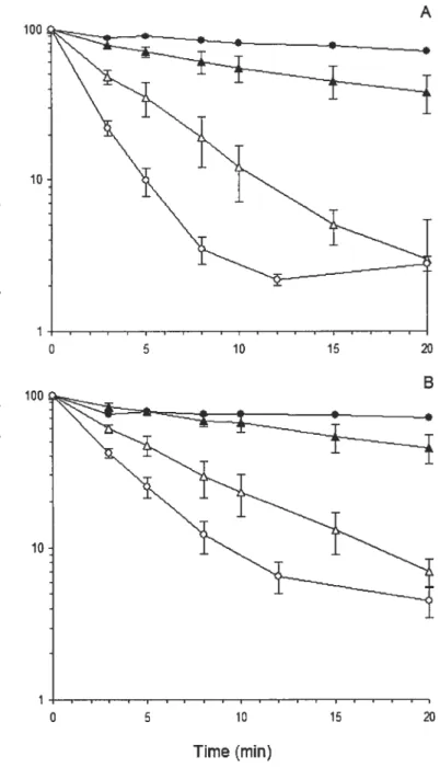
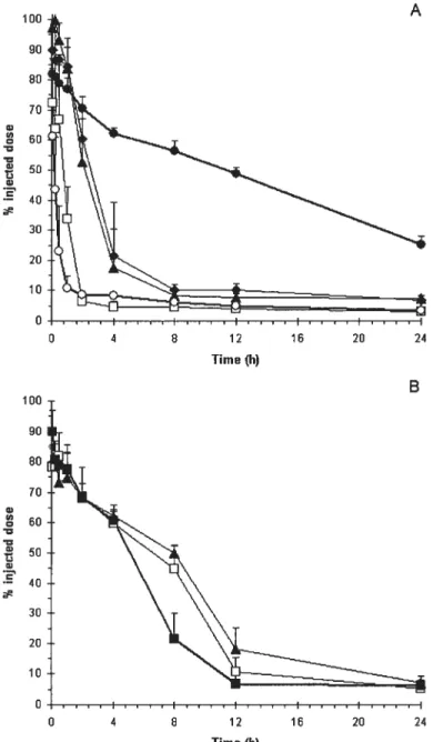
figure 6: Radioactivity in plasma of triolein (TO) and cholesteryl oleate (CO) labels aller injection of emulsions stabilized by mixtures of sphingomyelin (5M) with egg phosphatidylcholine (egg PC). TO-CO-cholesterol emulsions stabilized with mixtures of SM and egg PC were injected intravenously in conscious rats. Plotted are the data for labeled TO (A) and CO (B) incorporated in the emulsions remaining in the plasma at 3, 5, 8, 12 and 20 min aller injection. Results are means ± S.E. of at least four
experiments for each observation. SM 100%
(.),
SM/egg PC 50/50 (Â), SM/egg PC 25/75 (A), egg PC 100% (o). Adapted with permission from Elsevier Ref. [21]Copyright 1992 73
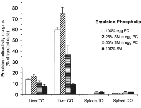
f igure 7 : Radioactivity in the liver and spleen of triolein (TO) and cholesteryl oleate (CO) labels aller injection of emulsions stabilized by mixtures of sphingomyelin
(SM) with egg phosphatidylcholine (egg PC). TO-CO-cholesterol emulsions stabilized with mixtures of SM and egg PC were injected intravenously in conscious rats. Organ uptakes of radioactive TO and CO labels in the emulsions were measured 20 min afier injection. Resuits are means± S.E. of at least four experiments for each
observation. By analysis of variance the differences between groups were statisticaliy significant with P < 0.01 for liver TO, P< 0.00 1 for liver CO, P <0.01 for spleen TO
and P < 0.025 for the spleen CO. Adapted with permission from Elsevier Ref. [21]
Copyright 1992 74
Figure 8: Blood concentration-time profile of stealth liposomes and different lipid nanocapsule formulations prepared by the conventional (A) or post-insertion method (B). Mean ± SD (n =3 to 5). A: PEGylated liposomes
(.),
plain lipid nanocapsules(o), PEGylated lipid nanocapsules with 1.7 mol% 1 ,2-distearoyl-sn-glycero-3-phosphatidylethanolamine-N-monomethoxy-[pofyethylene glycol] (PEG2000-DSPE) (o), PEGylated lipid nanocapsules with 1.4 mol% PEG5000-DSPE (À), and PEGylated lipid nanocapsules with 3.4 mol% PEG2000-DSPE (+). B. PEGylated lipid nanocapsules with 6 mol% PEG2000-DSPE
(.),
PEGylated lipid nanocapsules with 6 mol% PEG5000-DSPE (o), and PEGylated lipid nanocapsules with 10 mol% PEG2000-DSPE (À). Formulations were injected intravenously at a dose of 2 mg lipids/rat. Adapted with permission from Springer Science and Business Media Ref. [381xii Figure 9: Liver uptake and serum decay ofthe control and human recombinant (rec) apo E-enriched emulsion-iododeoxyuridine-oleoyl ([3HJIDU-012) in rats, in the absence or presence of lactoferrin. Control and rec-apo E-enriched emulsions, double-labelled with [1-’4C]cholesteryl oleate ([14C]CO) and [3H]IDU-012 were injected into fasted anaesthetized rats. A, B, C. At the indicated times, the liver uptake and serum decay of [‘4C]CO (A) and [3H]IDU-012 (B) were determined. The liver
uptake and serum decay of rec-apo E-enriched emulsions were also determined afler preinjection of lactoferrin (C). D: At 30 min afier injection of emulsion-rec-apo E IDU-012, the liver was perfused. Total liver (L) association was determined and parenchymal (PC), endothelial (EC), and Kupffer (KC) ceils were subsequently isolated. Values are means± s.d. of three experiments. Adapted with permission from
Macmilian Publishers Ltd. Ref. [60] Copyright 1995 76
Figure 10: The main manufacmring steps involved in the production of intravenous
emulsions 77
Figure 11: Surface pressure versus molecular area (A) and surface pressure versus modulus of compressibility (B) plots of HSPC and ESM at the air/water interface.
Subphase conditions: PBS pH 7.4, 25°C 102
Figure 12: Surface pressure versus molecular area (A) and surface pressure versus modulus ofcompressibility (B) plots ofPS-80, HSPC/PS-80 (1:3, w/w) and E$M/PS
figure 13: Elimination profile of emulsions from blood (A) and distribution to 316-f10 melanoma (B) and muscle (C) after i.v. injection in C57BL/6 mice. Each mouse
was
administered 5.4 mg of lipids (exciuding DSPE-PEG) in a 100-pi injection volume. Mean + SD (n 4-5 mice/group). TC/PS-80/HSPC (ri), TC/PS-$0/ESM (D),and
TC/PS-$0/ESM!(lO mol%)DSPE-PEG 2000(.).
Statistically significant differences between plain and PEGylated emulsions are indicated. *p< 0.05 104Figure 14: Elimination profile of emulsions from blood (A) and distribution to C26 colon adenocarcinoma (B)
and
muscle (C) after i.v. injection in Balb!C mice. Each mouse was administered 5.4 mg of lipids (exciuding DSPE-PEG) in a 100-pi injection volume. Mean + SD (n = 4-5 mice/group). TC/PS-$0/ESM()
and TC/PS$0/ESM/( 10 mol%)DSPE-PEG 2000
(.),
TC/PS-$0/ESM/( 15 mot%)DSPE-PEG 2000 (b), TC/PS-$0/ESM/(10 mol%) DSPE-PEG 5000 (I:I), TC/PS-80/ESM/(l0 mol%)D$PE-4-armPEG().
Statistically significant differences between plain andPEGylated emulsions are indicated. p< 0.05 105
Figure 15: Effect of DSPE-PEG derivatives on the tissue distribution of the emulsions in Balb/C mice inoculated with C26 colon adenocarcinoma afier 2 h (A), 6 h (B),
and
12 h (C) post i.v. injection. Mean+ SD (n 4-5 mice/group). TC/P$-$0/ESM
() and
TC/PS-$0/ESM/( 10 mol%)DSPE-PEG 2000 (i), TC/PS-80/ESM/( 15 mol%)DSPE
PEG 2000
(),
TC/PS-$0/ESM/(10 mol%) DSPE-PEG 5000 (El), TC/PS-80/E$M/(10xiv
Figure 16: Influence of the proportions of TC, PS-$O and ESM on mean droplet diameter (A) and size distribution (B) of the emulsion. Sonication conditions were kept constant at medium intensity (72-$4 W) for 25 s. The weight ratio ofPS-$0/ESM was either 0.6
(.),
1 (o), 3 (À) or 7 (A). The extemal phase was citrate buffer at pH5.
(-
--)
indicates the range of acceptable diameter and PDI 110figure 17: Influence of sonication intensity and time on mean droplet size (A) and
size distribution (B). The proportion ofTC, PS-$0 and ESM was kept constant (5:3:1,
Abbreviations
surface pressure
A moleculararea
A molecular area at a particular surface pressure
Apo apolipoprotein
AUC area under the blood concentration-time curve
CHE cholesteryl hexadecyl ether
Chol cholesterol
CMC critical micelle concentration
CO cholesteryl oleate
C modulus of compressibility
DLVO Derjaguin, Landau, Vervey, and Overbeek theory of
colloidal stability
DMPC I ,2-dimyristoyl-sn-glycero-3 -phosphatidylcholine DOPC 1 ,2-dioleoyl-sn-glycero-3 -phosphatidylcholine DOPE 1 ,2-dioleoyl-sn-glycero-3-phosphatidylethanolamine DOTAP I ,2-dioleoyl-sn-glycero-3-trimethylammonium propane DPPC I ,2-dipalmitoyl-sn-glycero-3 -phosphatidylcholine DSPC 1 ,2-distearoyl-sn-glycero-3 -phosphatidylcholine DSPE 1 ,2-distearoyl-sn-glycero-3-phosphatidylethanolamine
xvi DSPE-4-armPEG 1 ,2-distearoyl-sn-glycero-3
-phosphatidylethanolamine-N- [(pentaerythritol polyoxyethylene) glutaryl]
DSPE-PEG 1 ,2-distearoyl-sn-glycero-3 -phosphatidylcholine-N monomethoxy-[poly(ethylene glycol)]
EPR enhanced permeation and retention
E$M egg sphingomyelin
Gal galactosylated
HDL high density lipoprotein
HLB hydrophile-fipophile balance
HLBrequired hydrophile-lipophile balance required
HPMA hydroxypropylmethacrylamide
HSPC hydrogenated soybean phosphatidylcholine
IDU-012 iododeoxyuridine-oleoyl
LCT long-chain triglyceride
LL2 anti-B-cell lymphoma monoclonal antibody
LPL lipoprotein lipase
LUV large unilamellar vesicle
MCT medium-chain triglyceride
MLV multilamellar vesicle
MP$ mononuclear phagocyte system
MTD maximum tolerated dose
07W oil-in-water emulsion
PA phosphatic acid
PBS phosphate buffered saline
PC phosphatidylcholine
PE phosphatidylethanolamine
PEG poly(ethylene glycol)
PEG-b-PDLLA poly(ethylene glycol)-b- poly(D,L-Iactide)
PEG2000-DPPE poly(ethylene glycol)-modified 1 ,2-dipalmitoyl-sn-glycero-3 -phosphatidylethanolamine
PEG-DOPE N-(monomethoxy-poly(ethylene glycol)-succinyl)
phosphatidylethanolamine
PEG2000-POPE poly(ethylene glycol)-N- 1 -palmitoyl-2-oleoyl-sn-glycero-3 -phosphoethanolamine
PEO poly(ethylene oxide)
PEO-b-PPO-b-PEO poly(ethylene oxide)-b-poly(propylene oxide)-b poly(ethylene oxide) (poloxamer)
Pgp P-glycoprotein
POPC 1
-palmitoyl-2-oleoyl-sn-glycero-3-phosphatidyfcholine
PS phosphatidylserine
PS-8O poly(ethylene oxide) 20 sorbitan monooleate
xviii PSPC I -palmitoyf -2-stearoyl-sn-glycero-3 -phosphatidylcholine PVA polyvinylalcohol PVP polyvinylpyrrolidone PVP-b-PDLLA poly(N-vinylpyrrolidone)-b-poly(D,L-lactide)
RES reticuloendothelial system
SCT short-chain triglyceride
SM sphingomyelin
SMANC$ conjugate ofneocarzinostatin and poly(styrene
comaleic acid)
SUV small unilamellar vesicle
T transition temperature
TC tricaprylin
TO triolein
TPGS D-a-tocopheryl polyethyleneglycol 1000 succinate
xx
Acknowledgements
First and foremost, I would like to express my sincere gratitude to my research supervisor, Dr. Jean-Christophe Leroux. I thank him for giving me the opportunity to work in his lab on a very interesting and important project in the field of drug delivery. I truly appreciate his guidance, availability, support, and dedication to the project. I have benefited a great deal from his vast knowledge and experience.
I would also like to thank my co-director, Dr. Christine Allen, for her
assistance and support throughout the project. It was a pleasure to collaborate with her.
Special thanks to Pascal Delmas and Bioxel Pharma for their direction, financial contribution and assistance.
I also express my gratitude to Dr. Suzanne Giasson and her research group,
especially Benoît Liberrelle and franck Petriat for their useful suggestions with the Langmuir balance experiments. I would also like to thank everyone in Dr. Leroux’s group in particular François Plourde, Dr. Mohamed Nabil Khalid, and Renita D’Souza. It was a pleasure working with such a dedicated group of people and I feel very privileged to have been apart ofthis team.
I am grateful for the postgraduate scholarship given to me by the National
Sciences and Engineering Research Council of Canada (NSERC).
2
1.
Introduction
Conventional, low-molecular weight therapeutics ofien have the ability to traverse across various biological membranes and compartments, showing littie or no selectivity for diseased tissues over healthy ones [1]. This poor specfficity for the target site ofien leads to undesirable side-effects and low proportions of the administered dose reaching the intended site of action in the body. As a resuit, higher doses ofien need to be administered to achieve therapeutic concentrations at the target site. To circumvent this non-specific drug delivery, substantial efforts have been made to alter the pharmacokinetic and tissue distribution of drugs by incorporating or attaching them to colloïdal systems such as liposomes, micelles, macromolecular prodrugs, polymeric nanoparticles, and emulsions. By including or linking them to colloidal carriers the distribution of the therapeutic agent no longer depends on the physicochemical properties of the drug molecule but instead is contingent on the features ofthe carrier.
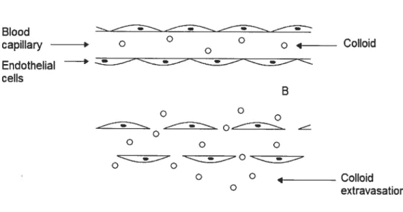
One way in which colloids achieve selectivity is a resuit of their large size which restricts extravasation to locations in the body with permeable vasculature (see figure 1). Solid tumors and sites of infection or inflammation ofien have porous blood capillaries, which allow for the passage of nano-sized colloidal drug carrier across the endothelium and into the extravascular space (passive targeting). Given that the majority of the vascular endothelium is continuous with tight junctions between neighboring endothelial ceils, active compounds associated with colloïdal carriers are prevented from reaching the extravascular space of most tissues in the body reducing many ofthe adverse side effects caused by drugs in the free form [2].
A BiooU capiIary o ° o 4 Colloid Endothelial —-ceNs B o o —-- ____;;—__ o o o o ---—-——o O 4 CoNoid O extravasation
figure 1: Capillary structure of normal tissues with a continuous endothelial lining (A) and tumors possessing enhanced vascular permeability (B).
4 In addition to the selectivity imparted by size, targeting moieties that are specific for determinants found primarily or in high amounts on the membrane of target celis can be attached to the surface ofthe carrier to enhance specificity (active targeting).
Another application of colloids is in the controlled release of therapeutics, whereby they act as reservoirs that release the encapsulated drug into the blood stream slowly. Such sustained release systems can maintain therapeutic drug levels in the blood, reducing the frequency of administration. Furthermore, colloidal drug delivery systems can also overcome efflux pumps such as P-glycoprotein (Pgp) by changing the pathway in which drugs enter the ceil. Intracellular intemalization of colloids by endocytosis Iocates the drug in an endosome/lysosome, which reduces interactions with Pgp compared to drugs in the free form that traverse across the ceil membrane by diffusion [3]. Other advantages of colloidal drug delivery systems include protection of the encapsulated drug from premature degradation and enhanced intracellular delivery of certain therapeutic compounds. For instance, free or un encapsulated genetic material requires a carrier to enter the ceil because of the unfavorable electrostatic interactions between DNA and ceil membranes.
Depending on their size and the physicochemical properties of the surface, colloids can be rapidly taken up by the ceils of the mononuclear phagocyte system (MPS) and quickly removed from the systemic circulation. Such systems are ideal for macrophage targeting. On the other hand, colloids can exhibit long-circulating properties in blood and target sites in the body other than MPS tissues.
Indeed, colloidal drug delivery systems offer many benefits over drugs in the free form. These systems, however, vary in terms of physicochemical properties and
thus have different advantages and drawbacks. An overview of the most common colloidal drug delivery systems is briefly presented in the following sections.
2.
Liposomes
Among the various colloidal drug delivery systems investigated, liposomes are the most widely studied. They can be formed from either synthetic lipids or lipids originating from biological membranes. Liposomes typically range in size from 50—
10,000 nm and are classified as either small unilamellar vesicles (SUVs), large unilamellar vesicles (LUVs) or multilamellar vesicles (MLV5). LUVs have diameters usually between 100 to 500 nm and are bigger than SUVs [4]. They are composed of an aqueous muer core surrounded by a single lipid bilayer. In contrast, MLVs contain several concentric lipid bilayers and vary in size from 100 to 10,000 nm [4]. These unique structures permit the encapsulation of either hydrophilic or hydrophobic compounds. The behavior of these systems in the host depends largeÏy on size, bilayer rigidity and surface charge [5, 6].
Based on composition and therapeutic application, liposomes are generally categorized into 4 major types which are conventional, ‘stealth’ or long-circulating, immunoliposomes (targeted), and cationic liposomes [7]. Conventional liposomes are usually quickly removed from the systemic circulation by the MP$ and thus are more appropriate for macrophage targeting, local depot or antigen delivery (vaccination)
[7]. Conversely, ‘stealth’ or long-circulating liposomes evade detection by the MPS
and tend to extravasate into tissues with enhanced vascular permeability (i.e. solids tumors and sites of infection or inflammation) [8-10]. The most widely used method to produce liposomes with enhanced blood residence times is to coat the surface of
6
the carrier with poly(ethylene glycol) (PEG). This polymer provides long-circulating properties by reducing interactions with plasma proteins and ceil surfaces because of its highly hydrated and flexible polymer chains [1 1, 12].
The specificity of encapsulated drugs towards the intended location can be enhanced by attaching ligands such as antibodies, antibody fragments, peptides, carbohydrates, vitamins or hormones onto the surface of the carrier that are specific for certain sites on the membrane oftarget celis [13]. If the encapsulated drug is to be delivered to non-MPS tissues, PEG can be grafied onto the surface of the liposome along with the targeting ligands to reduce uptake by macrophages [14]. Apart from being carriers for conventional drugs, cationic liposome complexes are being developed to protect genetic material (e.g. DNA and RNA) from degradation in the blood stream and enhance transfection into the celI [15].
As a resuit of the large effort in developing liposomes as drug delivery vehicles, several liposome formulations are presently on the market, such as
Doxil®/CaelyxTM, Myocet®, DepoCyt®, and AmBisome®, just to name a few [16].
Liposomal products currently on the market offer treatment for a wide range of illnesses including cancer, Kaposi’s sarcoma, ftmgal infections, and meningitis.
Despite the many advantages of liposomal drug delivery systems there are several drawbacks, which include poor efficiency to load hydrophobic moÏecules as a result ofthe limited solubility in the lipid bilayer and poor stability during storage due to hydrolysis and/or oxidation of the lipids in the bilayer. Formulation stability can be improved by preparing the liposomes with saturated lipids and displacing the air with an inert gas such as argon.
3.
Micelles
Micelles are core-sheÏl structures formed from amphiphilic molecules such as low-molecular weight surfactants or block copolymers. These amphiphilic molecules self-assemble in aqueous solvents at concentrations above the critical micelle concentration (CMC). The hydrophobic core provides a cargo space for poorly-water soluble compounds, while the hydrophilic corona permits solubilization in aqueous media [17]. As a resuit of their structure, micelles can considerably enhance the solubility of hydrophobic molecules in water and possibly protect the sequestered dmg from chemical and/or enzymatic degradation in the host.
Micelles can be classified as either Iow-molecular weight surfactant micelles or polymeric micelles depending on the molecular weight of the amphiphile. Low molecular weight surfactant micelles typically have high CMC values and Iow core viscosity, resulting in poor stability upon dilution in solution and in the blood stream after intravenous (i.v.) administration [1$]. In addition, many low-molecular weight surfactants cause adverse side reactions. for instance, Cremophor® EL has been associated with severe hypersensitivity reactions in many patients [19J. These side effects were also observed with polysorbate 80, but to a much lesser extent [20].
Compared to surfactant micelles, polymeric micelles have several advantages, such as reduced toxicity, higher drug loading capacity and greater stability upon dilution due to the lower CMC values, for example, poly(N-vinylpyrrolidone)-b poly(D,L-lactide) (PVP-b-PDLLA) with 27 and 37% DLLA have CMCs of 10 and 6 mg/L, respectively [21]. These values are considerably lower than those of common
8 fow molecular weight surfactants such as Cremophor® EL and polysorbate $0 with CMCs of 90 and 100 mg/L, respectively [22].
PEG is commonly used as the hydrophilic segment of the block copolymer to prolong systemic circulation time and target sites other than the celis ofthe MP$ [23]. Attaching targeting ligands to the hydrophilic block of polymeric micelles can potentially improve drug delivery, although research on these systems is flot well advanced yet [18]. Presently, a novel injectable polymeric micelle formulation of paclitaxel (Genexol®, PEG-b-PDLLA) is being evaluated in Phase II clinical trials in patients with advanced breast and non-small celi lung cancers [24]. This formulation was reported to have lower toxicity and enhanced efficacy in mice compared to the commercial low-molecular weight micelle formulation ofpaclitaxel (Taxol®) [25].
Micelles can also improve the delivery of genetic material by electrostatic complexation of polyanionic DNA with a cationic segment of a block copolymer, linked to a non-ionic, hydrophilic block. Neutralization of the oppositely-charged polyions produces a water-insoluble segment, which forms the core of the micelle in aqueous solvents [26]. The hydrophilic corona solubilizes the complex in aqueous media and enhances stability in biological fluids. $uch systems are referred to as polyion complex micelles.
The main drawbacks of micelles as drug delivery systems include rapid drug leakage from the micelle and dissociation if diluted below the CMC. As a resuit, the kinetics of micelle dissociation and drug diffusion are important parameters to control.
4.
Macromolecular prodrugs
Macromolecular prodrugs (also referred to as drug-polymer conjugates) are delivery systems in which therapeutic agents are covalently linked to a polymer. The polymer can be either naturally-occurring (e.g. starch amylase, pullulans, chitosan) or synthetic (e.g. PEG, poly-amino acids, hydroxypropylmethacrylamide (HPMA), polyvinylalcohol (PVA), polyvinylpyrrolidone (PVP)) [27]. These drug-polymer conjugates are water soluble and generally have high molecular weights (> 40 kDa) to overcome renal excretion and achieve extended plasma haif-lives. These polymeric drugs can attain half-lives in the order of hours as opposed to a few minutes for drugs in the free form [28]. Due to their relatively large size, drug-polymer conjugates cannot diffuse through the celi membrane and thus are usually taken up by endocytosis.
for this system to be efficacious the drug must be released from the polymer at the target site, however, there are exceptions whereby the prodrug is active without cleavage. In general, release of the drug from the polymer can be either pH-triggered or enzymatic [27]. Prodrugs with pH-sensitive bonds can be cleaved in acidic conditions such as the extracellular space of solid tumors and/or the endosomal or lysosomal compartments after internalization by the celi [27]. In the case of enzyme dependent release, the linker attaching the drug to the polymer is usually a peptide spacer such as Gly-Phe-Leu-Gly, which is susceptible to cleavage by intracellular enzymes [29]. Ideally, the macromolecular prodrug should remain stable in the blood circulation until it reaches the target site. Similar to other colloidal drug carriers, targeting moieties can be attached to macromolecular prodrugs to improve selectivity
10
for the target site [30]. Examples of macromolecular prodrugs presently on the market include PEG-interferon alpha, conjugate of neocarzinostatin and poly(styrene comaleic acid) (SMANCS), and PEG-L-asparaginase [27].
Macromolecular prodrugs have two major disadvantages. first, for drugs that are inactive as a conjugate, cleavage from the macromolecule must occur sufficiently fast at the target site in order to achieve greater efficacy than the free drug. Second, non-biodegradable polymers larger than 40 kDa cannot be eliminated by renal filtration and thus will remain in the patient.
5.
Polymeric nanoparticles
Polymeric nanoparticles consist of a dense polymer matrix, which can physically entrap hydrophobic compounds. These particulates range in diameter from
10 — 1,000 nm and can be prepared from either natural or synthetic polymers [2$].
Nanopartic1es are interesting dmg delivery systems due to the dense polymer core, which can considerably sustain or control the release of physically entrapped molecules [31]. The release rate of the drug molecules from the nanoparticle is controlled by the diffusion of the drug through the polymer matrix and the erosion of the nanoparticle [32]. As a resuit of the polymeric matrix, nanoparticles are usually more stable than liposomes and micelles in the presence of biological fluids. In general, without proper surface modification nanoparticles are quickly removed from the systemic circulation by the MP$. Incorporating PEG at the surface greatly enhances the residence time of the nanoparticles in the blood stream.
The preparation of nanoparticles ofien requires organic solvents, which presents a disadvantage for the use of this type of carrier in the clinic. Another
potential problem is the biodegradability and toxicity of the degradation products. Consequently, the polymer must be carefully selected.
6.
Emulsions
Emulsions are heterogeneous mixtures of two immiscible liquids (i.e. ou and water), whereby the one phase is dispersed as fine droplets in the other. The addition of an emulsifier or surfactant provides kinetic stability to the preparation by reducing the interfacial tension and increasing droplet-droplet repulsion through electrostatic and/or steric repulsive forces [33]. Similar to other drug delivery vectors, emulsions can protect the encapsulated drug against hydrolysis and enzymatic degradation in the blood compartment, lower the toxicity of cytotoxic compounds, and can also provide a certain level of selectivity towards target tissues, increasing the therapeutic index of many drugs [34]. The application of emulsions as intravenous drug delivery systems
wiII be discussed in more detail in Chapter 2.
The goal of the present work was to develop long-circulating emulsions and characterize their accumulation into solid tumors. The performance of the emulsions was assessed in vivo in mice bearing either B16 melanoma or C26 colon
adenocarcinoma. The experimental section and resuits of this study are provided in Chapter 3 in the form of a scientific article. A summary of these findings as well as additional data on formulation optimization and stability are presented and discussed in Chapter 4. Finally, the concluding remarks and perspectives are given in Chapter 5.
12
7.
Referen ces
1. Non, A., Kopecek, J. (2005). Intracellular targeting of polymer-bound drugs for cancer chemotherapy. Adv Drug Deliv Rev, 57, 609-636.
2. Jain, R. K. (1988). Determinants of tumor blood flow: a review. Cancer Res, 4$, 2641-265$.
3. St’astny, M., Strohaim, J., Plocova, D., Ulbrich, K., Rihova, B. (1999). A possibility to overcome P-glycoprotein (PGP)-mediated multidrug resistance
by antibody-targeted drugs conjugated to
N-(2-hydroxypropyl)methacrylamide (HPMA) copolymer carrier. Eur J Cancer, 35, 459-466.
4. Ulrich, A. S. (2002). Biophysical aspects of using liposomes as delivery vehicles. Biosci Rep, 22, 129-150.
5. Allen, T. M., Austin, G. A., Chonn, A., Lin, L., Lee, K. C. (1991). Uptake of liposomes by cultured mouse bone marrow macrophages: influence of liposome composition and size. Biochim Biophys Acta, 1061, 56-64.
6. Allen, T. M., Chonn, A. (1987). Large unilamellar liposomes with low uptake into the reticuloendothelial system. FEBS Lett, 223, 42-46.
7. Storm, G., Crommelin, D. J. A. (1992). Liposomes: quo vadis? F$TT, 1, 19-31.
8. Gabizon, A., Papahadjopoulos, D. (1988). Liposome formulations with prolonged circulation time in blood and enhanced uptake by tumors. Froc Nati AcadSci USA, 85, 6949-6953.
9. Metselaar, J. M., Storm, G. (2005). Liposomes in the treatment of inflammatory disorders. Expert Opin Drug Deliv, 2, 465-476.
10. Papahadjopoulos, D., Allen, T. M., Gabizon, A., Mayhew, E., Matthay, K., Huang, S. K., Lee, K. D., Woodle, M. C., Lasic, D. D., Redemann, C., et al. (1991). Sterically stabilized liposomes: improvements in pharmacokinetics and antitumor therapeutic efficacy. Froc Nati Acad Sci U S A, 88,
11460-11464.
11. Lundberg, B. (1994). Preparation of drug - carrier emulsions stabilized with
phosphatidylcholine - surfactant mixtures. Journal of Fharmaceutical
Sciences, 83, 72-75.
12. Blume, G., Cevc, G. (1993). Molecular mechanism of the lipid vesicle longevity in vivo. Biochim Biophys Acta, 1146, 157-16$.
13. Sapra, P., Allen, T. M. (2003). Ligand-targeted liposomal anticancer drugs. Frog Ltpid Res, 42, 439-462.
14. Allen, T. M. (2002). Ligand-targeted therapeutics in anticancer therapy. Nat Rev Cancer, 2, 750-763.
15. Lasic, D. D., Templeton, N. S. (1996). Liposomes in gene delivery. Adv Drug Deliv Rev, 20, 22 1-266.
16. Allen, T. M., Cullis, P. R. (2004). Drug delivery systems: entering the mainstream. Science, 303, 18 18-1822.
17. Allen, C., Maysinger, D., Eisenberg, A. (1999). Nano-engineering block copolymer aggregates for drug delivery. Colloids and Surfaces B: Biointerfaces, 16, 3-27.
18. Le Garrec, D., Ranger, M., Leroux, J. C. (2004). Micelles in anticancer drug delivery. Am JDrug Deliv, 2, 15-42.
19. Nuijen, B., Bouma, M., Schellens, J. H. M., Beijnen, J. H. (2001). Progress in the deve!opment of alternative pharmaceutical formulations of taxanes. Investigational New Drugs, 19, 143-153.
20. Hennenfent, K. L., Govindan, R. (2005). Nove! formulations of taxanes: a review. Old wine in a new bottie. Annals of OncoÏogy, 1-15.
21. Le Garrec, D., Gori, S., Luo, L., Lessard, D., Smith, D. C., Yessine, M. A., Ranger, M., Leroux, J. C. (2004). Poly(N-vinylpyrrolidone)-block-poly(D,L lactide) as a new polymeric solubilizer for hydrophobic anticancer drugs: in vitro and in vivo evaluation. JControl Release, 99, 83-101.
22. van Zuylen, L., Verweij, J., Sparreboom, A. (2001). Role of formulation vehicles in taxane pharmacology. InvestNew Drugs, 19, 125-141.
23. Gaucher, G., Dufresne, M. H., Sant, V. P., Kang, N., Maysinger, D., Leroux, J. C. (2005). Block copo!ymer micelles: preparation, characterization and application in drug delivery. JControl Release, 109, 169-188.
24. Kim, T. Y., Kim, D. W., Chung, J. Y., Shin, S. G., Kim, S. C., Heo, D. S., Kim, N. K., Bang, Y. J. (2004). Phase I and pharmacokinetic study of Genexo!-PM, a cremophor-free, po!ymeric micel!e-formu!ated pac!itaxel, in patients with advanced ma!ignancies. Clin Cancer Res, 10, 3708-37 16.
25. Kim, S. C., Kim, D. W., Shim, Y. H., Bang, J. S., Oh, H. S., Wan Kim, S., Seo, M. H. (2001). In vivo evaluation of polymeric micellar pac!itaxel formulation: toxicity and efficacy. J Control Release, 72, 191-202.
26. Kakizawa, Y., Kataoka, K. (2002). Block copo!ymer micelles for de!ivery of gene and related compounds. Adv Drug Deliv Rev, 54, 203-222.
27. Greish, K., fang, J., Inutsuka, T., Nagamitsu, A., Maeda, H. (2003). Macromolecular therapeutics: advantages and prospects with special emphasis on solid tumour targeting. Clin Pharmacokinet, 42, 1089-1105.
28. Maeda, H. (2001). SMANCS and po!ymer-conjugated macromolecular drugs: advantages in cancer chemotherapy. Adv Drug Deliv Rev, 46, 169-185.
29. Rihova, B., Etrych, T., Pechar, M., Jelinkova, M., Stastny, M., Hovorka, O., Kovar, M., U!brich, K. (2001). Doxombicin bound to a HPMA copo!ymer carrier through hydrazone bond is effective also in a cancer celi une with a !imited content of lysosomes. J Control Release, 74, 225-23 2.
30. Lu, Z. R., Shah, J. G., Sakuma, S., Kopeckova, P., Kopecek, J. (2002). Design of novel bioconjugates for targeted drug delivery. J Control Release, 78, 165-173.
31. Roney, C., Kuikarni, P., Arora, V., Antich, P., Bonte, F., Wu, A., Mallikarjuana, N. N., Manohar, S., Liang, H. F., Kulkarni, A. R., Sung, H. W., Sairam, M., Aminabhavi, T. M. (2005). Targeted nanoparticles for drng de!ivery through the b!ood-brain barrier for Alzheimer’s disease. J Control Release, 108, 193-214.
14
32. Soppimath, K. S., Aminabhavi, T. M., Kuikarni, A. R., Rudzinski, W. E. (2001). Biodegradable polymeric nanoparticles as drug delivery devices. J Control Release, 70, 1-20.
33. friberg, S. E., Quencer, L. G., Hilton, M. L. Theory of Emulsions. In Lieberman, H. A., Rieger, M. M., and Banker, G. S. (eds), Pharmaceutical Dosage Forms, Marcel Dekker, Inc., New York, 1996, pp. 53-90.
34. Barratt, G. (2003). Colloidal drug carriers: achievements and perspectives. CeÏÏ Mol Lfe Sci, 60, 2 1-37.
CHAPTER 2: PRINCIPLES IN THE
DEVELOPMENT 0F INTRAVENOU$ LIPID
EMUL$IONS
Joanna Rossi and Jean-Christophe Leroux*
Canada Research Chair in Drug Delivery, faculty of Pharmacy, University of Montreal, P.O. Box 6128, Downtown Station, Montreal (PQ) CANADA H3C 3J7
Role of Lipid Excipients in Modifying Oral and Parenteral Drug Delivery, Edited by Kishor M. Wasan, Copyright © 2007 John Wiley & Sons, Inc. (in press)
16
1.
Introduction
Emulsions can be defined as heterogeneous mixtures of two immiscible liquids, in which one phase is dispersed as fine droplets in the other. Small ou droplets dispersed in a continuous water phase is termed an ‘oil-in-water’ (olw) emulsion. The opposite ofthis system is a ‘water-in-oil’ (w/o) emulsion, whereby the water phase is dispersed in an oily extemal medium. Among these types, only 01w emulsions can be used for intravenous administration [1]. Emulsions are thermodynamically unstable systems that will eventually destabilize into two separate phases. A third component, the surfactant or emulsifier, is added to stabilize the preparation by reducing the interfacial tension and increasing droplet-droplet repulsion through electrostatic andlor steric repulsive forces [2]. The addition of an emu1siing agent however, only provides kinetic stability. Even though emulsions
are unstable systems, surface active agents may provide stability for several years,
making the system useful for practical application [2].
Lipid emulsions have traditionally been used for parenteral nutrition to deliver essential fatty acids to patients unable to acquire them in food. Due to the successfiil induction of Iipid emulsions in parenteral nutrition, there has been increasing interest in developing emulsions as carriers for lipophilic drugs. Many intravenous lipid emulsion formulations are commercially available (Table 1) and a number of others
are in clinical phase or in preclinical development (Table 2). Lipid emulsions are
promising carriers for drug delivery due to their biocompatibility, reasonable stability, ability to solubilize high quantities of hydrophobie compounds and relative ease of manufacture at an industrial seale [3, 4]. In addition, emulsions can protect the
encapsulated drug against hydrolysis and enzymatic degradation in the blood compartment, reduce drug loss in infusion sets, lower the toxicity of cytotoxic compounds, and reduce the incidence of irritation and pain upon injection [1, 4]. They can also provide a certain level of selectivity towards target tissues, increasing the therapeutic index of many drugs [5]. However, afier intravenous injection, lipid emulsions can acquire apolipoproteins and be metabolized as natural fats or be recognized as foreign bodies and taken up by the ceils of the mononuclear phagocyte system (MPS; also known as the reticuloendothelial system (RES)) [6]. Evading the MPS or natural fat metabolism is necessary when the encapsulated drug is to be delivered to non-MPS organs or liver parenchymal ceils, respectively. 11e in vivo fate of lipid emulsions can be controlled to a certain extent by altering the physicochemical properties of the carrier such as, droplet size, composition and surface properties. This chapter will discuss the main factors to consider when developing emulsions for intravenous injection.
2.
Emulsion stability
Emulsions are thermodynamically unstable systems and will inevitably break apart into separate ou and water phases. Emulsion instability is caused by the increase in surface free energy tAG) as small droplets are formed as a resuit of the enhanced surface area (AA). Adding a surfactant to the mixture reduces the interfacial tension (y0) at the oil-water interface facilitating globule rupture during emulsification and stabilizes the preparation (Eqn 1).
1$
It is important to state that surfactants only provide the emulsions with kinetic stability, which delays the destabilization process. Nevertheless, surface active agents can provide stability for several years, which is long enough for the system to be useful for practical purposes [2]. Emulsions that are thermodynamically stable are known as microemulsions. They are clear or transiucent systems and do flot require much energy input during emulsification. In contrast, emulsions are cloudy and require a greater amount of energy for emulsification [7]. The theory behind the formation of microemulsions is beyond the scope of this chapter.
2.1.
Destabijization processes
Emulsion destabilization can be characterized by three separate processes: flocculation, coalescence and Ostwald ripening. Coalescence and Ostwald ripening are irreversible processes which Iead to an increase in droplet size, requiring a large energy input to re-disperse the droplets. flocculation, on the other hand, is reversible and occurs when droplets aggregate to form a clump of many individual droplets. The aggregated droplets move together as a cluster but each droplet stiil retains its separate identity. The interactions holding the droplets together are weak and can be broken by mild agitation. Even though floccules can be easily re-dispersed, they may eventually fuse together to form single, larger globules. The fusion of droplets is irreversible and is termed coalescence. Ostwald ripening, which also increases droplet size, occurs in polydisperse formulations wherein the smaller droplets are more soluble in the continuous phase than the larger ones. In this process, the oil from the smaller droplets dissolves in the aqueous phase and diffuses towards the larger droplets. This transfer of ou causes the big droplets to grow, while the smaller ones
decrease in size. As the small droplets continue to shrink, the Ostwald ripening effect is enhanced. The progressive increase in droplet size over time will eventually lead to complete phase separation. Adding too much surfactant may promote Ostwald ripening as the excess surfactant will form micelles which enhance the solubility of the ou in the aqueous phase. Ostwald ripening can be reduced by increasing the viscosity of the continuous phase, decreasing polydispersity, or adding a third component which has a lower solubility in the continuous phase than the oil [8, 9].
Depending on the density differences between the dispersed and continuous phases, individual droplets or floccules can cream or sediment. If the dispersed phase is lower in density than the continuous phase, the droplets or floccules will rise to the surface producing a highly concentrated layer of dispersed phase, which is known as a cream. In the case where the dispersed phase is higher in density than the continuous phase, a sediment will form at the bottom of the formulation. for o/w emulsions, creaming usually occurs since the oil phase is typically less dense than the aqueous phase. The rate of creaming or sedimentation can be linked to the size of the droplet by Stokes’ equation (Eqn 2). According to this equation the limiting velocity ofa falling sphere (u) is:
(2)
9v
where a is the radius of the droplet, Ap is the density difference between the dispersed and continuous phases, y js the viscosity of the continuous phase and g is the acceleration due to gravity. Stokes’ equation implies that droplets will rise or settie faster if the droplet size or the density difference between the dispersed and continuous phases is large, while an increase in continuous phase viscosity will slow
20 down the separation process. As a resuit, creaming or sedimentation can be delayed by reducing dropiet size, decreasing the density differences between the two phases and increasing the viscosity of the continuous phase. Not much emphasis, however, is being placed on density adjustments to produce stable emulsions since there are a limited number of oils approved for intravenous administration and these oils have similar densities.
$ubmicron emuisions have coiloidal properties and as a resuit are less susceptible than coarse emuisions to the gravitational forces in Stokes’ equation [10]. Nanosized droplets are subjected to random Brownian motion and consequentiy are less inclined to cream or sediment. Brownian motion, however, does flot provide complete protection against instability since droplets may aggregate or coalesce upon random collisions. Stability against these collisions depends on the attractive and repulsive forces acting on the droplets. Typicaily, emulsions are stabiiized by either electrostatic or steric repulsive forces (or a combination ofthe two).
2.2.
Electrostatic stabilization
The balance between attractive Van der Waais forces and electrostatic repuisive forces is described in the theory of coiloidal stabiiity, termed DLVO afier its developers Derjaguin, Landau, Vervey, and Overbeek. If the net force is attractive, the droplets wiil either flocculate or coalesce. In contrast, if the net force is repulsive, the particles wiil repel each other and the system is stable. The attractive interaction between particles arises from Van der Waals forces and is experienced by ail particies. Van der Waals forces dominate at short separation distances and the strength of this attractive force can be determined from the magnitude of the Hamaker
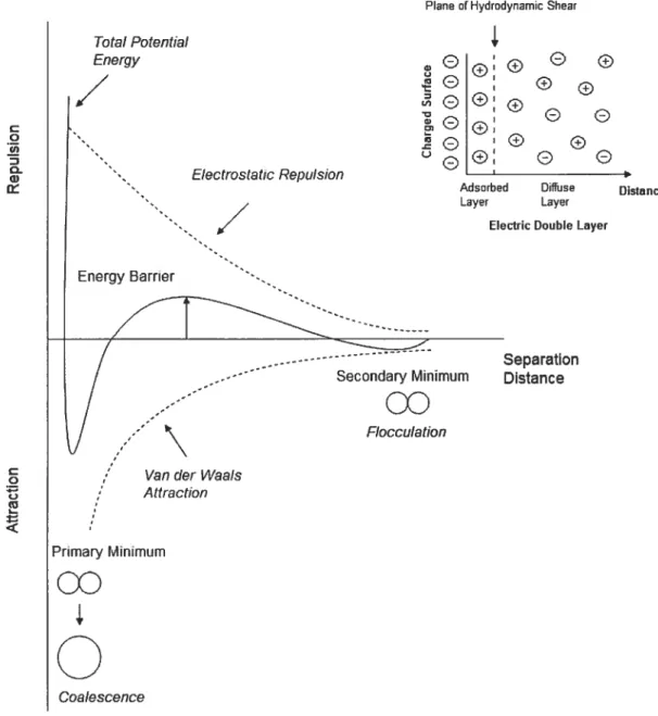
constant (A). Emulsions can overcome the attractive Van der Waals forces through electrostatic repulsion with charged emulsifying agents. Electrostatic repulsion is provided by the electric layer surrounding the droplet. The electric double-layer is characterized by an adsorbed double-layer of fixed counterions and a diffuse double-layer of ions that move freely with the fluid. Two approaching particles will experience a repulsive force as the electric double-layers overlap. The total potential of interaction between two droplets is the sum of the attractive van der Waals forces and the electrostatic repulsive forces (Eqn 3);
V=V+J’Ç, (3)
where VT is the total interaction potential, VA represents the attractive van der Waals forces and VR signifies the electrostatic repulsive forces. The potential energy of interaction between two droplets as a function of separation distance is illustrated in Figure 2. The repulsive barrier generated by the electric double-layer corresponds to the maximum in the curve. The height of the energy barrier determines the stability of the emulsion and depends on the ionization of the surfactants.
for the system to be stable, the energy barrier must be high enough sucli that
the droplets do flot have enough kinetic energy to surpass it and reach the primary minimum. At the primary minimum (maximum attractive potential) droplet coalescence readily occurs. f locculation takes place at the secondary minimum and contrary to coalescence, is reversible by providing a small amount of kinetic energy to overcome the weak attractive forces holding the droplets together. flocculated droplets are prevented from coatescing as a resuit of this repulsive energy barrier. If the flocculated droplets have enough energy to surpass the energy barrier they will
22 easily reach the primary minimum and coalesce. The strength of the electrostatic forces can be quantified by measuring the zeta potential, which is the potential at the plane of hydrodynamic shear. Generally, emulsions are stabilized by electrostatic repulsive forces if the zeta potential is greater than ± 30 mV [2, 8, 1 1]. An emulsion stabilized by electric double-layer repulsion can be destabilized if the concentration of electrolytes is increased above a critical value. Adding electrolytes to an emulsion decreases the electric double-layer repulsion potential, while the van der Waals attractive potential remains unchanged. As electrolyte concentration increases, the repulsive forces stabilizing the colloid become weaker until the net force is attractive and stability is lost.
2.3.
Steric stabilization
Emulsions can also be stabilized by steric repulsion by grafiing long-chain polymers at the emulsion interface. Steric repulsion is a non-DLVO interaction that occurs due to the unfavorable overlap of the polymer chains as two particles approach each other [8, 12]. Steric stabilization occurs at short inter-droplet separation distances and can provide a strong barrier against coalescence [8]. Optimal steric repulsion can be achieved at high polymer surface density as desorption and chain rearrangement is minimized [8].
3.
Elimination mechanisms for lïpid emulsions
Afier intravenous injection, lipid emulsions may be metabolized in a manner similar to chylomicrons or might be recognized as foreign bodies and removed by the ceils of the MPS. The mechanism of elimination from the body depends on the
physicochemical properties of the emulsion. Both mechanisms of elimination can occur for a given lipid emulsion, however, one process may be favored over another. This section describes the two primary pathways of lipid emulsion elimination from the body.
3.1.
Lipid emulsions metabolized as endogenous chylomicrons
Depending on the composition and surface properties, lipid emulsions may be recognized as chylomicrons and be elirninated via the fat metabolism pathway. Chylomicrons are endogenous emulsions produced by the enterocytes of the small intestine afier dietary lipids are ingested. They are rich in triglycerides and possess apolipoproteins A-I, A-IV and 3-48 prior to entering the blood circulation (Figure 3) [13]. Chylomicrons are secreted into the lymph and enter the systemic circulation through the thoracic duct. After entering the blood, chylomicrons obtain Apo C-II and Apo E from the high-density lipoproteins (HDLs) and release Apo A-IV. In the capillaries of adipose tissues and muscle, lipoprotein lipase (LPL) located on endothelial ceils adsorb onto the mature chylomicron and hydrolyze the triglycerides to fatty acids [14]. The fatty acids are then absorbed mainly by adipose tissues and muscle. During lipolysis, a substantial amount of phospholipid, Apo A and Apo C are transferred to the HDLs and the size of the chylomicron is reduced considerably. The remuant chylomicrons composed of mainly Apo B-48, Apo E and cholesterol are quickly removed from the blood by the liver. The uptake of remuant chylomicrons by the liver occurs via two Apo E-specific recognition sites on parenchymal ceils, which are the low-density lipoprotein receptor (LDLr) and the remuant receptor [15-17].
24 Injectable lipid emulsions differ from chylomicrons in that they do flot have apolipoproteins on the surface prior to entering the blood stream, although they may acquire them afier systemic injection. Emulsions rich in triglycerides are known to acquire apolipoproteins (Apo C-I, C-II, C-III, E and possibly Apo A-IV), mainly from HDLs, soon afier injection into the systemic circulation and are metabolized in a pathway comparable to that described for chylomicrons [15, 18, 19]. Among the apolipoproteins acquired Apo C-II and Apo E are essential for LPL activation and uptake of remnant emulsions by the liver, respectively [19].
Elimination of the lipid emulsion via the pathway of natural fat metabolism may be desirable when the liver parenchymal ceÏÏs are the target site. On the other hand, if the target site is flot the liver then apolipoprotein adsorption onto the emulsion should be avoided. The metabolism of lipid emulsions as natural fats is strongly dependent on the type of emulsifier [20, 21], the presence of cholesterol [22] and the chain length ofthe triglyceride ou [23].
3.2.
Elimination by the mononuclear phagocyte system
Ifthe body recognizes the lipid emulsions as foreign, they will be captured by the ceils ofthe MPS, mainly the Kupffer celis ofthe liver and the macrophages ofthe spleen, and removed from the systemic circulation. The MPS takes up the emulsions via endocytosis and localizes them in the lysosomal compartment where they are degraded by enzymes [24]. The extent of emulsion clearance from the systemic circulation is enhanced by the adsorption cf opsonins (proteins) onto the colloid surface. The bound proteins then interact with the receptors on monocytes and macrophages, facilitating endocytosis. Carriers that become bound te opsonins will be
rapidly cleared from the blood and prevented from reaching the target site(s) [24]. Immunoglobins and complement components such as Clq and C3 fragments (C3b, iC3b) are well known opsonins.
A major challenge in drug delivery using colloidal nano-carriers is to avoid clearance by the celis of the MPS when the target sites are non-MPS tissues. Overloading or saturating the MPS with large injections volumes has been shown to enhance the circulation time of lipid emulsions [25]. However, temporary impairment of the MP$ may pose a health hazard to the patient [26J. Altematively, the clearance rate of carriers from the blood can be altered by modifying the physicochemical properties of the emulsion, such as droplet size [27, 28] and surface characteristics [29]. This will be discussed in detail in section 4.
4.
Biodistribution of lipid emulsions
The biodistribution of an emulsion after systemic injection is dependent primarily on the droplet size, composition and surface properties. A certain specificity towards the target site can be achieved by controlling the physicochemical properties of the emulsion. The principle factors that influence the biodistribution of emulsions has already been very thoroughly reviewed in a book chapter by Nishikawa [6]. This section provides a brief overview of these factors and has been updated with some recent work.
4.1.
Effect of lipid emulsion size
It is well known that droplet size greatly influences the uptake of the emulsions by the MP$ [27, 28]. In general, larger particles are more susceptible to
26 uptake by the MPS and are cleared more quickly from the systemic circulation. The influence of droplet size on the in vivo biodistribution of lipid emulsions was explored by Takino et al. [27]. The authors compared the biodistribution of large (250 nm) and small (100 nm) lipid emulsions composed of egg phosphatidylchoÏine (egg PC):soybean ou = 1:1. [‘4C]Cholesteryl oleate ([14C]CO), a highly lipophilic
compound (log P = 18.3) that does flot undergo lipolysis by LPL and remains
associated with the emulsion, was incorporated into each emulsion to track the ehmination of the whole droplet [30]. The large egg PC emulsion was rapidly eliminated from the blood with 60% of the injected emulsion recovered in the liver within 10 min. The small egg PC emulsion, however, remained in the blood for longer and accumulated less in the liver. Similarly, Lundberg et al. [28] reported that droplet size influenced emulsion clearance rate from plasma. They observed that the smallest emulsion (50 nm) survived the longest in plasma, whereas the larger emulsions (100 and 175 nm) were cleared more rapidly (figure 4). The influence of emulsion-like lipid nanocapsule size (20, 50 and 100 nm) on the extent of complement activation and macrophage uptake was evaluated by Vonarbourg et al. [31]. Similar to emulsions, Iipid nanocapsules are core-sheil structures with an oily internal phase that is stabilized by a monolayer of emulsifiers. They differ from lipid emulsions in the physicochemical properties of the hydrophilic/hydrophobic interface. In lipid nanocapsules, the emulsifiers form a semi-rigid sheil, while the interface is more fluid in emulsions. The authors observed that larger lipid nanocapsules were stronger activators of the complement and were taken up more by macrophages than the smaller ones.
The size of the lipid emulsion was also shown to influence lipolysis. Kurihara et al. [32] found that the rate of lipolysis was much faster for the small sized emulsions (‘- 100 nm) in vitro compared to the larger ones (225-416 nm). However, afier intravenous injection of these formulations in rats, they observed that the small sized emulsions remained in plasma longer than the larger ones, which is consistent with the studies of Takino [27] and Lundberg [2$]. Consequently, even though small emulsions were better substrates for LPL, large emulsions were cleared from the blood faster, which suggests a greater uptake by the MPS.
Droplet size also determines the ability of the emulsion to escape the systemic circulation through the blood capillaries and reach the extravascular space. Capillary walls are composed of a single layer of endothelial celis surrounded by a basement membrane. They are classified into three types, continuous (intact), fenestrated or discontinuous (sinusoidal), based on their wall structure [33]. Both fenestrated and discontinuous capillaries have pores in the endothelium, while continuous ones have tightjunctions between adjacent endothelial ceils [34]. Continuous capillaries have an intact subendothelial basement membrane and can be found in most regions of the body such as the skin, connective tissue, skeletal and cardiac muscle, alveolar capillaries of the lung, and the brain [33]. In fenestrated capillaries, the pores (fenestrae) are approximately 40-80 nm in diameter and they can be either open (unobstructed) or covered by a thin diaphragm [33]. These capillaries have a continuous subendothelial basement membrane and are situated in the intestinal mucusa, pancreas, glomerulus, peritubular capillaries, endocrine glands, the choroid plexus ofthe brain and the ciliary body ofthe eye [33]. Discontinuous capillaries, on
2$
the other hand, have large gaps between endothelial ceils and are located in the liver, spleen and bone marrow [33]. The basal membrane is either absent, which is the case for the liver or discontinuous (spleen and bone marrow) [34J. The largest pore size in the capillary endothelium is believed to be approximately 100 nm [35]. Nanoscopic drug carriers are generally too large to diffuse across the capillaries of continuous endothelium. Their best opportunity to escape the systemic circulation is through the gaps between the endothelial ceils of discontinuous capillaries. Consequently, colloidal drug carriers tend to accumulate in the liver, spleen and bone marrow.
Control over carrier size can impart some selectivity for the extravascular space of tumoral sites, reducing anticancer drug toxicity towards healthy tissues. This selectivity can be achieved by taking advantage ofthe difference in capiflary structure between tumors and normal tissues. Tumor vasculature is often characterized as porous or “leaky” allowing enhanced permeation of colloidal particles across the endothelium and into the extravascular space. In addition, tumors have poor lymphatic drainage allowing colloids to be retained in the tissue for longer periods of time [36]. This increased permeation and retention of colloids is called the enhanced permeation and retention (EPR) effect [37]. The optimum size range for colloidal particle accumulation in tumors is generally accepted to be approximately 5 0-200 nm [38]. Particles in this size range can be convected from the blood vessel into the extravascular space through the porous vasculature of the tumor. Depending on the porosity of the tumor capillaries, particles above 200 nm may not pass through the pores and will be eliminated more quickly by the MPS. On the other hand, particles
less than 50 nm will easily extravasate through the discontinuous endothelium of the liver, spleen and bone marrow.
As a rule of thumb, for successffil accumulation of dmg in the tumor by the EPR effect, the concentration of colloidal carriers in the plasma must remain high for more than 6 hours [39]. The progressive extravasation of the carrier into the tumor tissue over several hours wiii resuit in increasing concentrations of anticancer drug in the vicinity of the cancer ceils. Kurihara et al. [40] demonstrated that lipid emulsions under 230 nm in diameter could deliver more RS-1541, a highly lipophilic antitumor agent (13-0-palmitoyl-rhizoxin), to the tumor site (M5076 sarcoma ceils) than larger dropiets (Figure 5). The low concentrations of RS-1541 detected in the tumor for the larger emuisions is most likeiy due to the impermeability of the leaky tumor capiilaries to large particles and their faster removal rates from blood. It was also observed that emulsions greater than 220 nm reduced the toxicity of R$-1541 as shown by the higher maximum tolerated dose (MTD) with increasing size (Table 3). Ail emulsions regardless of size (70 — 380 nm) suppressed tumor growth and
improved survival at the MTD. The medium sized emulsions (220 nm), however, displayed the highest antitumor activity at the MTD due to the permeability of the tumor vasculature for the emulsions and reduced toxicity, permifting the injection of a higher dose. Hence, lipid emulsions can augment the delivery of cytotoxic compounds to tumoral sites and reduce systemic toxicity by suitable selection of the droplet size.
30
4.2.
Effect of
lipid emulsion composition and emulsifiers
Composition ofthe oit phase
The composition of the internai phase has aiso been shown to alter the biodistribution of lipid emulsions. Lutz et al. [41] observed that lipid emuisions composed of medium-chain triglycerides (MCTs) were cleared from plasma more quickly than those prepared with long-chain triglycerides (LCT5). This is probably due to the faster hydrolysis of MCTs by LPL and hepatic lipases compared to LCTs as a resuit of the greater solubility and mobility of shorter chain triglycerides at the oil/water emulsion interface [42].
Adding free cholesterol bas also been shown to alter the metabolism of triglyceride emuisions. Maranhao et al. [22] observed that emulsions with low free cholesterol content (< 4, % w/w) were metabolized in a maimer similar to chylomicrons, as shown by the faster removal rate of triglycerides from the blood than CO due to LPL mediated hydrolysis of the ou and greater uptake of CO than triglycerides by the liver. In contrast, emulsions with high free cholesterol (>16, % w/w) displayed similar triglyceride and CO removal rates from blood and equal uptake by the liver. The group also observed that emulsions containing high ftee cholesterol bound less Apo A-I, Apo A-IV and Apo C and more Apo E in vitro. Apo C-II us essential for LPL binding and activation and hinders liver uptake, while Apo E facilitates emulsion uptake by the liver. Hence, the presence of free cholesterol may modify the metabolism of the droplets by altering the binding of apolipoproteins onto the surface.