T
T
H
H
È
È
S
S
E
E
En vue de l'obtention du
D
D
O
O
C
C
T
T
O
O
R
R
A
A
T
T
D
D
E
E
L
L
’
’
U
U
N
N
I
I
V
V
E
E
R
R
S
S
I
I
T
T
É
É
D
D
E
E
T
T
O
O
U
U
L
L
O
O
U
U
S
S
E
E
Délivré par l’Université Toulouse III – Paul Sabatier Spécialité : Sciences et Génie des Matériaux
JURY
Zeger HENS Professeur Université de Gand Président
Jose Francisco MARCO Professeur Université Autonome de Madrid Rapporteur
Andre VANTOMME Professeur Université de Louvain(KUL) Rapporteur
Antoine VAN ALBOOM Professeur Université de Gand Examinateur
Eddy DE GRAVE Professeur Université de Gand Co-directeur de thèse
Christophe LAURENT Professeur Université de Toulouse Directeur de thèse
Ecole doctorale : Sciences de la Matière
Unité de recherche : Centre Interuniversitaire de Recherche et d’Ingénierie des Matériaux Directeurs de Thèse : Ch. Laurent et E. De Grave
Présentée et soutenue par
Valdirene Gonzaga de Resende
le 3 avril 2009
Synthesis and characterization of Fe-containing oxide powders and carbon nanotube-Fe-oxide nanocomposites
T
T
H
H
È
È
S
S
E
E
En vue de l'obtention du
D
D
O
O
C
C
T
T
O
O
R
R
A
A
T
T
D
D
E
E
L
L
’
’
U
U
N
N
I
I
V
V
E
E
R
R
S
S
I
I
T
T
É
É
D
D
E
E
T
T
O
O
U
U
L
L
O
O
U
U
S
S
E
E
Délivré par l’Université Toulouse III – Paul Sabatier Spécialité : Sciences et Génie des Matériaux
JURY
Zeger HENS Professeur Université de Gand Président
Jose Francisco MARCO Professeur Université Autonome de Madrid Rapporteur
Andre VANTOMME Professeur Université de Louvain(KUL) Rapporteur
Antoine VAN ALBOOM Professeur Université de Gand Examinateur
Eddy DE GRAVE Professeur Université de Gand Co-directeur de thèse
Christophe LAURENT Professeur Université de Toulouse Directeur de thèse
Ecole doctorale : Sciences de la Matière
Unité de recherche : Centre Interuniversitaire de Recherche et d’Ingénierie des Matériaux Directeurs de Thèse : Ch. Laurent et E. De Grave
Présentée et soutenue par
Valdirene Gonzaga de Resende
le 3 avril 2009
Synthesis and characterization of Fe-containing oxide powders and carbon nanotube-Fe-oxide nanocomposites
Faculteit Wetenschappen
Vakgroep WE05 Subatomaire en Stralingsfysica
Onderzoeksgroep NUMAT
Synthesis and characterization of Fe-containing oxide powders and
carbon nanotube-Fe-oxide nanocomposites
Valdirene Gonzaga de Resende
Proefschrift voorgelegd tot het behalen van de academische graad van
Doctor in de Wetenschappen - Chemie
Promotor: Prof. Dr. E. De Grave
Copromotor: Prof. Dr. Ch. Laurent
Academiejaar 2008-2009
This work would not have been possible without the support and encouragement of my promoter, Dr. Eddy De Grave, under whose supervision I chose this topic and began the thesis work. Dr. Christophe Laurent, co-promoter of the thesis, has also been abundantly helpful, and has assisted me in numerous ways. I am grateful also to Dr. Robert Vandenberghe for his insightful comments.
Many thanks to Dr. Geraldo Magela da Costa (Universidade Federal de Ouro Preto – Brazil) for his assistance and endless patience before and during my Mater studies and for helping me in part of the syntheses and characterization of samples involved in my PhD research, to Dr. Sigrid Eeckhout (European Synchrotron Radiation Facility – ESRF, France) and Dr. Gabriele Giuli (Università di Camerino – Italy) for performing and interpreting the Fe K-edge XANES measurements.
I would also like to thank the Bijzonder Onderzoeksfonds (BOF) program of the UGent that has supported me during my four years of research, and the Fonds voor Wetenschappelijk Onderzoek – Vlaanderen (FWO) that generously provided the financial means to keep the Mössbauer lab running in optimum conditions.
I cordially express my gratitude for the kind support by the technical staff of the Department of Subatomic and Radiation Physics: Mr. Rudi Verspille, Mr. Philippe Van Auwegem, Mrs. Linda Schepens, Mrs. Daniëlla Lootens, Mrs. Brigitte Verschelden, Mr. Patrick Sennesael, Mr. Christophe Schuerens and Mr. Roland De Smet.
I am also grateful to all people that helped me during my stays at the Université-Paul Sabatier: Dr. Anne Cordier, Dr. Alain Peigney, Dr. Alicia Weibel, Dr. Felipe Legorreta García, Mr. Lucien Datas and Mr. Julien Gurt Santanach.
All my lab buddies from the Department of Subatomic and Radiation Physics, Julieth Alexandra Mejía Gómez, Carlos Andrés Palacio Gómez, Khaled Mostafa, Abdurazak Alakrami, Caroline Van Cromphaut,... it has been a real pleasure to work with you and to discuss about our research endeavours.
Many thanks to Alexa, Carlos and Francine, for their friendship, help and encouragements in all these years. ¡Muchísimas gracias! Dank u wel!
I would like to address special thanks to my husband, Orlando, who deserves my deepest gratitude for his unconditional support, love and encouragement.
Last, but not least, I wish to express my deepest and most sincere gratitude towards my family. Their love and efforts, on which I have relied throughout my life, have made it possible to reach this stage of my scientific education.
Summary ……… i
Samenvatting ……….. iii
Résumé ………... vii
PART I Synthesis and characterization of Fe-containing oxide powders and carbon nanotube-Fe-oxide nanocomposites 1 1 Introduction ……….. 3
2 Literature Survey ………. 5
2.1 Carbon nanotubes ………... 5
2.2 Synthesis of CNTs by CCVD method ………... 6
2.2.1 Formation of carbon filaments ……… 7
2.2.2 Formation of CNTs ……….. 8
2.3 Synthesis of CNT-nanocomposite powders by the method developed by CIRIMAT …..…...……… 11
2.4 About Fe and Fe-carbide species ....………... 16
3 Methodology ……….. 19 3.1 Synthesis procedures ……….. 19 3.2 Characterization ……….. 22 3.2.1 Mössbauer spectroscopy ……….. 23 3.2.2 X-ray diffraction ……….. 24 3.2.3 Thermal analysis ……….. 25
3.2.4 Specific surface area measurements …………... 25
3.2.5 Carbon analysis ………... 26
3.2.6 Raman spectroscopy ………... 26
3.2.7 Electron microscopy ………... 27
Paper I: Synthesis of -(Al1-xFex)2O3 solid solutions from
oxinate precursors and formation of carbon nanotubes from the solid solutions using methane or ethylene as carbon source ... 35
Paper II: Presence of metallic Fe nanoclusters in α-(Al,Fe)2O3
solid solutions ……… 53
Paper III: Catalytic chemical vapor deposition synthesis of single- and double-walled carbon nanotubes from
-(Al1-xFex)2O3 powders and self-supported foams .... 63
Paper IV: In situ high-temperature Mössbauer spectroscopic study of carbon nanotube-Fe-Al2O3 nanocomposite
powder ………. 75
Paper V: Surface composition of carbon nanotubes-Fe-alumina nanocomposite powders: an integral low-energy electron Mössbauer spectroscopic study ………. 89
Paper VI: Integral low-energy electron Mössbauer spectroscopic studies of the surfaces of carbon nanotube-nanocomposite powders ……….. 97
Paper VII: Fe-substituted mullite powders for the in-situ synthesis of carbon nanotubes by catalytic chemical vapor
deposition ……….. 105
Bibliography ………... 119 Author’s publications not included in thesis .……….. 123
Summary
The goal of this PhD research project was the synthesis and characterization of a number of Fe-containing oxide systems and of the carbon nanotube-nanocomposites obtained by subjecting these oxide materials to a catalytic chemical vapor deposition (CCVD) process using methane or ethylene as carbon source. Various analytical techniques have been applied to characterize the involved samples, such as carbon analysis, BET gas adsorption measurements, X-ray diffraction, thermal analysis (TGA and DTA), transmission and scanning electron microscopy, and Raman and Mössbauer spectroscopy.
First, the synthesis of -(Al1-xFex)2O3 (x = 0.02, 0.05, 0.07 and 0.10) solid solution
powders with high specific surface area (200-230 m2/g) by the decomposition of mixed oxinate [(Al1-xFex)(C9H6ON)3] in air at 800 °C and the potential of these
powders as catalytic materials for the synthesis of carbon nanotubes were investigated. The nanocomposite powders prepared by reduction in H2-CH4 at
1000°C contain carbon nanotubes which are mostly double-walled. However, a fair amount of undesirable carbon nanofibers, hollow carbon particles and metal particles covered by carbon layers were found to be present. By contrast, for nanocomposite powders prepared by reduction in N2-C2H4 at 800°C the carbon deposit is much more
abundant and homogeneous and consists in multi-walled carbon nanotubes which however contain a significant proportion of defects.
The syntheses of α-(Al,Fe)2O3 solid solution powders were performed by the
calcination in air of the different -(Al1-xFex)2O3 solid solutions at high temperatures
(~1100 °C). It was for the first time evidenced and proved by different characterization techniques the presence of metallic iron nanoclusters consisting of only a few number of atoms within the α-(Al,Fe)2O3 solid solution grains. The
formation of these nanoclusters is thought to be a consequence of the phase transition which implies structural rearrangement on both the cationic and anionic sublattices.
A comparison of the potential interest of both α-(Al,Fe)2O3 powders and
self-supported foams, as starting materials for the synthesis of carbon nanotubes was performed. Contrary to some expectations, using foams do not lead to an easier reduction of the solid solutions and thus to the formation of more α-Fe and/or -Fe-C potentially active nanoparticles for the formation of carbon nanotubes. There is thus no gain in the quantity of carbon nanotubes. However, using foams strongly favors the selectivity of the method towards walled carbon nanotubes (60 %
single-walled and 40 % double-single-walled carbon nanotubes) compared to what is obtained using powders (5 % single-walled, 65 % double-walled and 30 % multi-walled carbon nanotubes).
The thermal stability in air of a carbon nanotube-Fe-Al2O3 nanocomposite
powder was studied. The as prepared nanocomposite powder contained -Fe, Fe3C,
-Fe-C, (Al,Fe)2O3 and a weakly abundant Fe2+ phase as shown by the Mössbauer
results. The results have shown that Fe3C decomposes at relatively low temperatures,
while α-Fe and -Fe-C remain quite stable up to 670 °C and 850 °C, respectively. The oxidation of the Fe3C and Fe particles results in the formation of hematite
(α-Fe2O3).
Information about the composition of the surface layers of the grains that form the carbon nanotube-Fe-Al2O3 nanocomposite powders was obtained for the first
time from a comparative study of the transmission and emission Mössbauer spectra, the latter ones acquired by the Integral Low-energy Electron Mössbauer Spectroscopy (ILEEMS) technique. Several samples were prepared by reduction of an -Al1.8Fe0.2O3 powder in a H2-CH4 atmosphere and changing only the maximum
temperature (800 – 1070 °C). The nature of the iron species (Fe3+, Fe3C, -Fe,
-Fe-C) is correlated to their location in the material. In addition, carbon nanotubes-Fe-MgAl2O4 nanocomposite powders were studied by transmission and emission
Mössbauer spectroscopy as well. The studies have demonstrating that emission Mössbauer spectroscopy is a promising tool complementing transmission Mössbauer spectroscopy for the investigation of the location of the metal Fe and iron-carbide nanoparticles in the carbon nanotube-nanocomposite materials.
Finally, iron-substituted mullite powders [(Al,Fe)6Si2O13] were prepared by the
combustion route and calcined in air (800 – 1400 °C) in order to increase their crystallinity. The thermal treatment resulted in a mixture [(Al,Fe)6Si2O13] and
α-(Al,Fe)2O3. The influence of the calcination temperature of the material on the
formation of nanocomposite powders by reduction in H2-CH4 at 1050 °C was
Samenvatting
Het doel van deze doctoraatsstudie omvatte de synthese en karakterisering van een aantal Fe-houdende oxidesystemen en van de (samengestelde) koolstofnanobuisjes-nanocomposietpoeders bekomen door deze systemen te onderwerpen aan een CCVD-proces (catalytic chemical vapor deposition) met methaan of ethyleen als bron voor koolstof. Diverse analytische technieken werden toegepast, zoals analyses voor de bepaling van het koolstofgehalte, BET-metingen ter bepaling van de specifieke oppervlakte, X-straal diffractiemetingen, thermische analyses (TGA en DTA), transmissie en scanning elektronenmicroscopie, raman- en mössbauer-spectroscopie.
In eerste instantie werden -(Al1-xFex)2O3-mengpoeders (x = 0.02, 0.05, 0.07 en
0.10) en met hoge specifieke oppervlakte van 200-230 m²/g bereid. De synthese geschiedde door thermische decompositie aan de lucht van de corresponderende (Al,Fe)-oxinaten (Al1-xFex)(C9H6ON)3 bij 800 °C. Het potentieel van die
-aluminapoeders als katalysators voor de productie van koolstofnanobuisjes werd bestudeerd. De nanocomposietpoeders bekomen door reductie in een (H2-CH4
)-atmosfeer bij 1000 °C bevatten naast koolstofnanobuisjes, die hoofdzakelijk van het dubbelwandig type zijn, een aanzienlijke hoeveelheid aan ongewenste koolstofvormen zoals nanovezels, holle koolstofdeeltjes en koolstoflagen die metallische deeltjes omsluiten. Indien daarentegen reductie van de -(Al1-xFex)2O3
-poeders wordt uitgevoerd bij 800 °C in een (N2-C2H4)-atmosfeer, dan blijkt het aantal
nanobuisjes ten aanzien van ongewenste koolstofvormen talrijker te zijn en hun morfologie vertoont een hogere graad van homogeniteit. Echter is het merendeel van de koolstofbuisjes meerwandig en structurele defecten zijn overvloedig aanwezig.
In een volgend stadium werden -(Al,Fe)2O3-poeders bereid door uitgloeiing in
lucht bij relatief hoge temperatuur (~1100 °C) van de voormelde -(Al1-xFex)2O3
-samenstellingen. Voor het eerst werd vastgesteld en met behulp van diverse karakteriseringstechnieken ondubbelzinnig aangetoond dat bij de transformatie van - naar -alumina intragranulaire nanoclusters gevormd worden bestaande uit een klein aantal Fe-atomen. Er wordt aangenomen dat de herschikking van de kationen en anionen die met de -fasetransitie gepaard gaat aan de basis ligt van de clustervorming.
Vervolgens werd een vergelijkende studie uitgevoerd in hoeverre -(Al,Fe)2O3
zijn voor de synthese van koolstofnanobuisjes. In tegenstelling tot de verwachtingen blijkt de aanwending van schuimen niet te leiden tot een meer performante reductie, en dus ook niet tot de vorming van een groter aantal nanodeeltjes van -Fe en
-Fe-C die geacht worden de katalysators te zijn voor de vorming van de nanobuisjes en zodoende de kwaliteit van die buisjes bepalen. In vergelijking met poeders blijkt het gebruik van schuimen het aantal nanotubes niet te doen toenemen, maar wel de selectiviteit te bevorderen voor de vorming van enkelwandige nanobuizen boven meerwandige, nl. 60% enkelwandige en 40% dubbelwandige, tegenover 5% enkelwandige, 65% dubbelwandige en 30% meerwandige na reductie van de poeders.
De thermische stabiliteit in lucht van een koolstofnanobuis-(Fe-Al2O3
)-nanocomposietpoeder werd vervolgens in detail bestudeerd. Met behulp van mössbauerspectroscopie werd vastgesteld dat het betrokken materiaal is samengesteld uit -Fe, Fe3C, -(Fe-C), (Al,Fe)2O3 en, in geringe mate, een Fe2+-fase.
De resultaten hebben uitgewezen dat Fe3C reeds ontbindt bij een relatief lage
temperatuur, terwijl -Fe en -(Fe-C) stabiel blijven tot temperaturen van respectievelijk 670 °C en 850 °C waarna ze oxideren tot hematiet ( -Fe2O3).
Voor het eerst werd unieke informatie ingewonnen omtrent de samenstelling van de oppervlaktelagen van de korrels die de bouwstenen zijn van de samengestelde koolstofnanobuisjes-(Fe-Al2O3)-nanocomposietpoeders. Dit geschiedde in een
vergelijkend studie van transmissie- en emissie-mössbauerspectra, waarbij deze laatste bekomen werden met behulp van Integral Low-Energy Electron Mössbauer Spectroscopy (ILEEMS), die in de eerste plaats gevoelig is voor oppervlaktespecies. Meerdere specimens werden bereid door reductie van een -Al1.8Fe0.2O3-powder in
een (H2-CH4)- atmosfeer waarbij de eindtemperatuur (800 – 1070 °C) werd
gevarieerd. Er werd vastgesteld dat de aard van de ijzerspecies (Fe3+, Fe3C, -Fe,
-Fe-C) gecorreleerd is aan hun locatie in de composietkorrels. Verder werden eveneens een aantal koolstofnanobuisjes-Fe-MgAl2O4-nanocomposietpoeders
onderzocht met behulp van zowel transmissie- als emissiemössbauerspectroscopie. Er werd aangetoond dat ILEEMS een veelbelovende techniek is die, in combinatie met de conventionele transmissie-mössbauerspectroscopie, waardevolle informatie verstrekt omtrent de locatie van de metallische ijzerdeeltjes en de ijzer-carbidedeeltjes in de betrokken ijzerhoudende koolstofnanobuisjes-nanocomposieten.
Tenslotte werden Fe-gesubstitueerd mulliet poeders [(Al,Fe)6Si2O13] bereid door
zeer snelle verbranding van een oplossing van de gepaste Fe-, Al- en Si-zouten met ureum. Om de graad van kristalliniteit van het aldus bekomen poeder te bevorderen, werden stalen ervan uitgegloeid in lucht bij diverse temperaturen in het gebied 800 °C tot 1400 °C. Deze thermische behandelingen resulteerden in de vorming van een
mengsel van (Al,Fe)6Si2O13 en -(Al,Fe)2O3. Vervolgens werd de invloed bepaald
van de calcinatietemperatuur op de vorming van nanocomposietpoeders die werden gevormd in een (H2-CH4)-atmosfeer bij 1050 °C.
Résumé
Le but de cette thèse de Doctorat est la synthèse et la caractérisation de plusieurs oxydes contenant du fer et de nanocomposites contenant des nanotubes de carbone (NTC), préparés par dépôt chimique catalytique en phase vapeur (CCVD) en utilisant les oxydes comme matériaux catalytiques et le méthane ou l’éthylène comme source de carbone. Différentes techniques analytiques ont été utilisées pour caractériser les matériaux, comme l’analyse élementaire du carbone, l’adsorption de gaz (méthode BET), la diffraction des rayons X (DRX), l’analyse thermogravimétrique (ATG), l’analyse thermique différentielle (ADT), la microscopie électronique à balayage (MEB) et en transmission (MET), la spectroscopie Raman et la spectroscopie Mössbauer.
Tout d’abord, des poudres de solutions solides -(Al1-xFex)2O3 (x = 0,02; 0,05;
0,07 et 0,10) de grande surface spécifique (200-230 m2/g) ont été préparées par décomposition sous air à 800°C des oxinates mixtes [(Al1-xFex)(C9H6ON)3]. Leur
potentiel comme matériaux catalytiques pour la synthèse des NTC a été étudié. Les poudres nanocomposites préparées par réduction sous H2-CH4 à 1000°C contiennent
des NTC qui sont principalement biparois. Cependant, une assez forte proportion d’espèces carbonées indésirables (nanofibres, nanoparticules creuses) et de particules métalliques recouvertes de carbone est également observée. En revanche, dans les poudres nanocomposites préparées par réduction sous N2-C2H4 à 800°C, le dépôt
carboné est bien plus abondant et homogène, et consiste en des NTC multi-parois contenant cependant une proportion significative de défauts.
La synthèse des poudres de solutions solides α-(Al,Fe)2O3 a été faite par
calcination sous air des solutions solides -(Al1-xFex)2O3 à température élevée
(~1100°C). Différentes techniques de caractérisation ont mis en évidence et démontré, pour la première fois, la présence de nanoclusters de fer métallique ne contenant que quelques atomes au sein des grains de solution solide α-(Al,Fe)2O3. Il
est proposé que la formation de ces nanoclusters est une conséquence de la transition de phase , qui implique un réarrangement structural à la fois sur les sous-réseaux cationique et anionique.
Une comparaison de l’intérêt potentiel des solutions solides α-(Al,Fe)2O3 sous forme de poudres et de mousses autosupportées comme matériaux
catalytiques pour la synthèse des NTC a été effectuée. Contrairement à certaines attentes, l’utilisation des mousses ne permet pas une réduction plus poussée des solutions solides et donc ne permet pas la formation de plus de nanoparticules de
α-Fe et/ou -Fe-C potentiellement actives pour la formation des NTC. On n’obtient donc pas plus de NTC. Cependant, l’utilisation des mousses favorise fortement la sélectivité de la méthode en faveur des NTC monoparois (60% monoparois et 40% biparois) par rapport à ce qui est obtenu à partir des poudres (5% monoparois, 65% biparois et 30% multiparois).
La stabilité thermique sous air d’une poudre nanocomposite NTC-Fe-Al2O3 a été
étudiée. L’analyse des résultats de spectroscopie Mössbauer a montré que la poudre contient initialement plusieurs espèces du fer : -Fe, Fe3C, -Fe-C, (Al,Fe)2O3 ainsi
qu’une phase du Fe2+ en faible proportion. Il a été montré que Fe
3C décompose à
température relativement basse tandis que α-Fe et -Fe-C sont stables jusqu’à respectivement 670°C and 850°C. L’oxydation des particules de Fe3C et de Fe
provoque la formation d’hématite.
Une étude comparative des spectres Mössbauer en transmission et en émission, ces derniers étant obtenus par la technique Integral Low-energy Electron Mössbauer Spectroscopy (ILEEMS), a permis pour la première fois d’obtenir des informations sur la composition des couches de surface des grains de poudre nanocomposite NTC-Fe-Al2O3. Plusieurs échantillons ont été préparés par réduction d’une poudre
-Al1,8Fe0,2O3 sous H2-CH4, en changeant uniquement la température maximale (800
– 1070°C). La nature des espèces du fer (Fe3+, Fe
3C, -Fe, -Fe-C) a été corrélée à
leur localisation dans le matériau. De plus, des poudres nanocomposites NTC-Fe-MgAl2O4 ont été également étudiées par spectroscopie Mössbauer en transmission et
en émission. Les résultats ont démontré que la spectroscopie Mössbauer en émission est une technique complémentaire prometteuse pour la localisation des nanoparticules de fer et carbure de fer dans de tels matériaux nanocomposites.
Enfin, des poudres de mullite substituée par du fer [(Al,Fe)6Si2O13] ont été
préparées par combustion et calcinées sous air à différentes températures (800 – 1400°C) pour accroître leur cristallinité. Ce traitement thermique résulte en la formation d’un mélange de [(Al,Fe)6Si2O13] et de α-(Al,Fe)2O3. L’influence de la
température de calcination du matériau sur la formation des poudres nanocomposites par réduction sous H2-CH4 (20 mol% CH4) à 1050°C a été étudiée.
Part I
Synthesis and characterization of
Fe-containing oxide powders and
carbon nanotube-Fe-oxide
nanocomposites
1
Introduction
Carbon nanotubes (hereafter denoted as CNTs) are one of the most widely investigated nanomaterial (about 15 papers in peer-reviewed journals each day) because they show excellent thermal, electrical and mechanical properties besides that they are very small, hollow and monodimensional.
Single-walled, double-walled and multi-walled CNTs can be produced by the decomposition of a carbonaceous gas at temperatures from 600 °C to 1200 °C on very small transition metal particles. To form catalytic particles that are nanometric in size at the relatively high temperature required for the production of CNTs is a challenge.
In most of the papers available in literature, the CNTs are well-characterized by several techniques, but not a lot of studies have concentrated on the catalytic materials itself. Fe-containing catalysts are one of the most common materials used in the CNTs synthesis. They usually produce CNTs with high efficiency. 57Fe Mössbauer spectroscopy offers several advantages for the study of Fe-containing compounds. The spectra, and the parameters derived from these, are sensitive to electronic, magnetic and structural characteristics of the probed material and as such,
57Fe Mössbauer spectroscopy is a useful technique for phase identification and
quantification of mixtures of Fe-containing materials. Therefore, 57Fe Mössbauer spectroscopy technique, applied in the Department of Subatomic and Radiation
Physics (NUMAT team) at the University of Ghent (UGent), can accurately
characterize iron phases present in the powders before and after formation of CNTs. Although Fe species are often involved in CNTs formation, only relatively few CNT-related studies report on Mössbauer data regarding the species present before and after CNTs synthesis.
The Centre Interuniversitaire de Recherche et d’Ingénierie des Matériau (CIRIMAT) at Université Paul-Sabatier (UPS) in Toulouse (France) worked out an original and easily done catalytic chemical vapor deposition method. It is based on selective reduction in H2/CH4 atmosphere at high temperature (800-1070 °C) of
on the surface of the oxide grains and are therefore of a size adequate to be immediately active for the formation of CNTs.
The goals of this work are to perform a profound investigation of Fe/Al2O3 solid
solutions and Fe/mullite powders, and the derived CNT nanocomposites by 57Fe Mössbauer spectroscopy and to correlate the results with those obtained by other techniques with the aim to obtain information on the formation of the CNTs, thermal stability, and surface composition of the CNT-nanocomposite powders. The other techniques apart from Mössbauer spectroscopy were applied at CIRIMAT laboratory during several exchange visits (totally about six months).
This thesis is structured as follows: in the next chapter a literature survey is given. Chapter 3 contains the methodology applied in this work. In Chapter 4 general conclusions are presented. The second part of this work (Part II) contains the original papers, published or submitted, in which a detailed discussion of the results is given.
2
Literature Survey
In the first part of this chapter a brief introduction of what are carbon nanotubes (CNTs) is given. After details of the catalytic chemical vapor deposition (CCVD) synthesis method of CNTs are presented in Sections 2.2 and 2.3. The last section (2.4) of this chapter concerns the Fe and Fe-carbide species.
2.1 Carbon nanotubes
Iijima reported the observation of multi-walled CNTs in 1991 [1]. Two years later, two independent groups, Iijima and Ichihashi [2] and Bethune et al. [3] reported the growth of single-walled CNTs. Since then many laboratories around the world have been studying these forms of carbon.
The structure of the CNT is a hexagonal lattice (graphene sheet) rolled into a seamless cylindrical tube (Figure 2.1a), sometimes with a cap at each end of the cylinder, such that the two caps can be joined to form a fullerene (Figure 2.1b). These tubes may exist inside other cylinders, with an interlayer distance of ~0.34 nm. A tube that is composed of only one cylinder is named as single-walled CNT (SWNT). A double-walled nanotube (DWNT) is composed of two layers (Figure 2.1c) and multi-walled CNTs (MWNT) are built up by multiple coaxial carbon cylinders. Additionally, some pentagonal and heptagonal defects can cause cylinders to bend, change diameter, or twist.
Figure 2.1 Schematic illustrations of a SWNT without (a) and with caps at each end
of the cylinder with half of a fullerene molecule (b), and of a DWNT (c).
(a) (b)
A major feature of the CNT structure is the hexagon pattern (honeycomb) that repeats itself periodically in space. As a result of the periodicity, each atom is bonded to three neighboring atoms. This structure is mainly due to the process of sp2 hybridization during which three hybrid sp2 orbitals are formed at 120 ° to each other within a plane. This covalent bond, referred to as the -bond, is a strong chemical bond. It results in the high stiffness and high strength of a CNT. Additionally, the out-of-plane bond (the π-bond) is relatively weak and contributes to the interaction between the layers in MWNT, and between SWNT’s in SWNT bundles.
The CNTs have diameters in the range between fraction of nanometers and tens of nanometers and lengths up to several tens of micrometers. They are considered as nearly one-dimensional structure according to their high length-to-diameter ratio (103 - 105). Moreover, they also have a large external specific surface area (1315 m2/g for a SWNT closed and isolated). More information about CNTs can be found in several textbooks [4-9].
The remarkable and unique properties of the CNTs have placed them right among the hottest topics of material science. They exhibit excellent mechanical, thermal and electrical properties. These nanoscale carbon structures are potentially interesting for many applications in various fields such as nanoelectronics, energy storage (Li-batteries, supercapacitors), mechanical reinforcement of composite materials (metallic, ceramic and polymer matrices), removing of electrical charges, detection of gases, electromechanical actuators, etc.
2.2 Synthesis of CNTs by CCVD method
The catalytic chemical vapor deposition method is based on the decomposition of a carbonaceous gas on small metal particles. The metal generally used for these reactions are transition metals, such as Fe, Co, and Ni. Additives such as Mo and W are sometimes used. The carbonaceous gas can be either hydrocarbons (CH4, C2H4,
C2H2 and C6H6, generally mixed with H2, N2 or Ar) or CO. The key parameters
controlling the formation of CNTs are the CCVD parameters (hydrocarbons species, gas pressure and flow rate, maximum temperature and treatment duration) on the one hand and the catalytic material parameters (nature and content of the transition metal, additives, specific surface area, powder or foam shaping, etc) on the other hand. CCVD has emerged as the main synthesis route for CNTs because it is based on low-cost technology, can be upscaled and the multitude of parameters, as described above, can be fine-tuned and almost always result in reaching the synthesis objectives in terms of yield and selectivity [8]. Moreover, the CCVD method allows the localized and/or oriented formation of CNTs.
2.2.1 Formation of carbon filaments
As reviewed by Monthioux and Kuznetsov [10], the formation of carbon filaments by catalytic decomposition of carbonaceous gases on metal particles has been known since the XIXth century and hollow filaments have first been reported in 1952 [11]. The formation mechanisms have been hotly debated over the years and this subsection concerns the main formation mechanisms that have been reported earlier than the observation of MWNTs by Iijima [1].
In 1976 Oberlin et al. [12] proposed a formation mechanism for tubular carbon filaments. The carbon filament is formed by a catalytic process involving the surface diffusion of carbon species around the metal particle (Figure 2.2a). New metal hydrocarbon species dissociate on its edges and the carbon layers develop by lateral growth following the external surface of the metal particle, thus producing a carbon shell(Figure 2.2b). This growth exerts a force strong enough to remove the catalytic particle from the substrate (Figure 2.2c). When the catalytic particle is covered by carbon layers at the tip, the diffusion stops and the growth ends (Figure 2.2d). The hollow channel in the centre is due to the fact that no carbon supply can reach the back of the particle.
Figure 2.2 Schematic representation of the formation mechanism of a tubular carbon
filament, adapted from Oberlin et al. [12].
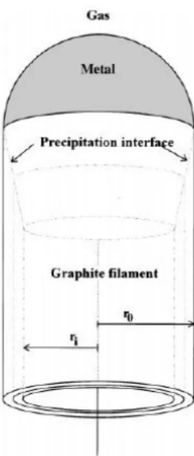
Another explanation for the formation mechanism of the carbon filaments was given by Tibbetts [13]. In this model the author assumed that molecular decomposition and carbon solution occur at one side of the catalytic particle, creating a gradient of concentration in the particle. Consequently, this particle becomes super-saturated and the carbon diffuses to the back face of the particle where precipitation occurs (Figure 2.3).
Figure 2.3 Model for the formation of carbon filaments, showing the inner and the
outer diameters and the precipitation interface. After Tibbetts [13].
2.2.2 Formation of CNTs
Yacamán et al. [14] were the first to report that the hollow carbon filaments prepared for decades were indeed similar to Iijima’s MWNTs [1]. Catalyst particles with diameter higher then 10 nm tend to form MWNTs or carbon nanofibers (henceforward called as CNF). Amelinckx et al. [15] have proposed a model directly related to the well-known Vapor-Liquid-Solid (VLS) mechanism [16] which is not really different from earlier mechanisms [12, 13]. A small metallic particle rests on a larger one that acts as a support (Figure 2.4a). The small particle is lifted away from the support by the deposition of graphene sheets formed from carbon diffusion through the catalyst and through the base (Figure 2.4b and c). The outer diameter of the tube becomes equal to the particle size (Figure 2.4d). A layer of graphite covering the small particle and inhibiting further tip growth of the tube (Figure 2.4e). Figure 2.4f shows tubular layers fed by the supporting particle grown beyond the small particle. The small particle is already covered by a graphite layer during the initial stage (Figure 2.4g); further growth occurs by extrusion through the base and diffusion occurs along the graphite surface (Figure 2.4h). Such a mechanism where the catalyst particle is lifted away is known as tip-growth.
Figure 2.4 Formation model of MWNTs proposed by Amelinckx et al. [15].
Since the diameter of the CNT matches that of the particle, the critical point for the synthesis of SWNTs and DWNTS is the control of the metal particle (catalyst) diameter to a still lower value, i.e., 0.4 - 5 nm. Several hypotheses for the formation of CNTs have been proposed, but an agreement has not yet been reached because that the conditions of synthesis are very diverse, and the phenomenon is quite fast and difficult to observe in situ. However, one of the most probable mechanism (the so-called yarmulke mechanism) was proposed by Dai et al. [17] and accounts for the formation of SWNTs and DWNTs. In this model, a nanometric metal particle (Figure 2.5a) contains a very high fraction of surface atoms and the surface energy per atom is very high. An excess of carbon assembles on the metal particle surface to form a graphene cap (the yarmulke) with its edges strongly chemisorbed to the metal (Figure 2.5b). Because the basal plane of graphite has an extremely low surface energy (10 – 20 times smaller than most metals), the total surface energy diminishes. Newly arriving carbon will continue to assemble on the surface of the catalyst. There are three places for additional carbon to go:
1) The original surface shell can continue to grow around the particle, which if continued would result in over-coating and deactivation of the catalytic particle (Figure 2.5c).
2) A second cap can form underneath the first, spaced by roughly the interspacing of graphite. As additional caps form, older caps are forced to lift
up by forming a cylindrical tube whose open end remains chemisorbed onto the catalytic particle (Figure 2.5d and e).
3) Carbon can add to the cylindrical section of a growing layer. Once the smallest yarmulke has formed, insertion of a new carbon between the tube edge and the catalytic particle is the best solution, as long as, complete over-coating is avoided (Figure 2.5f).
Figure 2.5 Schematic illustration of the yarmulke mechanism adapted by Flahaut et
al. [18] from the description of Dai et al. [17].
By contrast to the mechanisms described above for carbon filaments and MWNTs, the yarmulke mechanism is a base-growth mechanism with no particle lift-off. Using surface-energy calculations, Hafner et al. [19] have shown that particles over 3 nm in diameter will get fully covered particle (Figure 2.5c) and thus not give rise to the formation of a CNT. The experimental works of CIRIMAT, more particularly concerning the synthesis of DWNTs [18, 20-22], have confirmed the yarmulke mechanism. With the aim to perform a study of the distribution of the number of walls and the diameter of the CNTs, hundreds of high-resolution transmission electron microscopy images were collected for isolated CNTs. It was found firstly that a particle ≤ 5 nm in diameter produced a CNT with one or two walls, and secondly, that in the same sample, the inner diameter of DWNTs was lower than the diameter of SWNTs, giving evidence that the outer wall is the first one as proposed in the yarmulke mechanism. It was further found that metal particles with diameter between 6 and 10 nm are generally covered by carbon layers forming
the so-called nanoencapsulated particles and that still larger particles produced MWNTs. Furthermore, very large particles lead to the formation of carbon CNF or nanocapsules.
2.3 Synthesis of CNT-nanocomposite powders by the method
developed by CIRIMAT
There are two main methods to prepare the catalytic material. The most common one [17, 23] is the impregnation of a powder substrate with a solution of a transition metal salt. After drying and calcination, one obtains a dispersion of particles of the catalytic metal oxide on the substrate. The so-obtained catalytic material can be used for the CCVD treatment, although most authors apply a pre-reduction treatment in order to first obtain metal particles on which the catalytic decomposition of the carbon source will lead to CNT formation. The main advantage of this method is that it is extremely versatile (you can use any substrate and metallic salt). However, the usual particles size is relatively large, which is convenient for the synthesis of MWNTs only. In order to decrease the size of the metal particles, one has to decrease the metal loading which results in a poor yield of the eventual SWNTs and DWNTs.
To overcome this, the Nanocomposite and Carbon Nanotubes team of CIRIMAT has proposed a method [24] where the metal particles are formed in situ during the CCVD treatment, at a relatively high temperature, by selective reduction of solid solutions powders based on Al2O3, MgO, MgAl2O4, etc. This method is the one
applied for the syntheses of the various CNT-nanocomposite powders presented in this work and will be discussed in this section. Note that nanocomposite powders were obtained on purpose in order to further prepare dense nanocomposite materials with very homogeneous CNTs dispersions. However, for CNT-metal-MgO powders, the MgO matrix and most of the metal catalyst can be easily dissolved by soaking the powder in an aqueous solution of a non-oxidative acid such as HCl in mild conditions (room temperature, diluted HCl), thus allowing the preparation of a suspension of undamaged CNTs [25].
The method is based on the synthesis of a catalytic material consisting in an oxide solid solution containing transition-metal ions such as Fe3+, Fe2+, Co2+ and Ni2+, for example (Al1-xFex)2O3, Mg1-xMxAl2O4, Mg1-xMxO with M = Fe, Co, Ni [25-28].
Because of the very homogenous dispersion of the transition-metal ions into the oxide solid solution, it is possible to produce very small catalytic metal particles by the selective reduction in a H2/CH4 at the high temperature required for the
decomposition of hydrocarbon (typically 1000°C for CH4). The method is
Figure 2.6 Illustration of the formation of the CNT-nanocomposite-metal oxide
powders by the CIRIMAT method where the metal particles are formed in situ during the CCVD treatment.
The metallic nanoparticles formed upon reduction on the surface of the oxide grains are active for the catalytic decomposition of CH4 and have a size adequate for
the formation of CNTs. The metal-oxide powder grains are densely covered by a network of CNTs which are very long (up to 100 µm) and flexible (Figure 2.7) and tend to form small bundles along their length.
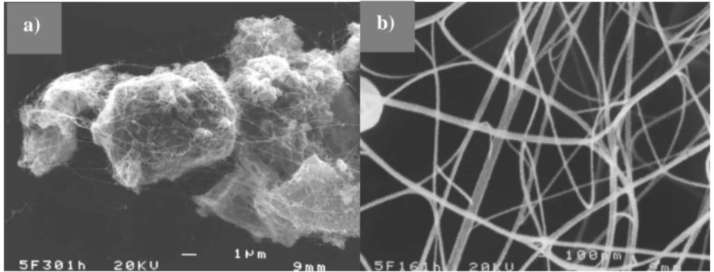
Figure 2.7 SEM image of a CNT-Fe-Al2O3 nanocomposite powder (a); higher
magnification image of the CNT network showing flexible filaments (b) [24].
As mentioned above in the presentation of the yarmulke mechanism, the presence of metal particles with a diameter too high for the formation of CNTs will result in
the presence of undesirable carbon species such as CNF, bamboo filaments, ribbons, carbon layers encapsulating catalyst particles and carbon particles (CP). Typical SEM and high-resolution TEM images of these carbon species are shown in Figure 2.8 and 2.9.
Figure 2.8 Typical SEM images showing bamboos, CNFs, ribbons and carbon
particles [29].
Tremendous efforts by the same group of researchers have been devoted to improving the synthesis of CNTs, i.e., to increase the CNT quantity and the carbon quality (CNT versus other carbon species) and to obtain a high selectivity on the number of walls, with a special emphasis on SWNTs and DWNTs. Note that using catalytic materials in the form of foams, as opposed to powders, allows a significant increase in the production of CNTs and favors the formation of SWNTs over DWNTs, although the exact reason for this is not fully explained yet [22, 30].
In the following, we will review the main results obtained with (Al1-xFex)2O3
powders. The effects upon the obtained CNTs-oxide nanocomposite powders of the various parameters related to the catalyst material as well as parameters related to the reduction treatment have been systematically investigated: iron content [31],
b) CNFs 100 nm a) CNF Bamboo 100 nm d) Particles (nanocapsules) 100 nm c) Ribbons 100 nm
allotropic form and specific surface area of the solid solution [26, 32], composition of the H2/CH4 atmosphere [33], reduction temperature Tr [21], and time spent at Tr [18].
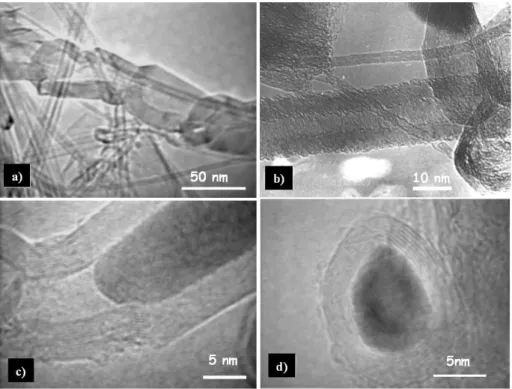
Figure 2.9 Typical high-resolution TEM images of (a) an bamboo; (b) CNF; (c) a
filament containing a metal particle; and (d) a metal particle encapsulated by carbon layers [34].
The influence of the metal content on the synthesis of CNT-Fe-Al2O3
nanocomposite powders has been studied by Peigney et al. [31]. The nanocomposite powders were prepared by the reduction of α-(Al1-xFex)2O3 solid solutions with
various amounts of Fe (x = 0.02, 0.05, 0.1, 0.15 and 0.2). The authors found that the highest quantity of CNTs was obtained using α-Al1.8Fe0.2O3 as the catalytic material,
i.e. the maximum Fe amount (x = 0.1) allowing to retain the monophase solid solution. By contrast, an increase in Fe content (>0.1) induced a phase partitioning and the formation of mixtures of Al2O3- and Fe2O3-rich solid solutions. The Fe2O3
-rich (hematite) particles produce upon reduction too large Fe particles, which result in the formation of nanofibers, CNTs with large diameters or end up encapsulated by carbon layers.
Laurent et al. [26] found it necessary to use a stable α-Al1.8Fe0.2O3 solid solution
obtain carbon essentially in the form of CNTs. The crystallization of these latter compounds during the reduction provokes the entrapment of carbon within the grains. Laurent et al. [32] prepared α-Al1.8Fe0.2O3 by calcination between 1025 and
1100 °C and the obtained powders were reduced in a H2-CH4 gas mixture at high
temperature. Calcination at only 1050 °C or 1025 °C resulted in an α-Al1.8Fe0.2O3
with a higher specific surface area. It was observed by these authors that the high specific surface area is beneficial to both the quantity and the quality parameters of the nanocomposite powders prepared from the α-Al1.8Fe0.2O3 solid solution. By
contrast, a higher specific surface area arising from a lower grain size (after some grinding for example) is not beneficial because the better packing of the solid solution powder hampers the formation of the CNTs.
Peigney et al. [33] have investigated the influence of the composition of H2-CH4
gas mixtures (range 0 to 45 mol% CH4) on the synthesis of CNT-Fe-Al2O3
nanocomposite powders prepared by selective reduction of α-Al1.9Fe0.1O3. Methane
concentrations ranging between 9 and 18 mol% gave the best results.
Peigney et al. [21] produced SWNTs and DWNTs with an average diameter close to 2.5 nm by the reduction of an α-Al1.8Fe0.2O3 powder in H2-CH4 atmosphere
applying different reduction temperatures Tr (in the range between 800 °C to 1070
°C). The authors found that increasing the Tr increases the reduction yield and thus
favors the formation of surface metal particles, thus producing more CNTs. Flahaut et al. [18] reported the influence of time spent at Tr on synthesis of CNTs. The
authors observed that increasing the dwell at the maximum temperature reached during the reduction process results in the production of CNTs with more walls.
At the end of the nineties, the research collaboration between CIRIMAT and NUMAT (at University of Ghent) on the study of carbon-free metal-oxide nanocomposites was therefore extended to such compounds. Indeed, 57Fe Mössbauer spectroscopy has shown to be a useful, complimentary technique to examine the Fe-containing CNT-nanocomposite powders as well as the starting oxides. This technique, being specific for Fe phases, was believed to be able to provide unique formation about the Fe-containing particles. Various oxide solid-solutions such as (Al1-xFex)2O3, Mg1-xMxAl2O4, Mg1-xMxO, Zr1-xMxO2 with M = Fe, Co, Ni, were
considered [21, 35-39]. Common to all CNT-Fe-oxide nanocomposite powders investigated by Mössbauer spectroscopy, is the presence of three characteristic Fe-phases: α-Fe, Fe3C(cementite) and a -Fe-C phase [21, 36, 40-44]. A lot of work was
devoted to correlate the identified Fe-phases to the carbon species in the CNT-Fe-oxide nanocomposite powders. One of the conclusions at the time was that the particles active for the formation of CNTs are probably Fe-C alloys but are detected as Fe3C in post-reaction Mössbauer spectroscopy analysis. More details about these
2.4 About Fe and Fe-carbide species
Iron has three allotropic forms, known as alpha, gamma and delta. In ordinary temperature and pressure conditions, iron crystallizes in the body-centered cubic (bcc) crystal structure (α-Fe). Between 912 °C and 1394 °C, it crystallizes in the face-centered cubic (fcc) structure ( -Fe), and between 1394 °C and 1538 °C iron has again a bcc structure ( -Fe).
Because both α-Fe and -Fe are bcc, they are identical in structure but are differentiated by name: α-Fe (ferrite) is of great practical importance, while -Fe is relatively unimportant. In earlier times it was believed that another phase, -Fe, existed but this was shown to be α-Fe above its Curie temperature (770 °C). The α-Fe transforms from ferromagnetic to paramagnetic state at 770 °C while maintaining the bcc structure. The old denomination -Fe corresponding to paramagnetic bcc iron is no longer used. At room temperature bulk ferromagnetic α-Fe is characterized by a sextet of well-known Mössbauer parameters: Bhf = 33.0 T, = 0.00 mm/s and 2 Q =
0.00 mm/s. Paramagnetic α-Fe is characterized by a singlet and no quadrupole splitting is detected.
The Fe-C binary [45] system is usually described in terms of Fe and the metastable carbide Fe3C (cementite), as well as in terms of Fe and stable graphite.
Carbon is soluble to only a very small amount in α-Fe (up to about 0.022 wt.% at 727 °C. On the other hand, -Fe has a much higher solubility for carbon than α-Fe (up to 2.14 wt.% at 1147 °C) due to the presence of large interstices in the fcc structure. The temperature of 727 °C and a carbon concentration of 0.76 wt.% is a eutectoid decomposition in the Fe-C diagram, where -Fe transforms to a mixture of α-Fe and Fe3C. Several works on CNT-nanocomposite systems prepared by CCVD method
[21,33,37,41] reported the presence of -Fe-C nanoparticles on these powders besides others Fe and Fe-carbide nanoparticles. The spectrum of the -Fe-C phase was composed by a singlet with an isomer shift of ~ -0.12 mm/s at 295 K. Coquay et al. [40] obtained a Mössbauer spectrum of a CNT-nanocomposite powder at 4.2 K and observed that the present -Fe-C showed an antiferromagnetic coupling. The spectrum was fitted using a weak-hyperfine-field of ~1.8 T.
More than ten iron-carbides are mentioned in the literature [45]. Among them, Fe3C being the most common and stable one at room temperature. It has an
orthorhombic unit cell and two inequivalent crystallographic Fe-sites [46, 47], which are magnetically and electronically very similar. Bi et al. [48] have shown the differences between the Mössbauer parameters of the two Fe-sites of Fe3C which
increase with decreasing temperature (Table 2.1). The authors have performed Mössbauer measurements between 12 K and room temperature on bulk Fe3C and
Fe3C nanoparticles (as small as 5 nm). Interesting, no superparamagnetism was
observed even for the smallest particles measured at room temperature.
Table 2.1 Room temperature and 12 K Mössbauer parameters for nanoparticle and
bulk Fe3C [48]. Nanoparticle Fe3C Bulk Fe3C T (K) Bhf (T) 2 Q (mm/s) (mm/s) Bhf (T) 2 Q (mm/s) (mm/s) Fe (site i) 21.0 0.02 0.19 21.1 0.01 0.19 300 Fe (site ii) 19.8 0.02 0.19 20.6 -0.01 0.18 average 20.4 0.02 0.19 20.8 0.0 0.19 Fe (site i) 25.5 0.01 0.32 25.8 0.0 0.34 12 Fe (site ii) 23.7 0.02 0.31 24.2 0.0 0.32 average 24.6 0.02 0.32 25.0 0.0 0.33
3
Methodology
3.1 Synthesis procedures
All details about the synthesis procedures used for the catalytic materials as well as for the CNT nanocomposites by reduction of these catalytic materials are outlined in the papers that are reproduced in Part II. The synthesis flowcharts are briefly presented in what follows.
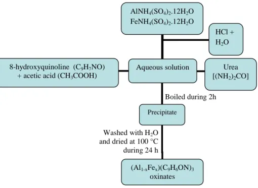
The procedure for the synthesis of the mixed oxinates powders, (Al1-xFex)(C9H6ON)3, is shown in Figure 3.1 (see paper I).
Figure 3.1 Flow-chart showing the synthesis of the (Al1-xFex)(C9H6ON)3
oxinate powders. AlNH4(SO4)2.12H2O FeNH4(SO4)2.12H2O Aqueous solution HCl + H2O 8-hydroxyquinoline (C9H7NO)
+ acetic acid (CH3COOH)
Urea [(NH2)2CO] Boiled during 2h Precipitate Washed with H2O and dried at 100 °C during 24 h (Al1-xFex)(C9H6ON)3 oxinates
The preparation of the -(Al1-xFex)2O3 powders by decomposition of the
(Al1-xFex)(C9H6ON)3 powders and the subsequent synthesis of CNT-Fe-Al2O3
nanocomposites by reduction of the former compounds are shown in Figure 3.2 (see paper I).
Figure 3.2 Flow-chart of the CNT-Fe-Al2O3 nanocomposites synthesis
from -(Al1-xFex)2O3 powders.
-(Al1-xFex)2O3 oxide powders were prepared by heating the -(Al1-xFex)2O3 solid
solutions at high temperatures (1100 or 1120 °C) in air (Figure 3.3). Further, the -powders were used to prepare self-supported -(Al1-xFex)2O3 oxide foams by
impregnation of a polyurethane foam with a slurry (oxide plus a dispersant) followed by a calcination at 600 °C. The -(Al1-xFex)2O3 oxide powders and foams were
submitted to CCVD treatment to obtain CNT-Fe-Al2O3 nanocomposites as shown in
Figure 3.3 (see paper III).
(Al1-xFex)(C9H6ON)3 oxinates -(Al1-xFex)2O3 powders Calcination in air at 800 °C CNT-Fe-Al2O3 nanocomposite powders CNT-Fe-Al2O3 nanocomposite powders CCVD treatment N2-C2H4 800 °C CCVD treatment H2-CH4 1000 °C
Figure 3.3 Flow-chart of the CNT-Fe-Al2O3 nanocomposites synthesis from
-(Al1-xFex)2O3 powders and -(Al1-xFex)2O3 foams.
An Fe/mullite powder was prepared by combustion route. After, several batches of the so-obtained combustion powder were calcined in air at selected temperatures between 800 °C and 1400 °C. The obtained powders were used for the synthesis of CNT-Fe-mullite nanocomposites by reduction in H2-CH4 gas mixture (Figure 3.4),
see paper VII.
-(Al1-xFex)2O3 powders -(Al1-xFex)2O3 powders Calcination in air at 1100 °C or 1120 °C Self-supported -(Al1-xFex)2O3 foams CNT-Fe-Al2O3 nanocomposite powders Preparation of self-supported foams CCVD treatment H2-CH4 1025 °C CNT-Fe-Al2O3 nanocomposite foams CCVD treatment H2-CH4 1025 °C
Figure 3.4 Flow-chart of the CNT-Fe-mullite nanocomposites synthesis from
Fe0.6Al5.4Si2O13 powders.
3.2 Characterization
Several techniques such as Mössbauer spectroscopy (transmission and emission), X-ray diffraction, specific surface area measurements, carbon analysis, Raman spectroscopy, and scanning and transmission electron microscopy have been intensively used for the characterization of the various samples considered in this research project. All mentioned techniques, except Mössbauer spectroscopy, were
Al(NO3)3.9H2O Fe(NO3)3.9H2O Colloidal SiO2 NH4NO3 Urea (NH2)2CO H2O Aqueous solution Combustion synthesis Calcination at 800, 1000, 1100, 1200, 1300 and 1400 °C Fe0.6Al5.4Si2O13 powders CCVD treatment H2-CH4 1050 °C CNT-Fe-mullite nanocomposites
applied in CIRIMAT laboratories (UPS, Toulouse) in collaboration with local research teams. Selected groups of samples have additionally been investigated by Fe
K-edge XANES (in collaboration with Dr. S.G. Eeckhout (ESRF Grenoble, France)
and Dr. G. Giuli (Dipartimento di Scienze della Terra and INFM, Università di Camerino, Italy).
3.2.1 Mössbauer spectroscopy
Mössbauer spectroscopy is a technique based on the recoil-free emission and subsequent absorption of -rays by certain isotopes. The phenomenon of emission of a -photon without loss of energy due to recoil of the nucleus (and without thermal broadening) is known as the Mössbauer effect, after R.L. Mössbauer who discovered and explained the effect in 1958. Its unique feature is the production of monochromatic electromagnetic radiation with an extremely narrow energy spectrum, so that it can be used to resolve minute energy differences.
The electromagnetic interactions between the nucleus and the surrounding charge distribution have a significant effect on the nuclear energy levels. Three interactions are of major importance, respectively the electric monopole, the electric quadrupole and the magnetic dipole interaction. In the Mössbauer spectra they give rise to three parameters: the isomer shift , the quadrupole splitting EQ or shift Q, and the
magnetic hyperfine field Bhf, respectively. Their values are characteristic of the
compound being studied and very often can be decisive as to the precise nature of that compound.
In this work two different variants of 57Fe Mössbauer spectroscopy have been applied. The first one is the transmission technique whereby the 14.4 keV -quanta emitted by a standard source and transmitted through an absorber of the investigated material are counted. The energy of the incident -quanta as experienced by the absorber nuclei is modulated through the Doppler effect by subjecting the source to a continuously oscillating velocity in a range –v0 to +v0. A Mössbauer spectrum is then
obtained by measuring the transmission as a function of the source velocity, which commonly is of the order of several mm/s in the case of 57Fe. In addition, for many samples emission Mössbauer spectra have been acquired as well. For this purpose use was made of Integral Low-Energy Electron Mössbauer Spectroscopy (ILEEMS). This method counts the low-energy (< 15 eV) Auger and shake-off electrons that are emitted by the absorber nuclei, which have been excited by resonant absorption of incident 14.4 keV -quanta. Acquisition of data is also performed as a function of source velocity. Emission spectra yield information similar to that obtained from transmission spectra, but the information primarily refers to Fe species present in the top surface layers of the substance being examined.
Transmission Mössbauer spectra were collected at temperatures between 4.2 K and 295 K, whereas ILEEMS spectra were obtained exclusively at 295 K. Accumulation of the data was made in 1024 channels. The spectrometers have been calibrated by collecting at room temperature the spectrum of a standard hematite (α-Fe2O3) powder or a standard metallic iron foil. The isomer shifts quoted in this
thesis are referenced with respect to α-Fe at room temperature. In-situ high temperature transmission Mössbauer spectroscopy was applied at selected temperatures between 295 K and 1123 K.
The Mössbauer spectra were generally fitted with Lorentzian line shapes and, where found necessary, some spectra components were described by a model-independent hyperfine field or quadrupole splitting distribution with Lorentzian-shaped elemental spectra (for instance in the case of Fe3C component) [49]. In that
case, the hyperfine-parameter values quoted in this thesis refer to maximum-probability values.
3.2.2 X-ray diffraction (XRD)
X-ray diffraction involves interaction of electromagnetic radiation with a wavelength ( ) of around 0.1 nm, with the atoms in the solid. As the distances between the atoms in a crystal structure are comparable with the wavelength of the radiation, crystals can diffract X-rays. The incidence angles (θ), the wavelength of the rays and the lattice spacings (dhkl) are related by the Bragg equation:
2
hklsin
n
d
It is a versatile, non-destructive technique that reveals detailed information about the composition and structure of the crystallized phases present in the materials, allows to calculate lattice parameters of these phases and can also provide information of their degree of crystallinity and on the size of the crystallites.
For identification of crystalline phases, XRD patterns were recorded in the range 10-70o (2 ) using a Bruker D4 Endeavor diffractrometer equipped with a Cu K radiation tube ( = 1.5406 Å). Counts were registered every 0.02° (2 ).
In order to calculate the cell parameters, XRD patterns were recorded in the range 20-70° (2θ) using a Seifert 3003 TT diffractometer equipped with a CuKα radiation
tube. Counts were registered every 0.02° (2θ). The cell parameters were calculated by the Rietveld method using the “Fullprof” software.


![Figure 2.4 Formation model of MWNTs proposed by Amelinckx et al. [15].](https://thumb-eu.123doks.com/thumbv2/123doknet/2232452.16119/24.748.146.616.96.461/figure-formation-model-mwnts-proposed-amelinckx-et-al.webp)