THESE
THESE
En vue de l'obtention du
DOCTORAT DE L’UNIVERSITÉ DE TOULOUSE
DOCTORAT DE L’UNIVERSITÉ DE TOULOUSE
Délivré par l'Université Toulouse III - Paul Sabatier Discipline ou spécialité : Physiopathologie
JURY
Pr. Bertrand Perret
Pr. Françoise Dignat-George, Rapporteur Dr. Jean-Max Pasquet, Rapporteur
Dr Christian Gachet, Examinateur Pr Pierre Sié, Examinateur
Pr Bernard Payrastre, Directeur de thèse
Présentée et soutenue par Thibault LHERMUSIER Le 5 janvier 2012
Titre :
Régulation plaquettaire : Ciblage de la proteine kinase Syk dans les HIT et rôle du transporteur lipidique ABCA1 dans les fonctions plaquettaires.
Remerciements
Je remercie tout d’abord l’ensemble des membres du jury de m’avoir fait l’honneur de juger ce travail :
Merci au Professeur Bertrand Perret d’avoir accepté de présider ce jury. Mes premières années de Médecine ont été marquées par votre enseignement en Biochimie. Votre présence m’honore.
Merci au Professeur Françoise Dignat-George d’avoir pris le temps de lire et juger ce travail. Merci au Docteur Jean Max Pasquet d’avoir accepté d’évaluer cette thèse en qualité de rapporteur.
Merci au Docteur Christian Gachet d’avoir accepté d’être examinateur de ce travail de thèse. Merci au Professeur Pierre Sié d’avoir accepté de juger ce travail dont il est en partie à l’origine. Je souhaite que notre collaboration étroite entre les services de cardiologie et d’hémostase se poursuive.
Merci au Professeur Bernard Payrastre pour son encadrement scientifique et ses conseils. Ton perfectionnisme et ton abnégation sont un modèle pour moi.
Ces années, enrichissantes et difficiles, sont à l’origine d’une expérience sans pareil. Difficiles parce qu’il est douloureux pour le clinicien que je suis de s’éloigner des patients et de se plonger dans le casse tête chinois de la science.
Enrichissantes car j’ai découvert la rigueur et l’exigence de la recherche au sein d’une équipe dynamique et chaleureuse. Vous m’avez aussi montré le plaisir d’avancer dans ce monde complexe mais passionnant qu’est la recherche.
Mes remerciements vont bien sûr à Bernard Payrastre pour m’avoir accueilli au sein de l’équipe et dans son bureau.
Travailler à tes cotés m’a permis de répondre à des questions centrales qui animent l’équipe au cours de tes absences (rares) : Il n’est pas là Bernard ? A quelle heure revient-il ? Est-il là demain ? Ceci souligne à quel point ton avis et tes conseils sont recherchés au sein de cette équipe. Je reste impressionné par l’étendue de tes connaissances et ta capacité à les mobiliser. J’admire ton « bon sens paysan » dans la science, je pense que tu apprécieras le terme.
Je remercie tous les membres de l’équipe plaquette qui ont donné (beaucoup) de leur temps et parfois (un peu) de leur sang pour faire avancer ce projet.
Cédric, ton esprit aiguisé et ta gentillesse sans limite m’ont beaucoup aidé.
Gaëtan, j’ai pu apprécié ton ardeur et ta rigueur au travail. Il est agréable de travailler à tes côtés.
Ashraf, ton expérience m’a été d’une grande aide.
Bien évidemment, je remercie Marie Pierre Gratacap qui m’a initié à la préparation des plaquettes, petits êtres parfois bien capricieux, et dont les conseils ont été particulièrement utiles à mes travaux.
Merci à Valérie et Marie Cécile.
Mes remerciements vont également à Jérôme VR qui s’est largement investi dans ces travaux. Au delà de notre amitié, j’espère que nous aurons la possibilité de continuer à travailler ensemble.
Je remercie le service de Cardiologie de Rangueil. Bien que cet investissement ait perturbé mon activité hospitalière, vous avez su garder une attitude bienveillante et continué à m’encourager.
Léopold, Gaspard et Anselme, mes pierres précieuses.
Emilie, toujours à mes côtés au soleil et dans la tempête, à la ville et à la montagne. Le skipper de la famille.
A ma cabane au Canada, avec toi bien sûr.
Aux amis de toujours et pour toujours, Nicolas, Yann, Anaïs, Xavier et Caroline. Ma deuxième famille.
Mes remerciements vont aussi à mes parents pour leur amour et à ma belle famille pour son soutien et ses encouragements.
RESUME
Les plaquettes sanguines jouent un rôle essentiel dans le maintien de l’intégrité vasculaire. Outre leur rôle physiologique conduisant à la formation du clou hémostatique en cas de brèche vasculaire, elles sont impliquées dans des pathologies thrombotiques comme l’athérothrombose ou les thrombopénies induites par l’héparine (TIH), ce qui en fait des cibles pharmacologiques de choix dans ces situations. Nous avons étudié les mécanismes mis en jeux par les plaquettes dans deux situations pathologiques ; les TIH avec la tyrosine kinase Syk comme acteur majeur et la maladie de Tangier résultant d’une mutation perte de fonction du transporteur de lipide ABCA1.
Les TIH sont des complications rares mais graves des traitements à l’héparine dues au développement d’anticorps dirigés contre le complexe héparine-‐facteur 4 plaquettaire (PF4). Ces anticorps induisent la clairance des plaquettes (thrombopénie) et activent les récepteurs Fc à la surface des plaquettes (FcγRIIA) et des monocytes (FcγRI et FcγRII) conduisant à l’agrégation plaquettaire et l’expression du facteur tissulaire par les monocytes responsable des complications thrombotiques. Nous montrons que Syk est une kinase essentielle à l’activation des plaquettes (signalisation, sécrétion, agrégation, microparticules) par les anticorps dirigés contre le complexe héparine-‐PF4 présents dans le sérum de patients atteints de TIH. Parallèlement, l’inhibition de Syk bloque fortement l’expression du facteur tissulaire et l’activité procoagulante des monocytes induites par les anticorps TIH. Les inhibiteurs de Syk, en développement clinique avancé en immunothérapie, apparaissent donc prometteurs dans le traitement des TIH.
ABCA1 est impliqué dans le transport réverse du cholestérol et de phospholipides vers les lipoprotéines de haute densité. Son déficit est associé à de profondes anomalies du bilan lipidique mais aussi à un phénotype hémorragique. Grâce à un modèle de souris déficientes en ABCA1, nous avons caractérisé son rôle dans l’activation plaquettaire. Contrairement à ce que l’on aurait pu penser, ABCA1 n’est pas impliqué dans l’exposition des phosphatidylsérines et l’activité procoagulante des plaquettes activées, et ne régule pas le contenu en cholestérol des membranes plaquettaires. Par contre, un défaut de taille des plaquettes et de leurs réponses fonctionnelles sont observés en son absence. ABCA1 apparaît comme un régulateur des mécanismes de signalisation pouvant expliquer le phénotype hémorragique rapporté dans la maladie de Tangier.
L’ensemble de ces travaux a permis de caractériser des acteurs moléculaires de l’activation plaquettaire dans des situations pathologiques et de proposer de nouvelles stratégies thérapeutiques.
ABSTRACT
Platelet activation at sites of vascular injury is essential for haemostasis. Next to this critical role in bleeding arrest, platelets are involved in thrombotic diseases such atherothrombosis or heparin-‐induced thrombocytopenia (HIT). Therefore, they are targets of antithrombotic therapies. We have studied the mechanisms of platelet activation in two pathological situations; HIT with the tyrosine kinase Syk as a key player and Tangier disease resulting of a loss of function of the ABCA1 lipid transporter. HIT is a prothrombotic and potentially devastating complication of heparin therapy due to formation of platelet-‐activating antibodies directed against complexes of platelet factor 4 (PF4) and heparin. These antibodies contribute to clear platelet from the circulation and activate, by their Fc fragment, FcγRIIA receptors on platelet surface or FcγRI and FcγRII receptors on monocyte, leading to platelet aggregation and tissue factor expression by monocytes. We have shown that Syk activation is crucial for platelet activation (signalization, secretion, aggregation, microparticle generation) initiated by anti-‐PF4/heparin antibodies from sera of patients suffering from HIT. Interestingly, Syk inhibition also prevented tissue factor expression and procoagulant activity of moncytic cells induced by these antibodies. We propose that Syk inhibitors, initially developed as a potential treatment of autoimmune disease, may be of therapeutic interest in the treatment of HIT.
ABCA1 has been demonstrated to be crucial in the reverse cholesterol transport pathway by loading cholesterol and phospholipids into ApoA-‐I to generate high density lipoproteins. Its defect is associated to circulating lipid disturbance but also to hemorrhagic diathesis. Surprisingly, using ABCA1 knock-‐out mice, we have demonstrated that this transporter is neither implicated in phosphatidyserine exposure, a mechanism leading to the procoagulant activity of activated platelets, nor in the control of platelet membranes cholesterol content. However, mouse platelets deficient for ABCA1 have an increased size and a defect in response to low doses of agonists such as thrombin and collagen. Our data indicate that ABCA1 is involved in the efficiency of platelet signal transduction, particularly in the activation of Akt.
Sommaire
Table des illustrations... 4
Abréviations ... 5
Partie 1 : Revue bibliographique... 7
Chapitre 1 Présentation générale des plaquettes sanguines. ...8
I. Morphologie et caractéristique des plaquettes sanguines...8
A. L’asymétrie de la membrane plasmique plaquettaire : régulation et rôle de l’exposition des PS dans la coagulation ... 9
1. Régulation de l’asymétrie de la membrane plasmique ... 9
2. Rôle de l’exposition des PS dans la coagulation ...11
Review Article : Platelet membrane phospholipid asymmetry: from the characterization of a scramblase activity to the identification of an essential protein mutated in Scott syndrome. ... 13
B. Les organelles plaquettaires...15
1. Les granules denses, α et les lysosomes...15
2. Les mitochondries...16
C. Impact des lipides circulants sur la réactivité plaquettaire...17
II. Les grandes étapes de l’activation plaquettaire lors d’une lésion vasculaire. ...18
A. Les principales protéines adhésives et leurs récepteurs ...20
1. Le vWF et son récepteur le complexe GPIb-‐IX-‐V ...20
2. Le collagène et ses récepteurs, l’intégrine α2β1 et GPVI ...23
3. La fibronectine...28
4. Le fibrinogène ...28
B. Les agonistes solubles et leurs récepteurs ...29
1. La thrombine et ses récepteurs ...29
2. Le Thromboxane A2 et ses récepteurs...31
3. L’ADP et ses récepteurs ...32
4. Le fragment Fc des Ig et le récepteur FcγRIIa ...33
C. Les voies de signalisation intraplaquettaires...34
1. Voies de signalisation dépendantes des protéines G hétérotrimériques...34
2. Voies de signalisation dépendantes des ITAM – l’exemple de FcγRIIa...36
D. L’intégrine αIIbβ3 et la signalisation « inside-‐out » et « outside-‐in » ...38
1. L’intégrine αIIbβ3...38
2. La signalisation « inside-‐out »...39
Chapitre 2 : Le transporteur ABCA1... 42
I. Présentation des transporteurs ABC ...42
A. Appartenance à la superfamille des transporteurs ABC ...43
1. La présence de motifs spécifiques de la famille des ABC ...43
2. Une double activité moléculaire...43
3. Une organisation en domaines ...43
II. Structure du transporteur ABCA1...43
III. Expression et distribution tissulaire de ABCA1...44
IV. Fonction...44
V. ABCA1 et maladie de Tangier ...47
A. Phénotype de la maladie de Tangier...47
B. Génétique de la maladie de Tangier ...47
VI. Rôle de ABCA1 dans l’exposition des PS ? ...48
VII. ABCA1 et signalisation ...48
Chapitre 3 : La plaquette en pathologie humaine... 50
I. Plaquette et athérothrombose...50
A. Rôle des plaquettes dans l’athérogénèse et dans l’inflammation...50
B. La plaquette comme cible antithrombotique ...51
1. Médicaments antithrombotiques d’aujourd’hui ...51
a. Aspirine...51
b. Inhibiteurs du récepteur P2Y12 ...52
Clopidogrel...52
Prasugrel ...54
Ticagrelor...54
Elinogrel ...54
c. Anti-‐GP IIb/IIIa (αIIbβ3)...54
2. Les nouvelles cibles antithrombotiques ...55
a. La voie du TXA2 ...55
b. La voie de la thrombine ...55
c. La voie du collagène ...55
C. Plaquettes et développement de thérapies ciblant les acteurs de la signalisation...56
II. Plaquette et thrombopénie immunoallergique à l’héparine...59
A. Epidémiologie ...59
B. Aspects cliniques ...59
C. Physiopathologie...62
1. Le facteur 4 plaquettaire (PF4) ...62
a. PF4 et thrombose. ...62
b. PF4 et chémotactisme ...63
2. Interactions héparine – PF4...63
3. Réponse immunitaire...64
4. Activation plaquettaire par les anticorps anti PF4 ...64
Partie 2 : Résultats...67
ARTICLE 1 : ABCA1 deficiency affects platelet activation and signaling but spares calcium-induced PS exposure and platelet lipid composition ... 68
I. Introduction...69
II. Discussion et perspectives...71
ARTICLE 2 : The Syk-kinase inhibitor R406 impairs platelet activation and monocyte tissue factor expression triggered by heparin-PF4 complex directed antibodies ... 77
I. Introduction...78
II. Discussion et perspectives...80
Partie 3 : Conclusion générale ...83
Table des illustrations
Figure 1 : forme typique « discoïde » des plaquettes de souris au repos (A) et modifications de
forme induites par la stimulation (B) ... 8
Figure 2 : Organisation générale des membranes plaquettaires et représentation schématique des différentes protéines membranaires impliquées dans l’asymétrie membranaire... 10
Figure 3 : Représentation schématique de la structure de la plaquette et des principaux récepteurs membranaires. ... 16
Figure 4 : Résumé des grandes étapes de l’activation plaquettaire lors d’une brèche de la paroi vasculaire dans des conditions de flux artériel ... 19
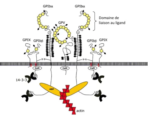
Figure 5 : Structure du complexe GPIb-IX-V... 21
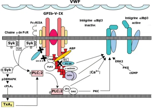
Figure 6 : Voies de signalisation organisées autour du complexe GPIb-V-IX engagé. ... 23
Figure 7 : Représentation schématique de l’intégrine α2β1... 24
Figure 8 : Voies de signalisation dépendante de l’intégrine α2β1... 25
Figure 9 : représentation schématique du récepteur GPVI ... 26
Figure 10 : Représentation schématique de la voie de signalisation dépendante de la glycoprotéine GPVI. ... 27
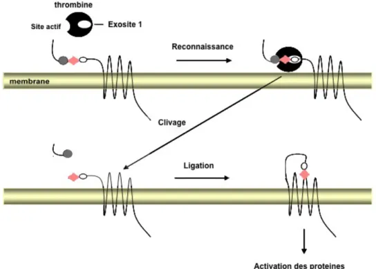
Figure 11 : Mécanisme d’activation de PAR1. ... 30
Figure 12 : Représentation schématique de l’activation des récepteurs PARs dans les plaquettes humaines et murines. ... 31
Figure 13 : Représentation schématique du récepteur FcγRIIa . ... 33
Figure 14 : Exemple de quelques voies de signalisation en aval des RCPG ... 35
Figure 15 : Voies de signalisation dépendante de la glycoprotéine GPVI ... 37
Figure 16 : Représentation schématique de la tyrosine kinase Syk ... 39
Figure 18 : Modèle schématique de stimulation plaquettaire soutenue et d’activation d’αIIbβ3 par la thrombine (figure du haut) et le collagène (figure du bas). ... 41
Figure 19 : Organisation en domaine du transporteur ABCA1 ... 44
Abréviations
ADP Adénosine 5’-diphosphate
ADN Acide désoxyribonucléique
AMPc adénosine 5’-monophosphate cyclique
ATP Adénosine 5’-triphosphate
ARNm Acide ribonucléique messager
DAG Diacylglycérol
ECGF Endothelial Cell Growth Factor
EGF Epidermal Growth Factor
FAK Focal Adhesion Kinase
FcγR Chaînes γ des récepteurs aux fragments constants des immunoglobulines
GEF Guanosine Nucleotide Exchange Factor
GMPc Guanosine 5’-monophosphate cyclique
GP Glycoprotéine
GDP Guanosine Diphosphate
GTP Guanosine Triphosphate
HDL High Density Lipoprotein
IGF1 Insulin Growth Factor 1
IP3 Inositol 1,4,5-trisphosphate
ITAM Immunoreceptor Tyrosine-based Activation Motif
IL1-β Interleukine 1 β
JAK2 Janus Kinase 2
KO Knock-Out
LDL Low Density Lipoprotein
MAPK Mitogen Activated Protein Kinase
MLC Myosin Light Chain
PA Phosphatidic Acid
PAI-1 Plasminogen Activator Inhibitor
PAR Protease-activated receptors
PC Phosphatidylcholine
PDGF Platelet Derived Growth Factor
PDK1 Phosphoinositide-dependent kinase 1
PDK2 Phosphoinositide-dependent kinase 2
PE Phosphatidyléthanolamine
PF4 Platelet Factor 4
PI3K Phosphoinositide 3-kinase
PKA Protein Kinase cAMP-dependent
PKB Protein Kinase B PKC Protein Kinase C PLA2 Phospholipase A2 PLC Phospholipase C PLCγ2 Phospholipase Cγ2 PON1 Paraoxonase 1 PS Phosphatidylsérine
PSGL-1 P-Selectine glycoprotein ligand-1
PtdIns Phosphatidylinositol
PtdIns3P Phosphatidylinositol 3-monophosphate
PtdIns4P Phosphatidylinositol 4-monophosphate
PtdIns5P Phosphatidylinositol 5-monophosphate
PtdIns(3,4)P2 Phosphatidylinositol 3,4-bisphosphate
PtdIns(4,5)P2 Phosphatidylinositol 4,5-bisphosphate
PtdIns(3,5)P2 Phosphatidylinositol 3,5-bisphosphate
PtdIns(3,4,5)P3 Phosphatidylinositol 3,4,5-trisphosphate
RCPG Récepteur Couplé aux Protéines G hétérotrimériques
SCO Système Canaliculaire Ouvert
SH2 Src Homology 2
SH3 Src Homology 3
SM Sphyngomyéline
STD Système Tubulaire Dense
TGFβ Transforming Growth Factor β
TNFα Tumor Necrosis Factor α
TXA2 Thromboxane A2
VASP Vasodilator-Stimulated Phosphoprotein
VCAM
VEGF Vascular Epidermal Growth Factor
Chapitre 1 Présentation générale des plaquettes sanguines.
Les plaquettes sanguines sont de petites cellules anucléés provenant de la fragmentation du cytoplasme de grandes cellules de la moelle osseuse hématopoïétique, les mégacaryocytes (Hartwig and Italiano, 2003). Elles sont relarguées dans le sang où leur nombre varie dans des conditions physiologiques de 150 000 à 400 000/µl. Leur durée de vie est 7 à 12 jours avant leur élimination par phagocytose des macrophages du système réticulo-histiocytaire au sein du foie, de la rate et la moelle osseuse.
Les plaquettes sont les acteurs essentiels du maintien de l’intégrité vasculaire. Leur fonction majeure est d’assurer l’hémostase dite « primaire » conduisant à la formation d’un thrombus riche en plaquettes ou « thrombus blanc » (Furie and Furie, 2008). Cependant, on s’aperçoit aujourd’hui qu’elles ont également un rôle important dans les processus de cicatrisation, de régulation de l’inflammation, d’angiogénèse et de séparation des vaisseaux lymphatiques et sanguins (Leslie, 2010).
I. Morphologie et caractéristique des plaquettes sanguines
Les plaquettes circulantes au repos se présentent sous la forme d’éléments discoïdes de 1,5 à 4 µm de diamètre et 0,5 à 2 µm d’épaisseur. Lors de leur activation, elles passent d’une forme discoïde à une forme sphérique et émettent des prolongements membranaires de type filopodes et lamellipodes (Figure 1).
A. L’asymétrie de la membrane plasmique plaquettaire : régulation et rôle de l’exposition des PS dans la coagulation
La membrane plaquettaire présente une structure classique, avec 2 feuillets lipidiques, externe et interne, de composition différente.
D’un point de vue lipidique, la membrane plasmique est composée de glycérophospholipides (phosphatidylsérine PS, phosphatidyléthanolamine PE, phosphatidylcholine PC) et de sphyngolipides (sphyngomyélines SM) ; chaque classe présentant de nombreuses espèces moléculaires variant par la nature des chaînes de carbones (longueur, branchement et degré d’insaturation). Un autre constituant lipidique essentiel est le cholestérol, molécule rigide et compacte assurant l’homéostasie de la fluidité membranaire et la formation de microdomaines appelés radeaux lipidiques (rafts) ayant un rôle important dans la signalisation plaquettaire (Bodin et al., 2003). Les membranes plaquettaires sont relativement riches en cholestérol et en SM, éléments favorables à la formation des microdomaines membranaires (Bodin et al., 2003, Bodin et al., 2005, Bodin et al., 2001).
La membrane plasmique contient une grande quantité de glycoprotéines qui sont ancrées dans la bicouche phospholipidique ou traversent cette dernière grâce à des domaines hydrophobes.
1. Régulation de l’asymétrie de la membrane plasmique
Comme la plupart des membranes plasmiques des cellules de mammifères, la membrane plaquettaire présente une distribution asymétrique des phopholipides entre les deux feuillets. Les PC et les SM prédominent sur le feuillet externe alors que les PE et les PS prédominent dans le feuillet interne.
Figure 2 : Organisation générale des membranes plaquettaires et représentation schématique des différentes protéines membranaires impliquées dans l’asymétrie membranaire (d’après Heemskerk et al., 2002).
Il a été démontré que la cellule dépensait de l’énergie à la fois pour créer cette asymétrie membranaire et pour la maintenir, suggérant l’existence de protéines spécifiques dont la fonction serait d’assurer un transfert de lipides d’un feuillet à l’autre (mouvements de flip-flop) (Seigneuret and Devaux, 1984). En effet, des protéines assurent le passage de certains lipides du feuillet externe au feuillet interne (flippases) alors que d’autres prennent en charge des mouvements inverses (floppases) (Figure 2). Si la fonction biologique de ces transporteurs est connue de longue date, leur identification biochimique est plus récente. La P4 ATPase est une flippase qui assure le transfert des PS vers le feuillet interne des membranes plasmiques, rôle indispensable au maintien de l’asymétrie membranaire (Tang et al., 1996). De la même façon, la floppase ABCC1, initialement connue sous le nom de multidrug résistance protein (MRP1) assure le transfert des phospholipides sur le feuillet externe des membranes plasmiques (Kamp and Haest, 1998, Dekkers et al., 1998). Des transporteurs de la superfamille ABC sont donc impliqués dans le transport de phospholipides entre les deux feuillets de la membrane plasmique.
Cette asymétrie peut cependant être perdue lors de certains événements comme l’apoptose ou l’activation plaquettaire. En effet, au cours d’une forte activation plaquettaire, il
Il existe une ou des protéines dont la fonction est d’éliminer, de façon plus ou moins réversible, l’asymétrie membranaire en assurant des mouvements aléatoires entre les deux feuillets. Cette ou ces protéines, dénommées scramblases, ont été caractérisées dans les années 70 mais sont restées non identifiées pendant près de 30 ans. Récemment, la protéine TMEM 16F a été identifiée comme jouant un rôle majeur dans l’exposition Ca2+- dépendante des PS sur le feuillet externe lors de l’activation plaquettaire (Suzuki et al., 2010).
2. Rôle de l’exposition des PS dans la coagulation
La transformation du fibrinogène soluble en un réseau fibreux insoluble de fibrine constitue l’étape clef de la coagulation. Cette transformation nécessite l’action enzymatique de la thrombine. La thrombine dérive du clivage de la prothrombine inactive par le complexe prothrombinase composé de la protéase Xa, du cofacteur Va, d’ion Ca2+ et de phospholipides anioniques (figure 2). Après stimulation de la plaquette par certains agents, induisant notamment une forte augmentation de la concentration cytosolique de Ca2+ (thrombine plus collagène essentiellement), un remodelage membranaire rapide se produit, avec translocation des amino-phospholipides sur le feuillet exoplasmique, bourgeonnement de la membrane et émission de microparticules. L’exposition des PS à la surface des plaquettes activées et des microparticules leur confère un potentiel procoagulant qui repose sur la formation du complexe enzymatiques prothrombinase à la surface membranaire. L’exposition des PS sur le feuillet externe des plaquettes est donc une étape importante pour la coagulation, permettant par ailleurs de focaliser la formation de thrombine au niveau de la brèche vasculaire.
L’importance de ce mécanisme est illustrée par l’existence du syndrome de Scott, caractérisé par des hémorragies dues à un défaut d’exposition plaquettaire des PS. Le fait que le défaut d’exposition rapide des PS se manifeste également dans les érythrocytes et lymphocytes B des patients atteints du syndrome de Scott stimulés par le ionophore calcique démontre que le mécanisme de translocation de PS à la surface cellulaire n’est pas spécifiquement plaquettaire. Des signaux comportant une étape commune, défective dans les cellules « Scott », seraient donc induits par les agonistes forts des plaquettes et par les ionophores calciques dans toutes les cellules hématopoïétiques. La redistribution des PS sur le feuillet externe de la membrane plasmique et la production de microparticules surviennent aussi lors de l’apoptose, et jouent dans ce cas un rôle dans la reconnaissance et l’élimination des cellules mortes par le système réticulo-endothélial. Les voies de signalisation déclenchées au cours de l’apoptose impliquent des processus en cascade qui conduisent, à court ou moyen
terme, à l’activation de caspases, à des réactions mitochondriales spécifiques, à l’exposition des PS puis à la dégradation de l’ADN. La translocation membranaire des PS lors de l’apoptose pourrait résulter de signalisations différentes de celles mises en jeu lors de l’externalisation de PS procoagulante, car les lignées de lymphocytes B Scott en culture exposent largement les PS sous l’effet d’inductions proapoptotiques (Kozian et al., 2005).
L’asymétrie membranaire et sa régulation, notamment par l’activité scramblase (encore un peu mystérieuse) et sa répercussion sur l’hémostase sont repris plus en détail dans la revue générale ci dessous.
Platelet membrane phospholipid asymmetry: from the
characterization of a scramblase activity to the identification of an essential
protein mutated in Scott syndrome.
Thibault Lhermusier, Hugues Chap, Bernard Payrastre
REVIEW ARTICLE
Platelet membrane phospholipid asymmetry: from the
characterization of a scramblase activity to the identification of
an essential protein mutated in Scott syndrome
T . L H E R M U S I E R , * H . C H A P ! and B . P A Y R A S T R E * "
*Inserm, U1048 and Universite´ Toulouse 3, I2MC, 31432 Toulouse Cedex 04; !Inserm, U1043 and Universite´ Toulouse 3, CPTP2, CHU-Purpan 31024 Toulouse Cedex 03; and "Hoˆpital de Toulouse, Laboratoire d#He´matologie, 31059 Toulouse Cedex 04, France
To cite this article: Lhermusier T, Chap H, Payrastre B. Platelet membrane phospholipid asymmetry: from the characterization of a scramblase activity to the identification of an essential protein mutated in Scott syndrome. J Thromb Haemost 2011; 9: 1883–91.
Summary. Like all eukaryotic cells, platelets maintain plasma membrane phospholipid asymmetry in normal blood circula-tion via lipid transporters, which control transbilayer move-ment. Upon platelet activation, the asymmetric orientation of membrane phospholipids is rapidly disrupted, resulting in a calcium-dependent exposure of the anionic phospholipid, phosphatidylserine (PS), at the outer platelet surface. This newly-exposed PS surface is a major component of normal hemostasis because it supports platelet procoagulant function. Binding of blood clotting enzyme complexes to this negatively-charged membrane surface allows a dramatic increase in the rate of conversion of zymogens to active serine proteases, which in turn produce a burst of thrombin leading to the formation of a fibrin clot and further platelet activation. Cells have the capacity to catalyze transbilayer phospholipid exchange via ATP-requiring translocase enzymes (flippases and floppases), which control unidirectional phospholipid transport against a concentration gradient. They also use an energy-independent, calcium-dependent scramblase activity to govern the bidirec-tional exchange of phospholipids between the two leaflets of the bilayer; this activity is essential for PS exposure during platelet activation. Scramblase activity, biochemically characterized in the 1980s, is deficient in patients with Scott syndrome, a rare inherited bleeding disorder with defective platelet procoagulant activity. Despite considerable efforts, the platelet scramblase protein remained elusive for years but a significant advance has recently been made with the identification of TMEM16F, a membrane protein essential for calcium-dependent PS exposure whose loss of function mutations are found in Scott syndrome. This review recalls historical aspects of platelet membrane asymmetry characterization, summarizes the mechanisms and
roles of PS exposure following platelet activation and discusses the recent identification of TMEM16F and its significance in the scrambling process.
Keywords: phosphatidylserine, phospholipid asymmetry, plate-let procoagulant activity, Scott syndrome, scramblase. Historical background: from phospholipid transverse distribution to molecular recognition of scramblase The concept of membrane phospholipid asymmetry in mammalian cells was first established in erythrocytes and then extended to platelets, using either chemical labeling of aminophospholipids (i.e. phosphatidylserine (PS) and phos-phatidylethanolamine (PE) [1,2]) or addition of exogenous phospholipases to act selectively on the external layer of the membrane under non-lytic conditions [3–5]. As a general rule, the exterior surface of plasma membranes is almost exclusively made up of choline phospholipids (i.e. sphingomyelin [SPH] and phosphatidylcholine [PC]), whereas the majority of PE and all of the PS, together with phosphoinositides, are localized in the inner leaflet (Fig. 1A).
As illustrated in Fig. 1(B), the asymmetric distribution of phospholipids established at the end of the 1970s brought a strong argument to the view that most of the reactions involving phospholipids in activatable cells such as platelets take place in the inner leaflet of the plasma membrane. These have since been shown to include phospholipase C (PLC)-mediated generation of the second messengers inositol 1,4,5-trisphosphate (IP3) and diacylglycerol (DAG) from phosphatidylinositol 4,5-bisphosphate (PIP2) [6], activation of protein kinase C by PS, DAG and calcium [7], liberation of arachidonic acid from glycerophospholipids confined to the
ability to activate coagulation via the so-called platelet factor-3. As a first attempt to understand platelet behavior in this context, Zwaal et al. [10] noticed that resting intact platelets cleverly offered an inert surface to the plasma coagulation system, owing to the virtual absence on the cell exterior of the most procoagulant lipid, PS. In their conclusion, the authors
wrote: $This raises the question whether during or after
formation of the primary hemostatic plug, either (partial) lysis of platelets or an as yet unknown mechanism which translo-cates PS through the plasma membrane is necessary to provide a catalytic lipid surface for interacting coagulation factors#.
This was the starting point of a quest lasting more than 30 years, which began with the concept of membrane phos-pholipid asymmetry and recently led to the molecular identi-fication of TMEM16F as an essential component of platelet scramblase activity [11]. Throughout this quest, four main steps can be identified, which are detailed below, each of which not only corresponded to a fundamental conceptual advance, but also provided new experimental clues to facilitate further progress.
Activated platelets disrupt membrane phospholipid asymmetry following appropriate stimulation
Using both purified coagulation factors and external phospho-lipases, Bevers et al. [12,13] observed that platelets activated with thrombin plus collagen were able to expose the procoag-ulant phospholipid PS on their surface (Fig. 1C). This active process occurred in the absence of cell lysis and corresponded with a transfer of PS (and PE) from the inner to the outer leaflet of the membrane, corroborating the aforementioned prediction by Zwaal et al. [10].
These studies introduced two novelties into the field: (i) the use of coagulation factors to detect surface exposure of procoagulant phospholipids, thus offering a convenient and sensitive tool to investigate this type of membrane modifica-tion; and (ii) the notion that PS exposure requires a large increase in the concentration of free cytoplasmic calcium. This can be achieved in vitro by calcium ionophores or, more physiologically, by the combination of thrombin plus collagen. Significant calcium influx can also be produced by membrane pore-forming agents such as the complement proteins C5b-9
Fig. 1. Schematic view of phospholipid transverse distribution in the plasma membrane of resting and stimulated platelets. (A) In resting platelets, the external membrane surface consists almost exclusively of SPH and PC and exerts no procoagulant activity. In contrast, PS, PE and phosphoinositides are confined to the inner leaflet of the membrane (shown in red). (B) Upon relatively weak platelet stimulation with thrombin alone at low concentrations, a number of metabolic modifications involve phospholipids of the inner leaflet, including PIP2 hydrolysis by PLC and subsequent activation of PKC by PS,
DAG and Ca2+, arachidonic acid (AA) liberation by PLA
2, as well as PIP3 production by phosphoinositide 3-kinases (not shown). However, this calcium
mobilization is not sufficient to promote phospholipid scrambling. (C) When platelets are strongly activated by a combination of thrombin and collagen, a large calcium influx occurs through specific channels (not shown) and promotes phospholipid scrambling, with the subsequent appearance of procoagulant phospholipids (mainly PS), on the external surface. PS, phosphatidylserine; SPH, sphingomyelin; PC, phosphatidylcholine; PE, phosphatidylethanolamine; PI, phosphatidylinositol; PIP, phosphatidylinositol monophosphate; PIP2, phosphatidylinositol bisphosphate; IP3, inositol trisphosphate; THR, thrombin; COLL, collagen; PLC, phospholipase C; DAG, 1,2 diacyl sn glycerol; PKC, protein kinase C; PLA2, phospholipase A2.
[14]. These different methods to increase calcium were subse-quently applied to a number of studies from this group and others. Simultaneous activation of platelets with thrombin and
collagen has led to the characterization of so-called
$coated-platelets#, which correspond to a small fraction of total blood platelets (mainly the youngest ones), exposing on their surface
not only PS but also various modified proteins secreted from
a-granules [15]. It has also been proposed that these coated platelets have an elevated ability to promote thrombin generation and possibly thrombosis.
The discovery of aminophospholipid translocase
Another major development in the understanding of mem-brane phospholipid asymmetry was the identification of an ATP-dependent activity able to transfer aminophospholipids, mainly PS, from the outer to the inner leaflet of the red blood cell membrane [16]. This led to the view that some specialized proteins are able to transport phospholipids between the two halves of the membrane. As discussed in various reviews [17,18] and as a summary of an impressive number of studies, Fig. 2 illustrates our current knowledge of translocases responsible for the establishment and maintenance of phospholipid asym-metry in membranes. These include a flippase, the former aminophospholipid translocase now identified as a P4 ATPase [19,20], and a floppase, whose activity on phospholipids is supported by the product of the gene ABCC1, initially known as multidrug resistance protein MRP1 [21,22]. The molecular mechanism of action of these ATP-dependent translocases is still unclear, because structural data are not yet available [23– 25]. However, phospholipid movements catalyzed by these proteins have been followed using phospholipid analogues bearing a short chain fatty acid linked to either a spin label or a fluorescent probe (NBD-phospholipids). This allows
sponta-neous intercalation of the phospholipids in the external leaflet of the membrane and their internalization can be assessed by either the decrease in extraction of these NBD-phospholipids by bovine serum albumin or through monitoring the reduction of fluorescent signal quenching initiated by external non-permeable agents. This experimental strategy has been applied to stimulated cells and has led to the concept that a scramblase
stimulated bylM concentrations of cytoplasmic free calcium
allows the reciprocal migration of phospholipids between the two halves of the membrane [26], as illustrated in Fig. 2.
Scott syndrome
This disorder was first described in 1979 by Weiss et al. [27] in a patient with a history of bleeding episodes and impaired platelet procoagulant activity (Mrs M.A. Scott, 1939-1996). In the mid 1980s, Scott syndrome was recognized as a very rare, moderately severe bleeding disorder characterized by a defect in membrane lipid scrambling in both activated platelets [28] and other hematologic lineages such as red cells and lymphocytes [29,30]. However, it is noteworthy that most patients do not present with any clinical symptoms other than bleeding episodes. Platelet count and structure are normal in Scott syndrome and no obvious alterations in platelet secretion, aggregation, metabolism, granule content, adhesion to suben-dothelium or coagulation factor levels are observed [31,32]. Scott platelets do, however, fail to promote fibrin formation in the presence of coagulation factors. When investigating this further, it was noted that prothrombin was not efficiently cleaved during the clotting of whole blood; however, in contrast to deficiencies in coagulation factors this reduced prothrombin cleavage was not restored by the addition of normal plasma. Phospholipases were also used to analyze the phospholipid composition of the outer membrane leaflet and led to the suggestion that, unlike normal platelets, Scott platelets do not have altered phospholipid composition in their outer membrane surface upon activation by a mixture of thrombin plus collagen [28]. These results were confirmed using annexin V binding experiments, which show a lack of PS exposure upon Scott platelet activation [33]. A similar defect was also observed in a canine model presenting a deficiency in platelet procoagulant activity [34].
The recognition of Scott syndrome as an inherited disorder [33,35] initiated an avid search for the gene encoding the hypothetical scramblase.
PS exposure in apoptosis and microvesicle formation
In parallel with studies on platelets and erythrocytes, a major
ATP ADP ATP ADP
PS, PE PL PL Flippase (P4 ATPase) Floppase (ABCC1) PL Ca2+ Scramblase (TMEM16F ?)
apoptotic cells, external PS exposure leads to the release of microvesicles (also called membrane microparticles), formed by evagination of the plasma membrane. This occurs concurrently with, and possibly as a consequence of, membrane phospho-lipid scrambling [18,39], although the two events might also be unrelated [40]. In addition to promoting the procoagulant activity, loss of phospholipid asymmetry renders the mem-branes of both microvesicles and remnant cells susceptible to the action of secretory type II phospholipase A2, resulting in a number of potential consequences such as transcellular signal-ing via lysophosphatidic acid or arachidonic acid metabolites [41–44]. This latter point warrants further investigation in the future.
PS exposure: linking platelet activation to thrombin generation and coagulation
While the central role of platelet activation in hemostasis has long been recognized, its implication in the control of coagulation was only characterized more recently [45,46]. The fact that the plasma membrane surface of activated platelets provides a critical platform for the activation of blood clotting enzyme complexes to generate thrombin was proposed in the 1980s [47]. When potently activated by soluble agonists, molecules of the subendothelial matrix or a combination of the two, particularly collagen plus thrombin, platelets lose their plasma membrane phospholipid asymmetry and PS is exposed at the surface of both platelets and platelet-derived micropar-ticles [13]. At physiological pH, surface exposure of PS provides a negatively-charged membrane platform necessary for the interaction of the non-enzymatic protein factor (F) Va. Factor Va then binds FXa with high affinity to form the prothrom-binase complex that efficiently activates thrombin by cleavage of two peptide bonds in prothrombin [48–50]. Through this process, activated platelets express specific and saturable binding sites for FVa and FXa [51,52]. The most widely-accepted view is that the negatively-charged membrane binds FVa and, in the presence of calcium ions, this factor undergoes a conformational change to create a high-affinity binding site for FXa, thereby forming the prothrombinase complex [53,54]. When compared with the activity of FXa alone, incorporation of FXa into the prothrombinase complex increases the rate of prothrombin cleavage by five to 10 orders of magnitude [51,55]. Assembly of the prothrombinase complex results in local propagation of blood coagulation because prothrombin is rapidly converted to thrombin [56]. Thrombin then cleaves fibrinogen to produce an insoluble fibrin clot and further activates platelets to amplify the reaction.
Thus, the anionic platelet membrane surface plays a critical role in the amplification of prothrombin cleavage and focuses thrombin production to the site of platelet activation, making platelet PS exposure a key regulator of hemostasis.
In vivo, in addition to providing a membrane surface needed for assembly of the prothrombinase complex, activated plate-lets secrete FVa, a key component of that complex, from their a-granules. Furthermore, activated platelets shed
microparti-cles that also support prothrombinase complex formation by increasing the procoagulant surface area [57].
Overall these data indicate that a deficiency in PS exposure disrupts the pathway from platelet activation to prothrombin activation, with bleeding consequences, as seen in Scott syndrome.
Regulation of PS exposure in platelets
The role of calcium, calpain, the cytoskeleton and lipid rafts
In agonist-activated platelets, surface exposure of PS is a process controlled by calcium and may also involve calpain activity and cytoskeletal organization. Following an increase in cytosolic calcium, flippase is inhibited but this in itself is not sufficient to provide a rapid PS exposure because activation of phospholipid scrambling is also required. Whilst scrambling activity is not ATP-dependent, it requires a sustained increase in cytosolic calcium [26]. Cytosolic calcium acts as a switch for the scrambling machinery so lowering the calcium levels stops the scrambling process [58]. Most platelet agonists induce PLC activation and IP3 production, leading to calcium mobilization, but this is not sufficient to switch on the scrambling process. It appears that an influx of calcium via store-operated calcium entry (SOCE) is mandatory [59]. There is evidence that the transient receptor potential channel TRPC1 is involved in the control of PS exposure in HEL cells, a lineage with mega-karyoblastic properties [60,61]; however, TRPC1-deficient mouse platelets have a normal calcium response, indicating that in fact this channel is not a major regulator of calcium influx upon platelet stimulation. More recent reports strongly implicate the SOCE machinery, including the Orai 1 channel and the STIM1 calcium sensors, in PS exposure following collagen activation of mouse platelets [62,63]. However, using STIM1- and Orai 1-deficient platelets, Gilio et al. [63] observed that an as-yet-uncharacterized (non-SOCE) mechanism of calcium entry initiated by thrombin receptors also contributes to PS exposure.
Cytosolic calcium modulates many cell functions, including actin cytoskeleton dynamics, by impacting on actin-severing proteins, actin-crosslinking proteins, calmodulin-regulated enzymes and proteases such as calpain. In 1985, Comfurius and colleagues had already correlated PS exposure with the cleavage of cytoskeletal proteins such as Actin-Binding Protein (ABP) [64]. Consistent with this observation, the calcium-dependent protease calpain, known to hydrolyze ABP and spectrin, was then proposed to participate in platelet PS exposure [65]. However, more recent data indicate that, at least upon stimulation of the collagen receptor GPVI under platelet adhesion conditions, calpain-mediated protein cleavage is involved in membrane blebbing and vesiculation rather than in PS exposure per se [66]. The interactions between the cytoskeleton and membrane glycoproteins may have a general structural role impacting on PS exposure, as recently shown for the platelet receptor for von Willebrand factor, the GPIb-V-IX complex, which seems to modulate platelet procoagulant
activity through the intracellular cytoskeleton-anchoring por-tions of GpIb [67].
The lateral organization of the plasma membrane, particu-larly cholesterol and sphingolipid-rich microdomains, also appears important for phospholipid scrambling. This is based upon the observation that cholesterol extraction and disruption of lipid rafts by methyl-b-cyclodextrin impairs phospholipid scrambling in HEL cells [61]. These membrane microdomains appear to be important for calcium signaling in general and particularly the SOCE machinery but it is not clear whether membrane fluidity and lateral organization directly impact on the scramblase activity, on regulatory elements, or both.
Although the scrambling activity does not require ATP hydrolysis, there is evidence that prolonged ATP depletion results in the loss of scrambling activity, suggesting that the scramblase machinery may require phosphorylation to be active [68].
Apoptotic vs. agonist-induced platelet PS exposure
As previously mentioned, PS exposure is a hallmark of apoptotic cells. Like in activated platelets, PS exposure in apoptotic cells results from inhibition of phospholipid translo-case activity and activation of scramblase activity. Interestingly, besides PS exposure some features of a strong platelet activation that is required for procoagulant activity resemble those occurring in apoptotic cells, including membrane bleb-bing and microparticle production. Moreover, platelets express many apoptotic regulators such as members of the Bcl-2 protein family and caspases. These observations raise several questions. (i) Is the apoptotic machinery involved in the regulation of the procoagulant function of platelets? (ii) Is PS exposure regulated differently during procoagulant activation of platelets than during platelet apoptosis? (iii) Do apoptotic platelets have procoagulant activity? In a recent report, Schoenwaelder and colleagues [69] provided a significant piece of information to start answering these questions. They observed that two distinct pathways regulate platelet PS exposure depending on whether it is induced by either the apoptotic BH3 mimetic compound ABT-737 (a selective inhibitor of anti-apoptotic proteins) or as part of the agonist (convulxin plus thrombin)-mediated platelet procoagulant function. Indeed, in mouse platelets, agonist-induced PS exposure appears unchanged in the absence of the apoptotic proteins Bak associated protein K) and Bax (Bcl-2-associated protein X) or following caspase inhibition but is abolished by extracellular calcium chelators or by prostaglan-din E1 (PGE1) and theophylin treatment (which inhibits platelet activation by raising cAMP levels). Conversely,
that two distinct molecular mechanisms regulating PS exposure in platelets exist. This is consistent with a previous study demonstrating that, in Scott syndrome, PS exposure in apoptotic lymphocytes is normal whereas calcium-induced PS exposure is absent [30]. This observation also suggests that TMEM16F is not involved in PS exposure during apoptosis in these cells. Whether the role of PS exposure at the surface of apoptotic platelets can have an impact on thrombin generation in vivo or is exclusively a signal for elimination of these platelets remains to be established. In this context, it is noteworthy that the clearance of PS-rich platelet microparticles from the circulation involves the PS binding protein lactadherin, deficiency of which causes a hypercoagulable state in mice [70]. Identification of TMEM16F as an essential component of the calcium-dependent scramblase activity deficient in Scott syndrome
Throughout the quest to identify the platelet scramblase, few candidates have been proposed. A strong candidate was ABCA1, a member of the ATP-binding cassette (ABC) lipid transporters, which is required to load cholesterol and phos-pholipids onto apoA1 and is mutated in Tangier disease (a rare inherited disorder characterized by a severe reduction in the amount of HDL). Indeed, a missense mutation in the ABCA1 gene was reported in a Scott syndrome patient [71], suggesting that this transporter could be part of the machinery involved in PS exposure. However, although ABCA1 may play a role in platelet activation, several reports argue against its direct implication in lipid scrambling [26,72]. Another candidate, a plasma membrane protein called scramblase 1 or PLSCR1, can be eliminated because it has been observed to be present at normal levels in lymphocytes from a patient with Scott syndrome [73]. This was confirmed by the lack of hemostatic defect in mice bearing a knock-out of the scramblase 1 gene [74]. Thus, the identity of the scramblase remained elusive until recently, when Suzuki et al. [11] identified the protein TMEM16F (also called anoctamin 6) as an essential compo-nent for calcium-dependent PS exposure on the plasma membrane. Interestingly, using Ba/F3 murine B-cells and an annexin V-based fluorescence-activated cell-sorting strategy, this group isolated a subline exhibiting strong PS exposure in response to calcium ionophore treatment under low calcium conditions. A complementary DNA library was constructed from this subline and introduced into the parental Ba/F3 cell line. After repetitive sorting/expansion cycles a cell subline exhibiting strong PS exposure in the absence of calcium ionophore was isolated. Using expression cloning, the authors then identified a cDNA causing Ba/F3 to expose PS
of 106 kDa and is located on the plasma membrane with eight transmembrane domains (Fig. 3). Compared with wild-type TMEM16F, the TMEM16F expressed in the cell subline constitutively expressing PS exhibited a point mutation replacing aspartate 409 with a glycine (D409G). Consistent with a constitutively active scramblase function of this mutated TMEM16F (possibly through increased sensitivity to calcium), these cells also constitutively exposed PE on their surface.
To further characterize this protein the authors used RT-PCR to analyze the TMEM16F mRNA of a B-cell line from a patient with Scott syndrome and found that the 3¢ half, corresponding to exons 11–20, was smaller. Sequence analysis indicated that the cDNA of the patient lacked the 226 bp sequence corresponding to exon 13. Direct sequencing of the chromosomal DNA showed that the TMEM16F gene of the patient carried a G-to-T homozygous mutation in the splice acceptor site in intron 12, causing exon 13 to be skipped. The resulting frame shift led to a premature termination of the protein at the third transmembrane domain of TMEM16F (Fig. 3). Overall, these results demonstrate that TMEM16F is a critical component of the calcium-dependent phospholipid scrambling activity. Interestingly, another recent report de-scribes two novel mutations in the TMEM16F gene in another patient with Scott syndrome [75]. The scramblase defect in the platelets, erythrocytes and B lymphocytes of this patient had been previously characterized [76]. The mutations were iden-tified in intron 6 (G-to-A), disrupting the donor splice site consensus sequence, and in exon 11 as a single-nucleotide insertion predicting a frame shift and premature termination of translation at codon 411. It is predicted that the first mutation causes an in-frame deletion of 38 amino acids in the N-terminal cytoplasmic tail of TMEM16F and the second mutation causes the truncation of the protein between the second and third transmembrane domains (Fig. 3). It is noteworthy that a genome-wide linkage scan had previously localized the canine Scott deficiency to chromosome 27, a region syntenic with the centromeric region of human chromosome 12 where the TMEM16F gene is localized [77], suggesting, as in human Scott
syndrome, a potential contribution of the TMEM16F protein to this canine disease.
TMEM16F: a calcium-dependent scramblase or a key regulator of scrambling activity?
TMEM16F belongs to a family of proteins consisting of 10 members in humans. The founding member, TMEM16A, is a
calcium-activated Cl)channel [78]. The transmembrane
seg-ments of TMEM16 proteins are the regions showing maximal conservation and there is evidence that different members of the family have distinct functions [79]. TMEM16 proteins are abundantly expressed and many reports have demonstrated their physiological importance in various cell types, where they can control cell volume, proliferation and apoptosis. There is evidence that some members of the family such as TMEM16A are embedded in a protein network and that they probably operate in functional microdomains. The sequence of TME-M16A proteins shows no clear similarity to known domains involved in direct calcium binding such as EF-hand or motifs potentially involved in calmodulin binding. However, the N-terminal part of TMEM16A seems to have an important regulatory function. Interestingly, the increased calcium sensi-tivity of the D409G mutant of TMEM16F may suggest a role for this region in supporting calcium-mediated scramblase activation. Whether these regions of TMEM16F interact with a calcium sensor or directly bind calcium remains to be established.
Based on structural data it is difficult to conclude that TMEM16F is indeed the long-sought scramblase protein. The results of Suzuki et al. [11] do not exclude the possibility that TMEM16F is a critical regulator of the calcium-induced lipid scrambling processes rather than being the scramblase itself. It is particularly interesting to consider these data alongside very recent and unexpected results showing that opsin can rapidly transport lipids across the membrane with properties similar to the plasma membrane scramblase [80,81]. Opsin may catalyze lipids# movements by correctly positioning water molecules to allow lipid head groups to translocate through the core of the protein while the fatty acid chains remain outside the protein surface. This mechanism would explain the lack of head group
specificity of the scrambling process. Interestingly, the
b1-adrenergic receptor has the same property, suggesting that several serpentine receptors could have the ability to play the role of a scramblase. If this is the case one can imagine that potent negative regulators of the process can suppress lipid scrambling under normal conditions and that TMEM16F could be one of these regulators.
Concluding remarks
The exciting discovery of TMEM16F as a critical component of the calcium-dependent phospholipid scramblase activity and the identification of loss-of-function mutations in Scott syndrome will have important implications for future research in this field. Indeed, analysis of the structure/function
Fig. 3. Schematic representation of TMEM16F and its mutations in Scott syndrome patients. The mutations of TMEM16F identified in Scott syn-drome patients are indicated as follows: the G-to-T mutation predicting a termination of the protein in the third transmembrane domain is shown by a red arrow; the G-to-A mutation predicting the in-frame deletion of 38 amino acids in the N-terminal part is shown by the two green arrows; and the single-nucleotide insertion in exon 11 predicting the truncation of the protein between the second and third transmembrane domain is shown by the blue arrow.
relationship as well as studies of the interactome of TMEM16F and of the post-translational modifications of the protein should provide important information concerning the molec-ular mechanism of calcium-dependent phospholipid scram-bling and may reveal novel concepts in the biomembrane field. Moreover, defining the role of TMEM16F in other processes related to PS exposure and phospholipid scrambling, such as apoptosis or membrane blebbing and microparticle formation, will be of great interest. The fact that Scott syndrome is an extremely rare disease affecting mainly specific platelet func-tions raises fundamental quesfunc-tions such as whether there exists any compensatory mechanisms, whether there is a role for TMEMF16 in PS exposure in response to acute and strong stimulation such as that found during hemostasis, and whether there is a potential role of TMEMF16 in development. Investigating the possibility that some serpentine receptors could function as scramblases and identifying their regulators and links with TMEMF16 may contribute to answering these questions. The creation of conditional TMEMF16 knock-out/ in mouse lines should provide powerful models to investigate the exact role of platelet PS exposure and phospholipid scrambling in hemostasis and thrombosis in vivo. Thus, the fascinating story of the scramblase identification is not over; new avenues have now been opened with the investigation of the molecular aspects of its regulation and function.
Acknowledgements
This work was supported by grants from Inserm, ITMO Cancer and Re´gion Midi-Pyre´ne´es. T. Lhermusier was sup-ported by a grant from CHU-Toulouse. Thanks are due to P. Sie´ for critical reading of the review.
Disclosure of Conflict of Interests
The authors state that they have no conflict of interest. References
1 Bretscher MS. Asymmetrical lipid bilayer structure for biological membranes. Nature (New Biol) 1972; 236: 11–2.
2 Schick PK, Kurica KB, Chacko GK. Location of phosphatidyletha-nolamine and phosphatidylserine in the human platelet plasma mem-brane. J Clin Invest 1976; 57: 1221–6.
3 Verkleij AJ, Zwaal RFA, Roelofsen B, Comfurius P, Kastelijn D, van Deenen LLM. The asymmetric distribution of phospholipids in the human red cell membrane. A combined study using phospholipases and freeze–etch electron microscopy. Biochim Biophys Acta 1973; 323: 178–93.
4 Chap HJ, Zwaal RFA, van Deenen LL. Action of highly purified phospholipases on blood platelets. Evidence for an asymmetric
dis-7 Nakamura S, Yamamura H. Yasutomi Nishizuka: father of protein kinase C. J Biochem 2010; 148: 125–30.
8 Plantavid M, Perret BP, Chap H, Simon MF, Douste-Blazy L. Asymmetry of arachidonic acid metabolism in the phospholipids of the human platelet membrane as studied with purified phospholipases. Biochim Biophys Acta 1982; 693: 451–60.
9 Gratacap M-P, Martin V, Guillermet-Guibert J, Chicanne G,
Payrastre B. PI 3-kinaseb in the regulation of platelet signalling and
functions. Adv Enz Reg 2011; 51: 106–16.
10 Zwaal RFA, Comfurius P, van Deenen LLM. Membrane asymmetry and blood coagulation. Nature 1977; 268: 358–60.
11 Suzuki J, Umeda M, Sims PJ, Nagata S. Calcium-dependent phos-pholipid scrambling by TMEM16F. Nature 2010; 468: 834–8. 12 Bevers EM, Comfurius P, van Rijn JL, Hemker HC, Zwaal RFA.
Generation of prothrombin-converting activity and the exposure of phosphatidylserine at the outer surface of platelets. Eur J Biochem 1982; 122: 429–36.
13 Bevers EM, Comfurius P, Zwaal RFA. Changes in membrane phos-pholipid distribution during platelet activation. Biochim Biophys Acta 1983; 736: 57–66.
14 Sims PJ, Wiedmer T, Esmon CT, Weiss HJ, Shattil SJ. Assembly of the platelet prothrombinase complex is linked to vesiculation of the platelet plasma membrane. Studies in Scott syndrome: an isolated defect in platelet procoagulant activity. J Biol Chem 1989; 264: 17049– 57.
15 Dale GL. Coated-platelets: an emerging component of the procoagu-lant response. J Thromb Haemost 2005; 3: 2185–92.
16 Seigneuret M, Devaux PF. ATP-dependent asymmetric distribution of spin-labeled phospholipids in the erythrocyte membrane: relation to shape changes. Proc Natl Acad Sci USA 1984; 81: 3751–5.
17 Daleke DL. Regulation of transbilayer plasma membrane phospho-lipids asymmetry. J Lipid Res 2003; 44: 233–42.
18 Zwaal RFA, Comfurius P, Bevers EM. Surface exposure of phos-phatidylserine in pathological cells. Cell Mol Life Sci 2005; 62: 971–88. 19 Tang X, Halleck MS, Schlegel RA, Williamson P. A subfamily of P-type ATPases with aminophospholipid transporting activity. Science 1996; 272: 1495–7.
20 Paulusma CC, Elferink RP. P4 ATPases – The physiological relevance of lipid flipping transporters. FEBS Lett 2010; 584: 2708–16. 21 Kamp D, Haest CWM. Evidence for a role of multidrug resistance
protein (MRP) in outward translocation of NBD-phospholipids in the erythrocyte membrane. Biochim Biophys Acta 1998; 1372: 91–101. 22 Dekkers DWC, Comfurius P, Schroit AJ, Bevers EM, Zwaal RFA.
Transbilayer movement of NBD-labeled phospholipids in red blood cell membranes; outward-directed transport by the multidrug resis-tance protein 1 (MRP1). Biochemistry 1998; 37: 14833–7.
23 Lieke M, van der Velden L, van de Graaf SFJ, Klomp LWJ. Bio-chemical and cellular functions of P4 ATPases. Biochem J 2010; 431: 1– 11.
24 DeGorter MK, Conseil GI, Deeley RG, Campbell RL, Cole SPC. Molecular modeling of the human multidrug resistance protein 1 (MRP1/ABCC1). Biochem Biophys Res Commun 2008; 365: 29–34. 25 He SM, Li R, Kanwar JR, Zhou SF. Structural and functional
properties of human multidrug resistance protein 1 (MRP1/ABCC1). Curr Med Chem 2011; 18: 439–81.
26 Bevers EM, Williamson PL. Phospholipid scramblase: an update. FEBS Lett 2010; 584: 2724–30.
27 Weiss HJ, Vicic WJ, Lages BA, Rogers J. Isolated deficiency of platelet procoagulant activity. Am J Med 1979; 67: 206–13.